Next Issue
Volume 16, May
Previous Issue
Volume 16, March


Mar. Drugs, Volume 16, Issue 4 (April 2018) – 39 articles

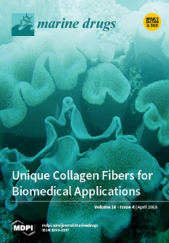
Cover Story (view full-size image): The current study elaborates on unique collagen fibers derived from Sarcophyton octocorals, their structure and biocompatibility in an in vitro model, suggesting that they are highly appropriate for a variety of biomedical applications. The in vitro results are promising for these fibers to serve as a scaffold that can be adapted for tissue regeneration and to support healing processes. The fibers can cope with the challenge to develop grafts for tissue regeneration that lies in the need to have a scaffold being able promote cell growth to form new tissue in a trauma-damaged site. The collagen fibers provide scaffolds featuring compatible mechanical properties to support new tissues and to facilitate the desired physiological activity. The study used natural materials to develop a bio-composite, made of the Sarcophyton-derived collagen, while being embedded in alginate hydrogel. View this paper
  • Issues are regarded as officially published after their release is announced to the table of contents alert mailing list.
  • You may sign up for e-mail alerts to receive table of contents of newly released issues.
  • PDF is the official format for papers published in both, html and pdf forms. To view the papers in pdf format, click on the "PDF Full-text" link, and use the free Adobe Reader to open them.
Order results
Result details
Section
Select all
Export citation of selected articles as:
15 pages, 2737 KiB  
Article
Bioactive Compounds from Posidonia oceanica (L.) Delile Impair Malignant Cell Migration through Autophagy Modulation
by Manuela Leri, Matteo Ramazzotti, Marzia Vasarri, Sara Peri, Emanuela Barletta, Carlo Pretti and Donatella Degl’Innocenti
Mar. Drugs 2018, 16(4), 137; https://doi.org/10.3390/md16040137 - 21 Apr 2018
Cited by 26 | Viewed by 5249
Abstract
Posidonia oceanica (L.) Delile is a marine plant with interesting biological properties potentially ascribed to the synergistic combination of bioactive compounds. Our previously described extract, obtained from the leaves of P. oceanica, showed the ability to impair HT1080 cell migration by targeting [...] Read more.
Posidonia oceanica (L.) Delile is a marine plant with interesting biological properties potentially ascribed to the synergistic combination of bioactive compounds. Our previously described extract, obtained from the leaves of P. oceanica, showed the ability to impair HT1080 cell migration by targeting both expression and activity of gelatinases. Commonly, the lack of knowledge about the mechanism of action of phytocomplexes may be an obstacle regarding their therapeutic use and development. The aim of this study was to gain insight into the molecular signaling through which such bioactive compounds impact on malignant cell migration and gelatinolytic activity. The increase in autophagic vacuoles detected by confocal microscopy suggested an enhancement of autophagy in a time and dose dependent manner. This autophagy activation was further confirmed by monitoring pivotal markers of autophagy signaling as well as by evidencing an increase in IGF-1R accumulation on cell membranes. Taken together, our results confirm that the P. oceanica phytocomplex is a promising reservoir of potent and cell safe molecules able to defend against malignancies and other diseases in which gelatinases play a major role in progression. In conclusion, the attractive properties of this phytocomplex may be of industrial interest in regard to the development of novel health-promoting and pharmacological products for the treatment or prevention of several diseases. Full article
Show Figures

Figure 1

12 pages, 2638 KiB  
Article
Eurotiumins A–E, Five New Alkaloids from the Marine-Derived Fungus Eurotium sp. SCSIO F452
by Wei-Mao Zhong, Jun-Feng Wang, Xue-Feng Shi, Xiao-Yi Wei, Yu-Chan Chen, Qi Zeng, Yao Xiang, Xia-Yu Chen, Xin-Peng Tian, Zhi-Hui Xiao, Wei-Min Zhang, Fa-Zuo Wang and Si Zhang
Mar. Drugs 2018, 16(4), 136; https://doi.org/10.3390/md16040136 - 21 Apr 2018
Cited by 34 | Viewed by 6354
Abstract
Three new prenylated indole 2,5-diketopiperazine alkaloids (13) with nine known ones (513), one new indole alkaloid (4), and one new bis-benzyl pyrimidine derivative (14) were isolated and characterized from the marine-derived [...] Read more.
Three new prenylated indole 2,5-diketopiperazine alkaloids (13) with nine known ones (513), one new indole alkaloid (4), and one new bis-benzyl pyrimidine derivative (14) were isolated and characterized from the marine-derived fungus Eurotium sp. SCSIO F452. 1 and 2, occurring as a pair of diastereomers, both presented a hexahydropyrrolo[2,3-b]indole skeleton. Their chemical structures, including absolute configurations, were elucidated by 1D and 2D NMR, HRESIMS, quantum chemical calculations of electronic circular dichroism, and single crystal X-ray diffraction experiments. Most isolated compounds were screened for antioxidative potency. Compounds 3, 5, 6, 7, 9, 10, and 12 showed significant radical scavenging activities against DPPH with IC50 values of 13, 19, 4, 3, 24, 13, and 18 µM, respectively. Five new compounds were evaluated for cytotoxic activities. Full article
Show Figures

Figure 1

16 pages, 2836 KiB  
Article
The Protective Role of Sulfated Polysaccharides from Green Seaweed Udotea flabellum in Cells Exposed to Oxidative Damage
by Fernando Bastos Presa, Maxsuell Lucas Mendes Marques, Rony Lucas Silva Viana, Leonardo Thiago Duarte Barreto Nobre, Leandro Silva Costa and Hugo Alexandre Oliveira Rocha
Mar. Drugs 2018, 16(4), 135; https://doi.org/10.3390/md16040135 - 20 Apr 2018
Cited by 37 | Viewed by 4134
Abstract
Seaweed is a rich source of bioactive sulfated polysaccharides. We obtained six sulfated polysaccharide-rich fractions (UF-0.3, UF-0.5, UF-0.6, UF-0.7, UF-1.0, and UF-2.0) from the green seaweed Udotea flabellum (UF) by proteolytic digestion followed by sequential acetone precipitation. Biochemical analysis of these fractions showed [...] Read more.
Seaweed is a rich source of bioactive sulfated polysaccharides. We obtained six sulfated polysaccharide-rich fractions (UF-0.3, UF-0.5, UF-0.6, UF-0.7, UF-1.0, and UF-2.0) from the green seaweed Udotea flabellum (UF) by proteolytic digestion followed by sequential acetone precipitation. Biochemical analysis of these fractions showed that they were enriched with sulfated galactans. The viability and proliferative capacity of 3T3 fibroblasts exposed to FeSO4 (2 µM), CuSO4 (1 µM) or ascorbate (2 mM) was not affected. However, these cells were exposed to oxidative stress in the presence of FeSO4 or CuSO4 and ascorbate, which caused the activation of caspase-3 and caspase-9, resulting in apoptosis of the cells. We also observed increased lipid peroxidation, evaluated by the detection of malondialdehyde and decreased glutathione and superoxide dismutase levels. Treating the cells with the ultrafiltrate fractions (UF) fractions protected the cells from the oxidative damage caused by the two salts and ascorbate. The most effective protection against the oxidative damage caused by iron was provided by UF-0.7 (1.0 mg/mL); on treatment with UF-0.7, cell viability was 55%. In the case of copper, cell viability on treatment with UF-0.7 was ~80%, but the most effective fraction in this model was UF-2.0, with cell viability of more than 90%. The fractions, mainly UF-0.7 and UF-2.0, showed low iron chelating activity, but high copper chelating activity and total antioxidant capacity (TAC). These results suggested that some of their protective mechanisms stem from these properties. Full article
(This article belongs to the Special Issue Development and Application of Herbal Medicine from Marine Origin)
Show Figures

Figure 1

14 pages, 12488 KiB  
Article
A Low Molecular Weight Protein from the Sea Anemone Anemonia viridis with an Anti-Angiogenic Activity
by Erwann P. Loret, José Luis, Christopher Nuccio, Claude Villard, Pascal Mansuelle, Régine Lebrun and Pierre Henri Villard
Mar. Drugs 2018, 16(4), 134; https://doi.org/10.3390/md16040134 - 19 Apr 2018
Cited by 8 | Viewed by 4677
Abstract
Sea anemones are a remarkable source of active principles due to a decentralized venom system. New blood vessel growth or angiogenesis is a very promising target against cancer, but the few available antiangiogenic compounds have limited efficacy. In this study, a protein fraction, [...] Read more.
Sea anemones are a remarkable source of active principles due to a decentralized venom system. New blood vessel growth or angiogenesis is a very promising target against cancer, but the few available antiangiogenic compounds have limited efficacy. In this study, a protein fraction, purified from tentacles of Anemonia viridis, was able to limit endothelial cells proliferation and angiogenesis at low concentration (14 nM). Protein sequences were determined with Edman degradation and mass spectrometry in source decay and revealed homologies with Blood Depressing Substance (BDS) sea anemones. The presence of a two-turn alpha helix observed with circular dichroism and a trypsin activity inhibition suggested that the active principle could be a Kunitz-type inhibitor, which may interact with an integrin due to an Arginine Glycin Aspartate (RGD) motif. Molecular modeling showed that this RGD motif was well exposed to solvent. This active principle could improve antiangiogenic therapy from existing antiangiogenic compounds binding on the Vascular Endothelial Growth Factor (VEGF). Full article
(This article belongs to the Collection Marine Compounds and Cancer)
Show Figures

Figure 1

15 pages, 10289 KiB  
Review
Recent Synthesis and Discovery of Brefeldin A Analogs
by Seung-Mann Paek
Mar. Drugs 2018, 16(4), 133; https://doi.org/10.3390/md16040133 - 18 Apr 2018
Cited by 20 | Viewed by 6718
Abstract
The recent development of analogs of brefeldin A (BFA), a fungal metabolite, for the improvement of BFA apoptosis-inducing activity is described. BFA has been isolated from various soil or, more recently, marine fungi and has shown versatile beneficial activities. More importantly, the apoptosis-inducing [...] Read more.
The recent development of analogs of brefeldin A (BFA), a fungal metabolite, for the improvement of BFA apoptosis-inducing activity is described. BFA has been isolated from various soil or, more recently, marine fungi and has shown versatile beneficial activities. More importantly, the apoptosis-inducing activity of BFA in cancer cells highlights the possibility of further developing this natural product as an anticancer agent. Besides its biological importance, its structural features have also gathered tremendous interest from both medicinal and synthetic chemists. By a medicinal chemistry and total synthesis approach, numerous analogs from BFA have been developed to improve its inferior bioavailability and its antiproliferative ability. In this review, the recent medicinal chemistry efforts in relation to the production of BFA analogs are extensively presented. Full article
(This article belongs to the Special Issue Connection of Marine Natural Products and Cell Apoptosis)
Show Figures

Figure 1

10 pages, 1186 KiB  
Article
Pharmacokinetic and Tissue Distribution of Fucoidan from Fucus vesiculosus after Oral Administration to Rats
by Olga N. Pozharitskaya, Alexander N. Shikov, Natalya M. Faustova, Ekaterina D. Obluchinskaya, Vera M. Kosman, Heikki Vuorela and Valery G. Makarov
Mar. Drugs 2018, 16(4), 132; https://doi.org/10.3390/md16040132 - 18 Apr 2018
Cited by 91 | Viewed by 6826
Abstract
Fucus vesiculosus L., known as bladderwrack, belongs to the brown seaweeds, which are widely distributed throughout northern Russia, Atlantic shores of Europe, the Baltic Sea, Greenland, the Azores, the Canary Islands, and shores of the Pacific Ocean. Fucoidan is a major fucose-rich sulfated [...] Read more.
Fucus vesiculosus L., known as bladderwrack, belongs to the brown seaweeds, which are widely distributed throughout northern Russia, Atlantic shores of Europe, the Baltic Sea, Greenland, the Azores, the Canary Islands, and shores of the Pacific Ocean. Fucoidan is a major fucose-rich sulfated polysaccharide found in Fucus (F.) vesiculosus. The pharmacokinetic profiling of active compounds is essential for drug development and approval. The aim of the study was to evaluate the pharmacokinetics and tissue distribution of fucoidan in rats after a single-dose oral administration. Fucoidan was isolated from F. vesiculosus. The method of measuring anti-activated factor X (anti-Xa) activity by amidolytic assay was used to analyze the plasma and tissue concentrations of fucoidan. The tissue distribution of fucoidan after intragastric administration to the rats was characterized, and it exhibited considerable heterogeneity. Fucoidan preferentially accumulates in the kidneys (AUC0–t = 10.74 µg·h/g; Cmax = 1.23 µg/g after 5 h), spleen (AUC0–t = 6.89 µg·h/g; Cmax = 0.78 µg/g after 3 h), and liver (AUC0–t = 3.26 µg·h/g; Cmax = 0.53 µg/g after 2 h) and shows a relatively long absorption time and extended circulation in the blood, with a mean residence time (MRT) = 6.79 h. The outcome of this study provides additional scientific data for traditional use of fucoidan-containing plants and offers tangible support for the continued development of new effective pharmaceuticals using fucoidan. Full article
(This article belongs to the Special Issue Marine Bioactive Natural Product Studies in Europe)
Show Figures

Graphical abstract

10 pages, 8683 KiB  
Article
Marine Longilenes, Oxasqualenoids with Ser-Thr Protein Phosphatase 2A Inhibition Activity
by Francisco Cen-Pacheco, Claudia Pérez Manríquez, María Luisa Souto, Manuel Norte, José Javier Fernández and Antonio Hernández Daranas
Mar. Drugs 2018, 16(4), 131; https://doi.org/10.3390/md16040131 - 17 Apr 2018
Cited by 6 | Viewed by 3225
Abstract
The red seaweed Laurencia viridis is a rich source of oxygenated secondary metabolites that were derived from squalene. We report here the structures of three novel compounds, (+)-longilene peroxide (1), longilene (2), and (+)-prelongilene (3) that were [...] Read more.
The red seaweed Laurencia viridis is a rich source of oxygenated secondary metabolites that were derived from squalene. We report here the structures of three novel compounds, (+)-longilene peroxide (1), longilene (2), and (+)-prelongilene (3) that were isolated from this alga, in addition to other substances, 4 and 5, resulting from their acid-mediated degradation. The effect of compounds 1 and 3 against Ser-Thr protein phosphatase type 2A (PP2A) was evaluated, showing that (+)-longilene peroxide (1) inhibited PP2A (IC50 11.3 μM). In order to explain the interaction between PP2A and compounds 1 and 3, molecular docking simulations onto the PP2A enzyme-binding region were used. Full article
(This article belongs to the Special Issue Pre-Clinical Marine Drug Discovery)
Show Figures

Graphical abstract

10 pages, 4264 KiB  
Article
Antartin, a Cytotoxic Zizaane-Type Sesquiterpenoid from a Streptomyces sp. Isolated from an Antarctic Marine Sediment
by Dayoung Kim, Eun Ju Lee, Jihye Lee, Alain S. Leutou, Yern-Hyerk Shin, Bomi Choi, Ji Sun Hwang, Dongyup Hahn, Hyukjae Choi, Jungwook Chin, Sung Jin Cho, Yong Deog Hong, Jaeyoung Ko, Chi Nam Seong, Katherine N. Maloney, Dong-Chan Oh, Inho Yang, Hayoung Hwang and Sang-Jip Nam
Mar. Drugs 2018, 16(4), 130; https://doi.org/10.3390/md16040130 - 16 Apr 2018
Cited by 17 | Viewed by 4596
Abstract
Antartin (1), a new zizaane-type sesquiterpene, was isolated from Streptomyces sp. SCO736. The chemical structure of 1 was assigned from the interpretation of 1D and 2D NMR in addition to mass spectrometric data. The relative stereochemistry of 1 was determined by [...] Read more.
Antartin (1), a new zizaane-type sesquiterpene, was isolated from Streptomyces sp. SCO736. The chemical structure of 1 was assigned from the interpretation of 1D and 2D NMR in addition to mass spectrometric data. The relative stereochemistry of 1 was determined by analysis of NOE data, while the absolute stereochemistry was decided based on a comparison of experimental and calculated electronic circular dichroism (ECD) spectra. Antartin (1) showed cytotoxicity against A549, H1299, and U87 cancer cell lines by causing cell cycle arrest at the G1 phase. Full article
(This article belongs to the Special Issue Natural Products from Marine Actinomycetes)
Show Figures

Figure 1

12 pages, 18465 KiB  
Article
Biochemical Characterization and Degradation Pattern of a Unique pH-Stable PolyM-Specific Alginate Lyase from Newly Isolated Serratia marcescens NJ-07
by Benwei Zhu, Fu Hu, Heng Yuan, Yun Sun and Zhong Yao
Mar. Drugs 2018, 16(4), 129; https://doi.org/10.3390/md16040129 - 15 Apr 2018
Cited by 29 | Viewed by 4406
Abstract
Enzymatic preparation of alginate oligosaccharides with versatile bioactivities by alginate lyases has attracted increasing attention due to its featured characteristics, such as wild condition and specific products. In this study, AlgNJ-07, a novel polyM-specific alginate lyase with high specific activity and pH stability, [...] Read more.
Enzymatic preparation of alginate oligosaccharides with versatile bioactivities by alginate lyases has attracted increasing attention due to its featured characteristics, such as wild condition and specific products. In this study, AlgNJ-07, a novel polyM-specific alginate lyase with high specific activity and pH stability, has been purified from the newly isolated marine bacterium Serratia marcescens NJ-07. It has a molecular weight of approximately 25 kDa and exhibits the maximal activity of 2742.5 U/mg towards sodium alginate under 40 °C at pH 9.0. Additionally, AlgNJ-07 could retain more than 95% of its activity at pH range of 8.0–10.0, indicating it possesses excellent pH-stability. Moreover, it shows high activity and affinity towards polyM block and no activity to polyG block, which suggests that it is a strict polyM-specific alginate lyase. The degradation pattern of AlgNJ-07 has also been explored. The activity of AlgNJ-07 could be activated by NaCl with a low concentration (100–300 mM). It can be observed that AlgNJ-07 can recognize the trisaccharide as the minimal substrate and hydrolyze the trisaccharide into monosaccharide and disaccharide. The TLC and ESI-MS analysis indicate that it can hydrolyze substrates in a unique endolytic manner, producing not only oligosaccharides with Dp of 2–5 but also a large fraction of monosaccharide. Therefore, it may be a potent tool to produce alginate oligosaccharides with lower Dps (degree of polymerization). Full article
Show Figures

Figure 1

15 pages, 24061 KiB  
Article
Structural Characterization and Interaction with RCA120 of a Highly Sulfated Keratan Sulfate from Blue Shark (Prionace glauca) Cartilage
by Qinying Li, Guoyun Li, Xiaoliang Zhao, Xindi Shan, Chao Cai, Jing Zhao, Fuming Zhang, Robert J. Linhardt and Guangli Yu
Mar. Drugs 2018, 16(4), 128; https://doi.org/10.3390/md16040128 - 14 Apr 2018
Cited by 4 | Viewed by 4664
Abstract
As an important glycosaminoglycan, keratan sulfate (KS) mainly exists in corneal and cartilage, possessing various biological activities. In this study, we purified KS from blue shark (Prionace glauca) cartilage and prepared KS oligosaccharides (KSO) through keratanase II-catalyzed hydrolysis. The structures of [...] Read more.
As an important glycosaminoglycan, keratan sulfate (KS) mainly exists in corneal and cartilage, possessing various biological activities. In this study, we purified KS from blue shark (Prionace glauca) cartilage and prepared KS oligosaccharides (KSO) through keratanase II-catalyzed hydrolysis. The structures of KS and KSO were characterized using multi-dimensional nuclear magnetic resonance (NMR) spectra and liquid chromatography-mass spectrometry (LC-MS). Shark cartilage KS was highly sulfated and modified with ~2.69% N-acetylneuraminic acid (NeuAc) through α(2,3)-linked to galactose. Additionally, KS exhibited binding affinity to Ricinus communis agglutinin I (RCA120) in a concentration-dependent manner, a highly toxic lectin from beans of the castor plant. Furthermore, KSO from dp2 to dp8 bound to RCA120 in the increasing trend while the binding affinity of dp8 was superior to polysaccharide. These results define novel structural features for KS from Prionace glauca cartilage and demonstrate the potential application on ricin-antidote exploitation. Full article
(This article belongs to the Special Issue Marine Drugs Interact with Functional Proteins)
Show Figures

Graphical abstract

15 pages, 5537 KiB  
Article
Terpenoids from the Soft Coral Sinularia sp. Collected in Yongxing Island
by Guo-Fei Qin, Xu-Li Tang, Yan-Ting Sun, Xiang-Chao Luo, Jing Zhang, Leen Van Ofwegen, Ping-Jyun Sung, Ping-Lin Li and Guo-Qiang Li
Mar. Drugs 2018, 16(4), 127; https://doi.org/10.3390/md16040127 - 13 Apr 2018
Cited by 30 | Viewed by 4476
Abstract
Three new sesquiterpenoids (sinuketal (1), sinulins A and B (2 and 3)) and two new cembranoids (sinulins C and D (4 and 5)), as well as eight known sesquiterpenoids (6–13) and eight known cembranoids ( [...] Read more.
Three new sesquiterpenoids (sinuketal (1), sinulins A and B (2 and 3)) and two new cembranoids (sinulins C and D (4 and 5)), as well as eight known sesquiterpenoids (6–13) and eight known cembranoids (14–21), were isolated from the Xisha soft coral Sinularia sp. Their structures were elucidated by extensive spectroscopic analysis. Compound 1 possesses an unprecedented isopropyl-branched bicyclo [6.3.0] undecane carbon skeleton with unique endoperoxide moiety, and a plausible biosynthetic pathway of it was postulated. According to the reported biological properties of endoperoxide, the antimalarial, cytotoxic, antiviral, and target inhibitory activities of 1 were tested. Compound 1 showed mild in vitro antimalarial activity against Plasmodium falciparum 3D7, weak cytotoxic activities toward Jurkat, MDA-MB-231, and U2OS cell lines, inhibitory effects against influenza A viruses H1N1 and PR8, as well as mild target inhibitory activity against acetylcholinesterase. The other compounds were evaluated for cytotoxicities against HeLa, HCT-116, and A549 tumor cell lines and target inhibitory activities against protein tyrosine phosphatase 1B (PTP1B). Compound 20 exhibited cytotoxicities against HeLa and HCT-116, and compounds 5, 11, and 15 showed mild target inhibitory activities against PTP1B. Full article
Show Figures

Graphical abstract

14 pages, 2375 KiB  
Article
Astaxanthin Restrains Nitrative-Oxidative Peroxidation in Mitochondrial-Mimetic Liposomes: A Pre-Apoptosis Model
by Camila M. Mano, Thais Guaratini, Karina H. M. Cardozo, Pio Colepicolo, Etelvino J. H. Bechara and Marcelo P. Barros
Mar. Drugs 2018, 16(4), 126; https://doi.org/10.3390/md16040126 - 12 Apr 2018
Cited by 15 | Viewed by 4048
Abstract
Astaxanthin (ASTA) is a ketocarotenoid found in many marine organisms and that affords many benefits to human health. ASTA is particularly effective against radical-mediated lipid peroxidation, and recent findings hypothesize a “mitochondrial-targeted” action of ASTA in cells. Therefore, we examined the protective effects [...] Read more.
Astaxanthin (ASTA) is a ketocarotenoid found in many marine organisms and that affords many benefits to human health. ASTA is particularly effective against radical-mediated lipid peroxidation, and recent findings hypothesize a “mitochondrial-targeted” action of ASTA in cells. Therefore, we examined the protective effects of ASTA against lipid peroxidation in zwitterionic phosphatidylcholine liposomes (PCLs) and anionic phosphatidylcholine: phosphatidylglycerol liposomes (PCPGLs), at different pHs (6.2 to 8.0), which were challenged by oxidizing/nitrating conditions that mimic the regular and preapoptotic redox environment of active mitochondria. Pre-apoptotic conditions were created by oxidized/nitr(osyl)ated cytochrome c and resulted in the highest levels of lipoperoxidation in both PCL and PCPGLs (pH 7.4). ASTA was less protective at acidic conditions, especially in anionic PCPGLs. Our data demonstrated the ability of ASTA to hamper oxidative and nitrative events that lead to cytochrome c-peroxidase apoptosis and lipid peroxidation, although its efficiency changes with pH and lipid composition of membranes. Full article
(This article belongs to the Special Issue Connection of Marine Natural Products and Cell Apoptosis)
Show Figures

Graphical abstract

15 pages, 58554 KiB  
Article
Anticancer Activity of Anthopleura anjunae Oligopeptides in Prostate Cancer DU-145 Cells
by Zong-Ze Wu, Guo-Fang Ding, Fang-Fang Huang, Zui-Su Yang, Fang-Miao Yu, Yun-Ping Tang, Ying-Lu Jia, Yuan-Yuan Zheng and Rui Chen
Mar. Drugs 2018, 16(4), 125; https://doi.org/10.3390/md16040125 - 12 Apr 2018
Cited by 22 | Viewed by 4961
Abstract
Anthopleura anjunae anti-tumor peptide (AAP-H) is a pentapeptide from the sea anemone Anthopleura anjunae with an amino acid sequence of Tyr-Val-Pro-Gly-Pro that is obtained by alkaline protease enzymatic hydrolysis extraction. In this study, we investigated the inhibitory effects of AAP-H on prostate cancer [...] Read more.
Anthopleura anjunae anti-tumor peptide (AAP-H) is a pentapeptide from the sea anemone Anthopleura anjunae with an amino acid sequence of Tyr-Val-Pro-Gly-Pro that is obtained by alkaline protease enzymatic hydrolysis extraction. In this study, we investigated the inhibitory effects of AAP-H on prostate cancer DU-145 cell proliferation using a methylthiazolyldiphenyl-tetrazolium bromide assay. Cell morphology was analyzed by hematoxylin-eosin staining, acridine orange/ethidium bromide fluorescence staining, Hoechst 33258 fluorescence staining, and scanning electron microscopy. The mitochondrial membrane potential was determined by flow cytometry following JC-1 staining. The cell apoptosis rate was measured by Annexin V-fluorescein isothiocyanate and propidium iodide staining followed by flow cytometric analysis, and the expression of apoptosis-associated proteins was assayed by Western blotting. The results demonstrated that AAP-H induced significant reductions in the number of viable cells and increased cell death in both a dose-dependent and time-dependent manner, with an IC50 of approximately 9.605 mM, 7.910 mM, and 2.298 mM at 24 h, 48 h, and 72 h, respectively. The morphologic characteristics of apoptotic cells were observed after treatment with AAP-H. The mitochondrial membrane potential was markedly decreased, and apoptosis increased after AAP-H treatment. Pro-apoptotic proteins, such as Bax, cytochrome-C, caspase-3, and caspase-9 were increased, but Bcl-2 was decreased. These findings suggest that AAP-H has moderate inhibitory effects on prostate cancer DU-145 cells, and the mechanism might involve the mitochondria-mediated apoptotic pathway. Therefore, AAP-H is a candidate anti-prostate cancer drug or health-care food. Full article
Show Figures

Figure 1

11 pages, 3638 KiB  
Article
The Structure of the Lipid A from the Halophilic Bacterium Spiribacter salinus M19-40T
by Clara Barrau, Flaviana Di Lorenzo, Rodolfo Javier Menes, Rosa Lanzetta, Antonio Molinaro and Alba Silipo
Mar. Drugs 2018, 16(4), 124; https://doi.org/10.3390/md16040124 - 11 Apr 2018
Cited by 8 | Viewed by 3866
Abstract
The study of the adaptation mechanisms that allow microorganisms to live and proliferate in an extreme habitat is a growing research field. Directly exposed to the external environment, lipopolysaccharides (LPS) from Gram-negative bacteria are of great appeal as they can present particular structural [...] Read more.
The study of the adaptation mechanisms that allow microorganisms to live and proliferate in an extreme habitat is a growing research field. Directly exposed to the external environment, lipopolysaccharides (LPS) from Gram-negative bacteria are of great appeal as they can present particular structural features that may aid the understanding of the adaptation processes. Moreover, through being involved in modulating the mammalian immune system response in a structure-dependent fashion, the elucidation of the LPS structure can also be seen as a fundamental step from a biomedical point of view. In this paper, the lipid A structure of the LPS from Spiribacter salinus M19-40T, a halophilic gamma-proteobacteria, was characterized through chemical analyses and matrix-assisted laser desorption ionization (MALDI) mass spectrometry. This revealed a mixture of mono- and bisphosphorylated penta- to tri-acylated species with the uncommon 2 + 3 symmetry and bearing an unusual 3-oxotetradecaonic acid. Full article
Show Figures

Figure 1

15 pages, 18934 KiB  
Article
Holotoxin A1 Induces Apoptosis by Activating Acid Sphingomyelinase and Neutral Sphingomyelinase in K562 and Human Primary Leukemia Cells
by Seong-Hoon Yun, Eun-Hye Sim, Sang-Heum Han, Jin-Yeong Han, Sung-Hyun Kim, Alexandra S. Silchenko, Valentin A. Stonik and Joo-In Park
Mar. Drugs 2018, 16(4), 123; https://doi.org/10.3390/md16040123 - 10 Apr 2018
Cited by 16 | Viewed by 4823
Abstract
Marine triterpene glycosides are attractive candidates for the development of anticancer agents. Holotoxin A1 is a triterpene glycoside found in the edible sea cucumber, Apostichopus (Stichopus) japonicus. We previously showed that cladoloside C2, the 25(26)-dihydro derivative of holotoxin A1, [...] Read more.
Marine triterpene glycosides are attractive candidates for the development of anticancer agents. Holotoxin A1 is a triterpene glycoside found in the edible sea cucumber, Apostichopus (Stichopus) japonicus. We previously showed that cladoloside C2, the 25(26)-dihydro derivative of holotoxin A1, induced apoptosis in human leukemia cells by activating ceramide synthase 6. Thus, we hypothesized that holotoxin A1, which is structurally similar to cladoloside C2, might induce apoptosis in human leukemia cells through the same molecular mechanism. In this paper, we compared holotoxin A1 and cladoloside C2 for killing potency and mechanism of action. We found that holotoxin A1 induced apoptosis more potently than cladoloside C2. Moreover, holotoxin A1 induced apoptosis in K562 cells by activating caspase-8 and caspase-3, but not by activating caspase-9. During holotoxin A1-induced apoptosis, acid sphingomyelinase (SMase) and neutral SMase were activated in both K562 cells and human primary leukemia cells. Specifically inhibiting acid SMase and neutral SMаse with chemical inhibitors or siRNAs significantly inhibited holotoxin A1–induced apoptosis. These results indicated that holotoxin A1 might induce apoptosis by activating acid SMase and neutral SMase. In conclusion, holotoxin A1 represents a potential anticancer agent for treating leukemia. Moreover, the aglycone structure of marine triterpene glycosides might affect the mechanism involved in inducing apoptosis. Full article
Show Figures

Graphical abstract

22 pages, 5587 KiB  
Article
Toxicological Investigations on the Sea Urchin Tripneustes gratilla (Toxopneustidae, Echinoid) from Anaho Bay (Nuku Hiva, French Polynesia): Evidence for the Presence of Pacific Ciguatoxins
by Hélène Taiana Darius, Mélanie Roué, Manoella Sibat, Jérôme Viallon, Clémence Mahana iti Gatti, Mark W. Vandersea, Patricia A. Tester, R. Wayne Litaker, Zouher Amzil, Philipp Hess and Mireille Chinain
Mar. Drugs 2018, 16(4), 122; https://doi.org/10.3390/md16040122 - 06 Apr 2018
Cited by 43 | Viewed by 5977
Abstract
The sea urchin Tripneustes gratilla (Toxopneustidae, Echinoids) is a source of protein for many islanders in the Indo-West Pacific. It was previously reported to occasionally cause ciguatera-like poisoning; however, the exact nature of the causative agent was not confirmed. In April [...] Read more.
The sea urchin Tripneustes gratilla (Toxopneustidae, Echinoids) is a source of protein for many islanders in the Indo-West Pacific. It was previously reported to occasionally cause ciguatera-like poisoning; however, the exact nature of the causative agent was not confirmed. In April and July 2015, ciguatera poisonings were reported following the consumption of T. gratilla in Anaho Bay (Nuku Hiva Island, Marquesas archipelago, French Polynesia). Patient symptomatology was recorded and sea urchin samples were collected from Anaho Bay in July 2015 and November 2016. Toxicity analysis using the neuroblastoma cell–based assay (CBA-N2a) detected the presence of ciguatoxins (CTXs) in T. gratilla samples. Gambierdiscus species were predominant in the benthic assemblages of Anaho Bay, and G. polynesiensis was highly prevalent in in vitro cultures according to qPCR results. Liquid chromatography–tandem mass spectrometry (LC-MS/MS) analyses revealed that P-CTX-3B was the major ciguatoxin congener in toxic sea urchin samples, followed by 51-OH-P-CTX-3C, P-CTX-3C, P-CTX-4A, and P-CTX-4B. Between July 2015 and November 2016, the toxin content in T. gratilla decreased, but was consistently above the safety limit allowed for human consumption. This study provides evidence of CTX bioaccumulation in T. gratilla as a cause of ciguatera-like poisoning associated with a documented symptomatology. Full article
(This article belongs to the Special Issue Marine Invertebrate Toxins)
Show Figures

Figure 1

16 pages, 19626 KiB  
Article
Fucoidan Rescues p-Cresol-Induced Cellular Senescence in Mesenchymal Stem Cells via FAK-Akt-TWIST Axis
by Jun Hee Lee, Chul Won Yun, Jin Hur and Sang Hun Lee
Mar. Drugs 2018, 16(4), 121; https://doi.org/10.3390/md16040121 - 06 Apr 2018
Cited by 38 | Viewed by 4778
Abstract
Mesenchymal stem cells (MSCs) are a source for cell-based therapy. Although MSCs have the potential for tissue regeneration, their therapeutic efficacy is restricted by the uremic toxin, p-cresol, in chronic kidney disease (CKD). To address this issue, we investigated the effect of [...] Read more.
Mesenchymal stem cells (MSCs) are a source for cell-based therapy. Although MSCs have the potential for tissue regeneration, their therapeutic efficacy is restricted by the uremic toxin, p-cresol, in chronic kidney disease (CKD). To address this issue, we investigated the effect of fucoidan, a marine sulfated polysaccharide, on cellular senescence in MSCs. After p-cresol exposure, MSC senescence was induced, as indicated by an increase in cell size and a decrease in proliferation capacity. Treatment of senescent MSCs with fucoidan significantly reversed this cellular senescence via regulation of SMP30 and p21, and increased proliferation through the regulation of cell cycle-associated proteins (CDK2, CDK4, cyclin D1, and cyclin E). These effects were dependent on FAK-Akt-TWIST signal transduction. In particular, fucoidan promoted the expression of cellular prion protein (PrPC), which resulted in the maintenance of cell expansion capacity in p-cresol-induced senescent MSCs. This protective effect of fucoidan on senescence-mediated inhibition of proliferation was dependent on the TWIST-PrPC axis. In summary, this study shows that fucoidan protects against p-cresol-induced cellular senescence in MSCs through activation of the FAK-Akt-TWIST pathway and suggests that fucoidan could be used in conjunction with functional MSC-based therapies in the treatment of CKD. Full article
(This article belongs to the Special Issue Marine Oligosaccharides and Polysaccharides)
Show Figures

Figure 1

13 pages, 4685 KiB  
Article
AlgM4: A New Salt-Activated Alginate Lyase of the PL7 Family with Endolytic Activity
by Guiyuan Huang, Qiaozhen Wang, Mingqian Lu, Chao Xu, Fei Li, Rongcan Zhang, Wei Liao and Shushi Huang
Mar. Drugs 2018, 16(4), 120; https://doi.org/10.3390/md16040120 - 06 Apr 2018
Cited by 37 | Viewed by 4317
Abstract
Alginate lyases are a group of enzymes that catalyze the depolymerization of alginates into oligosaccharides or monosaccharides. These enzymes have been widely used for a variety of purposes, such as producing bioactive oligosaccharides, controlling the rheological properties of polysaccharides, and performing structural analyses [...] Read more.
Alginate lyases are a group of enzymes that catalyze the depolymerization of alginates into oligosaccharides or monosaccharides. These enzymes have been widely used for a variety of purposes, such as producing bioactive oligosaccharides, controlling the rheological properties of polysaccharides, and performing structural analyses of polysaccharides. The algM4 gene of the marine bacterium Vibrio weizhoudaoensis M0101 encodes an alginate lyase that belongs to the polysaccharide lyase family 7 (PL7). In this study, the kinetic constants Vmax (maximum reaction rate) and Km (Michaelis constant) of AlgM4 activity were determined as 2.75 nmol/s and 2.72 mg/mL, respectively. The optimum temperature for AlgM4 activity was 30 °C, and at 70 °C, AlgM4 activity dropped to 11% of the maximum observed activity. The optimum pH for AlgM4 activity was 8.5, and AlgM4 was completely inactive at pH 11. The addition of 1 mol/L NaCl resulted in a more than sevenfold increase in the relative activity of AlgM4. The secondary structure of AlgM4 was altered in the presence of NaCl, which caused the α-helical content to decrease from 12.4 to 10.8% and the β-sheet content to decrease by 1.7%. In addition, NaCl enhanced the thermal stability of AlgM4 and increased the midpoint of thermal denaturation (Tm) by 4.9 °C. AlgM4 exhibited an ability to degrade sodium alginate, poly-mannuronic acid (polyM), and poly-guluronic acid (polyG), resulting in the production of oligosaccharides with a degree of polymerization (DP) of 2–9. AlgM4 possessed broader substrate, indicating that it is a bifunctional alginate lyase. Thus, AlgM4 is a novel salt-activated and bifunctional alginate lyase of the PL7 family with endolytic activity. Full article
Show Figures

Figure 1

22 pages, 3997 KiB  
Article
Bis-Indolyl Benzenoids, Hydroxypyrrolidine Derivatives and Other Constituents from Cultures of the Marine Sponge-Associated Fungus Aspergillus candidus KUFA0062
by Suradet Buttachon, Alice A. Ramos, Ângela Inácio, Tida Dethoup, Luís Gales, Michael Lee, Paulo M. Costa, Artur M. S. Silva, Nazim Sekeroglu, Eduardo Rocha, Madalena M. M. Pinto, José A. Pereira and Anake Kijjoa
Mar. Drugs 2018, 16(4), 119; https://doi.org/10.3390/md16040119 - 06 Apr 2018
Cited by 49 | Viewed by 6772
Abstract
A previously unreported bis-indolyl benzenoid, candidusin D (2e) and a new hydroxypyrrolidine alkaloid, preussin C (5b) were isolated together with fourteen previously described compounds: palmitic acid, clionasterol, ergosterol 5,8-endoperoxides, chrysophanic acid (1a), emodin (1b), [...] Read more.
A previously unreported bis-indolyl benzenoid, candidusin D (2e) and a new hydroxypyrrolidine alkaloid, preussin C (5b) were isolated together with fourteen previously described compounds: palmitic acid, clionasterol, ergosterol 5,8-endoperoxides, chrysophanic acid (1a), emodin (1b), six bis-indolyl benzenoids including asterriquinol D dimethyl ether (2a), petromurin C (2b), kumbicin B (2c), kumbicin A (2d), 2″-oxoasterriquinol D methyl ether (3), kumbicin D (4), the hydroxypyrrolidine alkaloid preussin (5a), (3S, 6S)-3,6-dibenzylpiperazine-2,5-dione (6) and 4-(acetylamino) benzoic acid (7), from the cultures of the marine sponge-associated fungus Aspergillus candidus KUFA 0062. Compounds 1a, 2a–e, 3, 4, 5a–b, and 6 were tested for their antibacterial activity against Gram-positive and Gram-negative reference and multidrug-resistant strains isolated from the environment. Only 5a exhibited an inhibitory effect against S. aureus ATCC 29213 and E. faecalis ATCC29212 as well as both methicillin-resistant S. aureus (MRSA) and vancomycin-resistant enterococci (VRE) strains. Both 1a and 5a also reduced significant biofilm formation in E. coli ATCC 25922. Moreover, 2b and 5a revealed a synergistic effect with oxacillin against MRSA S. aureus 66/1 while 5a exhibited a strong synergistic effect with the antibiotic colistin against E. coli 1410/1. Compound 1a, 2a–e, 3, 4, 5a–b, and 6 were also tested, together with the crude extract, for cytotoxic effect against eight cancer cell lines: HepG2, HT29, HCT116, A549, A 375, MCF-7, U-251, and T98G. Except for 1a, 2a, 2d, 4, and 6, all the compounds showed cytotoxicity against all the cancer cell lines tested. Full article
Show Figures

Graphical abstract

20 pages, 24757 KiB  
Review
Bioactive Compounds Isolated from Neglected Predatory Marine Gastropods
by Ashlin H. Turner, David J. Craik, Quentin Kaas and Christina I. Schroeder
Mar. Drugs 2018, 16(4), 118; https://doi.org/10.3390/md16040118 - 05 Apr 2018
Cited by 20 | Viewed by 7252
Abstract
A diverse range of predatory marine gastropods produce toxins, yet most of these molecules remain uncharacterized. Conus species have received the most attention from researchers, leading to several conopeptides reaching clinical trials. This review aims to summarize what is known about bioactive compounds [...] Read more.
A diverse range of predatory marine gastropods produce toxins, yet most of these molecules remain uncharacterized. Conus species have received the most attention from researchers, leading to several conopeptides reaching clinical trials. This review aims to summarize what is known about bioactive compounds isolated from species of neglected marine gastropods, especially in the Turridae, Terebridae, Babyloniidae, Muricidae, Buccinidae, Colubrariidae, Nassariidae, Cassidae, and Ranellidae families. Multiple species have been reported to contain bioactive compounds with potential toxic activity, but most of these compounds have not been characterized or even clearly identified. The bioactive properties and potential applications of echotoxins and related porins from the Ranellidae family are discussed in more detail. Finally, the review concludes with a call for research on understudied species. Full article
(This article belongs to the Collection Bioactive Compounds from Marine Invertebrates)
Show Figures

Figure 1

17 pages, 21629 KiB  
Article
Astaxanthin Promotes Nrf2/ARE Signaling to Inhibit HG-Induced Renal Fibrosis in GMCs
by Qing Chen, Jun Tao and Xi Xie
Mar. Drugs 2018, 16(4), 117; https://doi.org/10.3390/md16040117 - 05 Apr 2018
Cited by 56 | Viewed by 5290
Abstract
Oxidative stress is the main cause of diabetic nephropathy (DN) progression. Nuclear factor-erythroid 2-related factor 2 (Nrf2)/antioxidant response element (ARE) signaling is a crucial cellular defense system to cope with oxidative stress. Astaxanthin (AST) is a fat-soluble xanthophyll carotenoid with remarkable antioxidative capacity. [...] Read more.
Oxidative stress is the main cause of diabetic nephropathy (DN) progression. Nuclear factor-erythroid 2-related factor 2 (Nrf2)/antioxidant response element (ARE) signaling is a crucial cellular defense system to cope with oxidative stress. Astaxanthin (AST) is a fat-soluble xanthophyll carotenoid with remarkable antioxidative capacity. AST exerted renal protective in diabetic rats. This study aimed to determine whether AST could alleviate the pathological progress of DN by activating Nrf2/ARE signaling and diminishing the excessive oxidative stress and fibronectin (FN) accumulation in glomerular mesangial cells (GMCs) challenged with high glucose (HG). In the current study, we found that AST treatment alleviated the metabolic parameters, renal morphology and extracellular matrix (ECM) accumulation in streptozotocin-induced diabetic rats. Additionally, HG induced the adaptively activated Nrf2/ARE signaling and increased the expression of FN, intercellular adhesion molecule-1 (ICAM-1) and transforming growth factor-β1 (TGF-β1), as well as the intracellular reactive oxygen species (ROS) generation in GMCs. However, AST treatment strongly promoted the nuclear translocation and transcriptional activity of Nrf2 as well as upregulated the expression of superoxide dismutase (SOD1), NAD(P)H: quinone oxidoreductase (NQO1) and heme oxygenase-1 (HO-1), ultimately quenching the higher level of ROS and inhibiting the FN, ICAM-1 and TGF-β1 expression induced by HG. Collectively, our data suggest that the renoprotective effect of AST on DN depends on Nrf2/ARE signaling activation, which could be a potentially therapeutic strategy in the treatment of DN. Full article
Show Figures

Figure 1

11 pages, 2205 KiB  
Article
Specific Chemical and Genetic Markers Revealed a Thousands-Year Presence of Toxic Nodularia spumigena in the Baltic Sea
by Marta Cegłowska, Anna Toruńska-Sitarz, Grażyna Kowalewska and Hanna Mazur-Marzec
Mar. Drugs 2018, 16(4), 116; https://doi.org/10.3390/md16040116 - 04 Apr 2018
Cited by 11 | Viewed by 4367
Abstract
In the Baltic Sea, diazotrophic cyanobacteria have been present for thousands of years, over the whole brackish water phase of the ecosystem. However, our knowledge about the species composition of the cyanobacterial community is limited to the last several decades. In the current [...] Read more.
In the Baltic Sea, diazotrophic cyanobacteria have been present for thousands of years, over the whole brackish water phase of the ecosystem. However, our knowledge about the species composition of the cyanobacterial community is limited to the last several decades. In the current study, the presence of species-specific chemical and genetic markers in deep sediments were analyzed to increase the existing knowledge on the history of toxic Nodularia spumigena blooms in the Baltic Sea. As chemical markers, three cyclic nonribosomal peptides were applied: the hepatotoxic nodularin, which in the sea was detected solely in N. spumigena, and two anabaenopeptins (AP827 and AP883a) characteristic of two different chemotypes of this species. From the same sediment samples, DNA was isolated and the gene involved in biosynthesis of nodularin, as well as the phycocyanin intergenic spacer region (PC-IGS), were amplified. The results of chemical and genetic analyses proved for the first time the thousands-year presence of toxic N. spumigena in the Baltic Sea. They also indicated that through all this time, the same two sub-populations of the species co-existed. Full article
(This article belongs to the Special Issue Marine Bacterial Toxins)
Show Figures

Figure 1

15 pages, 5603 KiB  
Review
Total Synthesis of Bioactive Marine Meroterpenoids: The Cases of Liphagal and Frondosin B
by Yan Zong, Weijia Wang and Tao Xu
Mar. Drugs 2018, 16(4), 115; https://doi.org/10.3390/md16040115 - 31 Mar 2018
Cited by 11 | Viewed by 6065
Abstract
Liphagal and frondosin B are two marine-derived secondary metabolites sharing a very similar polyfused-benzofuran skeleton. The two tetracyclic meroterpenoids were isolated from marine sponges, both featuring a 6-5-7-6 fused ring system. A preliminary bioactive study shows that (+)-liphagal is a selective kinase (PI3K [...] Read more.
Liphagal and frondosin B are two marine-derived secondary metabolites sharing a very similar polyfused-benzofuran skeleton. The two tetracyclic meroterpenoids were isolated from marine sponges, both featuring a 6-5-7-6 fused ring system. A preliminary bioactive study shows that (+)-liphagal is a selective kinase (PI3K α) inhibitor, while (+)-frondosin B is shown to inhibit the binding of the cytokine interleukin-8 (IL-8) to its receptor, CX-CLR1/2. The unique structures and interesting biological profiles of these two meroterpenoids have attracted considerable attention from synthetic chemists. Herein we summarize the synthetic efforts with respect to (+)-liphagal and (+)-frondosin B during the past two decades. Full article
Show Figures

Figure 1

9 pages, 3754 KiB  
Article
Two New Diketomorpholine Derivatives and a New Highly Conjugated Ergostane-Type Steroid from the Marine Algal-Derived Endophytic Fungus Aspergillus alabamensis EN-547
by Sui-Qun Yang, Xiao-Ming Li, Xin Li, Lu-Ping Chi and Bin-Gui Wang
Mar. Drugs 2018, 16(4), 114; https://doi.org/10.3390/md16040114 - 31 Mar 2018
Cited by 18 | Viewed by 4196
Abstract
Chemical investigation of the marine algal-derived endophytic fungus Aspergillus alabamensis EN-547 resulted in the isolation of 4-epi-seco-shornephine A methyl ester (1) and 4-epi-seco-shornephine A carboxylic acid (2), two new secondary metabolites [...] Read more.
Chemical investigation of the marine algal-derived endophytic fungus Aspergillus alabamensis EN-547 resulted in the isolation of 4-epi-seco-shornephine A methyl ester (1) and 4-epi-seco-shornephine A carboxylic acid (2), two new secondary metabolites having a rare diketomorpholine motif, and 28-acetoxy-12β,15α,25-trihydroxyergosta-4,6,8(14),22-tetraen-3-one (3), a new highly conjugated ergostane-type steroid, together with four known metabolites (47). Their chemical structures were elucidated by detailed analysis of their NMR spectra, ECDs, HRESIMS, optical rotation, and X-ray crystallographic data, and by comparison with literature data as well. The antimicrobial activities of compounds 17 were evaluated. Full article
(This article belongs to the Special Issue Bioactive Compounds from Marine Microbes - II)
Show Figures

Figure 1

13 pages, 14065 KiB  
Article
Intraocular Penetration of a vNAR: In Vivo and In Vitro VEGF165 Neutralization
by Tanya A. Camacho-Villegas, María Teresa Mata-González, Walter García-Ubbelohd, Linda Núñez-García, Carolina Elosua, Jorge F. Paniagua-Solis and Alexei F. Licea-Navarro
Mar. Drugs 2018, 16(4), 113; https://doi.org/10.3390/md16040113 - 31 Mar 2018
Cited by 28 | Viewed by 5584
Abstract
Variable new antigen receptor domain (vNAR) antibodies are novel, naturally occurring antibodies that can be isolated from naïve, immune or synthetic shark libraries. These molecules are very interesting to the biotechnology and pharmaceutical industries because of their unique characteristics related to size and [...] Read more.
Variable new antigen receptor domain (vNAR) antibodies are novel, naturally occurring antibodies that can be isolated from naïve, immune or synthetic shark libraries. These molecules are very interesting to the biotechnology and pharmaceutical industries because of their unique characteristics related to size and tissue penetrability. There have been some approved anti-angiogenic therapies for ophthalmic conditions, not related to vNAR. This includes biologics and chimeric proteins that neutralize vascular endothelial growth factor (VEGF)165, which are injected intravitreal, causing discomfort and increasing the possibility of infection. In this paper, we present a vNAR antibody against human recombinant VEGF165 (rhVEGF165) that was isolated from an immunized Heterodontus francisci shark. A vNAR called V13, neutralizes VEGF165 cytokine starting at 75 μg/mL in an in vitro assay based on co-culture of normal human dermal fibroblasts (NHDFs) and green fluorescence protein (GFP)-labeled human umbilical vein endothelial cells (HUVECs) cells. In the oxygen-induced retinopathy model in C57BL/6:Hsd mice, we demonstrate an endothelial cell count decrease. Further, we demonstrate the intraocular penetration after topical administration of 0.1 μg/mL of vNAR V13 by its detection in aqueous humor in New Zealand rabbits with healthy eyes after 3 h of application. These findings demonstrate the potential of topical application of vNAR V13 as a possible new drug candidate for vascular eye diseases. Full article
(This article belongs to the Special Issue The Pharmacological Potential of Marine-Derived Peptides and Proteins)
Show Figures

Figure 1

12 pages, 2003 KiB  
Article
Cloning, Synthesis and Functional Characterization of a Novel α-Conotoxin Lt1.3
by Jinqin Chen, Li Liang, Huying Ning, Fengtao Cai, Zhuguo Liu, Longxiao Zhang, Liangyi Zhou and Qiuyun Dai
Mar. Drugs 2018, 16(4), 112; https://doi.org/10.3390/md16040112 - 31 Mar 2018
Cited by 16 | Viewed by 4103
Abstract
α-Conotoxins (α-CTxs) are small peptides composed of 11 to 20 amino acid residues with two disulfide bridges. Most of them potently and selectively target nicotinic acetylcholine receptor (nAChR) subtypes, and a few were found to inhibit the GABAB receptor (GABABR)-coupled [...] Read more.
α-Conotoxins (α-CTxs) are small peptides composed of 11 to 20 amino acid residues with two disulfide bridges. Most of them potently and selectively target nicotinic acetylcholine receptor (nAChR) subtypes, and a few were found to inhibit the GABAB receptor (GABABR)-coupled N-type calcium channels (Cav2.2). However, in all of α-CTxs targeting both receptors, the disulfide connectivity arrangement “C1-C3, C2-C4” is present. In this work, a novel α4/7-CTx named Lt1.3 (GCCSHPACSGNNPYFC-NH2) was cloned from the venom ducts of Conus litteratus (C. litteratus) in the South China Sea. Lt1.3 was then chemically synthesized and two isomers with disulfide bridges “C1-C3, C2-C4” and “C1-C4, C2-C3” were found and functionally characterized. Electrophysiological experiments showed that Lt1.3 containing the common disulfide bridges “C1-C3, C2-C4” potently and selectively inhibited α3β2 nAChRs and not GABABR-coupled Cav2.2. Surprisingly, but the isomer with the disulfide bridges “C1-C4, C2-C3” showed exactly the opposite inhibitory activity, inhibiting only GABABR-coupled Cav2.2 and not α3β2 nAChRs. These findings expand the knowledge of the targets and selectivity of α-CTxs and provide a new structural motif to inhibit the GABABR-coupled Cav2.2. Full article
(This article belongs to the Special Issue Marine Invertebrate Toxins)
Show Figures

Figure 1

30 pages, 47826 KiB  
Article
Production, Characterization and Biocompatibility Evaluation of Collagen Membranes Derived from Marine Sponge Chondrosia reniformis Nardo, 1847
by Marina Pozzolini, Sonia Scarfì, Lorenzo Gallus, Maila Castellano, Silvia Vicini, Katia Cortese, Maria Cristina Gagliani, Marco Bertolino, Gabriele Costa and Marco Giovine
Mar. Drugs 2018, 16(4), 111; https://doi.org/10.3390/md16040111 - 29 Mar 2018
Cited by 52 | Viewed by 5545
Abstract
Collagen is involved in the formation of complex fibrillar networks, providing the structural integrity of tissues. Its low immunogenicity and mechanical properties make this molecule a biomaterial that is extremely suitable for tissue engineering and regenerative medicine (TERM) strategies in human health issues. [...] Read more.
Collagen is involved in the formation of complex fibrillar networks, providing the structural integrity of tissues. Its low immunogenicity and mechanical properties make this molecule a biomaterial that is extremely suitable for tissue engineering and regenerative medicine (TERM) strategies in human health issues. Here, for the first time, we performed a thorough screening of four different methods to obtain sponge collagenous fibrillar suspensions (FSs) from C. reniformis demosponge, which were then chemically, physically, and biologically characterized, in terms of protein, collagen, and glycosaminoglycans content, viscous properties, biocompatibility, and antioxidant activity. These four FSs were then tested for their capability to generate crosslinked or not thin sponge collagenous membranes (SCMs) that are suitable for TERM purposes. Two types of FSs, of the four tested, were able to generate SCMs, either from crosslinking or not, and showed good mechanical properties, enzymatic degradation resistance, water binding capacity, antioxidant activity, and biocompatibility on both fibroblast and keratinocyte cell cultures. Finally, our results demonstrate that it is possible to adapt the extraction procedure in order to alternatively improve the mechanical properties or the antioxidant performances of the derived biomaterial, depending on the application requirements, thanks to the versatility of C. reniformis extracellular matrix extracts. Full article
(This article belongs to the Special Issue Collagen from Marine Biological Source and Medical Applications)
Show Figures

Figure 1

9 pages, 2403 KiB  
Communication
A New Breviane Spiroditerpenoid from the Marine-Derived Fungus Penicillium sp. TJ403-1
by Beiye Yang, Weiguang Sun, Jianping Wang, Shuang Lin, Xiao-Nian Li, Hucheng Zhu, Zengwei Luo, Yongbo Xue, Zhengxi Hu and Yonghui Zhang
Mar. Drugs 2018, 16(4), 110; https://doi.org/10.3390/md16040110 - 29 Mar 2018
Cited by 23 | Viewed by 4727
Abstract
Marine-derived fungi are a promising and untapped reservoir for discovering structurally interesting and pharmacologically active natural products. In our efforts to identify novel bioactive compounds from marine-derived fungi, four breviane spiroditerpenoids, including a new compound, brevione O (1), and three known [...] Read more.
Marine-derived fungi are a promising and untapped reservoir for discovering structurally interesting and pharmacologically active natural products. In our efforts to identify novel bioactive compounds from marine-derived fungi, four breviane spiroditerpenoids, including a new compound, brevione O (1), and three known compounds breviones I (2), J (3), and H (4), together with a known diketopiperazine alkaloid brevicompanine G (5), were isolated and identified from an ethyl acetate extract of the fermented rice substrate of the coral-derived fungus Penicillium sp. TJ403-1. The absolute structure of 1 was elucidated by HRESIMS, one- and two-dimensional NMR spectroscopic data, and a comparison of its electronic circular dichroism (ECD) spectrum with the literature. Moreover, we confirmed the absolute configuration of 5 by single-crystal X-ray crystallography. All the isolated compounds were evaluated for isocitrate dehydrogenase 1 (IDH1) inhibitory activity and cytotoxicity, and compound 2 showed significant inhibitory activities against HL-60, A-549, and HEP3B tumor cell lines with IC50 values of 4.92 ± 0.65, 8.60 ± 1.36, and 5.50 ± 0.67 µM, respectively. Full article
Show Figures

Graphical abstract

15 pages, 12527 KiB  
Article
Makaluvamine G from the Marine Sponge Zyzzia fuliginosa Inhibits Muscle nAChR by Binding at the Orthosteric and Allosteric Sites
by Denis S. Kudryavtsev, Ekaterina N. Spirova, Irina V. Shelukhina, Lina V. Son, Yana V. Makarova, Natalia K. Utkina, Igor E. Kasheverov and Victor I. Tsetlin
Mar. Drugs 2018, 16(4), 109; https://doi.org/10.3390/md16040109 - 28 Mar 2018
Cited by 8 | Viewed by 3914
Abstract
Diverse ligands of the muscle nicotinic acetylcholine receptor (nAChR) are used as muscle relaxants during surgery. Although a plethora of such molecules exists in the market, there is still a need for new drugs with rapid on/off-set, increased selectivity, and so forth. We [...] Read more.
Diverse ligands of the muscle nicotinic acetylcholine receptor (nAChR) are used as muscle relaxants during surgery. Although a plethora of such molecules exists in the market, there is still a need for new drugs with rapid on/off-set, increased selectivity, and so forth. We found that pyrroloiminoquinone alkaloid Makaluvamine G (MG) inhibits several subtypes of nicotinic receptors and ionotropic γ-aminobutiric acid receptors, showing a higher affinity and moderate selectivity toward muscle nAChR. The action of MG on the latter was studied by a combination of electrophysiology, radioligand assay, fluorescent microscopy, and computer modeling. MG reveals a combination of competitive and un-competitive inhibition and caused an increase in the apparent desensitization rate of the murine muscle nAChR. Modeling ion channel kinetics provided evidence for MG binding in both orthosteric and allosteric sites. We also demonstrated that theα1 (G153S) mutant of the receptor, associated with the myasthenic syndrome, is more prone to inhibition by MG. Thus, MG appears to be a perspective hit molecule for the design of allosteric drugs targeting muscle nAChR, especially for treating slow-channel congenital myasthenic syndromes. Full article
(This article belongs to the Special Issue Marine Invertebrate Toxins)
Show Figures

Figure 1

9 pages, 1762 KiB  
Article
New Eudesmane-Type Sesquiterpenoids from the Mangrove-Derived Endophytic Fungus Penicillium sp. J-54
by Liuming Qiu, Pei Wang, Ge Liao, Yanbo Zeng, Caihong Cai, Fandong Kong, Zhikai Guo, Peter Proksch, Haofu Dai and Wenli Mei
Mar. Drugs 2018, 16(4), 108; https://doi.org/10.3390/md16040108 - 28 Mar 2018
Cited by 26 | Viewed by 3498
Abstract
Four new eudesmane-type sesquiterpenoids, penicieudesmol A–D (14), were isolated from the fermentation broth of the mangrove-derived endophytic fungus Penicillium sp. J-54. Their structures were determined by spectroscopic methods, the in situ dimolybdenum CD method, and modified Mosher’s method. The [...] Read more.
Four new eudesmane-type sesquiterpenoids, penicieudesmol A–D (14), were isolated from the fermentation broth of the mangrove-derived endophytic fungus Penicillium sp. J-54. Their structures were determined by spectroscopic methods, the in situ dimolybdenum CD method, and modified Mosher’s method. The bioassays results showed that 2 exhibited weak cytotoxicity against K-562 cells. Full article
(This article belongs to the Special Issue Bioactive Compounds from Mangroves and Their-Associated Microbes)
Show Figures

Figure 1

Previous Issue
Next Issue
Back to TopTop