Article

Fucoxanthin and Its Metabolites in Edible Brown Algae Cultivated in Deep Seawater

by
Kanami Mori
1,
Takashi Ooi
1,
Masanori Hiraoka
2,
Naohiro Oka
3,
Hideyuki Hamada
4,
Mitsumasa Tamura
4 and
Takenori Kusumi
1,*
1
Graduate School of Pharmaceutical Sciences, Tokushima University, Tokushima 770-8505, Japan
2
Usa Marine Biological Institute, Kochi University, Usa, Tosa, Kochi 781-1164, Japan
3
The United Graduate School of Agricultural Sciences, Ehime University, Tarumi, Matsuyama, Ehime 790-8566, Japan
4
Kochi Prefectural Deep Seawater Laboratory, Muroto, Kochi 781-7101, Japan
*
Author to whom correspondence should be addressed.
Mar. Drugs 2004, 2(2), 63-72; https://doi.org/10.3390/md202063
Submission received: 21 April 2004 / Accepted: 8 May 2004 / Published: 25 May 2004

Abstract

:
Three metabolites of fucoxanthin were isolated from a brown alga, Scytosiphon lomentaria, and the structure of a new compound was determined by NMR. The content of fucoxanthin, a biologically active carotenoid, in four edible brown algae, cultivated in deep seawater, was studied.

Introduction

Deep seawater (DSW) is attracting biological and industrial interest because of the following advantages: (1) abundance of mineral nutrients, (2) cleanness (pathogen- and pollution-free), and (3) low temperature convenient for aquaculture. Kochi Prefectural DSW Laboratory is supplying about 1000 tons of DSW per day, and the water has been industrialized as a material for cosmetics, beverages, and foods such as miso (fermented soybean paste), tofu (bean curd), and dried fish. Recently, the laboratory has started a new project that aims to use DSW as a cultivation medium for edible seaweeds based on the above-mentioned merits. Actually, it turned out that a green alga, Enteromorpha plolifera (Müller) J. Agardh, which is a valuable seafood in Japan, grew rapidly in the tank supplied DSW throughout the year. The average daily growth rate was 40% [1]. Mass production of this alga (3 ton dry weight per year) is in progress at the East Muroto Fishery Cooperative in Kochi Prefecture.
During the course of our study to obtain pharmacologically active compounds from the algae that are cultivated in DSW, we focused our attention on fucoxanthin (FUCOX) (1) [2], a carotenoid commonly distributed in brown algae. FUCOX is a potent drug candidate and can be utilized as an excellent supplement like astaxanthin [3], since it acts as an antioxidant [4] and inhibits GOTO cells of neuroblastoma and colon cancer cells [5]. Recently, the apoptosis activity against HL-60 and Caco-2 cells has been reported for FUCOX [6].
This paper deals with the content of FUCOX in four brown algae cultivated in DSW in addition to the structure determination of a new compound (2) isolated from Scytosiphon lomentaria (Lingbye) Link.

Results and Discussion

At first, a brown alga, Scytosiphon lomentaria, cultivated in DSW, was selected as a model for analyzing the content of FUCOX (1). In order to obtain pure 1, the dichloromethane extract was subjected to flash column chromatography followed by preparative TLC and HPLC. By these procedures, pure 1 was obtained in 0.0081% yield from the crude extract together with compounds 2, 3, and 4. The identity of FUCOX was established by comparing its 1H- and 13C-NMR properties with those reported in the literature [2]. The structures of compounds 2, 3, and 4 will be described later.
Marinedrugs 02 00063f2

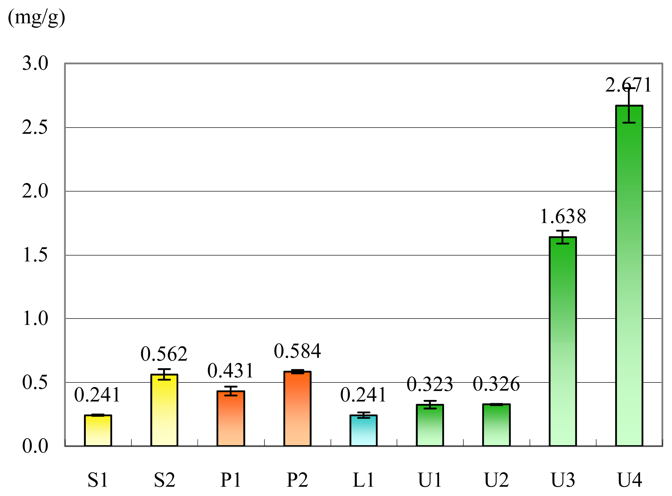
a) Content of FUCOX in the brown algae cultivated in DSW

The isolation yield of FUCOX (1) described above does not reflect the actual content of 1 in S. lomentaria because of the loss sustained during the prolonged isolation procedure. For the purpose of determining the exact content of 1, the methanol (MeOH) extract of S. lomentaria was subjected to HPLC and UV analyses. The HPLC (Mightsil® RP-18, 5 μm, 250 × 20 mm; 450 nm; MeOH-H2O= 95:5) showed many peaks, a few of which were overlapped with the signal due to 1. The UV spectrum showed a maximum at 450 nm ascribable to 1. This band, however, was obscured by those of concomitant chlorophylls. Therefore, we devised a quick and simple pretreatment method for separating 1 in the following manner.
The algal body of S. lomentaria was cut with a razor into pieces, which were stored in MeOH (30 mL/1 g wet body) in the dark for two days in a stoppered 50 mL Erlenmeyer flask (Extraction for 4 and 5 days gave the same result.). A 1.0 mL aliquot of the supernatant was taken out and diluted with distilled water (1.0 mL), and the mixture was charged on the top of Sep-Pak® Vac C18 (Waters, 1 cc), which had been washed with MeOH (2 mL) and then MeOH-H2O (1:1) (2 mL). The column was eluted successively with MeOH-H2O (1:1) (2 mL), MeOH-H2O (8:2) (2 mL), and MeOH-H2O (9:1) (4 mL), affording fractions 1, 2, and 3, respectively. TLC of fraction 3 shows one spot with bright orange color having the Rf value identical with that of authentic FUCOX (1). The HPLC (Mightsil® RP-18 GP, 3 μm, 250 × 4.6 mm; 450 nm; MeOH) also showed a single peak due to 1. The eluate (fraction 3) was diluted to 5.0 mL with MeOH-H2O (9:1), and the solution was analyzed by UV spectroscopy. The E value (1%, 1 cm) of 1197 at 451 nm determined for authentic FUCOX in MeOH-H2O (9:1) was used to calculate the amount of 1. The same experiment was performed three times, and the mean value was expressed as the weight of 1 [mg/g (wet algal body)]. In the case of S. lomentaria, the content of 1 was analyzed to be 0.241±0.005 mg/g. This value did not change significantly when other parts of the algal body were selected.
Figure 1 summarizes the amount of FUCOX, which was determined by this method, in 4 species of brown algae (Scytosiphon lomentaria, Petalonia binghamiae, Laminaria religiosa, and Undaria pinnatifida) cultivated in DSW.
It can be seen that the content of FUCOX in the young thallus (5–10 cm long) algae (S1, L1, P1, and U1 in Fig. 1) is in the range 0.24 – 0.43 mg/g and is not so different among the species. It is interesting that the content found in commercial dried U. pinnatifida (U2) is the same as that in the raw material (U1). This indicates that FUCOX is quite stable when mixed with the organic ingredients and survives the drying process and storage at ambient temperature, while pure FUCOX is susceptible to air oxidation. It is remarkable that the younger stages of the algae contain a higher content of FUCOX, S2/S1 = 2.3, P2/P1 = 1.35. Furthermore, examination of different stages in the life history of U. pinnatifida was indicative of different FUCOX contents. The extremely high content of FUCOX in the gametophytes of U. pinnatifida should be noted U3/U1 = 5.1, U4/U1 = 8.3. The biological significance of these facts is unknown.
Although comparison of the present results with the content of FUCOX in wild brown algae is needed and is in progress, it is important to point out that cultivation of the algae in DSW has the following merits in addition to the advantages described in the Introduction: (1) The algae can be harvested irrespective of the seasons. (2) Unialgal culture can be achieved in mass amounts.

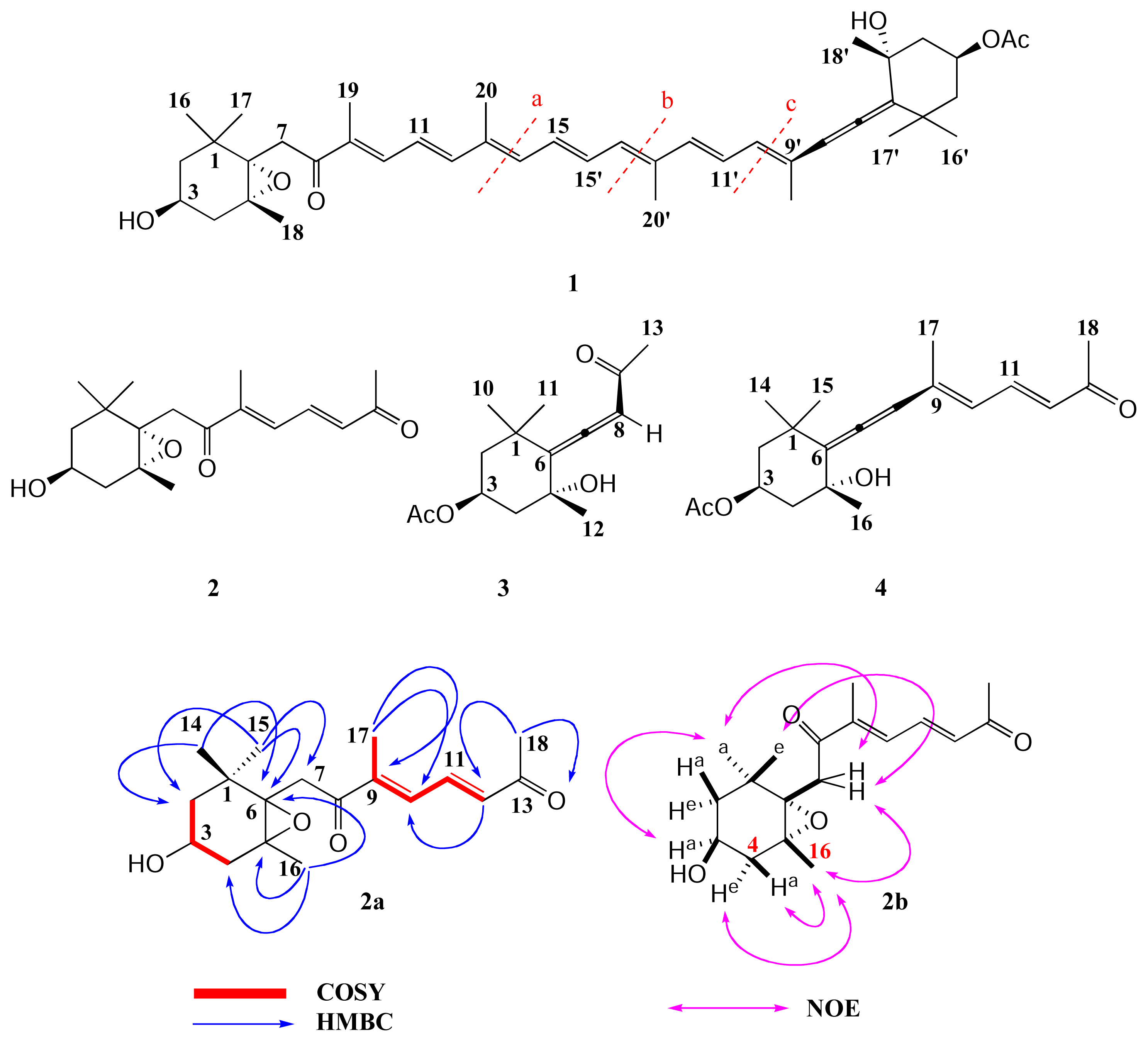
b) Structures of compounds 2, 3, and 4

Compounds 3 and 4 were identified as apo-9′-fucoxanthinone [7] and apo-13′-fucoxanthinone [8], respectively, by comparing their 1H- and 13C-NMR properties with those reported in the literature. The deacetyl derivative of compound 3 was first isolated as an allelopathic substance from a grasshopper [9] and is reported to exhibit cytotoxicity against murine lymphoma L-1210 and human epidoemoid carcinoma KB cells [7]. Compound 4 has been obtained from marine diatoms, and it shows a feeding deterrent effect in a copepod [8].
Compound 2, a new compound, gave a molecular ion at m/z 306.1813 indicating the molecular formula of C18H26O4. The 1H-NMR spectrum in C6D6 revealed the signals of five singlet methyls at δ 0.96 (H-15), 1.10 (H-14), 1.20 (H-16), 1.69 (H-17), and 1.88 (H-18), and three olefinic protons at 6.02 (d, 15.4 Hz, H-12), 6.66 (d, 11.1 Hz H-10), and 7.16 (dd, J=11.1, 15.4 Hz, H-11). The 1H-NMR spectrum also showed signals of three pairs of methylene protons at δ 1.36 (dd, J=9.1, 11.6 Hz, H-2) and 1.41 (ddd, J=1.8, 4.8, 11.6 Hz, H-2), 1.69 (dd, J=9.1, 13.6 Hz, H-4), and 2.24 (ddd, J=1.8, 4.8, 13.6 Hz, H-4), 2.56 (d, J=18.8 Hz, H-7) and 3.39 (d, J=18.8 Hz, H-7). From the triplet pattern of the methine signal at δ 3.81 (tt, J=4.8, 9.1 Hz, H-3), it was deduced that the oxymethine proton was axial, and, the orientation of the protons at 1.36 and 1.69 is axial, and that at 1.41 and 2.24 is equatorial. The equatorial protons at δ 1.41 (H-2) and 2.24 (H-4) are long-range coupled (W-coupling) with J=1.8 Hz. The 13C-NMR spectrum showed the signals of four olefinic carbons at δ 134.3 (C-10), 135.5 (C-12), 136.0 (C-11), and 142.9 (C-9), three oxygenated carbons at 63.9 (C-3), 65.8 (C-5), and 66.4 (C-6), and two carbonyl carbons at 195.8 (C-13) and 197.7 (C-8). The 1H-1H COSY and HSQC experiments allowed the complete assignment of the protons and carbons (Experimental). The proton networks of the compound were deduced by the 1H-1H COSY spectrum. The HMBC spectrum (see 2a) allowed the complete connectivity of the carbons, which revealed the planar structure of 2. In the NOESY spectrum, the cross-peaks depicted in 2b, the intensity of the NOE cross peaks between H-16 and H-4ax, and H-16 and H-4eq were almost the same, which allowed assignment of the relative stereochemistry of 2. If the epoxy oxygen takes the β-orientation, as the molecular models reveal, H-16 and H-4e are much closer than H-16 and H-4a, which would result in a more intense NOE peak of H-16/H-4e than that of H-16/H-4a. Compounds 2, 3, and 4 may be the metabolites of FUCOX (1) resulting from bond cleavage at the dotted lines a, c, and b shown in 1, respectively. Their absolute configurations, therefore, were deduced as depicted in the respective structures. The biological activity of 2 is now being examined.
Marinedrugs 02 00063f3

Conclusion

The content of fucoxanthin (1), a pharmacologically active carotenoid, in the edible brown algae, Scytosiphon lomentaria, Petalonia binghamiae, Laminaria religiosa, and Undaria pinnatifida, which are cultivated in deep seawater, has been determined by use of a newly developed pretreatment and UV analysis. It has been demonstrated that the content of 1 is remarkably higher in the younger stages of the algae than in the later stage. During the isolation procedure of 1 from S. lomentaria, three compounds, 2, 3, and 4 have been obtained. Compound 2 is a new compound, and its structure was elucidated by means of NMR spectroscopy.

Experimental

General remarks

1H- and 13C-NMR spectra were recorded on JEOL AL-400 (1H at 400 and 13C at 100 MHz, respectively) and Bruker AVANCE-400 (1H at 400 and 13C at 100 MHz, respectively) spectrometers. IR spectra were measured on a PERKIN-ELMER 1720 FT-IR spectrophotometer. LR-MS spectra were taken on a JEOL JMX-DX 303 spectrometer. HR-MS spectra were taken under electron impact (EI) conditions using a JEOL JMS-SX102A spectrometer by direct inlet. Optical rotations were determined for solutions in ethanol on a JASCO DIP-370 polarimeter. CD spectrum was measured on a JASCO J-600 spectrophotometer. For column chromatography, Kieselgel 60 (Merck) and Cosmosil® (Nakarai tesk) were used. For preparative TLC, Kieselgel 60 F254, 0.5 mm and 1.0 mm (Merck) were used. Preparative HPLC was performed on a JAI LC-908 and TOSOH SD-8023 instruments with Lichrosorb® Si 60 columns and a Mightsil® C-18 column. UV-VIS spectra were measured on a Beckman DU-650 spectrophotometer.

Mass culture and harvest of Scytosiphon lomentaria

Scytosiphon lomentaria was collected at Aikappu in Akkeshi, Hokkaido on 1 July 1989 by Dr. Kogame, Hokkaido University. The prostrate-branched filaments were kept as unialgal culture at the laboratory of Kochi Prefectural Deep Seawater Laboratory. In order to induce the erect thalli, the filaments were cultured as free-floating form in glass vessels containing 500 mL of PES medium with continuous aeration at 10 °C in short day length (10:14 h Light:Dark cycle) with white fluorescent light of 40–60 μmol m−2s−1. Several erect thallus germlings occurred in a filament and became ‘germling clusters’, each of which contained 10–100 germlings. When the germling clusters grew to more than 5 mm diameter, they were transplanted as free-floating forms to an outdoor tank and cultured with continuous aeration and deep seawater supply at a rate of 3 volume exchanges per day.
Other brown algae were cultivated essentially in the same manner.

Isolation of fucoxanthin (1) and compounds 2, 3, and 4 from S. lomentaria

The brown alga Scytosiphon lomentaria (Japanese name: Kayamonori; 15 kg wet weight) was obtained as unialgal culture at the Laboratory of Muroto DSW, Kochi Prefecture, Japan, in 2002. The whole plant was immediately soaked in MeOH and extracted at rt for 4 d. The MeOH extract was concentrated under a reduced pressure to give a residue. The residue was treated with H2O. The aqueous suspension was partitioned with hexane, dichloromethane (DCM), and ethyl acetate (EtOAc). A part of the DCM-soluble portion (1.6 g) was subjected to flash column chromatography (FCC) eluting with a mixed solvent (CHCl3-acetone-MeOH 100:10:1)-EtOAc (7:3) to give five fractions (Frs.1-1~1-5). Fraction 1-3 was applied to preparative TLC with 5% MeOH in hexane-EtOAc (1:1) to give five fractions (Frs.2-1~2-5). Fraction 2-3 was purified by preparative HPLC [5% MeOH in hexane-EtOAc (1:1)] to afford a new terpenoid (2, 2.5 mg). Fraction 1-4 was purified by preparative TLC with CHCl3-EtOAc (7:3) to afford fucoxanthin (1, 3.2 mg). Other part of the DCM-soluble portion (2.4 g) was subjected to FCC eluting with a mixed solvent (CHCl3-acetone-MeOH 100:10:1)-EtOAc (7:3) to give seven fractions (Frs.3-1~3-7). Fraction 3-4 was subjected to FCC eluting with hexane-EtOAc (1 : 1) to give seven fractions (Frs.4-1~4-7). Fraction 4-4 was purified by recycle HPLC [hexane-EtOAc (1:1)] to afford apo-9’-fucoxanthinone (3, 3.1 mg) and apo-13’-fucoxanthinone (4, 0.6 mg). Fraction 4-5 was purified by recycle HPLC [hexane-EtOAc (1:1)] to afford apo-9’-fucoxanthinone (3, 1.1 mg). Fraction 3-5 (930.2 mg) mainly consisting of fucoxanthin (1) was obtained. Fraction 4-6 was subjected to FCC eluting with 3% MeOH in hexane-EtOAc (1:1) to give seven fractions (Frs.5-1~5-7). Fraction 5-4 was applied to Sep-Pak® eluting with MeOH-distilled water (9:1) to give two fractions (Frs.6-1~2). Fraction 6-2 was purified by HPLC with MeOH-distilled water (95:5) to afford 1 (4.0 mg).

Fucoxanthin (1)

Red paste. λmax (EtOH): 449.5 [E (1%, 1 cm) 1174.1], 1H-NMR: (CDCl3, 400 MHz): δ 0.95 (s, Me-17), 1.02 (s, Me-16), 1.06 (s, Me-17’), 1.21 (s, H-18), 1.34 (s, H-18’), 1.36 (dd, J=8.7, 14.2 Hz, H-2ax), 1.37 (s, Me-16’), 1.41 (dd, J=10.4, 14.9 Hz, H-2’ax), 1.49 (dd, J=2.9, 14.2 Hz, H-2eq), 1.53 (dd, J=10.4, 14.9 Hz, H-4’ax), 1.77 (dd, J=8.7, 14.2 Hz, H-4ax), 1.80 (s, H-19’), 1.93 (s, H-19), 1.98 (s, H-20), 1.98 (s, H-20’), 2.00 (dd, J=2.9, 14.9 Hz, H-2’eq), 2.03 (s, Me, C-3’OAc), 2.29 (dd, J=2.9, 17.8 Hz, H-4’eq), 2.29 (dd, J=2.9, 17.8 Hz, H-4eq), 2.59 (d, J=20.4 Hz, H-7), 3.64 (d, J=20.4 Hz, H-7), 3.80 (m, H-3), 5.37 (tt, J=8.8, 12.0 Hz, H-3’), 6.04 (s, H-8’), 6.12 (d, J=11.6 Hz, H-10’), 6.26 (d, J=11.6 Hz, H-14’), 6.34 (d, J=11.6 Hz, H-12’), 6.40 (d, J=11.6 Hz, H-14), 6.58 (m, H-11), 6.66 (t, J=12.8 Hz, H-12), 6.67 (m, H-15), 6.71 (dd, J=12.0, 14.2 Hz, H-15’), 6.71 (t, J=12.0 Hz, H-11’), 7.14 (d, J=12.8 Hz, H-10). 13C-NMR: (CDCl3, 100 MHz): δ 11.7 (C-19), 12.6 (C-20), 12.80 (C-20’), 13.9 (C-19’), 21.0 (C-18), 21.3 (Me, C-3’OAc), 24.9 (C-16), 28.0 (C-17), 29.0 (C-16’), 31.1 (C-18’), 31.9 (C-17’), 35.0 (C-1’), 35.6 (C-1), 40.6 (C-7), 41.5 (C-4), 45.1 (C-4’), 45.2 (C-2’), 46.9 (C-2), 64.2 (C-3), 66.0 (C-5), 66.9 (C-6), 67.8 (C-3’), 72.6 (C-5’), 103.2 (C-8’), 117.3 (C-6’), 123.2 (C-11), 125.5 (C-11’), 128.4 (C-10’), 129.3 (C-15), 132.0 (C-14’), 132.4 (C-9’), 132.4 (C-15’), 134.3 (C-9), 135.3 (C-13), 136.6 (C-14), 137.0 (C-12’), 138.0 (C-13’), 139.0 (C-10), 144.9 (C-12), 170.4 (C-8), 197.7 (C=O, C-3’OAc), 202.2 (C-7’).

Compound 2

Yellow oil. [α]D +34.1° (c = 0.02, ethanol). IR 1669 cm−1 (-C=O), EIHRMS m/z 306.1813 (calcd for C18H26O4; 306.1831). 1H NMR: (C6D6, 400 MHz): δ 0.96 (s, H-15), 1.10 (s, H-14), 1.20 (s, Me-16), 1.36 (dd, J=9.1, 11.6 Hz, H-2ax), 1.41 (ddd, J=1.8, 4.8, 11.6 Hz, H-2eq), 1.69 (dd, J=9.1, 13.6 Hz, H-4ax), 1.69 (s, Me-17), 1.88 (s, H-18), 2.24 (ddd, J=1.8, 4.8, 13.6 Hz, H-4eq), 2.56 (d, J=18.8 Hz, H-7), 3.39 (d, J=18.8 Hz, H-7), 3.81 (tt, J=4.8, 9.1 Hz, H-3), 6.02 (d, 15.4 Hz, H-12), 6.66 (d, J=11.1 Hz H-10), 7.16 (dd, J=11.1, 15.4 Hz, H-11). 13C NMR: (C6D6, 100 MHz): δ ı ∼ ı (C-17), 21.1 (C-16), 25.1 (C-14), 27.7 (C-18), 28.2 (C-15), 35.2 (C-1), 41.7 (C-7), 42.1 (C-4), 47.7 (C-2), 63.9 (C-3), 65.8 (C-5), 66.4 (C-6), 134.3 (C-10), 135.5 (C-12), 136.0 (C-11), 142.9 (C-9), 195.8 (C-13), 197.7 (C-8).

Apo-9’-fucoxanthinone (3)

Orange oil. CD spectrum (EtOH): Δɛ 255 −6.07. 1H-NMR: (C6D6, 400 MHz): δ 0.94 (s, H-10), 1.01 (s, Me-12), 1.25 (ddd, J=2.0, 4.3, 12.4 Hz, H-2eq), 1.26 (dd, J=11.2, 12.4 Hz, H-4ax), 1.32 (s, Me-11), 1.76 (s, H-13), 1.91 (s, Me, C-3OAc), 1.95 (ddd, J=2.0, 4.3, 12.4 Hz, H-4eq), 2.21 (dd, J=11.2, 12.4 Hz, H-2ax), 5.59 (tt, J=4.3, 11.2 Hz, H-3), 5.81 (s, H-8). 13C-NMR: (C6D6, 100 MHz): δ 20.9 (Me, C-3OAc), 26.2 (C-11), 26.3 (C-13), 28.8 (C-12), 30.3 (C-10), 35.8 (C-1), 45.2 (C-4), 45.3 (C-2), 67.4 (C-3), 71.4 (C-5), 100.8 (C-8), 118.2 (C-6), 169.4 (C=O, C-3OAc), 196.0 (C-9), 209.0 (C-7).

Apo-13’-fucoxanthinone (4)

Yellow paste. 1H-NMR: δ (CD2Cl2, 400 MHz): 1.10 (s, Me-14), 1.36 (s, Me-15), 1.40 (s, H-16), 1.41 (dd, J=8.0, 10.0 Hz, H-2ax), 1.52 (dd, J=8.0, 10.0 Hz, H-4ax), 1.93 (s, H-17), 1.98 (ddd, J=1.9, 5.0, 10.0 Hz, H-2eq), 2.03 (s, Me, C-3OAc), 2.27 (s, H-18), 2.28 (ddd, J=1.9, 5.0, 10.0 Hz, H-4eq), 5.31 (m, H-3), δ 6.12 (s, H-8), 6.15 (d, J=15.6 Hz, H-10), 6.21 (d, J=10.5 Hz, H-12), 7.47 (dd, J=10.5, 15.6 Hz, H-11).
Figure 1. Content of fucoxanthin (1) in Scytosiphon lomentaria (S), Petalonia binghamiae (P), Laminaria religiosa (L), and Undaria pinnatifida (U). S1: young thallus (5–10 cm long) and S2: germlings of S. lomentaria. P1: young thallus (5–10 cm long) and P2: germlings of P. binghamiae. L1: young thallus (5–10 cm long) of L. religiosa. U1: young thallus (5–10 cm long), U2: commercial dried body (treated with freshwater), U3: female gametophyte, and U4: male gametophyte of U. pinnatifida. The mean values (n = 3 except for L1 and U2 [n = 6]).
Marinedrugs 02 00063f1

Acknowledgement

The authors are grateful for the following financial support: (1) A research grant from Faculty of Pharmaceutical Sciences, The University of Tokushima. (2) Ministry of Education, Science, Sports and Culture, Grants-in-Aid for Scientific Research on Priority Area (A)(2), 2001–2004, 12045250. (3) Scientific Research (A), 2003-, 14207096-00. (4) Grant of Entrustment, Kochi Prefecture.
  • Sample Availability: Samples are available from the authors.

References

  1. Hiraoka, M.; Ohno, M.; Dan, A.; Oka, N. Utilization of deep seawater for the mariculture of seaweeds in the Japan. The Japanese Journal of Phycology (Supplement). in press.
  2. Englert, G.; Bjoernland, T.; Liaaen-Jensen, S. 1D and 2D NMR study of some allenic carotenoids of the fucoxanthin series. Magn. Res Chem 1990, 28, 519–528. [Google Scholar]
  3. Miki, W. Biological functions and activities of animal carotenoids. Pure Appl. Chem 1991, 63, 141–146. [Google Scholar]
  4. Nomura, T.; Kikuchi, M.; Kubodera, A.; Kawakami, Y. Proton-donative antioxidant activity of fucoxanthin with 1, 1-diphenyl-2-picrylhydrazyl (DPPH). Biochem. Mol. Biol. Int 1997, 42, 361–370. [Google Scholar]Yan, X.; Chuda, Y.; Suzuki, M.; Nagata, T. Fucoxanthin as the major antioxidant in Hijikia fusiformis, a common edible seaweed. Biosci. Biotechnol. Biochem 1999, 63, 605–607. [Google Scholar]
  5. Kim, J. M.; Araki, S.; Kim, D. J.; Park, C. B.; Takasuka, N.; Baba-Toriyama, H.; Ota, T.; Nir, Z.; Khachik, F.; Shimidzu, N.; Tanaka, Y.; Osawa, T.; Uraji, T.; Murakoshi, M.; Nishino, H.; Tsuda, H. Chemopreventive effects of carotenoids and curcumins on mouse colon carcinogenesis after 1, 2-dimethylhydrazine initiation. Carcinogenesis 1998, 19, 81–85. [Google Scholar]
  6. Hosokawa, M. Fucoxanthin induces apoptosis in cancer cells. Bio Industry 2004, 1, 52–57. [Google Scholar]
  7. Doi, Y.; Ishibashi, M.; Yamaguchi, N.; Kobayashi, J. Isolation of apo-9′-fucoxanthinone from the cultured marine dinoflagellate Amphidinium sp. J. Nat. Prod 1995, 58, 1097–1099. [Google Scholar]
  8. Shaw, B. A.; Andersen, R. J.; Harrison, P. J. Feeding deterrence properties of apo-fucoxanthinoids from marine diatoms. I. Chemical structures of apo-fucoxanthinoids produced by Phaeodactylum tricornutum. Mar. Biol 1995, 124, 467–472. [Google Scholar]
  9. Meinwald, J.; Erickson, K.; Hartshorn, M.; Mainwald, C.Y.; Eisner, T. Defensive mechanisms of arthropods. XXIII. An allenic sesquiterpenoid from the grasshopper Romalea microptera. Tetrahedron Lett 1968, 2959–2962. [Google Scholar]

