Article

1-O-Sulfatobastadins-1 and -2 from Ianthella basta (Pallas).Antagonists of the yR1-FKBP12 Ca2+ Channel

by
Makoto N. Masuno
1,
Alexander C. Hoepker
1,
Isaac N. Pessah
2 and
Tadeusz F. Molinski
1,*
1
Department of Chemistry, University of California, Davis, CA 95616, USA
2
Department of Molecular Biosciences, University of California, Davis, CA 95616, USA
*
Author to whom correspondence should be addressed.
Mar. Drugs 2004, 2(4), 176-184; https://doi.org/10.3390/md204176
Submission received: 29 July 2004 / Accepted: 14 October 2004 / Published: 25 November 2004

Abstract

:
Two new sulfate monoesters of hemibastadins-1 and -2 were isolated from the marine sponge Ianthella basta (Pallas) from Guam. A third new compound was tentatively assigned the structure 34-O-sulfatobastadin-9. The 1-O-sulfatohemibastadins-1 and –2 were antagonists of the RyR1-FKBP12 Ca2+ channel under conditions where the known compound bastadin-5 exhibits potent agonism (EC50 2 μM).

Introduction

The release of Ca2+ from stores of the sarcoplasmic reticulum (SR) stimulates contraction of striated muscle fibers through ATP hydrolysis coupled to induced conformational changes of the actin-myosin protein complex. In 1994, we discovered that the known bromotyrosine tetramer, bastadin-5 (1)[1] from the marine sponge Ianthella basta (Pallas), is a potent modulator of Ca2+ release from the SR[2]. Bastadin-5 stimulates release of Ca2+ from the SR by binding to the RyR1-FKBP12 Ca2+ channel, a tetrameric heterodimeric channel protein (~2000 kDa) that is associated with the smaller 12 kDa immunophilin FKBP12 [3]. The mechanism of action of 1 is not fully understood, but it is known that binding occurs at a site on the SR junctional protein that is distinct from those of known effector molecules, including ATP and caffeine [2]. Bastadin-5 and -10 [4] also have been shown to relate ryanodine-sensitive and -insensitive Ca2+ efflux pathways in skeletal SR and BC3H1 cells [5]. Recently, we took the opportunity to examine the most polar fractions from extracts of the sponge Ianthella basta, collected in Guam, and found a complex mixture of bastadin O-sulfate esters from which we have isolated the new compounds 1-O-sulfatohemibastadin-1 (2) and 1-O-sulfatohemibastadin-2 (3), along with the known compound 32-O-sulfatobastadin-13 (4) [11] and a third new compound, the bastadin sulfate ester 34-O-sulfatobastadin-9 (5), The structures of the new compounds were established by interpretation of their spectral data and comparison with the parent phenols [6]. To our surprise, 2 and 3 exhibited antagonistic activity toward the RyR-1/FKBP12 complex (IC50 13 and 29 μM, respectively). This is the first report of antagonism of the SR channel by a bastadin analog, and suggests a bimodal mechanism of action upon a common, but as-yet unidentified, effector site of the RyR1-FKBP12 channel complex.
Marinedrugs 02 00176f2

Results and Discussion

Samples of freshly collected Ianthella basta (Pallas) were directly extracted in solvent (1:1 CH2Cl2-MeOH) and the concentrated extracts purified by silica flash chromatography using a gradient of MeOH in CH2Cl2. Pooled extracts that eluted with 3:1 CH2Cl2-MeOH were further purified by HPLC (reversed phase, C18, MeOH-H2O followed by C18, 70:30:0.05 H2O-CH3CN-TFA) to provide two new sulfated hemibastadins, 2 and 3, the known 4[11] and the novel 34-O-sulfatobastadin-9 (5). All compounds were readily soluble in MeOH and appreciably soluble in water, but insoluble in CHCl3.
Detailed MS analysis was used to secure compositions of the new compounds. For the purpose of clarity in the description that follows M is defined as the neutral acid, but the structures are depicted, here, as the Na+ salts of the sulfate half-esters. MALDI HRMS of 2 (m/z 594.8815 [M-H+2Na]+ Δ mmu +6.0) showed the presence of two Br atoms and secured the formula of the neutral compound as C17H16Br2N2O7S, which suggested a structure of almost half the molecular mass of the macrocyclic bastadins (e.g. 1[7]). Fragment ions in the ESI mass spectrum of 2 due to loss of SO3 implicated the presence of O-sulfate esters. The 1H- and 13C-NMR spectra of 2, although displaying similar chemical shifts and couplings as those of bastadin-5 [8], contained about half of the expected signals of the macrodilactams (Table 1). Lack of mirror symmetry in the NMR spectra, together with signals that accounted for a 3-bromo-4-tyramine and oxime-modified 3-bromotyrosine units suggested a ‘hemibastadin’ similar to the compounds from another sample of I. basta described by Capon and coworkers [6a]. The configuration of the ketoxime group in 2 was E, as is usually found in this series of compounds [9]. 1H-13C correlations observed in the HSQC and HMBC spectra of 2 were fully compatible with the proposed structure, including the position of the O-SO3H group which was assigned as follows. The 13C-NMR signals of phenols undergo well-described local changes in δ upon O-sulfation. Ragan has documented that the ipso carbon of a phenol undergoes a diamagnetic shift (Δδ −2.8~5.0 ppm upfield) while the ortho carbon signals suffer downfield shifts (Δδ +6.3–7.8 ppm) upon substitution of OH for O(SO3H) or the corresponding alkali metal salt O(SO3M)[10]. For example, changes in 13C shifts were used to place the O-sulfate esters in 34-O-sulfatobastadin-1 [11], 15,34-O-di-sulfatobastadin-7 and 4[8].
Critical 13C-NMR assignments that distinguished the signals of the two aryl rings were made on the basis of HMBC and nOe experiments (Figure 1). Because the substitution pattern in each trisubstituted phenyl ring of 2 was the same, we could simply compare the assigned 13C-NMR signals in each ring to determine which phenoxyl group was esterified as a sulfate ester.
The O-substituted para-carbon of the bromotyramine ring in 2 showed an upfield shift of the C1 13C signal in 2 (δ 149.7, s), with respect to the corresponding signal in the bromotyrosine ring at C1’ (δ 153.8 s) [12]. Conversely, the ortho13C-NMR signals of C2 (δ 116.7, s) and C6 (δ 123.4, d) were displaced in the paramagnetic direction (c.f. δ 110.5, s, C2’; 117.1, d, C6’). Thus, the O(SO3H) group is located at C1 of the 3-bromotyramine ring.
Obtaining reliable positive ion ESI and MALDI mass spectra of 3 was made difficult by the tendency of the compound to form dimers and trimers under standard conditions. Negative ion ESIMS, which gave better results with 3 and other bastadins, revealed discrete ions due to [M-H] (m/z 627) and [M-2H+Na] (m/z 649) and a 1:3:3:1 isotope pattern indicating three Br atoms. Eventually, positive ion MALDI HRMS provided a reliable pseudomolecular ion ([M–H+2Na]+m/z 672.7899, Δ+1.8 mmu) corresponding to the formula M of 3, C17H15Br3N2O7S. 1H-NMR showed replacement of one set of aryl ring signals with a two-proton singlet (δ 7.38, s, 2H) and simplification of the 13C-NMR spins to four signals indicating a local mirror plane of symmetry. Thus, compound 3 is a sulfate ester of hemibastadin-2 [6]. The O(SO3H) group was again located at C1 employing arguments similar to those used for 2.
The ESI mass spectrum of 5 showed an isotope pattern corresponding to the presence of four Br atoms and loss of SO3 that indicated the presence of a sulfate half ester. The formula C34H27Br4N4O11SNa2 for compound 5, (MALDI HRMS, m/z 1060.7840 [M–H+2Na]+ Δmmu = −8.6) was isomeric with the known sodium salt of 34-O-sulfatobastadin-13 (6) [11]. The 1H-NMR spectrum of 5 (d6-DMSO) revealed spin systems corresponding to four benzene rings; two trisubstituted, one tetrasubstituted and a 3,5-dibromo-4-phenoxyl ring (phenol numbering, δ 7.55, s, 2H, H28,30) that was identified from the presence of a local plane of symmetry (C1,4 axis). Exchangeable proton signals were observed for the two oxime groups (δ 11.87, s; 11.71, s), however, only one phenolic OH was evident (δ 9.30, s) which supported a phenolic O-sulfate ester. Consideration of the four possible parent bastadin skeleta that are isomeric with the parent of 5 (bastadin-9 [4], bastadin-13 [6], bastadin-18 [13] and bastadin-20 [8] allowed us to eliminate the latter two structures based on their lack of a symmetrically substituted 3,5-dibromobenzene ring. 34-O-Sulfatobastadin-13 (6) has been characterized by Wright and co-workers [11], however the 1H-NMR data for this compound differed considerably from those of 5 (both recorded in d6-DMSO). For example, the isochronous proton pair H27/31 in 5 resonated at lower field (δ 7.55, 2H) than 6 (δ 7.42, s, 2H [11]) and the exceptionally high field aryl doublet in 5 (δ 6.20, d, J= 2.0 Hz) is not found in 6 (the closest signal is H-8, δ 6.50, d, J= 2.0 Hz). The exclusion of 6 leaves only three undescribed isomers – 10-O-sulfatobastadin-13, 10-O-sulfatobastadin-9 and 34-O-sulfato- bastadin-9 – as remaining possibilities. The former two are eliminated because compound 5 lacks the downfield shifted 1H-NMR signal expected for an H-11 or H-16 proton ortho to an O-sulfate ester (Δδ~ −0.7 ppm, c.f. 2). Thus, we tentatively assigned the structure 34-O-sulfatobastadin-9 to 5. Unfortunately, limited sample precluded acquisition of a complete 13C-NMR spectrum, however, a partial set of HMBC correlations observed for 5 were consistent with the proposed structure.
Marinedrugs 02 00176f3
Compounds 1–3 were assayed for binding of [3H]-ryanodine to the high-affinity site of the ryanodine receptor in the RyR1-FKBP12 complex [15, 2]. Ryanodine binding is an indicator of the ‘open state’ of the SR Ca2+ channel. Bastadin-5 (1) showed the expected stimulation of ryanodine binding to the channel in the open state (EC50 2 μM) [2], however, both compounds 2 and 3 were antagonists and inhibited binding of [3H]-ryanodine (IC50 13 μM and 29 μM, respectively). This contrasts with 15,34-O-di-sulfatobastadin-7 and 4 which are weak agonists of the receptor (EC50 13.6 μM and 100 μM, respectively) [8a]. Given the similarity of the structures of 2 and 3 and their resemblance to a ‘truncated’ northern hemisphere of 1, it is likely that all three compounds interact with the same site, however, the mode of action with respect to Ca2+ channel opening are clearly distinct. Further investigations of these phenomena aimed at refinement of a model for bastadin-5-promoted Ca2+ release are underway in our laboratories.

Conclusions

Two new hemibastadin sulfate esters isolated from Ianthella basta were characterized. The compounds suppressed ryanodine binding with IC50’s 13 μM and 29 μM, respectively. This is the opposite effect of bastadin-5 (1), a potent agonist of Ca2+ release from the SR. Although 1–3 have structural features in common, and most likely a common binding motif, the mode of action suggests 2 and 3 inhibit channel opening. Thus, a broader structure-relationship emerges for bastadins that reveals a bimodal mechanism of action.

Experimental

General

NMR spectra were obtained using either Varian Inova 400 NMR or a Bruker DRX-600 spectrometer (equipped with a cryoprobe) operating at 399.771 MHz or 600.304 MHz for 1H and 100.531 MHz for 13C. Solvents used in extraction or chromatography were HPLC-grade or distilled from glass. ESIMS was carried out on a ThermoFinnigan Surveyor HPLC and LC Deca ion-trap by infusion in MeOH solution (0.1% HCOOH). HRMS measurements were provided by the University of California, Riverside Mass Spectrometry Facility. Other experimental procedures are described elsewhere [14].

Isolation of 1–5

The sponge Ianthella basta (Pallas) was collected by hand (2002) using scuba in Mangilao, Guam, and immediately steeped in CH2Cl2-MeOH. The organic extract was filtered and the filtrate concentrated to give a deep red gum. A portion of the extract (3.20 g) was dissolved in 1:1 CH2Cl2-MeOH and the solution combined silica (ca. 1g silica/100 mg) and concentrated to remove the solvent. The dry silica was slurried in CH2Cl2 and loaded onto the top of a silica column packed with CH2Cl2 and the column eluted with a gradient of MeOH in CH2Cl2. The fraction eluting with 3:1 CH2Cl2-MeOH was further separated by HPLC (reversed phase C18, Dynamax 10×250 mm, 60:40 H2O-MeOH, 3.0 mL/min) and the eluates monitored with a UV detector (λ 254 nm) to afford two fractions: ‘a’ (51.0 mg, tR = 5 min) and ‘b’ (111.7 mg, tR = 6.5 min). Purification of fraction ‘a’ on HPLC (RP C18, Dynamax 10 × 250 mm, 70:30:0.05 H2O-CH3CN-TFA, 4.0 mL/min) gave 1-O-sulfatohemibastadin-1 (2, 5.2 mg, tR = 6.5), 1-O-sulfatohemibastadin-2 (3, 7.3 mg, tR = 8 min), 5 (8.0 mg, tR =16 min), and 4[8] (23.4 mg, tR = 18 min).

Spectroscopic Data

1-O-Sulfatohemibastadin-1 (2): colorless solid; 1H-NMR and 13C-NMR (CD3OD): see Table 1; MALDI HRMS m/z 594.8815 [M-H+2Na]+; calcd. 594.8762 for C17H15 79Br2N2O7SNa2.
1-O-Sulfatohemibastadin-2 (3): colorless solid; UV (MeOH): λmax 204, 282 nm; IR (film) ν 3363 bs, 1678 s, 1211 s, 1145 cm−1; 1H-NMR and 13C-NMR (CD3OD): see Table 1; LR ESIMS (-ve ion) m/z 649 [M–2H+Na], 627 [M-H]; MALDI HRMS (+ve ion) m/z 672.7899 [M–H+2Na]+; calcd. 672.7862 for C17H14 79Br3N2O7SNa2.
34-O-Sulfatobastadin-9 (5): colorless solid; 1H-NMR (d6-DMSO) 11.87 (s, 1H, N-OH), 11.71 (s, 1H, N-OH), 9.33 (s, 1H, Ar-OH), 7.98 (t, J= 5.6 Hz, 1H, NH), 7.95 (t, J= 5.6 Hz, 1H, NH), 7.55 (s, 2H, H28/30), 7.49 (d, J=2.0 Hz, 1H), 7.11 (dd, J= 8.4, 2.0 Hz, 1H), 7.09 (d, J= 2.0 Hz, 1H), 6.84 (d, J= 8.4 Hz, 1H), 6.81 (dd, J= 8.4, 2.0 Hz, 1H), 6.75 (d, J=8.4 Hz, 1H), 6.71 (d, J= 2.0 Hz, 1H), 6.20 (d, J= 2.0 Hz, 1H), 3.58 (s, 2H), 3.53 (s, 2H), 3.41 (m, 2H), 3.21 (m, 2H), 2.66 (t, J= 7.2 Hz, 2H), 2.62 (t, J=7.2 Hz, 2H). MALDI HRMS m/z 1060.7840 [M–H+2Na]+; calcd. C34H27 79Br4N4O11SNa2 1060.7920.

[3H]-Ryanodine Binding Assay
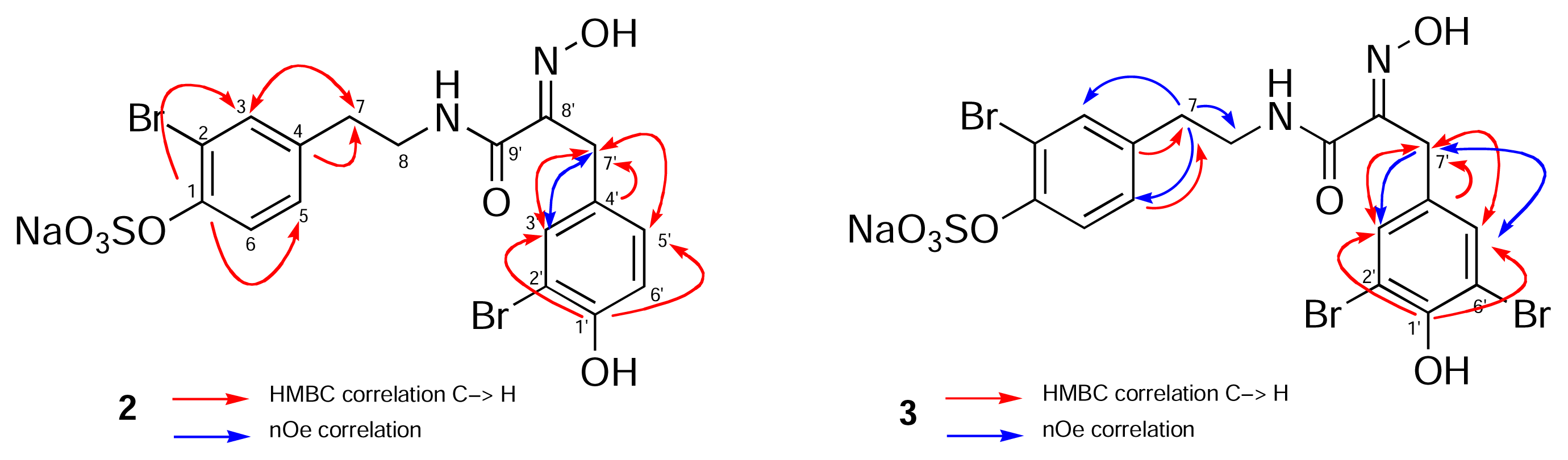
Specific binding of [3H]-ryanodine to high affinity sites on rabbit skeletal membrane vesicles [2,15] was determined by incubating SR protein (25 μg), containing the RyR1-FKBP12 complex, with [3H]ryanodine (1 nM) for 3.5 h at 37° C in binding assay buffer containing KCl (250 mM), NaCl (15mM), HEPES (20 mM), CaCl2 (20 μM) and at pH 7.4 (500 μL, final volume). The binding reaction was initiated by addition of the drug in DMSO (final DMSO conc. ~1%) to the complete assay medium and the incubation was terminated by filtration through Whatman GF/B glass fiber filters using a Brandel cell harvester (Gaithersburg, MD). Separation of bound and free [3H]-ryanodine was performed by washing the filters with ice-cold buffer (3 × 500 μL) containing Tris-HCl (20 mM), KCl (250 mM), NaCl (15 mM) at pH 7.4. Filters were placed in scintillation vials containing scintillant (5 mL). Bound radioactivity was measured by scintillation counting and corrected for background. Positive controls were bastadin-5 (EC50 2.0 μM) and PCB95 (2,2′,3,5′,6-pentachlorobiphenyl)[16] and nonspecific binding was determined in the presence of 100-fold unlabeled ryanodine.
Figure 1. Key HMBC (red arrows) and nOe correlations (blue arrows) for 2 and 3.
Marinedrugs 02 00176f1
Table 1. 1H-NMR (400 MHz) and 13C-NMR (100 MHz) data for 2 and 3a
#13C-NMR (2), δ1H-NMR (2) δ (mult., J Hz, Int.)HMBC13C-NMR (3) δ1H-NMR (3) δ (mult., J Hz, Int.)HMBC
1b149.7H3, H5, H6149.8
2116.7H3, H6116.7
3134.57.42 (d, J=2.0, 1H)H5, H7134.57.42 (d, J=2.0, 1H)-
4138.5H6, H7, H8138.5H7, H8
5129.77.09 (dd, J=8.4,2.0, 1H)H3, H7129.77.11 (dd, J=8.4,2.0, 1H)H7
6123.47.49 (d, J=8.4, 1H)123.57.50 (d, J=8.4, 1H)
735.42.75 (t, J=7.2, 2H)H3, H5, H835.42.76 (t, 2H, J=7.2)H8
841.73.43 (t, J=7.2, 2H)H741.83.43 (t, 2H, J=7.2)H7
1′b153.8H3′, H5′150.7H3′, H5′
2′110.5H3′, H6′112.0H3′
3′134.57.36 (d, J=2.0, 1H)H5′, H7′133.97.38 (s, 2H)H7′
4′130.6H6′, H7′132.3H7′
5′130.27.02 (dd, J=8.0, 2.0, 1H)H3′, H7′133.97.38 (s, 2H)H7′
6′117.16.76 (d, J=8.0, 1H)112.0H5′
7′28.63.78 (s, 2H)H3′, H5′28.43.78 (s, 1H)H3′, H5′
8′153.2H7′152.5H7′
9′165.8H7′, H8165.6H8, H7′
a1H-NMR chemical shifts recorded in CD3OD (99.5% atom D) are referenced to the residual solvent proton signal, δ 3.30 ppm. 1H- assignments are based on COSY, HSQC and chemical shift considerations;
bThe carbon chemical shifts of C1 and C1’ (–OSO3H vs.-OH) were assigned based on the expected upfield13C- shift of the ipso carbon upon sulfation of the OH (Ragan, [10]).

Acknowledgements

We thank Valerie Paul, University of Guam Marine Station (now at the Smithsonian Marine Station, Ft. Pierce), for I. basta extract and Rich Kondrat (UC Riverside Mass Spectrometry Facility) for HRMS results. We are grateful to John MacMillan and Jeff deRopp (UC Davis NMR Facility) for assistance with 600 MHz NMR measurements. Funding for the instruments was provided by NSF (Varian INOVA 400, CHE 9808183) and NIH (Bruker DRX-600, RR11973 and Thermo Finnigan LCMS, SIG S10 RR14701-01). ACH is grateful to the American Society of Pharmacognosy for an undergraduate research award. This work was supported by the National Institutes of Health (GM 57560).
  • Sample Availability: Contact authors.

References and Notes

  1. Kazlauskas, R.; Lidgard, R. O.; Murphy, P. T.; Wells, R. J.; Blount, J. F. Brominated Tyrosine-Derived Metabolites from the Sponge Ianthella basta. Aust. J. Chem 1981, 34, 765–786. [Google Scholar]
  2. Mack, M. M.; Molinski, T. F.; Buck, E. D.; Pessah, I. N. Novel Modulators of Skeletal Muscle FKBP12/Calcium Channel Complex from Ianthella basta: Role of FKBP in Channel Gating. J. Biol. Chem 1994, 269, 23236–23249. [Google Scholar]
  3. Clardy, J. The Chemistry of Signal Transduction. Proc. Nat. Acad. Sci. U.S.A 1995, 92, 56–61. [Google Scholar]
  4. Pordesimo, E. O.; Schmitz, F. J. New Bastadins from the Sponge Ianthella basta. J. Org. Chem 1990, 55, 4704–4709. [Google Scholar]
  5. Pessah, I. N.; Molinski, T. F.; Meloy, T. D.; Wong, P.; Buck, E. D.; Allen, P. D.; Mohr, F. C.; Mack, M. M. Bastadins Relate Ryanodine-Sensitive and -Insensitive Ca2+ Efflux Pathways in Skeletal SR and BC3H1 Cells. Am. J. Physiol 1997, 41, C601–C614. [Google Scholar]
  6. Butler, M. S.; Lim, T. K.; Capon, R. J.; Hammond, L. S. The Bastadins Revisited: New Chemistry from the Australian Marine Sponge Ianthella basta. J. Nat. Prod 1991, 44, 287–296. [Google Scholar]Pettit, G. R.; Butler, M. S.; William, M. D.; Filiatrault, M. J.; Pettit, R. K. Isolation and Structure of Hemibastadinols 1–3 from the Papua New Guinea Marine Sponge Ianthella basta. J. Nat. Prod 1996, 59, 927–934. [Google Scholar]
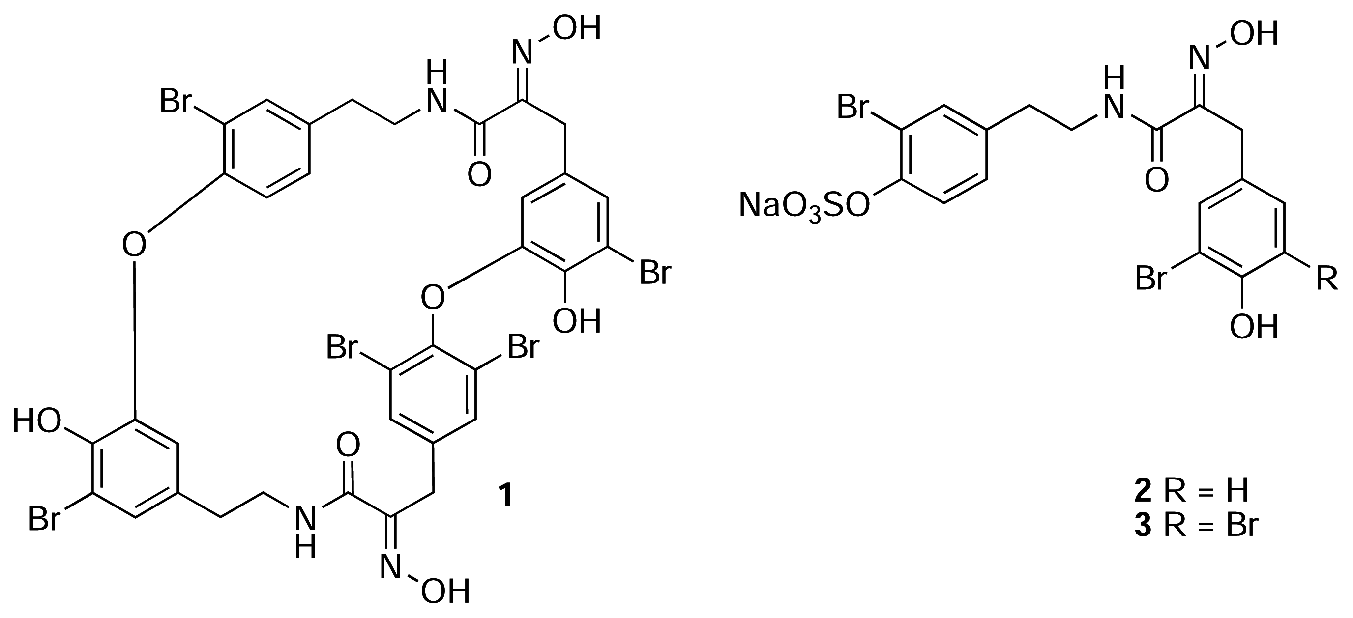
  7. For uniformity, we present the structures of all new compounds as the Na+salts of the sulfate-half esters as Na+is the most likely counter ion from extraction of marine invertebrates. The numbering scheme used here for 2 and 3 is adopted from Pettit (reference 6b) and differs from the numbering of the macrocyclic bastadins (eg. 1 reference [1]).
  8. Franklin, M. A.; Penn, S. G.; Lebrilla, C. B.; Lam, T. H.; Pessah, I. N.; Molinski, T. F. Bastadin 20 and Bastadin O-Sulfate Esters from Ianthella basta: Novel Modulators of the Ry(1)R FKBP12 Receptor Complex. J. Nat. Prod 1996, 59, 1121–1127. [Google Scholar]Franklin, M. A. M.Sc. Thesis, University of California, Davis, 1995.
  9. Unlike simple acyclic oximes which exhibit facile E-Z isomerism, the configuration of the C=N double bond in every cyclic bastadin is strictly E. A strong intramolecular H-bond between the amide NH and the N=C groups forms a rigid, planar 5-membered ring and places the OH group anti to the H-bonded donor electron pair on the imine nitrogen (viz. X-ray structures of bastadin-4 [1] and bastadin-10, Molinski, T. F.; Olmstead, M. M. unpublished results.). Both E/Z oxime isomers have been detected in related α-ketoximines from marine sponges and theE geometry has been correlated with an upfield 13C-NMR signal for the benzylic CH2 group (δ ~28 ppm, c.f. Z- δ ~35 ppm. Arabshahi, L.; Schmitz, F. J. Brominated Tyrosine Metabolites from an Unidentified Sponge. J. Org. Chem 1987, 52, 3584. [Google Scholar]). Compounds 2, 3 and 5 have E oximes.
  10. Ragan, M. A. Phenol Sulfate Esters: Ultraviolet, Infrared, Proton and Carbon-13 Nuclear Magnetic Resonance Spectroscopic Investigation. Can. J. Chem 1978, 56, 2681–2685. [Google Scholar]
  11. Gulavita, N. K.; Wright, A. E.; McCarthy, P. J.; Pomponi, S. A.; Kelly-Borges, M.; Chin, M.; Sills, M. A. Isolation and Structure Elucidation of 34-Sulfatobastadin 13, An Inhibitor of the Endothelin A Receptor, from a Marine Sponge of the Genus Ianthella. J. Nat. Prod 1993, 56, 1613–1617. [Google Scholar]
  12. This is also consistent with the C1’ 13C-NMR assignment (δ 154.0, s) of the parent alcohol, hemibastadin-1 [6b].
  13. Jaspars, M.; Rali, T.; Laney, M.; Schatzman, R. C.; Diaz, M. C.; Schmitz, F. J.; Pordesimo, E. O.; Crews, P. The Search for Inosine 5′-Phosphate Dehydrogenase (IMPDH) Inhibitors from Marine Sponges. Evaluation of the Bastadin Alkaloids. Tetrahedron 1994, 50, 7367–7374. [Google Scholar]
  14. Searle, P. A.; Richter, R. K.; Molinski, T. F. Bengazoles C-G From the Sponge Jaspis sp. Synthesis of the Side Chain and Determination of Absolute Configuration. J. Org. Chem 1996, 61, 4073–4079. [Google Scholar]
  15. Saito, A.; Seiler, S.; Chu, A.; Fleischer, S. Preparation and morphology of sarcoplasmic reticulum terminal cisternae from rabbit skeletal muscle. J. Cell Biol 1984, 99, 875–885. [Google Scholar]
  16. Wong, P. W.; Pessah, I. N. Ortho-substituted polychlorinated biphenyls alter calcium regulation by a ryanodine receptor-mediated mechanism: structural specificity toward skeletal-and cardiac-type microsomal calcium release channels. Mol. Pharmacol 1996, 49, 740–751. [Google Scholar]Wong, P. W.; Pessah, I. N. Noncoplanar PCB 95 alters microsomal calcium transport by an immunophilin FKBP12-dependent mechanism. Mol. Pharmacol 1997, 51, 693–702. [Google Scholar]

