Article

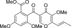
3-O-Methylfunicone, a Selective Inhibitor of Mammalian Y-Family DNA Polymerases from an Australian Sea Salt Fungal Strain

1
Laboratory of Food & Nutritional Sciences, Department of Nutritional Science, Kobe-Gakuin University, Nishi-ku, Kobe, Hyogo 651-2180, Japan
2
Cooperative Research Center of Life Sciences, Kobe-Gakuin University, Chuo-ku, Kobe, Hyogo 650-8586, Japan
3
Department of Applied Biological Science, Tokyo University of Science, 2641 Yamazaki, Noda, Chiba 278-8510, Japan
4
Nano-bioanalysis Team, Health Technology Research Center, Takamatsu, Kagawa 761-0395 Japan
*
Author to whom correspondence should be addressed.
Mar. Drugs 2009, 7(4), 624-639; https://doi.org/10.3390/md7040624
Submission received: 12 October 2009 / Revised: 18 November 2009 / Accepted: 19 November 2009 / Published: 23 November 2009
(This article belongs to the Special Issue Bioactive Compounds from Marine Microbes)

Abstract

:
We isolated a pol inhibitor from the cultured mycelia extract of a fungal strain isolated from natural salt from a sea salt pan in Australia, which was identified as 3-O-methylfunicone by spectroscopic analyses. This compound selectively inhibited the activities of mammalian Y-family DNA polymerases (pols) (i.e., pols η, ι and κ). Among these pols, human pol κ activity was most strongly inhibited, with an IC50 value of 12.5 μM. On the other hand, the compound barely influenced the activities of the other families of mammalian pols, such as A-family (i.e., pol γ), B-family (i.e., pols α, δ and ɛ) or X-family (i.e., pols β, λ and terminal deoxynucleotidyl transferase), and showed no effect on the activities of fish pol δ, plant pols, prokaryotic pols and other DNA metabolic enzymes, such as calf primase of pol α, human immunodeficiency virus type-1 (HIV-1) reverse transcriptase, human telomerase, T7 RNA polymerase, mouse IMP dehydrogenase (type II), human topoisomerases I and II, T4 polynucleotide kinase or bovine deoxyribonuclease I. This compound also suppressed the growth of two cultured human cancer cell lines, HCT116 (colon carcinoma cells) and HeLa (cervix carcinoma cells), and UV-treated HeLa cells exhibited lower clonogenic survival in the presence of inhibitor.

Graphical Abstract

1. Introduction

We have long been interested in the integrity of the genome of eukaryotes and its relation to cell differentiation. DNA replication, recombination and repair in eukaryotes are key systems to maintain these processes [1], and DNA polymerases (pols) have important roles in these. In this regard, we have concentrated our efforts on investigating eukaryotic pols associated with these processes [2].
The human genome encodes at least 15 pols to carry out cellular DNA synthesis [3,4]. Eukaryotic cells contain three replicative pols (α, δ and ɛ), mitochondrial pol γ, and at least eleven non-replicative pols [β, ζ, η, θ, ι, κ, λ, μ, ν, terminal deoxynucleotidyl transferase (TdT) and REV1] [35]. Pols have a highly conserved structure, which means that their overall catalytic subunits vary, on the whole, very little among species. Conserved structures usually indicate important, irreplaceable functions of the cell, the maintenance of which provides evolutionary advantages. Based on sequence homology, eukaryotic pols can be divided into four main families, A, B, X, and Y [6]. Family A includes mitochondrial pol γ, and pols θ and ν, and family B includes three replicative pols (α, δ, and ɛ) and pol ζ. Family X is pols β, λ, μ, and terminal deoxynucleotidyl transferase (TdT), and family Y includes pols η, ι, κ, and REV1. Because not all functions of eukaryotic pols have been fully elucidated, selective inhibitors of pol families are useful tools for distinguishing pols and clarifying their biological functions. We have therefore been searching for natural compounds that selectively inhibit each of these eukaryotic pols [715], and we succeeded in identifying novel compounds with pol inhibitory activity from marine microbiological products [1619].
In this study, we isolated fungal strains from Australian natural sea salt, and obtained 19 strains. These marine strains were screened, and a fungal strain displayed inhibitory activity against mammalian Y-family pols. The active compound purified from the strain, was identified as 3-O-methylfunicone, and the potential of this compound as an anti-cancer chemotherapy agent is discussed.

2. Results and Discussion

2.1. Purification of a DNA Polymerase Inhibitor, 3-O-Methylfunicone (Compound 1), from a Marine Fungal Strain

The 19 isolated and purified fungal strains from Australian sea salt were cultured, and each cultured mycelium was extracted with chloroform/methanol (v/v 1:1). The extracts were screened for inhibitory activity against mammalian pols, and the pol inhibitor (compound 1) was purified by silica gel column chromatography from the active mycelium extract of “AF1-2 strain”. From the 1H- and 13C-NMR spectral data, compound 1 was identified as 3-O-methylfunicone (Figure 1).

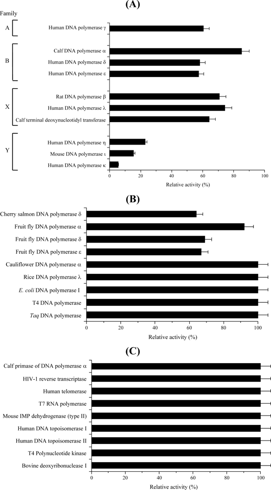
2.2. Effect of Isolated Compound 1 on the Activities of DNA Polymerases and Other DNA Metabolic Enzymes

First, the isolated compound 1 was investigated as to whether it inhibited the activities of ten mammalian pols, such as families A (pol γ), B (pols α, δ and ɛ), X (pols β and λ and TdT), and Y (pols η, ι and κ). As shown in Figure 2A, this compound at 100 μM was found to selectively inhibit the activity of the Y-family of pols, and within this family pol κ was the most sensitive. Compound 1 inhibited the activities of these pols dose-dependently, and 50% inhibition of pols η, ι and κ was observed at concentrations of 50.1, 34.3 and 12.5 μM, respectively (Figure 3); therefore, the inhibitory effect on mammalian Y-family pols ranked as follows: pol κ > pol ι > pol η. On the other hand, the other families (i.e., families A, B and X) of mammalian pols, fish pol (cherry salmon pol δ), insect pols (fruit fly pols α, δ and ɛ) hardly influenced the activities (Figure 2A and B), and higher plant pols (cauliflower pol α and rice pol λ), and prokaryotic pols (the Klenow fragment of E. coli pol I, T4 pol and Taq pol) had no effect (Figure 2B). When activated DNA (i.e., DNA digested by bovine deoxyribonuclease I [DNase I]) and dNTP was used as the DNA template-primer and nucleotide substrate instead of synthesized DNA [i.e., poly(dA)/oligo(dT)18] and dTTP, respectively, the inhibitory effects of the compound were unchanged (data not shown).
The same concentration (i.e., 100 μM) of this compound did not influence the activities of other DNA metabolic enzymes, such as calf primase pol α, human immunodeficiency virus type-1 (HIV-1) reverse transcriptase, human telomerase, T7 RNA polymerase, mouse inosine 5′-monophosphate (IMP) dehydrogenase (type II), human topoisomerases I and II, T4 polynucleotide kinase, and bovine DNase I (Figure 2C). These results suggest that compound 1 may be a selective inhibitor of mammalian Y-family pols in vitro.

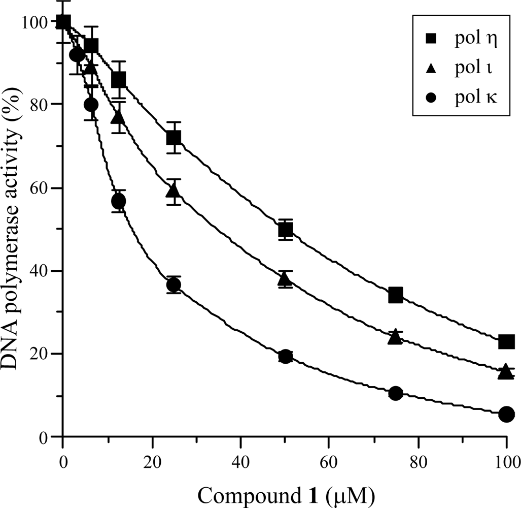
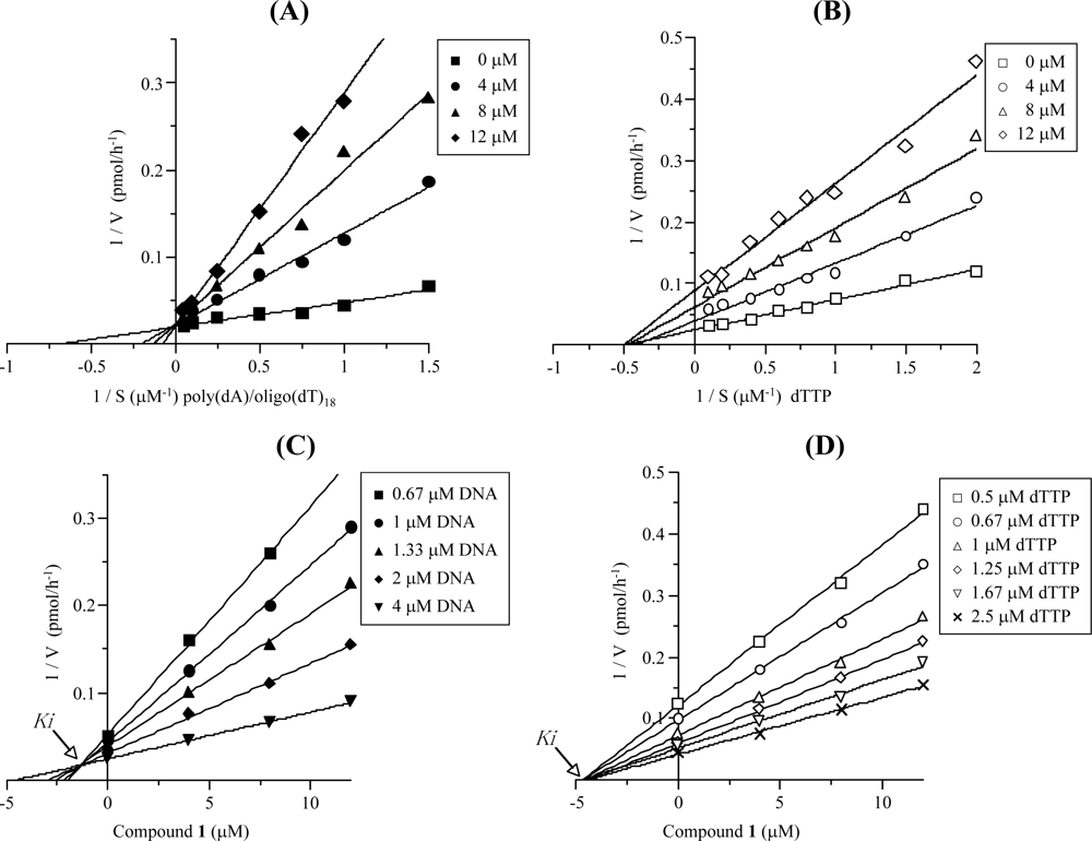
2.3. Mode of DNA Pol κ Inhibition by Compound 1

Next, to elucidate the mechanism of inhibition, the extent of inhibition as a function of DNA template-primer or dNTP substrate concentrations was studied (Figure 4). In kinetic analysis, poly(dA)/oligo(dT)18 and dTTP were used as the DNA template-primer and dNTP substrate, respectively. Double reciprocal plots of the results showed that compound 1 induced inhibition of human pol κ activity was competitive with the DNA template-primer and non-competitive with the dNTP substrate (Figure 4A and B). In the case of the DNA template-primer, the apparent maximum velocity (Vmax) was unchanged at 52.6 pmol/h, whereas the 295, 467, and 812% increases in Michaelis constant (Km) were observed in the presence of 4, 8, and 12 μM of compound 1, respectively (Figure 4A). The Km for the dNTP substrate was 2.00 μM, and the Vmax for the dNTP substrate decreased from 41.7 to 11.1 pmol/h in the presence of 12 μM compound 1 (Figure 4B). The inhibition constant (Ki) value, obtained from Dixon plots, was found to be 1.3 and 4.4 μM for the DNA template-primer and dTTP substrate, respectively (Figure 4C and D). When activated DNA and four dNTP substrates were used as the DNA template-primer and dNTP substrate, respectively, the inhibition of human pol κ by compound 1 was competitive with the DNA template-primer and non-competitive with the dNTP substrate (data not shown). These results suggest that compound 1 directly binds to the DNA template-primer-binding site of pol κ, whereas this compound may bind or interact with a site that is distinct from the dNTP substrate-binding site.
On the other hand, the inhibition of family B pols, such as pols α, δ and ɛ, by aphidicolin, which is a known classic pol inhibitor, was uncompetitive with activated DNA as a DNA template-primer and competitive with respect to the dNTP substrate [20]. Moreover, aphidicolin inhibited pol α by competing with dCTP but not by competing with the other three deoxynucleoside triphosphates [20]. In contrast, inhibition of pol κ by compound 1 was non-competitive with the four deoxynucleoside triphosphates (data not shown). The mode of the inhibitory effect of compound 1 on Y-family pols was quite different from the mode of the inhibitory effect of aphidicolin on B-family pols.

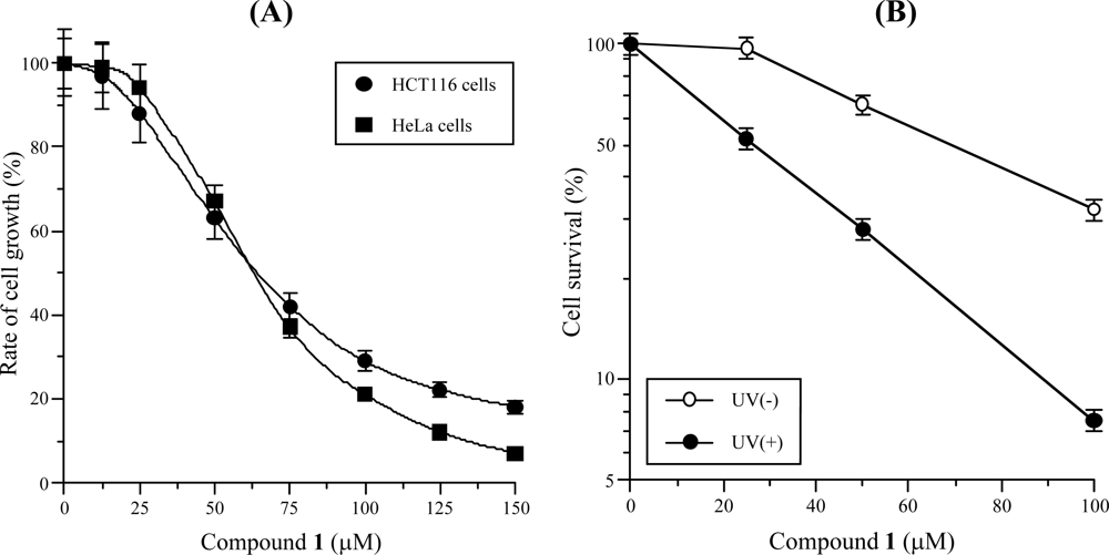
2.4. Inhibitory Effect of Isolated Compound 1 on Cultured Human Cancer Cells

Pols have recently emerged as important cellular targets for chemical intervention in the development of anti-cancer agents. The isolated compound 1 could therefore be useful in chemotherapy, and we investigated the growth inhibitory effect of this compound against two human cancer cell lines, a colon carcinoma cell line, HCT116, and a cervix carcinoma cell line, HeLa.
As shown in Figure 5A, compound 1 dose-dependently suppressed both HCT116 and HeLa cancer cell growth, and the LD50 value was 63.8 and 63.3 μM, respectively. Since the LD50 value of compound 1 for HCT116 cells was almost the same as that for HeLa cells, compound 1 might suppress all human cancer cell growth. The cell growth inhibition dose was approximately 1.3 to 5.1-fold higher than the enzyme inhibitory concentration, suggesting that the cause of the cancer cell influence might be the activity of pols. On the other hand, compound 1 did not influence the cell proliferation and growth of human normal cells such as HUVEC (human umbilical vein endothelial cells) and HDF (human dermal fibroblast) (data not shown); therefore, this compound might be a selective anti-cancer chemotherapy agent.
Y-family pols differ from other families in having low fidelity on undamaged templates and in their ability to replicate through damaged DNA. Members of this family are hence called translesion synthesis (TLS) pols [21]. Depending on the lesion, TLS pols can bypass the damage in an error-free or error-prone fashion, the latter resulting in elevated mutagenesis. Xeroderma pigmentosum variant (XPV) patients, for instance, have mutations in the gene encoding pol η, which is error-free for UV lesions. In XPV patients, alternative error-prone pols, e.g. pol ζ (B-family pol), are thought to be involved in mistakes which result in the cancer predisposition of these patients. Other members in humans are pols ι, κ, and Rev1 [6,21]. In E. coli, two TLS pols, pol IV (DINB) and pol V (UMUC), are known [21].
As shown in Figure 5B, compound 1 significantly enhanced HeLa cell UV-sensitivity, and the inhibiting clonogenic survival effect of 100 μM of the compound on 10 J/m2 UV exposed cells was 4.3-fold stronger than that on non-exposed cells. The LD50 value of compound 1 for UV-treated cell growth had the same potency as the IC50 values for mammalian Y-family pols. These results suggested that compound 1 inhibited the activities of family Y pols needed for DNA repair, thereby DNA damage must be augmented by this compound and UV-treatment, and then the cancer cells might undergo apoptotic cell death. It has been reported that pol η-deficient human fibroblast cells generated using siRNA were more than 3-fold sensitive to UV and DNA-targeting anticancer agents [22]. This supports the idea that compound 1 might selectively inhibit the activity of Y-family pols in the cells and that inhibitors of Y-family pols may have potential as anti-cancer drugs for clinical radiation therapy or cancer chemotherapy.

3. Conclusions

We obtained 3-O-methylfunicone (1) from a marine fungal strain isolated from Australian sea salt and showed it was a potent inhibitor specific to both mammalian Y-family pols and human cancer cell growth. Compound 1 has extremely high specificity for mammalian pol families, and significantly suppressed UV-treated human cancer cell growth and clonogenic survival. This compound could be a useful molecular tool as a Y-family pol-specific inhibitor in studies to determine the precise roles of the pol family in vitro. The products isolated from marine fungal strains, such as compound 1, may provide valuable information for developing a drug design strategy for anti-cancer chemotherapy agents.

4. Experimental

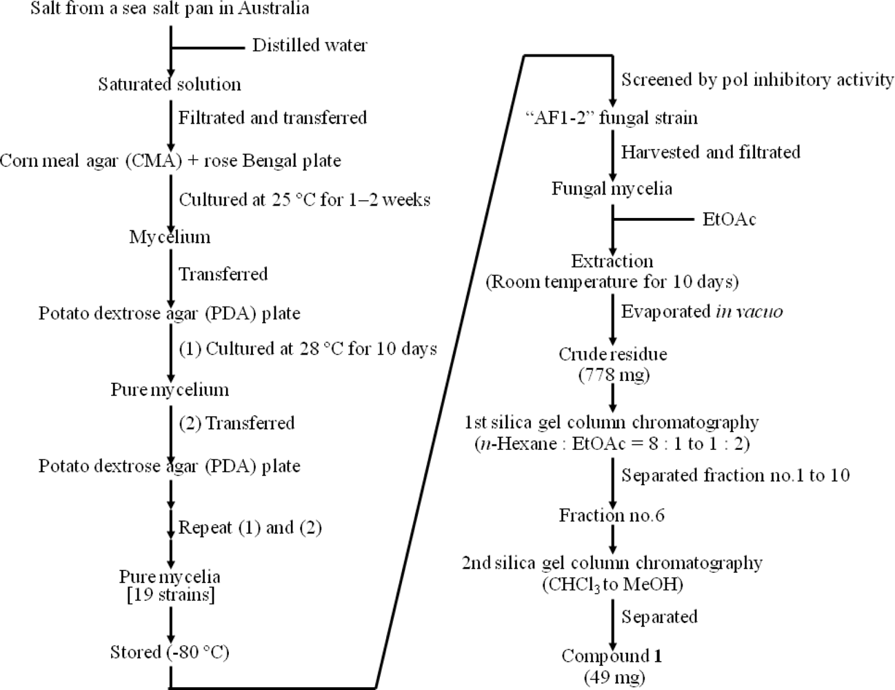
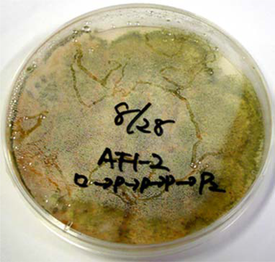
4.1. Isolation and Cultivation of Marine Fungal Strains from Australian Sea Salt

Natural salt (75 g) from an Australian sea salt pan was dissolved in distilled water (300 mL), and the saturated solution was filtered through a membrane filter (ADVANTEC, 0.2 μm, 47 mm). The filtered solution was placed on a corn meal agar (CMA) plate (Sigma-Aldrich K. K., Tokyo, Japan) containing rose Bengal (Junnsei Chemical Co., Ltd., Tokyo, Japan), and this plate was cultured for 1–2 weeks at 25 °C. Each grown mycelium on the plate was transferred to individual potato dextrose agar (PDA) (Difco Lab., KS, USA) plates, and cultured at 28 °C for 10 days. Each cultured fungus was transferred to a new PDA plate, and cultured at 28 °C for 10 days. Transformation and culture on PDA plates was repeated two to five times to obtain pure mycelium strains. The number of isolated and purified fungal strains was 19, which were stored at −80 °C (Figure 6, left).

4.2. Extraction and Purification of Compound from Marine Fungal Strains

The 19 isolated marine fungal strains were cultured in liquid potato dextrose (PD) medium, and each mycelium was extracted with chloroform/methanol (v/v 1:1). After evaporation of the solvent, the extracts were screened for inhibitory activity against mammalian pols. Pol catalyzes the addition of deoxyribonucleotides to the 3′-hydroxyl terminus of primed double-stranded DNA molecules and the assay method of pol inhibitors is shown in Figure 7. The mycelium extract of strain “AF1-2” displayed the greatest ability to inhibit DNA polymerase activity. An image of the AF1-2 fungal strain is shown in Figure 8. This strain was precultured by transferring a small agar piece to a 3L Erlenmeyer flask containing PD (2.4 g) in H2O (100 mL), and then fermented under static conditions in the dark for 3 days. The production culture was initiated by transferring the preculture to production medium [PD (2.4 g) and Daigo’s artificial seawater (36 g; Wako Chemical. Co. Ltd., Tokyo, Japan) in H2O (1 L)], and fermentation was carried out at room temperature under static conditions in the dark for 41 days. As shown in Figure 6 (right), the harvested fungal mycelia of AF1-2 were filtered from the cultured broth through cheesecloth, followed by homogenization in EtOAc, and extraction under static conditions at room temperature for 10 days. The extract was evaporated in vacuo to obtain crude residue (778 mg), and was separated by silica gel column chromatography (n-hexane/EtOAc = 8/1 to 1/2) to give fractions 1–10. Active fraction no. 6 was purified by a 2nd silica gel column chromatography (100% CHCl3 to 100% MeOH) to afford the active compound 1 (49 mg).

4.3. Structure Determination of Isolated Compound

1H- and 13C-NMR were recorded on a Bruker DRX400 instrument. Chemical shifts are reported in δ, parts per million (ppm), relative to TMS as an internal standard. Mass spectra were obtained on an API QSTAR Pulsar i spectrometer. The molecular formula of compound 1 was determined to be C20H20O8 by the high resolution mass spectrum. From the 1H- and 13C-NMR spectral data, compound 1 was identified as 3-O-methylfunicone (Figure 1). These spectroscopic data were consistent with those previously reported [23]. The analyzed data of compound 1 are shown below: 3-O-Methylfunicone (1). Colorless amorphous solid; 1H-NMR (CDCl3) δ: 8.49 (1H, d, J = 0.4 Hz), 7.09 (1H, d, J = 2.4 Hz), 6.64 (1H, d, J = 2.4 Hz), 6.61 (1H, dq, J =16.0, 6.4 Hz), 6.54 (1H, ddd, J = 16.0, 1.2, 0.4 Hz), 3.86 (3H, s), 3.81 (3H, s), 3.78 (3H s), 3.73 (3H, s), 1.97 (3H, dd, J = 6.4, 1.2 Hz); 13C-NMR (CDCl3) δC: 191.8, 172.4, 166.4, 160.7, 159.1, 157.3, 154.6, 144.1, 134.9, 129.7, 126.3, 126.1, 118.4, 105.2, 102.9, 60.5, 56.0, 55.5, 52.3, 18.9; HR ESIMS m/z 411.1058 [M+Na]+ (calcd for 411.1055).

4.4. Materials

Natural salt from a sea salt pan in Australia was supplied by the Research Institute of Salt and Sea Water Science (Kanagawa, Japan). Nucleotides, such as [3H]-deoxythymidine 5′-triphosphate (dTTP) (43 Ci/mmol), and chemically synthesized DNA template, such as poly(dA), were purchased from GE Healthcare Bio-Sciences (Little Chalfont, UK). DNA primer, such as oligo(dT)18, was customized by Sigma-Aldrich K. K. (Hokkaido, Japan). All other reagents were of analytical grade and were purchased from Wako Chemical Industries (Osaka, Japan).

4.5. Enzymes

Pol α was purified from calf thymus by immuno-affinity column chromatography, as described by Tamai et al. [24]. Recombinant rat pol β was purified from E. coli JMpβ5, as described by Date et al. [25]. The human pol γ catalytic gene was cloned into pFastBac. Histidine-tagged enzyme was expressed using the BAC-TO-BAC HT Baculovirus Expression System according to the supplier’s manual (Life Technologies, MD, USA) and purified using ProBoundresin (Invitrogen Japan, Tokyo, Japan) [26]. Human pols δ and ɛ were purified by nuclear fractionation of human peripheral blood cancer cells (Molt-4) using the second subunit of pols δ and ɛ-conjugated affinity column chromatography, respectively [27]. A truncated form of human pol η (residues 1–511) tagged with His6 at its C-terminal was expressed in E. coli cells and purified as described by Kusumoto et al. [28]. A recombinant mouse pol ι tagged with His6 at its C-terminal was expressed and purified by Ni-NTA column chromatography as described elsewhere (Masutani et al., in preparation). A truncated form of pol κ (residues 1–560) with 6 x His-tags attached at the C-terminus was overproduced in E. coli and purified as described by Ohashi et al. [29]. Recombinant human His-pol λ was overexpressed and purified according to a method described by Shimazaki et al. [30]. Fish pol δ was purified from the testis of cherry salmon (Oncorhynchus masou) [31]. Fruit fly pols α, δ and ɛ were purified from early embryos of Drosophila melanogaster, as described by Aoyagi et al. [32,33]. Pol α from a higher plant, cauliflower inflorescence, was purified according to the methods outlined by Sakaguchi et al. [34]. Recombinant rice (Oryza sativa L. cv. Nipponbare) His-pol λ was overexpressed and purified according to a method described by Uchiyama et al. [35]. Calf thymus TdT and bovine pancreas DNase I were obtained from Stratagene Cloning Systems (La Jolla, CA, USA). HIV-1 reverse transcriptase (recombinant) and the Klenow fragment of pol I from E. coli were purchased from Worthington Biochemical Corp. (Freehold, NJ, USA). T4 pol, Taq pol, T7 RNA polymerase and T4 polynucleotide kinase were purchased from Takara Bio (Tokyo, Japan). Purified human placenta DNA topoisomerases I and II were purchased from TopoGen, Inc. (Columbus, OH, USA).

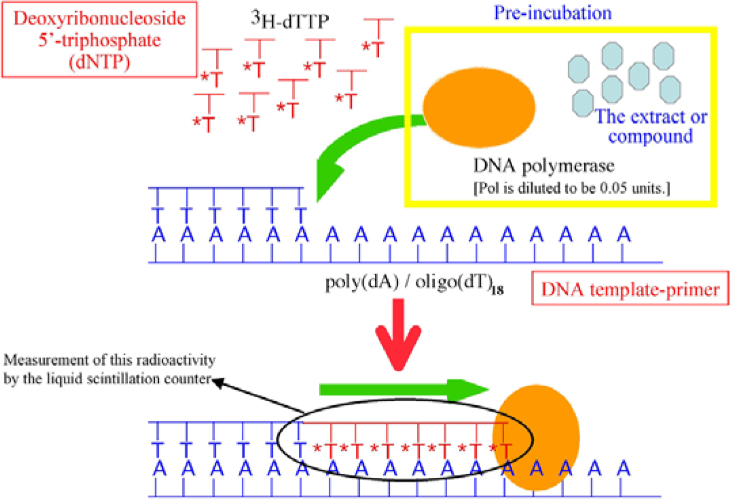
4.6. DNA Polymerase Assays

The reaction mixtures for pol α, pol β, plant pols and prokaryotic pols were described previously [7,8]; those for pol γ, and pols δ and ɛ were as described by Umeda et al. [26] and Ogawa et al. [36], respectively. The reaction mixtures for pols η, ι and κ were the same as for pol α, and the reaction mixture for pol λ was the same as for pol β. For pols, poly(dA)/oligo(dT)18 (A/T = 2/1) and dTTP were used as the DNA template-primer and nucleotide (i.e., dNTP) substrate, respectively. For HIV-1 reverse transcriptase, poly(rA)/oligo(dT)18 (A/T = 2/1) and dTTP were used as the DNA template-primer and nucleotide substrate, respectively. For TdT, oligo(dT)18 (3′-OH) and dTTP were used as the DNA primer and nucleotide substrate, respectively.
The compounds were dissolved in distilled dimethyl sulfoxide (DMSO) at various concentrations and sonicated for 30 sec. Aliquots of 4 μl sonicated samples were mixed with 16 μl of each enzyme (final amount 0.05 units) in 50 mM Tris-HCl (pH7.5) containing 1 mM dithiothreitol, 50% glycerol and 0.1 mM EDTA, and kept at 0 °C for 10 min. These inhibitor-enzyme mixtures (8 μl) were added to 16 μl of each of the enzyme standard reaction mixtures (final concentration of 50 mM Tris-HCl [pH 7.5], 1 mM dithiothreitol, 1 mM MgCl2, 15% glycerol, 10 μM poly(dA)/oligo(dT)18 and 10 μM [3H]-dTTP), and incubation was carried out at 37 °C for 60 min, except for Taq pol, which was incubated at 74 °C for 60 min. Activity without the inhibitor was considered 100%, and the remaining activity at each concentration of the inhibitor was determined relative to this value. One unit of pol activity was defined as the amount of enzyme that catalyzed the incorporation of 1 nmol dNTP (i.e., dTTP) into synthetic DNA template-primers in 60 min at 37 °C under the normal reaction conditions for each enzyme [7,8].

4.7. Other DNA Metabolic Enzymes Assays

The activities of calf primase of pol α, human telomerase, T7 RNA polymerase, mouse IMP dehydrogenase, human DNA topoisomerases I and II, T4 polynucleotide kinase and bovine DNase I were measured in standard assays according to the manufacturer’s specifications, as described by Tamiya-Koizumi et al. [37], Oda et al. [38], Nakayama et al. [39], Mizushina et al. [40], Ishimaru et al. [41], Soltis et al. [42] and Lu et al. [43], respectively.

4.8. Investigation of Growth on Cultured Human Cancer Cells

Human colon carcinoma cell line, HCT116, was a kind gift from Dr. Bert Vogelstein (Johns Hopkins University, Baltimore, MO, USA). Human cervix carcinoma cell line, HeLa, was obtained from Health Science Research Resources Bank (Osaka, Japan). The cells were maintained in McCoy’s 5A medium supplemented with 10% FBS, sodium bicarbonate (2 g/L) and streptomycin (100 μg/mL). The cells were cultured at 37 °C in standard medium in a humidified atmosphere of 5% CO2-95% air. The cytotoxicity of the compound was investigated as follows: high concentrations (10 mM) of the compounds were dissolved in DMSO and stored. Approximately 5 × 10³ cells per well were inoculated in 96-well micro plates, then the compound stock solution was diluted to various concentrations, and applied to each well. After incubation for 24 h, the survival rate was determined by MTT (3-(4,5-dimethylthiazol-2-yl)-2,5-diphenyl tetrazolium bromide) assay [44].

4.9. UV-Treated Clonogenic Assay

For clonogenic assay, 600 cells of HeLa were seeded on 10-cm plates and allowed to grow for 24 h before 10 J/m2 UV irradiation treatment. The cells were then treated with various concentrations of compound 1 for 24 h. The cells were then washed with PBS and fresh medium was added for an additional 7-day incubation in a humidified 10% CO2 incubator at 37 °C. After 7 days, the cultured cells were stained with 0.5% methylene blue in 50% ethanol to visualize the colonies. The number of colonies in each treatment group was counted, and the cell survival data were presented as percentage of the colony numbers of treated/untreated cells.

Acknowledgments

We greatly thank T. Hasegawa and Y. Noda of the Research Institute of Salt and Sea Water Science (Kanagawa, Japan) for the gift of natural salt from a sea salt pan in Australia (i.e., they could refer to Figure 6). We are grateful for the donations of calf pol α by M. Takemura of Tokyo University of Science (Tokyo, Japan), rat pol β, human pols δ and ɛ by K. Sakaguchi of Tokyo University of Science (Chiba, Japan), human pol γ by M. Suzuki of Nagoya University School of Medicine (Nagoya, Japan), and human pol λ by O. Koiwai of Tokyo University of Science (Chiba, Japan).
This work was supported in part by the “Academic Frontier” Project for Private Universities: matching fund subsidy from MEXT (Ministry of Education, Culture, Sports, Science and Technology), 2006–2010 (Y. M. and H. Y.). Y. M. acknowledges a Grant-in-Aid for Young Scientists (A) (No. 19680031) from MEXT and a Grant-in-Aid from The Salt Science Research Foundation, No. 09S3 (Japan). Y. M. and K. H. acknowledge a Grant from the Industrial Technology Research Program from NEDO (Japan).
  • Samples Availability: Available from the corresponding author.

References and Notes

  1. DePamphilis, ML. DNA Replication in Eukaryotic Cells; Cold Spring Harbor Laboratory Press: New York, NY, USA, 1996. [Google Scholar]
  2. Seto, H; Hatanaka, H; Kimura, S; Oshige, M; Tsuya, Y; Mizushina, Y; Sawado, T; Aoyagi, N; Matsumoto, T; Hashimoto, J; Sakaguchi, K. Purification and characterization of a 100 kDa DNA polymerase from cauliflower inflorescence. Biochem J 1998, 332, 557–563. [Google Scholar]
  3. Hubscher, U; Maga, G; Spadari, S. Eukaryotic DNA polymerases. Ann Rev Biochem 2002, 71, 133–163. [Google Scholar]
  4. Bebenek, K; Kunkel, TA. DNA repair and replication. In Advances in Protein Chemistry; Yang, W, Ed.; Elsevier: San Diego, CA, USA, 2004; Volume 69, pp. 137–165. [Google Scholar]
  5. Takata, K; Shimizu, T; Iwai, S; Wood, RD. Human DNA polymerase N (POLN) is a low fidelity enzyme capable of error-free bypass of 5S-thymine glycol. J Biol Chem 2006, 281, 23445–23455. [Google Scholar]
  6. Friedberg, EC; Feaver, WJ; Gerlach, VL. The many faces of DNA polymerases: Strategies for mutagenesis and for mutational avoidance. Proc Natl Acad Sci USA 2000, 97, 5681–5683. [Google Scholar]
  7. Mizushina, Y; Tanaka, N; Yagi, H; Kurosawa, T; Onoue, M; Seto, H; Horie, T; Aoyagi, N; Yamaoka, M; Matsukage, A; Yoshida, S; Sakaguchi, K. Fatty acids selectively inhibit eukaryotic DNA polymerase activities in vitro. Biochim Biophys Acta 1996, 1308, 256–262. [Google Scholar]
  8. Mizushina, Y; Yoshida, S; Matsukage, A; Sakaguchi, K. The inhibitory action of fatty acids on DNA polymerase β. Biochim Biophys Acta 1997, 1336, 509–521. [Google Scholar]
  9. Mizushina, Y; Takahashi, N; Ogawa, A; Tsurugaya, K; Koshino, H; Takemura, M; Yoshida, S; Matsukage, A; Sugawara, F; Sakaguchi, K. The cyanogenic glucoside, prunasin (D-mandelonitrile-β-D-glucoside), is a novel inhibitor of DNA polymerase β. J Biochem (Tokyo) 1999, 126, 430–436. [Google Scholar]
  10. Mizushina, Y; Ohkubo, T; Sugawara, F; Sakaguchi, K. Structure of lithocholic acid binding to the N-terminal 8-kDa domain of DNA polymerase β. Biochemistry 2000, 39, 12606–12613. [Google Scholar]
  11. Mizushina, Y; Kamisuki, S; Mizuno, T; Takemura, M; Asahara, H; Linn, S; Yamaguchi, T; Matsukage, A; Hanaoka, F; Yoshida, S; Saneyoshi, M; Sugawara, F; Sakaguchi, K. Dehydroaltenusin, a mammalian DNA polymerase α inhibitor. J Biol Chem 2000, 275, 33957–33961. [Google Scholar]
  12. Mizushina, Y; Kamisuki, S; Kasai, N; Shimazaki, N; Takemura, M; Asahara, H; Linn, S; Yoshida, S; Matsukage, A; Koiwai, O; Sugawara, F; Yoshida, H; Sakaguchi, K. A plant phytotoxin, solanapyrone A, is an inhibitor of DNA polymerase β and λ. J Biol Chem 2002, 277, 630–638. [Google Scholar]
  13. Mizushina, Y; Xu, X; Asahara, H; Takeuchi, R; Oshige, M; Shimazaki, N; Takemura, M; Yamaguchi, T; Kuroda, K; Linn, S; Yoshida, H; Koiwai, O; Saneyoshi, M; Sugawara, F; Sakaguchi, K. A sulphoquinovosyl diacylglycerol is a DNA polymerase ɛ inhibitor. Biochem J 2003, 370, 299–305. [Google Scholar]
  14. Kuriyama, I; Asano, N; Kato, I; Ikeda, K; Takemura, M; Yoshida, H; Sakaguchi, K; Mizushina, Y. Dipeptide alcohol-based inhibitors of eukaryotic DNA polymerase α. Bioorg Med Chem 2005, 13, 2187–2196. [Google Scholar]
  15. Kamisuki, S; Ishimaru, C; Onoda, K; Kuriyama, I; Ida, N; Sugawara, F; Yoshida, H; Mizushina, Y. Nodulisporol and Nodulisporone, novel specific inhibitors of human DNA polymerase λ from a fungus, Nodulisporium sp. Bioorg Med Chem 2007, 15, 3109–3114. [Google Scholar]
  16. Naganuma, M; Nishida, M; Kuramochi, K; Sugawara, F; Yoshida, H; Mizushina, Y. 1-Deoxyrubralactone, a novel specific inhibitor of families X and Y of eukaryotic DNA polymerases from a fungal strain derived from sea algae. Bioorg Med Chem 2008, 16, 2939–2944. [Google Scholar]
  17. Kimura, T; Nishida, M; Kuramochi, K; Sugawara, F; Yoshida, H; Mizushina, Y. Novel azaphilones, kasanosins A and B, which are specific inhibitors of eukaryotic DNA polymerases β and λ from Talaromyces sp. Bioorg Med Chem 2008, 16, 4594–4599. [Google Scholar]
  18. Nishida, M; Ida, N; Horio, M; Takeuchi, T; Kamisuki, S; Murata, H; Kuramochi, K; Sugawara, F; Yoshida, H; Mizushina, Y. Hymenoic acid, a novel specific inhibitor of human DNA polymerase λ from a fungus of Hymenochaetaceae sp. Bioorg Med Chem 2008, 16, 5115–5122. [Google Scholar]
  19. Kimura, T; Takeuchi, T; Kumamoto-Yonezawa, Y; Ohashi, E; Ohmori, H; Masutani, C; Hanaoka, F; Sugawara, F; Yoshida, H; Mizushina, Y. Penicilliols A and B, novel inhibitors specific to mammalian Y-family DNA polymerases. Bioorg Med Chem 2009, 17, 1811–1816. [Google Scholar]
  20. Oguro, M; Suzuki-Hori, C; Nagano, H; Mano, Y; Ikegami, S. The mode of inhibitory action by aphidicolin on eukaryotic DNA polymerase α. Eur J Biochem 1979, 97, 603–607. [Google Scholar]
  21. Prakash, S; Johnson, RE; Prakash, L. Eukaryotic translesion synthesis DNA polymerases: specificity of structure and function. Annu Rev Biochem 2005, 74, 317–353. [Google Scholar]
  22. Chen, YW; Cleaver, JE; Hanaoka, F; Chang, CF; Chou, KM. A novel role of DNA polymerase eta in modulating cellular sensitivity to chemotherapeutic agents. Mol Cancer Res 2006, 4, 257–265. [Google Scholar]
  23. Stefano, SD; Nicoletti, R; Milone, A; Zambardino, S. 3-o-Methylfunicone, a fungitoxic metabolite produced by the fungus. Penicillium pinophilum Phytochemistry 1999, 52, 1399–1401. [Google Scholar]
  24. Tamai, K; Kojima, K; Hanaichi, T; Masaki, S; Suzuki, M; Umekawa, H; Yoshida, S. Structural study of immunoaffinity-purified DNA polymerase α-DNA primase complex from calf thymus. Biochim Biophys Acta 1988, 950, 263–273. [Google Scholar]
  25. Date, T; Yamaguchi, M; Hirose, F; Nishimoto, Y; Tanihara, K; Matsukage, A. Expression of active rat DNA polymerase β in Escherichia coli. Biochemistry 1988, 27, 2983–2990. [Google Scholar]
  26. Umeda, S; Muta, T; Ohsato, T; Takamatsu, C; Hamasaki, N; Kang, D. The D-loop structure of human mtDNA is destabilized directly by 1-methyl-4-phenylpyridinium ion (MPP+), a parkinsonism-causing toxin. Eur J Biochem 2000, 267, 200–206. [Google Scholar]
  27. Oshige, M; Takeuchi, R; Ruike, R; Kuroda, K; Sakaguchi, K. Subunit protein-affinity isolation of Drosophila DNA polymerase catalytic subunit. Protein Expr Purif 2004, 35, 248–256. [Google Scholar]
  28. Kusumoto, R; Masutani, C; Shimmyo, S; Iwai, S; Hanaoka, F. DNA binding properties of human DNA polymerase η: implications for fidelity and polymerase switching of translesion synthesis. Genes Cells 2004, 9, 1139–1150. [Google Scholar]
  29. Ohashi, E; Murakumo, Y; Kanjo, N; Akagi, J; Masutani, C; Hanaoka, F; Ohmori, H. Interaction of hREV1 with three human Y-family DNA polymerases. Genes Cells 2004, 9, 523–531. [Google Scholar]
  30. Shimazaki, N; Yoshida, K; Kobayashi, T; Toji, S; Tamai, T; Koiwai, O. Over-expression of human DNA polymerase λ in E. coli and characterization of the recombinant enzyme. Genes Cells 2000, 7, 639–651. [Google Scholar]
  31. Yamaguchi, T; Saneyoshi, M; Takahashi, H; Hirokawa, S; Amano, R; Liu, X; Inomata, M; Maruyama, T. Synthetic Nucleoside and Nucleotides. 43. Inhibition of vertebrate telomerases by carbocyclic oxetanocin G (C.OXT-G) triphosphate analogues and influence of C.OXT-G treatment on telomere length in human HL60 cells. Nucleos Nucleot Nucleic Acids 2006, 25, 539–551. [Google Scholar]
  32. Aoyagi, N; Matsuoka, S; Furunobu, A; Matsukage, A; Sakaguchi, K. Drosophila DNA polymerase δ. Purification and characterization. J Biol Chem 1994, 269, 6045–6050. [Google Scholar]
  33. Aoyagi, N; Oshige, M; Hirose, F; Kuroda, K; Matsukage, A; Sakaguchi, K. DNA polymerase ɛ from Drosophila melanogaster. Biochem Biophys Res Commun 1997, 230, 297–301. [Google Scholar]
  34. Sakaguchi, K; Hotta, Y; Stern, H. Chromatin-associated DNA polymerase activity in meiotic cells of lily and mouse. Cell Struct Funct 1980, 5, 323–334. [Google Scholar]
  35. Uchiyama, Y; Kimura, S; Yamamoto, T; Ishibashi, T; Sakaguchi, K. Plant DNA polymerase λ, a DNA repair enzyme that functions in plant meristematic and meiotic tissues. Eur J Biochem 2004, 271, 2799–2807. [Google Scholar]
  36. Ogawa, A; Murate, T; Suzuki, M; Nimura, Y; Yoshida, S. Lithocholic acid, a putative tumor promoter, inhibits mammalian DNA polymerase β. Jpn J Cancer Res 1998, 89, 1154–1159. [Google Scholar]
  37. Tamiya-Koizumi, K; Murate, T; Suzuki, M; Simbulan, CG; Nakagawa, M; Takamura, M; Furuta, K; Izuta, S; Yoshida, S. Inhibition of DNA primase by sphingosine and its analogues parallels with their growth suppression of cultured human leukemic cells. Biochem Mol Biol Int 1997, 41, 1179–1189. [Google Scholar]
  38. Oda, M; Ueno, T; Kasai, N; Takahashi, H; Yoshida, H; Sugawara, F; Sakaguchi, K; Hayashi, H; Mizushina, Y. Inhibition of telomerase by linear-chain fatty acids: A structural analysis. Biochem J 2002, 367, 329–334. [Google Scholar]
  39. Nakayama, C; Saneyoshi, M. Inhibitory effects of 9-β-D-xylofuranosyladenine 5′-triphosphate on DNA-dependent RNA polymerase I and II from cherry salmon (Oncorhynchus masou). J Biochem (Tokyo) 1985, 97, 1385–1389. [Google Scholar]
  40. Mizushina, Y; Dairaku, I; Yanaka, N; Takeuchi, T; Ishimaru, C; Sugawara, F; Yoshida, H; Kato, N. Inhibitory action of polyunsaturated fatty acids on IMP dehydrogenase. Biochimie 2007, 89, 581–590. [Google Scholar]
  41. Ishimaru, C; Yonezawa, Y; Kuriyama, I; Nishida, M; Yoshida, H; Mizushina, Y. Inhibitory effects of cholesterol derivatives on DNA polymerase and topoisomerase activities, and human cancer cell growth. Lipids 2008, 43, 373–382. [Google Scholar]
  42. Soltis, DA; Uhlenbeck, OC. Isolation and characterization of two mutant forms of T4 polynucleotide kinase. J Biol Chem 1982, 257, 11332–11339. [Google Scholar]
  43. Lu, BC; Sakaguchi, K. An endo-exonuclease from meiotic tissues of the basidiomycete Coprinus cinereus: Its purification and characterization. J Biol Chem 1991, 266, 21060–21066. [Google Scholar]
  44. Mosmann, T. Rapid colorimetric assay for cellular growth and survival: application to proliferation and cytotoxicity assays. J Immunol Methods 1983, 65, 55–63. [Google Scholar]
Figure 1. Structure of compound 1 (3-O-methylfunicone).
Marinedrugs 07 00624f1
Figure 2. Effect of 3-O-methylfunicone (1) on the activities of various DNA polymerases and other DNA metabolic enzymes. (A) Mammalian pols, (B) fish, insect, plant and prokaryotic pols, and (C) other DNA metabolic enzymes. Compound 1 (100 μM) was incubated with each enzyme (0.05 units). % of relative activity. Enzymatic activities were measured as described in the Experimental section. Activities in the absence of the compounds were taken as 100%. Data are shown as the means ± SEM of four independent experiments.
Marinedrugs 07 00624f2
Figure 3. Mammalian Y-family DNA polymerase inhibition dose-response curves of 3-O-methylfunicone (1). Compound 1 was incubated with human pol η (square), mouse pol ι (triangle), and human pol κ (circle) (0.05 units of each). Pol activity was measured as described in the Experimental section. Activity in the absence of the compounds was taken as 100%. Data are shown as the means ± SEM of three independent experiments.
Marinedrugs 07 00624f3
Figure 4. Kinetic analysis of the inhibition of human pol κ by compound 1. (A) Human pol κ activity was measured in the absence (closed square) or presence of 4 μM (closed circle), 8 μM (closed triangle) or 12 μM (closed diamond) compound 1 using the indicated concentrations of the DNA template-primer. (B) Human pol κ activity was assayed with the indicated concentrations of the substrate dTTP in the presence of 4 μM (open circle), 8 μM (open triangle) or 12 μM (open diamond) or in the absence (open square) of compound 1. (C) and (D) The inhibition constants (Ki) were determined as 1.3 and 4.4 μM from a Dixon plot made on the basis of the same data for (A) and (B), respectively. Amount of human pol κ in the assay mixture was 0.05 units.
Marinedrugs 07 00624f4
Figure 5. (A) Human cancer cell growth inhibition dose-response curves of 3-O-methyl-funicone (1). Various concentrations (0 to 150 μM) of compound 1 were incubated with human colon carcinoma (HCT116) cells (circle) and human cervix carcinoma (HeLa) cells (square) for 24 h. Rate of cell growth was determined by MTT assay [44] as described in the Experimental section. Data are shown as the means ± SEM of five independent experiments. (B) Clonogenic survival of UV-treated HeLa cells by compound 1. Various concentrations (0 to 100 μM) of compound 1 were incubated with 10 J/m2 UV-treated (closed circle) or untreated (open circle) HeLa cells. After 7 days, the number of colonies in each treatment group was counted. Values are the means ± SEM of two independent experiments.
Marinedrugs 07 00624f5
Figure 6. (Left) Isolation methods of marine fungal strains from Australian sea salt. (Right) Purification procedure of a compound with pol inhibitory activity from mycelia of “AF1-2” fungal strain.
Marinedrugs 07 00624f6
Figure 7. Assay method of pol inhibitor.
Marinedrugs 07 00624f7
Figure 8. Marine fungal strain AF1-2.
Marinedrugs 07 00624f8

