Anticancer Effect and Structure-Activity Analysis of Marine Products Isolated from Metabolites of Mangrove Fungi in the South China Sea

Abstract
:1. Introduction
2. Results and Discussion
3. Experimental Section
3.1. Chemicals and reagents
3.2. Isolation of compounds from metabolites of marine-derived fungi
3.3. Tumor cell culture
3.4. Anticancer activity assays
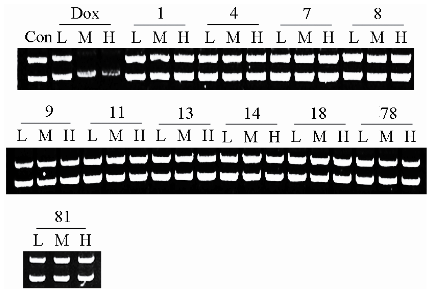
3.5. DNA binding assay
Acknowledgments
References and Notes
- Simmons, TL; Andrianasolo, E; McPhail, K; Flatt, P; Gerwick, WH. Marine natural products as anticancer drugs. Mol Cancer Ther 2005, 4, 333–342. [Google Scholar]
- Glaser, KB; Mayer, AM. A renaissance in marine pharmacology: from preclinical curiosity to clinical reality. Biochem Pharmacol 2009, 78, 440–448. [Google Scholar]
- Molinski, TF; Dalisay, DS; Lievens, SL; Saludes, JP. Drug development from marine natural products. Nat Rev Drug Discov 2009, 8, 69–85. [Google Scholar]
- Blunt, JW; Copp, BR; Hu, WP; Munro, MH; Northcote, PT; Prinsep, MR. Marine natural products. Nat Prod Rep 2009, 26, 170–244. [Google Scholar]
- Hong, K; Gao, AH; Xie, QY; Gao, H; Zhuang, L; Lin, HP; Yu, HP; Li, J; Yao, XS; Goodfellow, M; Ruan, JS. Actinomycetes for marine drug discovery isolated from mangrove soils and plants in China. Mar Drugs 2009, 7, 24–44. [Google Scholar]
- Schoffski, P; Dumez, H; Wolter, P; Stefan, C; Wozniak, A; Jimeno, J; Van Oosterom, AT. Clinical impact of trabectedin (ecteinascidin-743) in advanced/metastatic soft tissue sarcoma. Expert Opin Pharmacother 2008, 9, 1609–1618. [Google Scholar]
- Bugni, TS; Ireland, CM. Marine-derived fungi: a chemically and biologically diverse group of microorganisms. Nat Prod Rep 2004, 21, 143–163. [Google Scholar]
- Yanagihara, M; Sasaki-Takahashi, N; Sugahara, T; Yamamoto, S; Shinomi, M; Yamashita, I; Hayashida, M; Yamanoha, B; Numata, A; Yamori, T; Andoh, T. Leptosins isolated from marine fungus Leptoshaeria species inhibit DNA topoisomerases I and/or II and induce apoptosis by inactivation of Akt/protein kinase B. Cancer Sci 2005, 96, 816–824. [Google Scholar]
- Zhang, JY; Tao, LY; Liang, YJ; Yan, YY; Dai, CL; Xia, XK; She, ZG; Lin, YC; Fu, LW. Secalonic acid D induced leukemia cell apoptosis and cell cycle arrest of G(1) with involvement of GSK-3beta/beta-catenin/c-Myc pathway. Cell Cycle 2009, 8, 2444–2450. [Google Scholar]
- Zhang, JY; Wu, HY; Xia, XK; Liang, YJ; Yan, YY; She, ZG; Lin, YC; Fu, LW. Anthracenedione derivative 1403P-3 induces apoptosis in KB and KBv200 cells via reactive oxygen species-independent mitochondrial pathway and death receptor pathway. Cancer Biol Ther 2007, 6, 1413–1421. [Google Scholar]
- Dean, M; Rzhetsky, A; Allikmets, R. The human ATP-binding cassette (ABC) transporter superfamily. Genome Res 2001, 11, 1156–1166. [Google Scholar]
- Gillet, JP; Efferth, T; Remacle, J. Chemotherapy-induced resistance by ATP-binding cassette transporter genes. Biochim Biophys Acta 2007, 1775, 237–262. [Google Scholar]
- Singh, R; Sharma, M; Joshi, P; Rawat, DS. Clinical status of anti-cancer agents derived from marine sources. Anticancer Agents Med Chem 2008, 8, 603–617. [Google Scholar]
- Matsumoto, K; Akao, Y; Kobayashi, E; Ohguchi, K; Ito, T; Tanaka, T; Iinuma, M; Nozawa, Y. Induction of apoptosis by xanthones from mangosteen in human leukemia cell lines. J Nat Prod 2003, 66, 1124–1127. [Google Scholar]
- Koyama, J; Morita, I; Kobayashi, N; Osakai, T; Nishino, H; Tokuda, H. Correlation between reduction potentials and inhibitory effects on Epstein-Barr virus activation of poly-substituted anthraquinones. Cancer Lett 2005, 225, 193–198. [Google Scholar]
- Yan, Y; Su, X; Liang, Y; Zhang, J; Shi, C; Lu, Y; Gu, L; Fu, L. Emodin azide methyl anthraquinone derivative triggers mitochondrial-dependent cell apoptosis involving in caspase-8-mediated Bid cleavage. Mol Cancer Ther 2008, 7, 1688–1697. [Google Scholar]
- Greaves, M. Pharmacogenetics in the management of coumarin anticoagulant therapy: the way forward or an expensive diversion. PLoS Med 2005, 2, e342. [Google Scholar]
- Lin, Y; Wu, X; Feng, S; Jiang, G; Luo, J; Zhou, S; Vrijmoed, LL; Jones, EB; Krohn, K; Steingrover, K; Zsila, F. Five unique compounds: xyloketals from mangrove fungus Xylaria sp. from the South China Sea coast. J Org Chem 2001, 66, 6252–6256. [Google Scholar]
- Chen, WL; Qian, Y; Meng, WF; Pang, JY; Lin, YC; Guan, YY; Chen, SP; Liu, J; Pei, Z; Wang, GL. A novel marine compound xyloketal B protects against oxidized LDL-induced cell injury in vitro. Biochem Pharmacol 2009, 78, 941–950. [Google Scholar]
- Adlercreutz, H. Phyto-oestrogens and cancer. Lancet Oncol 2002, 3, 364–373. [Google Scholar]
- Dixon, RA. Phytoestrogens. Annu Rev Plant Biol 2004, 55, 225–261. [Google Scholar]
- Huang, Z; Cai, X; Shao, C; She, Z; Xia, X; Chen, Y; Yang, J; Zhou, S; Lin, Y. Chemistry and weak antimicrobial activities of phomopsins produced by mangrove endophytic fungus Phomopsis sp. ZSU-H76. Phytochemistry 2008, 69, 1604–1608. [Google Scholar]
- Tao, Y; Zeng, X; Mou, C; Li, J; Cai, X; She, Z; Zhou, S; Lin, Y. 1H and 13C NMR assignments of three nitrogen containing compounds from the mangrove endophytic fungus (ZZF08). Magn Reson Chem 2008, 46, 501–505. [Google Scholar]
- Guo, Z; She, Z; Shao, C; Wen, L; Liu, F; Zheng, Z; Lin, Y. (1)H and (13)C NMR signal assignments of paecilin A and B, two new chromone derivatives from mangrove endophytic fungus Paecilomyces sp. (tree 1–7). Magn Reson Chem 2007, 45, 777–780. [Google Scholar]
- Yang, RY; Li, CY; Lin, YC; Peng, GT; She, ZG; Zhou, SN. Lactones from a brown alga endophytic fungus (No. ZZF36) from the South China Sea and their antimicrobial activities. Bioorg Med Chem Lett 2006, 16, 4205–4208. [Google Scholar]
- Xia, XK; Huang, HR; She, ZG; Shao, CL; Liu, F; Cai, XL; Vrijmoed, LL; Lin, YC. (1)H and (13)C NMR assignments for five anthraquinones from the mangrove endophytic fungus Halorosellinia sp. (No. 1403). Magn Reson Chem 2007, 45, 1006–1009. [Google Scholar]
- Guo, Z; Shao, C; She, Z; Cai, X; Liu, F; Vrijimoed, LL; Lin, Y. 1H and 13C NMR assignments for two oxaphenalenones bacillosporin C and D from the mangrove endophytic fungus SBE-14. Magn Reson Chem 2007, 45, 439–441. [Google Scholar]
- Shao, CL; Guo, ZY; Xia, XK; Liu, Y; Huang, ZJ; She, ZG; Lin, YC; Zhou, SN. Five nitro-phenyl compounds from the South China Sea mangrove fungus. J Asian Nat Prod Res 2007, 9, 643–648. [Google Scholar]
- Fu, L; Liang, Y; Deng, L; Ding, Y; Chen, L; Ye, Y; Yang, X; Pan, Q. Characterization of tetrandrine, a potent inhibitor of P-glycoprotein-mediated multidrug resistance. Cancer Chemother Pharmacol 2004, 53, 349–356. [Google Scholar]
- Chen, LM; Wu, XP; Ruan, JW; Liang, YJ; Ding, Y; Shi, Z; Wang, XW; Gu, LQ; Fu, LW. Screening novel, potent multidrug-resistant modulators from imidazole derivatives. Oncol Res 2004, 14, 355–362. [Google Scholar]
- Tao, LY; Liang, YJ; Wang, F; Chen, LM; Yan, YY; Dai, CL; Fu, LW. Cediranib (recentin, AZD2171) reverses ABCB1- and ABCC1-mediated multidrug resistance by inhibition of their transport function. Cancer Chemother Pharmacol 2009, 64, 961–969. [Google Scholar]


| Compound | IC50 value (μmol/L) | |||||
|---|---|---|---|---|---|---|
| KB | KBv200 | MCF-7 | MCF-7/adr | A549 | LO2 | |
| Doxorubicin | 0.05 ± 0.003 | 3.21 ± 0.12 | 0.39 ± 0.02 | 22.33 ± 1.56 | 1.67 ± 0.09 | 0.11 ± 0.01 |
| Multi-substituent phenyl derivatives | ||||||
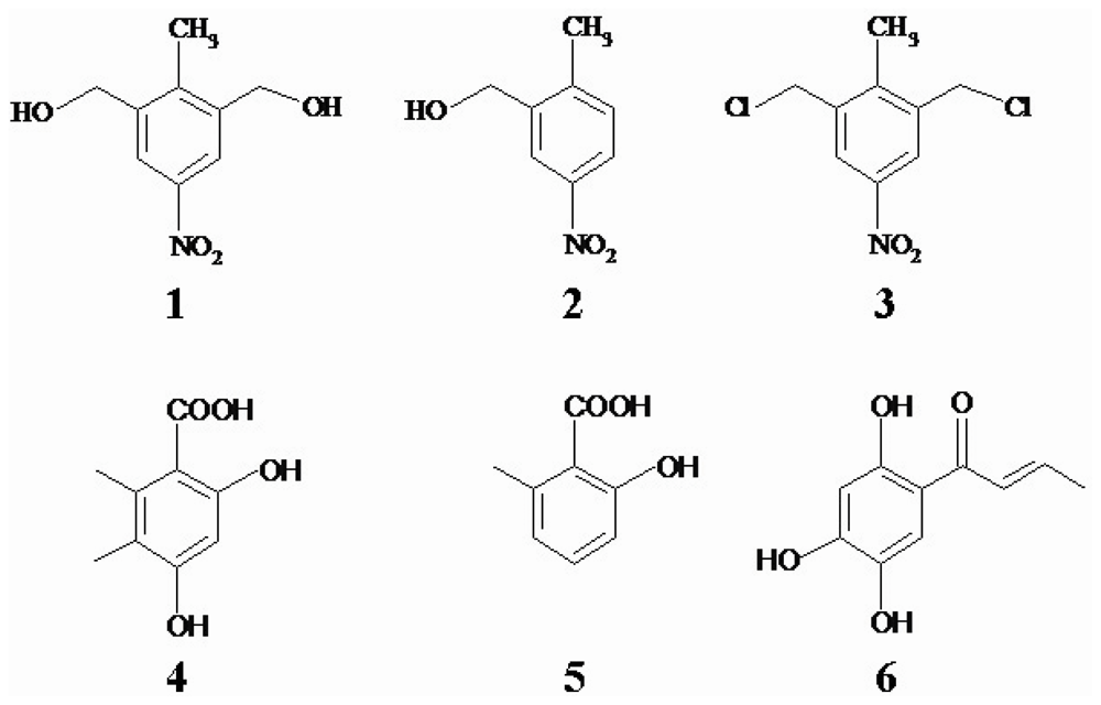
| 1 | 1.57 ± 0.08 | 2.67 ± 0.17 | 1.38 ± 0.07 | 2.34 ± 0.11 | 2.71 ± 0.15 | 6.59 ± 0.44 |
| 2 | >50 | >50 | >50 | >50 | >50 | >50 |
| 3 | >50 | >50 | >50 | >50 | >50 | >50 |
| 4 | 1.15 ± 0.06 | 6.74 ± 0.38 | 11.74 ± 0.86 | 35.67 ± 2.08 | 17.10 ± 1.03 | 16.48 ± 1.22 |
| 5 | >50 | >50 | >50 | >50 | >50 | >50 |
| 6 | >50 | >50 | >50 | >50 | >50 | >50 |
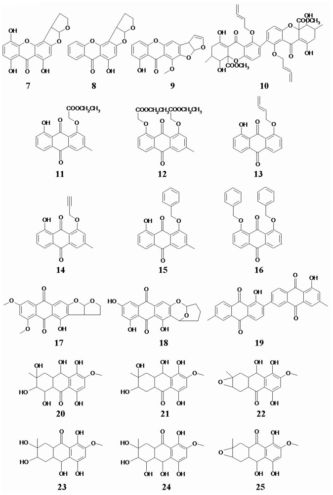
| Mangrove-derived quinones | ||||||
| 7 | 0.03 ± 0.001 | 9.08 ± 0.65 | 0.17 ± 0.01 | 31.56 ± 1.83 | 16.51 ± 0.88 | 47.35 ± 2.04 |
| 8 | 0.71 ± 0.03 | 17.20 ± 0.96 | 2.53 ± 0.14 | 9.37 ± 0.42 | >50 | >50 |
| 9 | 0.94 ± 0.01 | 47.98 ± 3.41 | 1.43 ± 0.09 | 31.60 ± 1.36 | >50 | >50 |
| 10 | >50 | >50 | >50 | >50 | >50 | >50 |
| 11 | 5.89 ± 0.37 | 18.94 ± 1.20 | 13.23 ± 0.84 | 35.34 ± 2.77 | 21.34 ± 1.60 | 28.88 ± 1.46 |
| 12 | >50 | >50 | >50 | >50 | >50 | >50 |
| 13 | 21.00 ± 1.35 | >50 | 16.32 ± 0.71 | >50 | 31.23 ± 2.02 | >50 |
| 14 | 11.86 ± 0.69 | >50 | 35.23 ± 1.87 | >50 | 23.53 ± 1.32 | 37.51 ± 1.64 |
| 15 | >50 | >50 | >50 | >50 | >50 | >50 |
| 16 | >50 | >50 | >50 | >50 | >50 | >50 |
| 17 | >50 | >50 | >50 | >50 | >50 | >50 |
| 18 | 28.19 ± 1.66 | 44.63 ± 2.57 | 17.22 ± 0.95 | 24.96 ± 1.06 | 33.89 ± 2.31 | >50 |
| 19 | >50 | >50 | >50 | >50 | >50 | >50 |
| 20 | >50 | >50 | >50 | >50 | >50 | >50 |
| 21 | >50 | >50 | >50 | >50 | >50 | >50 |
| 22 | >50 | >50 | >50 | >50 | >50 | >50 |
| 23 | >50 | >50 | >50 | >50 | >50 | >50 |
| 24 | >50 | >50 | >50 | >50 | >50 | >50 |
| 25 | >50 | >50 | >50 | >50 | >50 | >50 |
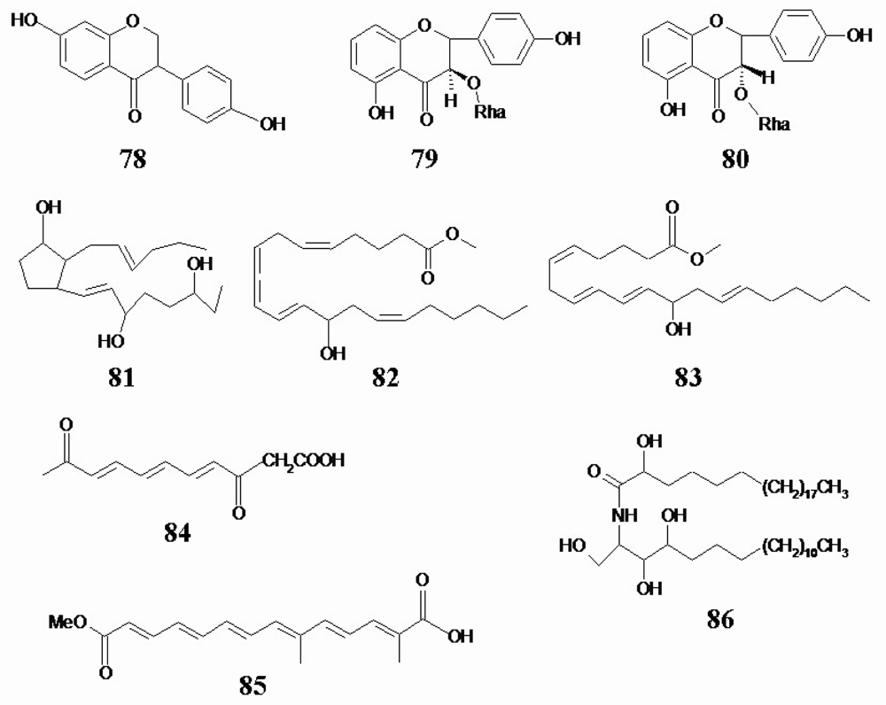
| Isoflavone analogs | ||||||
| 78 | 8.63 ± 0.57 | 9.37 ± 0.61 | 19.77 ± 0.89 | 24.95 ± 1.15 | 14.88 ± 0.64 | 33.62 ± 2.06 |
| 79 | >50 | >50 | >50 | >50 | >50 | >50 |
| 80 | >50 | >50 | >50 | >50 | >50 | >50 |
| Fatty acid derivatives | ||||||
| 81 | 0.37 ± 0.01 | 0.39 ± 0.02 | 0.41 ± 0.02 | 0.49 ± 0.02 | 0.34 ± 0.01 | 0.72 ± 0.03 |
| 82 | >50 | >50 | >50 | >50 | >50 | >50 |
| 83 | >50 | >50 | >50 | >50 | >50 | >50 |
| 84 | >50 | >50 | >50 | >50 | >50 | >50 |
| 85 | >50 | >50 | >50 | >50 | >50 | >50 |
| 86 | >50 | >50 | >50 | >50 | >50 | >50 |
© 2010 by the authors; licensee Molecular Diversity Preservation International, Basel, Switzerland This article is an open-access article distributed under the terms and conditions of the Creative Commons Attribution license (http://creativecommons.org/licenses/by/3.0/).
Share and Cite
Tao, L.-y.; Zhang, J.-y.; Liang, Y.-j.; Chen, L.-m.; Zheng, L.-s.; Wang, F.; Mi, Y.-j.; She, Z.-g.; To, K.K.W.; Lin, Y.-c.; et al. Anticancer Effect and Structure-Activity Analysis of Marine Products Isolated from Metabolites of Mangrove Fungi in the South China Sea. Mar. Drugs 2010, 8, 1094-1105. https://doi.org/10.3390/md8041094
Tao L-y, Zhang J-y, Liang Y-j, Chen L-m, Zheng L-s, Wang F, Mi Y-j, She Z-g, To KKW, Lin Y-c, et al. Anticancer Effect and Structure-Activity Analysis of Marine Products Isolated from Metabolites of Mangrove Fungi in the South China Sea. Marine Drugs. 2010; 8(4):1094-1105. https://doi.org/10.3390/md8041094
Chicago/Turabian StyleTao, Li-yang, Jian-ye Zhang, Yong-ju Liang, Li-ming Chen, Li-sheng Zheng, Fang Wang, Yan-jun Mi, Zhi-gang She, Kenneth Kin Wah To, Yong-cheng Lin, and et al. 2010. "Anticancer Effect and Structure-Activity Analysis of Marine Products Isolated from Metabolites of Mangrove Fungi in the South China Sea" Marine Drugs 8, no. 4: 1094-1105. https://doi.org/10.3390/md8041094