Article

Secondary Metabolites from a Marine-Derived Endophytic Fungus Penicillium chrysogenum QEN-24S

1
Key Laboratory of Experimental Marine Biology, Institute of Oceanology, Chinese Academy of Sciences, Nanhai Road 7, Qingdao 266071, China
2
Graduate School of the Chinese Academy of Sciences, Yuquan Road 19A, Beijing 100049, China
3
Institute for Pharmaceutical Biology and Biotechnology, Heinrich-Heine-University Duesseldorf, Universitaetsstreet 1, 40225 Duesseldorf, Germany
*
Author to whom correspondence should be addressed.
Mar. Drugs 2011, 9(1), 59-70; https://doi.org/10.3390/md9010059
Submission received: 8 November 2010 / Revised: 1 December 2010 / Accepted: 20 December 2010 / Published: 27 December 2010

Abstract

:
Penicillium chrysogenum QEN-24S, an endophytic fungus isolated from an unidentified marine red algal species of the genus Laurencia, displayed inhibitory activity against the growth of pathogen Alternaria brassicae in dual culture test. Chemical investigation of this fungal strain resulted in the isolation of four new (13 and 5) and one known (4) secondary metabolites. Their structures were identified as two polyketide derivatives penicitides A and B (1 and 2), two glycerol derivatives 2-(2,4-dihydroxy-6-methylbenzoyl)-glycerol (3) and 1-(2,4-dihydroxy-6-methylbenzoyl)- glycerol (4), and one monoterpene derivative penicimonoterpene (5). Penicitides A and B (1 and 2) feature a unique 10-hydroxy- or 7,10-dihydroxy-5,7-dimethylundecyl moiety substituting at C-5 of the α-tetrahydropyrone ring, which is not reported previously among natural products. Compound 5 displayed potent activity against the pathogen A. brassicae, while compound 1 exhibited moderate cytotoxic activity against the human hepatocellular liver carcinoma cell line.

Graphical Abstract

1. Introduction

Marine derived fungi have been proven to be a rich source of structurally unique and biologically active secondary metabolites [1]. In the past years, a number of new metabolites have been isolated and identified and their biological activities have been evaluated [24]. As part of our recently initiated program to assess the chemical and biological diversity of endophytic fungi derived from marine algae [512], Penicillium chrysogenum QEN-24S, an endophytic fungus obtained from the inner tissue of an unidentified marine red algal species of the genus Laurencia, attracted our attention. This fungus displayed inhibitory activity against the growth of pathogenic fungus Alternaria brassicae in our initial dual culture test (Figure 1). This phenomenon prompted us to examine the chemical metabolites of this fungal strain. The fungus P. chrysogenum was previously reported as the source of several β-lactam antibiotics, most significantly penicillin [13,14]. Besides the antibacterial activity, it was also reported that P. chrysogenum served as a source of bioactive substances with other activities such as antifungal, anti-HIV, and cytotoxic activity [15,16].
We wish to report herein the isolation and structure determination of four new compounds, namely, penicitides A and B (1 and 2), 2-(2,4-dihydroxy-6-methylbenzoyl)-glycerol (3), and penicimonoterpene (5) (Figure 2), from the rice culture fermentation of P. chrysogenum QEN-24S. In addition, a related known compound 1-(2,4-dihydroxy-6-methylbenzoyl)-glycerol (4) [17] was also isolated and identified. The structures of these compounds were established on the basis of extensive spectroscopic analysis. The absolute configuration at C-15 of penicitide A (1) was determined by application of the modified Mosher’s method. The inhibitory activity of these metabolites against two pathogens A. brassicae and Aspergullus niger was determined. In addition, the cytotoxic activity against seven tumor cell lines was also evaluated.

2. Results and Discussion

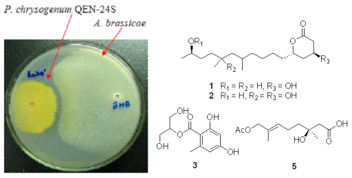
2.1. Bioassay-Guided Isolation

The endophyte P. chrysogenum QEN-24S displayed obvious activity against the pathogen fungus A. brassicae in our initial dual culture test (Figure 1). This endophyte was therefore submitted to a large scale fermentation for bioactive compounds isolation. The EtOAc extract derived from the rice culture of the fungal strain was suspended in MeOH-H2O (9:1, v/v) and was extracted with n-hexane to remove the non-polar fraction. The MeOH-soluble fraction displayed moderated inhibitory activity against the pathogenic fungus A. brassicae. This fraction was then subjected to various separation procedures by column chromatography on silica gel, Lobar LiChroprep RP-18, and Sephadex LH-20, to afford four new (13, and 5) and one known secondary metabolites (4), as shown in Figure 2. By detailed analysis of the spectroscopic data, the structure for the known compound 4 was determined as 1-(2,4-dihydroxy-6-methylbenzoyl)-glycerol (4) [17].

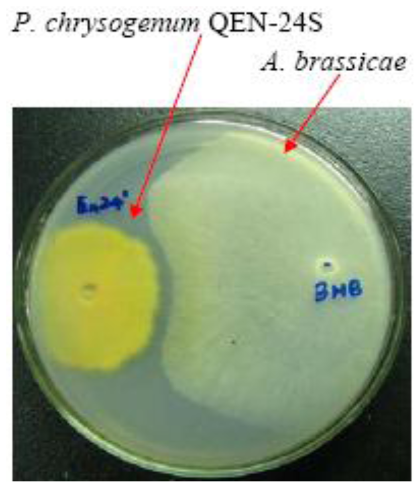
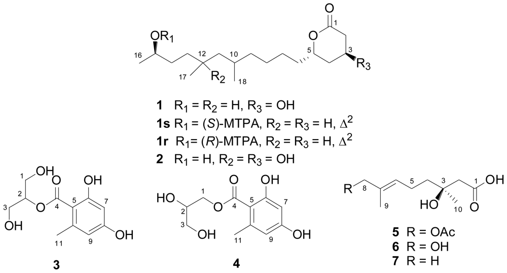
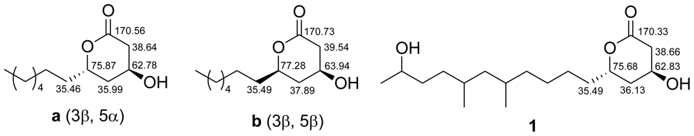
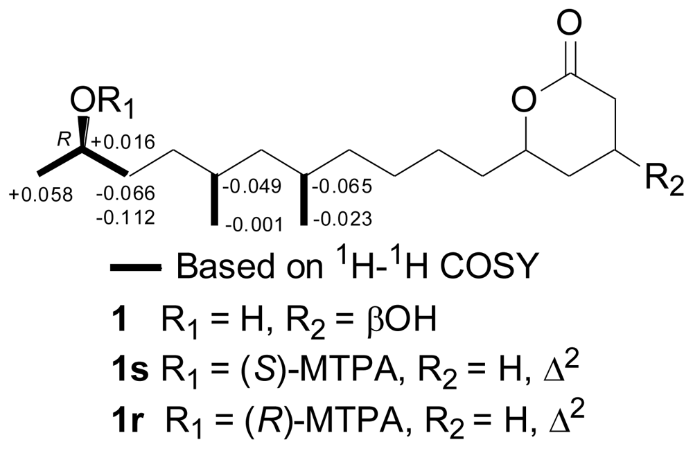
2.2. Structural Elucidation of the New Compounds

Compounds 1 and 2 were obtained as colorless oils. The IR spectrum of 1 showed absorption bands for OH (3405 cm−1) and C=O (1716 cm−1) functionalities in the molecules. The low-resolution ESI-MS displayed ion peaks at m/z 337 [M + Na]+ and 651 [2M + Na]+. Its molecular formula was determined as C18H34O4 on the basis of positive HR-ESI-MS, indicating two degrees of unsaturation. The 13C-NMR (Table 1) along with the DEPT experiments revealed the presence of 18 carbon atoms including one carbonyl carbon, five sp3 methines, nine sp3 methylenes, and three methyl groups. The remaining one unsaturation demonstrated that 1 is a monocyclic compound. The 1H-1H COSY correlations revealed the presence of a contiguous sequence of the proton signals comprising from H-2 to H-16, as shown in Figure 3. Further 1H-1H COSY correlations from H3-17 to H-12 and from H3-18 to H-10 unambiguously demonstrated two methyl groups H3-17 and H3-18 attached to C-12 and C-10, respectively. The HMBC correlations from H-2 and H-3 to C-1 established the linkage of C-1 to C-2 and C-3. Finally, the chemical shifts of C-5 (δC 75.68, d) and C-1 (δC 170.33, s) established the connection of C-1 and C-5 via an ester linkage to form a tetrahydropyran-2-one moiety as shown in Figure 3.
The relative configuration of the two chiral centers C-3 and C-5 in the tetrahydropyran ring was proposed by comparison of its 13C-NMR data with those published for its analogues [18] sharing common partial structures from C-1 to C-8 (Figure 4). The relative configurations of C-3 and C-5 were deduced as 3β and 5α, respectively, as shown in Figure 2, according to the corresponding chemical shifts as shown in Figure 4. This was supported by the fact that no NOE correlations could be observed between H-3 and H-5 in the NOESY experiment. However, the relative configuration for C-10, C-12, and C-15 in the side chain remains unknown. Based on the above evidence, the structure for compound 1 was established as 4β-hydroxy-6α-(10-hydroxy-5,7-dimethylundecyl)tetrahydropyran-2-one, which was named as penicitide A, as shown in Figure 2.
The absolute configuration at C-15 of penicitide A (1) was determined to be R by application of modified Mosher’s method, as shown in Figure 5. However, due to the dehydration between H-2 and OH-3 during the acylation, attempts to determine the absolute configurations for C-3 and C-5 failed.
Compound 2 was assigned the molecular formula C18H34O5, having one oxygen unit more than 1, on the basis of positive HR-ESI-MS experiments. Detailed comparison of the 1H- and 13C-NMR data (Table 1) revealed that the structure of 2 was very similar to 1. However, the methine carbon signal at δC 30.11 (C-12) in the 13C-NMR spectrum of 1 was replaced by an oxygenated quaternary carbon at δC 72.37 in 2. Accordingly, the methine proton at δH 1.45 in 1 disappeared in the 1H-NMR spectrum of 2. Moreover, the doublet signal at δH 0.85 (J = 6.4 Hz) for H3-17 in 1 was replaced by a downfield singlet at δH 1.15 in the 1H-NMR spectrum of 2. The above evidences indicated that an OH substituent at C-12 in 2. The HMBC correlations from H-11 and H3-17 to C-12 supported this conclusion. The configuration for the chiral centers at C-3 and C-5 was determined to be the same as that of 1 by NOESY experiment, as well as by detailed comparison of the NMR data with that of 1. The structure for compound 2 was therefore established as 4β-hydroxy-6α-(7,10-dihydroxy-5,7- dimethylundecyl)-tetrahydropyran-2-one as shown in Figure 2, which was named penicitide B.
Penicitides A and B (1 and 2) bear a unique 10-hydroxy- or 7,10-dihydroxy-5,7-dimethylundecyl moiety substituting at C-5 of the α-tetrahydropyrone ring. This structure feature is not reported previously among natural products. These compounds appear to be α-tetrahydropyrone polyketides that are derived from a mixed-precursor biosynthesis including acetate and propionate building blocks [19]. Propionate polyketides are well known components from marine invertebrates and actinomycetes [20,21], but they have been rarely isolated from fungi as far as we know.
Compounds 3 and 4 were obtained as a colorless oily mixture. Attempts to separate the two compounds by different CC steps as well as by semi-preparative HPLC with different solvent systems failed. Fortunately, compound 3 could be distinguished from 4, aided by 2D NMR experiments including 1H-1H COSY, HSQC, and HMBC, and by their different ratio (1:3) as indicated by the 1H- and 13C-NMR spectra. Most NMR signals are well-resolved. The structure of compound 4 was readily identified as 1-O-(2,4-dihydroxy-6-methylbenzoyl)-glycerol by detailed NMR spectral data analysis (Table 2) and comparison with our recent report [17]. Low-resolution ESI-MS displayed pseudo-molecular ion peaks at m/z 265 [M + Na]+ and 507 [2M + Na]+ for both compounds. The molecular formula was determined as C11H14O6 on the basis of positive HR-ESI-MS, suggesting five degrees of unsaturation. The structure of 3 was independently assigned by analysis of the 1H- and 13C-NMR data (Table 2) as well as by HSQC and HMBC correlations. The only difference between the two isomers was the connectivity of the glycerol moiety with the 2,4-dihydroxy-6-methylbenzoyl unit. The symmetric nature of the NMR data for the glycerol moiety (H-1/H-3 and C-1/C-3) in 3 as well as the observed 3J correlation from H-2 to C-4 in the HMBC spectrum (Figure 3) established the linkage of 2,4-dihydroxy-6-methylbenzoyl group to C-2 of the glycerol unit. Thus, the structure of compound 3 was assigned as 2-(2,4-dihydroxy-6-methylbenzoyl)-glycerol, as shown in Figure 2.
Compound 5 was obtained as colorless oil. Its IR spectrum showed absorption bands for OH (3178 cm−1) and C=O (1727 cm−1) functionalities in the molecules. The low-resolution ESI-MS displayed ion peaks at m/z 245 [M + H]+, 267 [M + Na]+, and 511 [2M + Na]+. Its molecular formula was determined as C12H20O5 on the basis of positive HR-ESI-MS. The planar structure was determined mainly by comparison with the literature reports of the known compounds (E)-3,8-dihydroxy-3,7- dimethyl-6-octenoic acid (6) [22] and (S,E)-3-hydroxy-3,7-dimethyl-6-octenoic acid (7) [23] as well as by the NOESY spectral data. Detailed comparison of the 1H-NMR data of 5 (Table 3) with that of 6 [22] revealed that these two structures were very similar, except an extra acetyl methyl signal at δH 2.07 was observed in the 1H-NMR spectrum of 5. Further analysis of the 13C-NMR data of 5 indicated that an extra acetyl unit was attached to the monoterpene skeleton of 6. The location of the acetyl unit was clearly determined at C-8 by the long range correlations from the acetoxyl methyl proton to C-8 in the HMBC spectrum. The observed NOE correlation from H-6 to H-8 in the NOESY spectrum revealed that the double bond was in trans-geometry. Finally, the absolute stereochemistry at C-3 was assigned to be S by comparison of the optical rotation of 5 ([α]D20 = +1.4, MeOH) with that of 7 ([α]D25 = +1.8, MeOH) [23].
Compound 5, which was named penicimonoterpene, pertains to a class of naturally occurring monoterpene derivatives named citronellic acid formed by the oxidation of citronellal with a carboxyl group at C-1 and an olefinic double bond at C-6 as shown in Figure 2, which has been isolated from ascomycete [23] and fungus [24].

2.3. Biological Activities of the Isolated Compounds

The biological activity of the isolated compounds 15 was examined in antifungal and cytotoxicity bioassays. In the initial antifungal screening, penicimonoterpene (5) displayed potent activity against pathogen A. brassicae with an inhibition zone of 17 mm in diameter at the concentration of 20 μg/disk, while penicitide A (1) displayed moderate activity with an inhibition zone of 6 mm in diameter at the concentration of 20 μg/disk (Table 4). The cytotoxicity evaluation was also performed and the results indicated that penicitide A (1) possessed activity against HepG2 cell line with the IC50 of 32 μg/mL. The other compounds displayed no appreciable activity.

3. Experimental Section

3.1. General

Column chromatography was performed on commercial silica gel (Qingdao Haiyang Chemical Group Co., China; 200–300 mesh), Lobar LiChroprep RP-18 (40–63 μm; Merck), and Sephadex LH-20 (Pharmacia). Optical rotations were measured on an Optical Activity AA-55 polarimeter. UV Spectroscopic data were obtained on a Lengguang Gold Spectrumlab 54. IR spectra were performed on a JASCO FT/IR-4100 Fourier Transform infrared spectrometer. NMR spectra were acquired on a Bruker Avance-500 spectrometer at 500 MHz for 1H and 125 MHz for 13C. MS data were recorded on a VG Autospec-3000 mass spectrometer.

3.2. Fungal Material

The endophytic fungus Penicillium chrysogenum QEN-24S was isolated, by use of a standard procedure as in our previous report [9], from the inner tissue of an unidentified marine red algal species of the genus Laurencia collected from the Weizhou Island of southern China sea. Fungal identification was carried out by use of a molecular biological protocol by DNA amplification and sequencing of the ITS region as in our previous report [9]. The sequence data derived from the fungal strain has been submitted to and deposited at GenBank with accession number GU985086. A BLAST search result showed that the sequence was similar (99%) to the sequence of P. chrysogenum (compared to AY373903.1 GI: 34809383). The strain is preserved at the Key Laboratory of Experimental Marine Biology, Institute of Oceanology of the Chinese Academy of Sciences, with accession number QEN-24S.
Mass growth of the fungus for the isolation and identification of new metabolites was carried out in Erlenmeyer flasks (1 L each). The fungus was grown on rice solid medium (to 100 g commercially available rice, 0.6 g of peptone and 100 mL of distilled water was added, then kept overnight prior to autoclaving) at room temperature under static conditions for 30 days.

3.3. Extraction and Isolation

The rice culture (10 flasks) of the fungal strain P. chrysogenum QEN-24S was extracted with EtOAc. The crude extract obtained was dried and partitioned between n-hexane and 90% MeOH. The 90% MeOH-soluble material (2 g) was subjected to column chromatography (CC) over silica gel, eluting with different solvents of increasing polarity from petroleum ether (PE) to MeOH to yield 10 fractions (Frs. 1–10) on the basis of TLC analysis. Fr. 5 (0.3 g) was further purified by CC on silica gel eluting with a CHCl3-MeOH gradient (from 80:1 to 1:1), Sephadex LH-20 (MeOH), and Lobar LiChroprep RP-18 with a H2O-MeOH gradient (from 1:1 to 0:1) to afford 1 (10.5 mg) and a CC-unseparable mixture of 3 and 4 (11.1 mg). Fr. 8 (0.4 g) was further purified by CC on silica gel by eluting with a CHCl3-MeOH gradient (from 80:1 to 5:1), Sephadex LH-20 (MeOH), and preparative TLC (plate: 20 × 20 cm, developing solvents: CHCl3-MeOH, 10:1) to afford 2 (5.4 mg) and 5 (12.4 mg).
Penicitide A (1). Colorless oil; [α]D20: +42.9 (c 0.14, MeOH); IR (KBr) νmax 3405, 2927, 2865, 1716, 1457, 1376, 1253, 1064, 937, 852, 725, 586 cm−1; 1H- and 13C-NMR data, see Table 1; ESI-MS m/z 337 [M + Na]+, 651 [2M + Na]+; HR-ESI-MS: m/z 337.2345 [M + Na]+ (calcd for C18H34O4Na+, 337.2354).
Penicitide B (2). Colorless oil; [α]D20: +100.0 (c 0.08, MeOH); IR (KBr) νmax 3394, 3297, 2931, 2861, 1724, 1635, 1458, 1377, 1057, 802, 489 cm−1; 1H- and 13C-NMR data, see Table 1; ESI-MS m/z 353 [M + Na]+, 683 [2M + Na]+; HR-ESI-MS: m/z 353.2297 [M + Na]+ (calcd for C18H34O5Na+, 353.2303).
2-(2,4-Dihydroxy-6-methylbenzoyl)-glycerol (3). Colorless oil; UV λmax (MeOH) nm (log ɛ): 214 (4.79), 264 (4.50); 1H- and 13C-NMR data, see Table 4; ESI-MS m/z 265 [M + Na]+, 507 [2M + Na]+; HR-ESI-MS: m/z 265.0689 [M + Na]+ (calcd for C11H14O6Na+, 265.0688).
Penicimonoterpene (5). Colorless oil; [α]D20: +1.4 (c 0.83, MeOH); IR (KBr) νmax 3178, 2969, 2938, 1727, 1442, 1381, 1238, 1025, 949, 852 cm−1; 1H- and 13C-NMR data, see Table 5; ESI-MS m/z 245 [M + H]+, 267 [M + Na]+, 511 [2M + Na]+; HR-ESI-MS: m/z 267.1203 [M + Na]+ (calcd for C12H20O5Na+, 267.1208).

3.4. Preparation of the (R)- and (S)-MTPA Ester Derivatives of Compound 1 [25]

(S)-(+)-α-Methoxy-α-(trifluoromethyl)phenylacetyl chloride (10 μL) and 4-(dimethylamino)-pyridine (2 mg) were added to penicitide A (1, 1.1 mg) which was dissolved in dry pyridine (400 μL). The mixture was kept at room temperature for 12 h. The acylation product was purified by preparative TLC on silica gel [eluent: petroleum ether/EtOAC (4:1, v/v)] to yield corresponding (R)-Mosher ester 1r. Treatment of 1 (1.2 mg) with (R)-MTPA-Cl (10 μL) as described above yielded the corresponding (S)-Mosher ester 1s.

3.5. Dual Culture Test

Dual culture test of P. chrysogenum QEN-24S against A. niger and A. brassicae was carried out as literature report [26]. Pathogens and endophyte P. chrysogenum QEN-24S were inoculated at the periphery of the PDA-medium plate (200 g potato, 20 g dextrose, 20 g agar, and 1 L sea water), and then cultured at 27 °C for 5 days.

3.6. Antifungal Assay

Antifungal assay against A. niger and A. brassicae was carried out using the well diffusion method [27]. Amphotericin B (AMPB) was used as positive control.

3.7. Cytotoxicity Assay

The cytotoxic activities against NCI-H460 (human non-small cell lung cancer), SMMC-7721 (human hepatoma), SW1990 (human pancreatic cancer), DU145 (human prostate carcinoma), HepG2 (human hepatocellular liver carcinoma), Hela (human epithelial carcinoma), and MCF-7 (human breast adenocarcinoma) cell lines were determined according to previously reported methods [28].

4. Conclusions

In summary, four new (13 and 5) and one known (4) secondary metabolites were characterized from the algal-derived endophytic fungus P. chrysogenum QEN-24S and their chemical structures were solved by spectroscopic and chemical analysis. Among these, penicimonoterpene (5) were natural inhibitors of pathogenic fungus A. brassicae. Penicitide A (1) also displayed moderate activity against A. brassicae. All of these results are consistent with the initial dual culture test, in which P. chrysogenum QEN-24S displayed obvious inhibitory activity against the growth of pathogenic fungus A. brassicae. In addition, compound 1 exhibited moderate selective cytotoxic activity against HepG2 tumor cell line.

Acknowledgements

This work was financial supported by programs from the Ministry of Science and Technology (2010CB833802 and 2007AA09Z446), from the National Science Foundation of China (30910103914 and 30970293), and from the Chinese Academy of Sciences (KSCX2-EW-G-12B).
  • Samples Availability: Available from the authors.

References

  1. Bugni, TS; Ireland, CM. Marine-derived fungi: A chemically and biologically diverse group of microorganisms. Nat Prod Rep 2004, 21, 143–163. [Google Scholar]
  2. Blunt, JW; Copp, BR; Hu, WP; Munro, MHG; Northcote, PT; Prinsep, MR. Marine natural products. Nat Prod Rep 2008, 25, 35–94. [Google Scholar]
  3. Blunt, JW; Copp, BR; Hu, WP; Munro, MHG; Northcote, PT; Prinsep, MR. Marine natural products. Nat Prod Rep 2009, 26, 170–244. [Google Scholar]
  4. Blunt, JW; Copp, BR; Munro, MHG; Northcote, PT; Prinsep, MR. Marine natural products. Nat Prod Rep 2010, 27, 165–237. [Google Scholar]
  5. Zhang, Y; Li, XM; Wang, BG. Nigerasperones A–C, new monomeric and dimeric naphtho-γ-pyrones from a marine alga-derived endophytic fungus Aspergillus niger EN-13. J Antibiot 2007, 60, 204–210. [Google Scholar]
  6. Zhang, Y; Wang, S; Li, XM; Cui, CM; Feng, C; Wang, BG. New sphingolipids with a previously unreported 9-methyl-C20-sphingosine moiety from a marine algous endophytic fungus Aspergillus niger EN-13. Lipids 2007, 42, 759–764. [Google Scholar]
  7. Zhang, Y; Li, XM; Proksch, P; Wang, BG. Ergosterimide, a new natural Diels-Alder adduct of a steroid and maleimide in the fungus Aspergillus niger. Steroids 2007, 72, 723–727. [Google Scholar]
  8. Zhang, Y; Li, XM; Wang, CY; Wang, BG. A new naphthoquinoneimine derivative from the marine algal-derived endophytic fungus Aspergillus niger EN-13. Chin Chem Lett 2007, 18, 951–953. [Google Scholar]
  9. Wang, S; Li, XM; Teuscher, F; Li, DL; Diesel, A; Ebel, R; Proksch, P; Wang, BG. Chaetopyranin, a benzaldehyde derivative, and other related metabolites from Chaetomium globosum, an endophytic fungus derived from the marine red alga Polysiphonia urceolata. J Nat Prod 2006, 69, 1622–1625. [Google Scholar]
  10. Gao, SS; Li, XM; Wang, BG. Perylene derivatives produced by Alternaria alternata, an endophytic fungus isolated from Laurencia species. Nat Prod Commun 2009, 4, 1477–1480. [Google Scholar]
  11. Cui, CM; Li, XM; Li, CS; Sun, HF; Gao, SS; Wang, BG. Benzodiazepine alkaloids from marine-derived endophytic fungus Aspergillus ochraceus. Helv Chim Acta 2009, 92, 1366–1370. [Google Scholar]
  12. Cui, CM; Li, XM; Li, CS; Proksch, P; Wang, BG. Cytoglobosins A–G, cytochalasans from a marine-derived endophytic fungus, Chaetomium globosum QEN-14. J Nat Prod 2010, 73, 729–733. [Google Scholar]
  13. Samson, RS; Hadlok, R; Stolk, AC. A taxonomic study of the Penicillium chrysogenum series. Antonie Van Leeuwenhoek 1977, 43, 169–175. [Google Scholar]
  14. Soper, QF; Whitehead, CW; Behrens, OK; Corse, JJ; Jones, RG. Biosynthesis of penicillins. VII. Oxy- and mercaptoacetic acids. J Am Chem Soc 1948, 70, 2849–2855. [Google Scholar]
  15. Bringmann, G; Lang, G; Gulder, TAM; Tsuruta, H; Muhlbacher, J; Maksimenka, K; Steffens, S; Schaumann, K; Stohr, R; Wiese, J; Imhoff, JF; Perovic-Ottstadt, S; Boreiko, O; Muller, WEG. The first sorbicillinoid alkaloids, the antileukemic sorbicillactones A and B, from a sponge-derived Penicillium chrysogenum strain. Tetrahedron 2005, 61, 7252–7265. [Google Scholar]
  16. Maskey, RP; Grun-Wollny, I; Laatsch, H. Isolation, structure elucidation and biological activity of 8-O-methylaverufin and 1,8-O-dimethylaverantin as new antifungal agents from Penicillium chrysogenum. J Antibiot 2003, 56, 459–463. [Google Scholar]
  17. Yan, HJ; Gao, SS; Li, CS; Li, XM; Wang, BG. Chemical constituents of a marine-derived endophytic fungus Penicillium commune G2M. Molecules 2010, 15, 3270–3275. [Google Scholar]
  18. Romeyke, Y; Keller, M; Kluge, H; Grabley, S; Hammann, P. Secondary metabolites by chemical screening-13. Enantioselective synthesis of δ-lactones from streptenol A, a chiral building block from streptomyces. Tetrahedron 1991, 47, 3335–3346. [Google Scholar]
  19. Oh, DC; Gontang, EA; Kauffman, CA; Jensen, PR; Fenical, W. Salinipyrones and pacificanones, mixed-precursor polyketides from the marine actinomycete Salinispora pacifica. J Nat Prod 2008, 71, 570–575. [Google Scholar]
  20. Hochlowski, JE; Faulkner, DJ. Antibiotics from the marine pulmonate Siphonaria diemenensis. Tetrahedron Lett 1983, 24, 1917–1920. [Google Scholar]
  21. Lindel, T; Jensen, PR; Fenical, W. Lagunapyrones A–C: cytotoxic acetogenins of a new skeletal class from a marine sediment bacterium. Tetrahedron Lett 1996, 37, 1327–1330. [Google Scholar]
  22. Diliberto, JJ; Srinivas, P; Overstreet, D; Usha, G; Burka, LT; Birnbaum, LS. Metabolism of citral, an α,β-unsaturated aldehyde, in male F344 rats. Drug Metab Dispos 1990, 18, 866–875. [Google Scholar]
  23. Fkyerat, A; Burki, N; Tabacchi, R. Enantioselective synthesis of 3-hydroxycitronellic acid isolated from Ceratocystis fimbriata sp. platani. Tetrahedron: Asymmetry 1996, 7, 2023–2028. [Google Scholar]
  24. Madyastha, MK; Renganathan, V. Biodegradation of acetates of geraniol, nerol and citronellol by P. incognita: Isolation and identification of metabolites. Indian J Biochem Biophys 1983, 20, 136–140. [Google Scholar]
  25. Wang, BG; Ebel, R; Wang, CY; Wray, V; Proksch, P. New methoxylated aryltetrahydronaphthalene lignans and a norlignan from Aglaia cordata. Tetrahedron Lett 2002, 43, 5783–5787. [Google Scholar]
  26. Imtiaj, A; Lee, TS. Antagonistic effect of three Trichoderma species on the Alternaria porri pathogen of onion blotch. World J Agric Sci 2008, 4, 13–17. [Google Scholar]
  27. Al-Burtamani, SKS; Fatope, MO; Marwah, RG; Onifade, AK; Al-Saidi, SH. Chemical composition, antibacterial and antifungal activities of the essential oil of Haplophyllum tuberculatum from Oman. J Ethnopharmacol 2005, 96, 107–112. [Google Scholar]
  28. Bergeron, RJ; Cavanaugh, PF, Jr; Kline, SJ; Hughes, RG, Jr; Elliott, GT; Porter, CW. Antineoplastic and antiherpetic activity of spermidine catecholamide iron chelators. Biochem Biophys Res Commun 1984, 121, 848–854. [Google Scholar]
Figure 1. P. chrysogenum QEN-24S inhibitory activity against growth of the pathogen fungus A. brassicae.
Marinedrugs 09 00059f1
Figure 2. Structures of the isolated compounds 15 and the reference compounds 6 and 7.
Marinedrugs 09 00059f2
Figure 3. Key 1H-1H COSY (bold lines) and HMBC (arrows) correlations of compounds 1, 3 and 5.
Marinedrugs 09 00059f3
Figure 4. Comparison of 13C-NMR chemical shifts (in CDCl3) of 1 with two analogues (a and b) to establish the relative configurations at C-3 and C-5 in 1.
Marinedrugs 09 00059f4
Figure 5. Values of ΔδH(SR) (measured in CDCl3) of the MTPA esters (α-methoxy-α-(trifluoro-methyl)phenylacetate) of compound 1.
Marinedrugs 09 00059f5
Table 1. 1H- and 13C-NMR data of compounds 1 and 2 a.
No.1 b2 c

1H13C1H13C
1170.33 s172.31 s
22.61 dd (17.6, 5.1)38.66 t2.54 dd (17.7, 3.5)37.81 t
2.73 dd (17.6, 3.7)2.71 dd (17.7, 4.7)
34.38 m62.83 d4.25 m61.96 d
41.73 m36.13 t1.72 m35.14 t
1.95 m1.93 m
54.68 tdd (7.9, 5.2, 3.0)75.68 d4.69 m76.46 d
61.59 m35.49 t1.61 m35.32 t
1.73 m1.70 m
71.41 m25.11 t1.36 m24.86 t
81.30 m26.39 t1.37 m26.49 t
91.08 m36.55 t1.19 m38.58 t
1.30 m1.40 m
101.54 m29.88 d1.66 m28.37 d
110.93 m44.79 t1.30 m48.71 t
1.22 m1.48 m
121.45 m30.11 d72.37 s
131.14 m32.67 t1.45 m38.19 t
1.35 m1.60 m
141.38 m36.55 t1.47 m33.19 t
1.48 m1.52 m
153.76 m68.56 d3.69 m67.85 d
161.19 d (6.9)23.54 q1.16 d (6.2)22.17 q
170.85 d (6.4)20.28 q1.15 s25.91 q
180.84 d (6.4)20.26 q0.97 d (6.6)21.01 q
aMeasured at 500 MHz for 1H and 125 MHz for 13C with reference to the solvent signals, δ in ppm and J in Hz;
bMeasured in CDCl3;
cMeasured in CD3OD.
Table 2. 1H- and 13C-NMR data of compounds 3 and 4 a.
No.34

1H13C1H13C
13.87 brs61.67 t4.35 dd (11.5, 6.0)
4.46 dd (11.5, 4.5)
67.38 t
25.20 m78.01 d4.03 m70.73 d
33.87 brs61.67 t3.66 d (5.4)64.28 t
4172.06 s172.43 s
5106.06 s105.68 s
6166.16 s166.29 s
76.23 d (2.5)101.68 d6.25 d (2.5)101.73 d
8163.26 s163.35 s
96.28 d (2.5)112.32 d6.29 d (2.5)112.37 d
10144.68 s144.93 s
112.51 s24.36 q2.51 s24.45 q
aMeasured in acetone-d6 at 500 MHz for 1H and 125 MHz for 13C with reference to the solvent signals, δ in ppm and J in Hz.
Table 3. 1H- and 13C-NMR data of compound 5 a.
No.1H13CNo.1H13C
1176.37 s7130.56 s
22.57 d (15.8)
2.50 d (15.8)
44.72 t84.44 s70.09 t
371.07 s91.66 s13.83 q
41.61 m41.12 t101.29 s26.53 q
52.14 m22.27 tOAc2.07 s20.92 q
65.44 t (6.9)128.84 d171.16 s
aMeasured in CDCl3 at 500 MHz for 1H and 125 MHz for 13C with reference to the solvent signals, δ in ppm and J in Hz.
Table 4. Inhibitory activity of compounds 15 against the pathogenic fungi A. niger and A. brassicae a.
AMPB b123 + 45
A. niger24++
A. brassicae18617
aThe diameter of the zone of inhibition is indicated in mm. To each disk, 20 μg of sample compound was loaded. The plus (+) means slight inhibition and the minus (−) means no inhibition;
bAMPB: amphotericin B was used as positive control.

