Communication

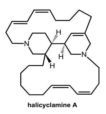
DedA Protein Relates to Action-Mechanism of Halicyclamine A, a Marine Spongean Macrocyclic Alkaloid, as an Anti-dormant Mycobacterial Substance

1
Graduate School of Pharmaceutical Sciences, Osaka University, Yamada-oka 1-6, Suita, Osaka 565-0871, Japan
2
Department of Chemistry, Faculty of Science, Lampung University, Jl. Prof. Dr. Sumantri Brodjonegoro No. 1, Bandar Lampung 35145, Indonesia
*
Authors to whom correspondence should be addressed.
Mar. Drugs 2011, 9(6), 984-993; https://doi.org/10.3390/md9060984
Submission received: 1 April 2011 / Revised: 25 May 2011 / Accepted: 3 June 2011 / Published: 8 June 2011

Abstract

:
A macrocyclic alkaloid, halicyclamine A, was re-discovered from an Indonesian marine sponge of Haliclona sp. 05A08 as an anti-dormant mycobacterial substance. To clarify action-mechanism of halicyclamine A, halicyclamine A-resistant strains were screened from the transformants of Mycobacterium smegmatis with the genomic DNA library of M. bovis BCG, which were constructed in the multi-copy shuttle cosmid pYUB145. Sequencing analysis of the cosmids isolated from the halicyclamine A-resistant transformants revealed that the responsible gene was involved in the genome region between 2920.549 kb and 2933.210 kb. Further experiments using the transformants over-expressing individual gene contained in the responsible region were executed, and the transformant, which over-expressed BCG2664 gene assigned as dedA gene, was found to become halicyclamine A-resistant. This evidence strongly suggested that DedA protein correlates with the action-mechanism of halicyclamine A as an anti-dormant mycobacterial substance.

Graphical Abstract

1. Introduction

Tuberculosis (TB) is one of the most common causes of morbidity and mortality in HIV-positive adults living in poverty [1]. Eight million new TB cases and two million deaths from TB are estimated each year [2]. It is now generally accepted that the requirement for a minimum six months treatment for TB is due to the difficulty in eradicating non-replicating persistent Mycobacterium tuberculosis. Therefore, new lead compounds, which are effective against M. tuberculosis in both active and dormant states, are urgently needed. Although physiology of latent M. tuberculosis infection is still unclear, hypoxic condition was found to induce dormant state of Mycobacterium sp., which has a drug susceptibility profile resembling that of the latent M. tuberculosis infection [35]. Recently, we have established a screening system searching for anti-dormant mycobacterial substances [68]. In this assay system, M. tuberculosis H37Ra under hypoxic conditions, became highly resistant against isoniazid, which is one of the first-line drugs for TB. Thus, the MIC value of isoniazid against M. tuberculosis H37Ra was 0.05 μg/mL under standard aerobic growth condition. While, the MIC value of isoniazid was more than 100 μg/mL under nitrogen atmosphere containing 0.2% oxygen. In the course of our search for anti-dormant mycobacterial substances from marine organisms by using this assay system, a macrocyclic alkaloid, halicyclamine A (Figure 1), has been re-discovered as an anti-dormant Mycobacterial substance from an Indonesian marine sponge of Haliclona sp 05A08 [6]. Halicyclamine A showed potent anti-microbial activity against M. smegmatis (MIC = 2.5 μg/mL), M. bovis BCG (MIC = 1.0 μg/mL), and M. tuberculosis H37Ra (MIC = 5.0 μg/mL) under standard aerobic growth condition as well as dormancy-inducing hypoxic condition [6]. On the other hand, M. Jaspars, et al. reported that the crude spongean extract containing halicyclamine A showed an inhibitory activity against human inosine 5’-monophosphate dehydrogenase (IMPDH) [9]. In order to clarify action-mechanism of halicyclamine A, we previously prepared the transformants of M. smegmatis, which over-expressed mycobacterial IMPDHs, since the transformant over-expressing target proteins often confer drug-resistance [10,11]. However, the IMPDH over-expressing strains showed the same MIC value against halicyclamine A with that of the wild-type M. smegmatis [6]. In this paper, our continued study of the action-mechanism of halicyclamine A is presented.

2. Results and Discussion

2.1. Isolation of the Halicyclamine A-Resistant Clones from the Transformants of M. smegmatis with Genomic DNA Library

To identify the gene that could confer a resistance to halicyclamine A, we prepared transformants of Mycobacterium smegmatis, which were transformed with the genomic DNA library of M. bovis BCG constructed in the multi-copy shuttle cosmid pYUB145. Then, halicyclamine A-resistant clones were screened from over the 4,000 transformants by the dish culture containing 5.0 μg/mL (2 × MIC value for M. smegmatis) of halicyclamine A. We could isolate two transformants, designated ICHO2004 and ICHO2005, as the resistant strains to halicyclamine A (Figure 2). Each resistant strain was cultured in the Middlebrook 7H9 broth containing hygromycin B, and the two cosmids (pYUB145_2004 and pYUB145_2005) were extracted from the ICHO2004 strain and ICHO2005 strain, respectively. Then, the pYUB145_2004 and pYUB145_2005 cosmids were found to contain 23.070 kb (coordinates from 2920.549 kb to 2943.619 kb) and 26.587 kb (coordinates from 2906.623 kb to 2933.210 kb) genome fragments, respectively. This data indicated that the gene for giving resistnace to halicyclamine A would be contained in the genome region of M. bovis BCG from 2920.549 kb to 2933.210 kb (12.661 kb) (Figure 3).

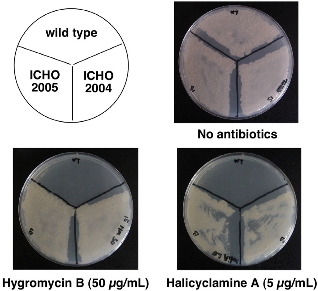
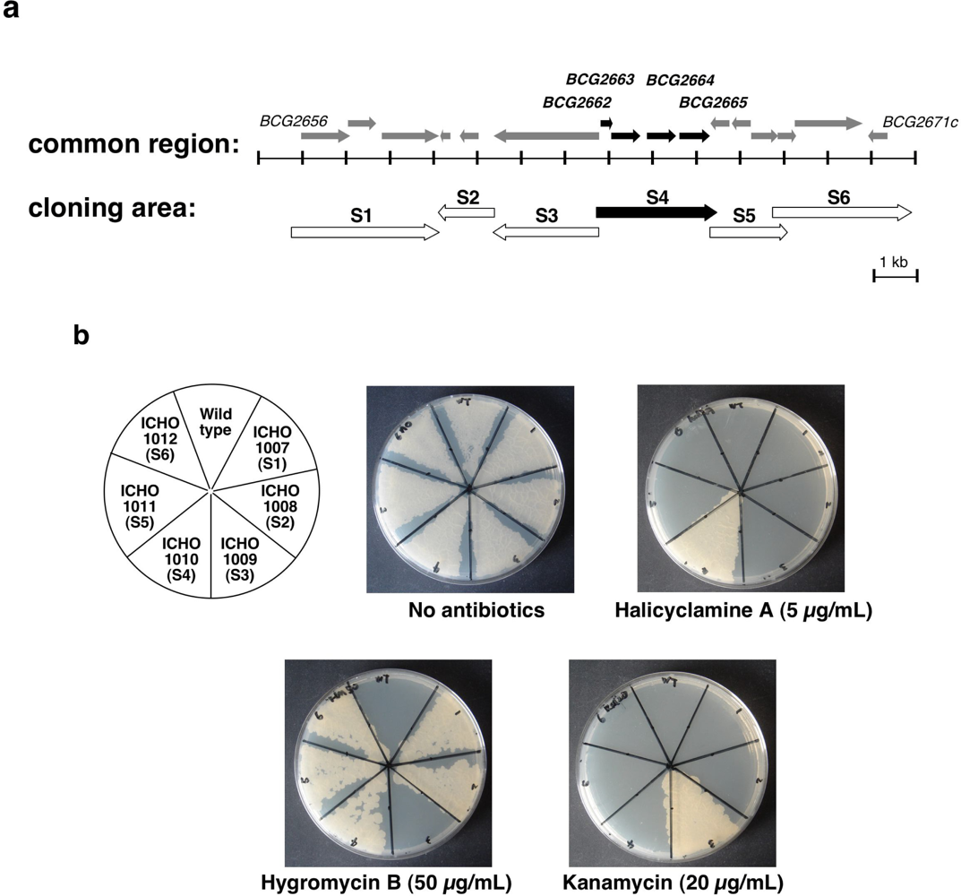
2.2. Second Selection of the Transformants of M. smegmatis Expressing the Candidate Region Showing Resistance to Halicyclamine A

To narrow down candidate genes expressing resistance to halicyclamine A, we then divided the 12.661 kb common region into six areas (S1–S6) as shown in Figure 4a and established six transformants (ICHO1007–ICHO1012) of M. smegmatis, which over-expressed each area. As shown in Figure 4b, all established transformants showed resistance against hygromycin B or kanamycin, which corresponds to the antibiotic-resistance genes in the plasmids of pMV206 or pMV261. The transformant of ICHO1010, which over-expressed the S4 area containing BCG2662, BCG2663, BCG2664 and BCG2665 genes, showed resistance against 5.0 μg/mL concentartion of halicyclamine A, whereas no resistance to halicyclamine A was observed in both wild-type and other transformants of M. smegmatis.

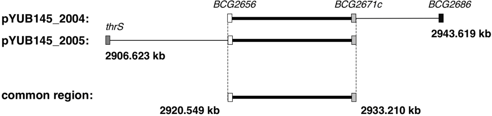
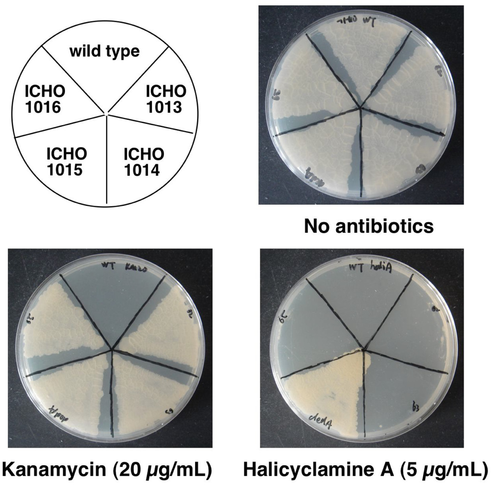
2.3. Identification of the Gene Causing Resistance to Halicyclamine A

To identify the gene causing resistance to halicyclamine A, four transformants (ICHO1013–ICHO1016) of M. smegmatis, which over-expressed each BCG2662, BCG2663, BCG2664 and BCG2665 gene, were prepared, and we observed growth of the four transformants on the 7H10 agar containing 5 μg/mL concentration of halicyclamine A. As a result, the only ICHO1015 strain, which over-expresses BCG2664 gene assigned as dedA gene, showed resistance to halicyclamine A (Figure 5). In addition, the MIC value of halicyclamine A against ICHO1015 strain was evaluated 5.0 μg/mL by agar dilution assay using 7H10 agar plate, whereas 2.5 μg/mL of halicyclamine A inhibited the growth of the wild type of M. smegmatis completely (data not shown) [12]. Although further study is necessary for the identification of the target molecule of halicyclamine A, these results strongly suggested that DedA protein correlates with the action mechanism of halicyclamine A as an anti-dormant mycobacterial substance.
DedA family gene exists in most bacterial genome and is speculated to encode integral membrane protein with completely unknown functions. M. smegmatis, M. bovis BCG and M. tuberculosis H37Rv have seven genes of dedA family in their genome, respectively. The homology between BCG2664 gene and one of dedA genes in M. tuberculosis H37Rv is 100%, while the homology with that in M.smegmatis is 38%. Although no precise function of DedA family protein has been reported, several studies suggested that DedA family protein might function as envelope biogenesis and maintenance. For instance, a DedA protein in Ralstonia metallidurans was shown to be required for resistance to selenite and was thought to be involved in transport function [13]. The yghB and yqjA genes, which belong to the dedA gene family, play redundant and critical roles in membrane biology that are essential for completion of cell division in E. coli [14]. From these observations, halicyclamine A possibly inhibits the function of envelope biogenesis and maintenance in Mycobacterium species by inhibition of DedA protein.

3. Experimental Section

3.1. Materials

Middlebrook 7H9 broth, Middlebrook 7H10 agar, Middlebrook OADC Enrichment, and Luria-Bertani (LB) broth were obtained from BD (Franklin Lakes, NJ, USA). DNA restriction enzymes and T4 DNA ligase were obtained from New England BioLabs INC. (Ipswich, MA, USA). Expand High Fidelity PCR System (Roche Applied Science, Mannheim, Germany) was used for PCR. Carbenicillin, hygromycin B, isoniazid, kanamycin and other chemicals were purchased from Sigma (St. Louis, MO, USA). Halicyclamine A was isolated from Indonesian marine sponge of Haliclona sp. 05A08 (Figure 1). Briefly, the methanol extract (43 g) of the dry sponge (200 g) were partitioned by the alkaloid extraction procedure. The alkaloid fraction (2.5 g) was subjected to silica gel column chromatography (eluted with CHCl3-MeOH containing 1.0% triethylamine) and HPLC [COSMOSIL Sugar-D column (Nacalai tesque, Kyoto, Japan); eluted with CHCl3-CH3CN-H2O] to isolate halicyclamine A (20% yield from the alkaloid fraction). Halicyclamine A was identified by ESI-TOF-MS and 2D-NMR analyses and comparison with authentic spectral data [9,15].

3.2. Bacterial Culture

Mycobacterium strains were grown in Middlebrook 7H9 broth containing 10% Middlebrook OADC enrichment, 0.2% glycerol, and 0.05% Tween 80, or on Middlebrook 7H10 agar containing 10% Middlebrook OADC enrichment and 0.5% glycerol. Escherichia coli DH5α was used for cloning and maintaining plasmid and grown in Luria-Bertani (LB) liquid medium. E. coli HB101 was used for construction of the genomic DNA library of M. bovis BCG and grown in LB liquid medium containing 0.3% maltose and 10 mM MgSO4. The concentrations of antibiotics used were 100 μg/mL (carbenicillin), 150 μg/mL (hygromycin B), and 40 μg/mL (kanamycin) for E. coli strains and 50 μg/mL (hygromycin B) and 20 μg/mL (kanamycin) for Mycobacterium strains, respectively.

3.3. Construction of Gnomic DNA Library and Transformation of M. smegmatis mc2155

The chromosomal DNA of M. bovis BCG Pasteur was prepared by the hexadecyltrimethylammonium bromide (CTAB) method [16]. The chromosomal DNA was then digested with restriction endonuclase Sau3AI to produce approximately 30 kb DNA fragments. Genomic DNA library was constructed in the E. coli-Mycobacterium shuttle cosmid, pYUB145, by using the double cos vector strategy as previously described [17]. Briefly, the left and right arms of pYUB145 were generated by digestion with restriction endonuclases of XbaI and BamHI. The fragments of genome were ligated to the both arms of pYUB145. The mixture of ligations were in vitro packaged with packaging mix of MaxPlax Lambda Packaging (Epicentre), and the resulting recombinant cosmids were transduced to E. coli HB101. The transformants of E. coli HB101 were selected on the LB agar plates containing carbenicillin. Over 3 × 10⁵ independent clones were pooled, and cosmids for transformation of M. smegmatis was obtained by large scale DNA preparation using a standard alkaline lysis method. In order to prepare transformants of M. smegmatis with genomic DNA library, M. smegmatis mc2155 were grown at 37 °C as described above until the optical density reached 0.8–1.0 at 600 nm. The cultures were centrifuged, and the resulting pellets were washed with 10% glycerol twice and re-suspended in the same solution (1/10th of the initial culture volume). The cell suspensions were mixed with gnomic DNA library and electroporated (2500 V, 25 μF, 1000 Ω). The resulting suspensions were incubated at 37 °C for 4 h and then plated on Middlebrook 7H10 agar containing 50 μg/mL of hygromycin B.

3.4. Isolation of Halicyclamine A-Resistant Clones from the Transformants of M. smegmatis with Genomic DNA Library and End Sequencing of Cosmids

Halicyclamine A-resistant clones were screened from over the 4 × 10³ transformants of M. smegmatis with the genomic DNA library by cultivating on the 7H10 agar containing 5.0 μg/mL concentration of halicyclamine A. Then, the resistant clones against halicyclamine A were grown in the 7H9 broth containing hygromycin B, and cosmids for end sequencing were isolated by using standard alkaline lysis method. The cosmids extracted from the halicyclamine A-resistant transformants were subjected to end sequencing (MACROGEN, South Korea). The primers P1 (5’-GTACGCCACCGCCTGGTTC-3’) and P2 (5′-GTGCCACCTGACGTCTAAG-3’), which were designed based on the sequence of cosmid vector pYUB415, were used to amplify the integrated end sequences of the cosmids. The obtained sequences were analyzed by BLAST search using the database of BCGList ( http://genolist.pasteur.fr/BCGList/) and Comprehensive Microbial Resource in J. Craig Venter Institute ( http://cmr.tigr.org/cgi-bin/CMR/CmrHomePage.cgi).

3.5. Preparation of the Transformants of M. smegmatis Over-Expressing the Candidate-Genes for Giving Resistance to Halicyclamine A

The gene regions or genes for candidate giving resistance to halicyclamine A were PCR amplified from the cosmids pYUB415_2004 or pYUB415_2005 by using the primer pairs as shown in Table 1. PCR was performed using a program of 30 cycles of 94 °C for 15 sec, 57 °C for 30 sec and 68 °C for 1 min/kb. Following cloning into pCR2.1-TOPO (Invitrogen) and sequencing, the cloned PCR fragment was excised using the primer-introduced restriction sites and cloned into the mycobacterial expression vector pMV261 having hsp60 promoter and kanamycin-resistant gene or promoter-less shuttle vector pMV206 having hygromycin B-resistant gene. Description of plasmids and name of transformants were listed as shown in Table 1. The transformation of M. smegmatis was performed by above mentioned method.

4. Conclusions

We previously found that a marine spongian macrocyclic alkaloid, halicyclamine A, showed potent anti-mycobacterial activity under standard aerobic growth conditions as well as dormancy-inducing hypoxic condition, although its action-mechanism was not clear. In this paper, we searched the gene, which gives a resistance to halicyclamine A, using the genomic DNA library of M. bovis BCG, since the transformants over-expressing target proteins often confer drug-resistance. Then, we finally found that a transformant of M. smegmatis, which over-expressed BCG2664 gene coded DedA protein, showed the resistance to halicyclamine A. This evidence strongly suggested that DedA protein correlates with the action-mechanism of halicyclamine A as an anti-dormant Mycobacterial substance.

Acknowledgments

The authors thank W. R. Jacobs, Jr. and C. Vilchèze (Albert Einstein College of Medicine) for their support concerning the genomic DNA library. This study was financially supported by the Hoansha Foundation, the Uehara Memorial Foundation, Grant-in-Aid for scientific research from the Ministry of Education, Culture, Sports, Science and Technology of Japan.
  • Samples Availability: Available from the authors.

References

  1. Idemyor, V. HIV and tuberculosis coinfection: inextricably linked liaison. J Natl Med Assoc 2007, 99, 1414–1419. [Google Scholar]
  2. Global Tuberculosis Control: Epidemiology, Strategy, Financing; WHO report 2009. World Health Organization: Geneva, Switzerland, 2009. Available online: http://www.who.int/tb/publications/global_report/2009/en/index.html (accessed on 25 May 2011).
  3. Wayne, LG; Hayes, LG. An in vitro model for sequential study of shiftdown of Mycobacterium tuberculosis through two stages of nonreplicating persistence. Infect Immun 1996, 64, 2062–2069. [Google Scholar]
  4. Lim, A; Eleuterio, M; Hutter, B; Murugasu-Oei, B; Dick, T. Oxygen depletion-induced dormancy in Mycobacterium bovis BCG. J Bacteriol 1999, 181, 2252–2256. [Google Scholar]
  5. Dick, T; Lee, BH; Murugasu-Oei, B. Oxygen depletion induced dormancy in Mycobacterium smegmatis. FEMS Microbiol Lett 1998, 163, 159–164. [Google Scholar]
  6. Arai, M; Sobou, M; Vilchèze, C; Baughn, A; Hashizume, H; Pruksakorn, P; Ishida, S; Matsumoto, M; Jacobs, WR, Jr; Kobayashi, M. Halicyclamine A, a marine spongean alkaloid as a lead for anti-tuberculosis agent. Bioorg Med Chem 2008, 16, 6732–6736. [Google Scholar]
  7. Arai, M; Ishida, S; Setiawan, A; Kobayashi, M. Haliclonacyclamines, tetracyclic alkylpiperidine alkaloids, as anti-dormant mycobacterial substances from a marine sponge of Haliclona sp. Chem Pharm Bull 2009, 57, 1136–1138. [Google Scholar]
  8. Pruksakorn, P; Arai, M; Kotoku, N; Vilchèze, C; Baughn, AD; Moodley, P; Jacobs, WR, Jr; Kobayashi, M. Trichoderins, novel aminolipopeptides from a marine sponge-derived Trichoderma sp., are active against dormant mycobacteria. Bioorg Med Chem Lett 2010, 20, 3658–3663. [Google Scholar]
  9. Jaspars, M; Pasupathy, V; Crews, P. A tetracyclic diamine alkaloid, halicyclamine A, from the marine sponge Haliclona sp. J Org Chem 1994, 59, 3253–3255. [Google Scholar]
  10. Banerjee, A; Dubnau, E; Quemard, A; Balasubramanian, V; Um, KS; Wilson, T; Collins, D; de Lisle, G; Jacobs, WR, Jr. inhA, a gene encoding a target for isoniazid and ethionamide in Mycobacterium tuberculosis. Science 1994, 263, 227–230. [Google Scholar]
  11. Telenti, A; Philipp, WJ; Sreevatsan, S; Bernasconi, C; Stockbauer, KE; Wieles, B; Musser, JM; Jacobs, WR, Jr. The emb operon, a gene cluster of Mycobacterium tuberculosis involved in resistance to ethambutol. Nature Med 1997, 3, 567–570. [Google Scholar]
  12. Biehle, JR; Cavalieri, SJ; Saubolle, MA; Getsinger, LJ. Evaluation of Etest for susceptibility testing of rapidly growing mycobacteria. J Clin Microbiol 1995, 33, 1760–1764. [Google Scholar]
  13. Ledgham, F; Quest, B; Vallaeys, T; Mergeay, M; Covès, J. A probable link between the DedA protein and resistance to selenite. Res Microbiol 2005, 156, 367–374. [Google Scholar]
  14. Thompkins, K; Chattopadhyay, B; Xiao, Y; Henk, MC; Doerrler, WT. Temperature sensitivity and cell division defects in an Escherichia coli strain with mutations in yghB and yqjA, encoding related and conserved inner membrane proteins. J Bacteriol 2008, 190, 4489–4500. [Google Scholar]
  15. Matsunaga, S; Miyata, Y; van Soest, RWM; Fusetani, N. Tetradehydrohalicyclamine A and 22-hydroxyhalicyclamine A, new cytotoxic bis-piperidine alkaloids from a marine sponge Amphimedon sp. J Nat Prod 2004, 67, 1758–1760. [Google Scholar]
  16. Connell, ND. Mycobacterium: isolation, maintenance, transformation, and mutant selection. Methods Cell Biol 1994, 45, 107–125. [Google Scholar]
  17. Balasubramanian, V; Pavelka, MS, Jr; Bardarov, SS; Martin, J; Weisbrod, TR; McAdam, RA; Bloom, BR; Jacobs, WR, Jr. Allelic exchange in Mycobacterium tuberculosis with long linear recombination substrates. J Bacteriol 1996, 178, 273–279. [Google Scholar]
Figure 1. Chemical structure of halicyclamine A.
Marinedrugs 09 00984f1
Figure 2. Halicyclamine A resistant-strains of M. smegmatis transformed with genomic DNA library. The wild-type strain and transformants (ICHO2004 and ICHO2005) of M. smegmatis were cultured in 7H9 broth, and the concentration of the each culture was adjusted to 1 × 10⁵ CFU/mL. The 10 μL aliquot of each culture was spread on a 7H10 agar plate in the presence or absence of halicyclamine A (5.0 μg/mL).
Marinedrugs 09 00984f2
Figure 3. Gene maps of the cosmids extracted from the halicyclamine A-resistant transformants.
Marinedrugs 09 00984f3
Figure 4. Growth of the transformants of M. smegmatis over-expressing candidate area showing resistance to halicyclamine A on the 7H10 agar plate containing halicyclamine A. (a) Gene map of the cloning area to prepare transformants of M. smegmatis. (b) Each strain (wild-type of M. smegmatis, ICHO1007, ICHO1008, ICHO1009, ICHO1010, ICHO1011 and ICHO1012) was cultured in the 7H9 broth, and then the cultures were adjusted to 1 × 10⁵ CFU/mL. The 10 μL aliquots of each culture were spread on the 7H10 agar plates in the presence or absence of halicyclamine A (5.0 μg/mL).
Marinedrugs 09 00984f4
Figure 5. Growth of the transformants of M. smegmatis over-expressing the candidate gene for giving resistance to halicyclamine A on the 7H10 agar plate containing halicyclamine A. Each strain (wild-type of M. smegmatis, ICHO1013, ICHO1014, ICHO1015 and ICHO1016) was cultured in the 7H9 broth, and then the cultures were adjusted to 1 × 10⁵ CFU/mL. The 10 μL aliquots of each culture were spread on the 7H10 agar plate in the presence or absence of halicyclamine A (5.0 μg/mL).
Marinedrugs 09 00984f5
Table 1. List of the transformants of M. smegmatis and description of plasmids.
StrainsPlasmidsDescription of plasmidsSequence of primers (5’ to 3’)
F; Forward primer, R; Reverse primer
ICHO1007pMV206Cloned S1 area# (2919991–2923343 bp of M. bovis BCG genome)*F:TCTAGAGCCACACCCTGATAGCATTG
R: AAGCTTTCGCGACGGCTGGAATG
ICHO1008pMV206Cloned S2 area # (2924490–2923316 bp of M. bovis BCG genome)*F:GGATCCAACTGTTTCCGCACGAGGAG
R:CTGCAGTCAACGGCGAACATTCCAGC
ICHO1009pMV261Cloned S3 area # (2926966–2924589 bp of M. bovis BCG genome)*F:GGATCCATCCAGGAGGTCGCAGATGTC
R: GAATTCGTGATGTTCGCGCCTAACGG
ICHO1010pMV206Cloned S4 area # (2926766–2929363 bp of M. bovis BCG genome)*F:CAGCTGATAGCTCTGCGCGTACGAC
R: TTCGAAGATCAGCAACGGGCTGGAC
ICHO1011pMV206Cloned S5 area # (2929454–2931112 bp of M. bovis BCG genome)*F: GGATCCTGCGGTATCGCTTGCCTTG
R: AAGCTTGCGCGACTACGCAGCTC
ICHO1012pMV206Cloned S6 area # (2930898–2933532 bp of M. bovis BCG genome)*F:GGATCCAAGCGGCTAACTGTAGGCCTG
R: AAGCTTTCAGCCCGGTGGTGTTGG
ICHO1013pMV261Cloned BCG2662 gene (2926980–2927222 bp of M. bovis BCGgenome)*F:GAATTCAGGACAACCCGCATGGTCG
R: AAGCTTTATCACTCAGCGCCGGTACG
ICHO1014pMV261Cloned BCG2663 gene (2927223–2927900 bp of M. bovis BCG genome)*F: GAATTCCCGGTCAGGCATTCTTTACCC
R:AAGCTTGAAAGTTCCAGTTGCGCACGAC
ICHO1015pMV261Cloned BCG2664 gene(2928096–2928752 bp of M. bovis BCG genome)*F: GAATTCCCGGTCAGGCATTCTTTACCC
R: AAGCTTCAAGCCGGTCGGTCACG
ICHO1016pMV261Cloned BCG2665 gene(2928915–2929361 bp of M. bovis BCG genome)*F: GGATCCATGCACCATGGGCCTGATCAC
R: GAATTCTACGGCCGGGATCAGCAAC
#;Name of area corresponds to the Figure 4a.
*;Coordinate of M. bovis BCG genome is based on the database of BCGList ( http://genolist.pasteur.fr/BCGList/).

