Article

Capilloquinol: A Novel Farnesyl Quinol from the Dongsha Atoll Soft Coral Sinularia capillosa

1
Department of Marine Biotechnology and Resources, National Sun Yat-sen University, Kaohsiung 804, Taiwan
2
Department of Microbiology, Kaohsiung Medical University, Kaohsiung 807, Taiwan
3
Centers for Asia-Pacific Ocean Research and Translational Biopharmaceuticals, National Sun Yat-sen University, Kaohsiung 804, Taiwan
*
Authors to whom correspondence should be addressed.
Mar. Drugs 2011, 9(9), 1469-1476; https://doi.org/10.3390/md9091469
Submission received: 25 July 2011 / Revised: 19 August 2011 / Accepted: 24 August 2011 / Published: 30 August 2011

Abstract

:
Capilloquinol (1), possessing an unprecedented farnesyl quinoid skeleton, was isolated from the Dongsha Atoll soft coral Sinularia capillosa. The structure of capilloquinol was elucidated by extensive analysis of spectroscopic data. The cytotoxicity and antiviral activity against human cytomegalovirus of 1 was evaluated in vitro.

1. Introduction

Numerous farnesyl quinones and quinols [1], possessing a wide range of structural diversity, have been obtained from marine organisms, including sponges [28], algae [8,9], ascidians [1012], and soft corals [13,14]. Many of these mixed C15 + C6 analogues are of considerable interest due to their cytotoxicity [4,6,7,12], anti-HIV-1 reverse transcriptase activity [4,8,15], and antimicrobial activity [4,6]. As part of a continuing search for bioactive substances from marine invertebrates, chromatographic separation on the acetone extract of the Dongsha Atoll soft coral S. capillosa (Figure 1) resulted in the isolation of capilloquinol (1) (Figure 2), an unprecedented farnesyl quinoid having novel carbon skeleton. The details of isolation and structural elucidation of metabolite 1 are discussed in this paper. The cytotoxicity against P-388 (mouse lymphocytic leukemia), HT-29 (human colon adenocarcinoma), and A-459 (human lung adenocarcinoma) cancer cell lines as well as antiviral activity against human cytomegalovirus (HCMV) of 1 were evaluated. Meanwhile, the posible biosynthetic pathway for 1 was postulated see below.

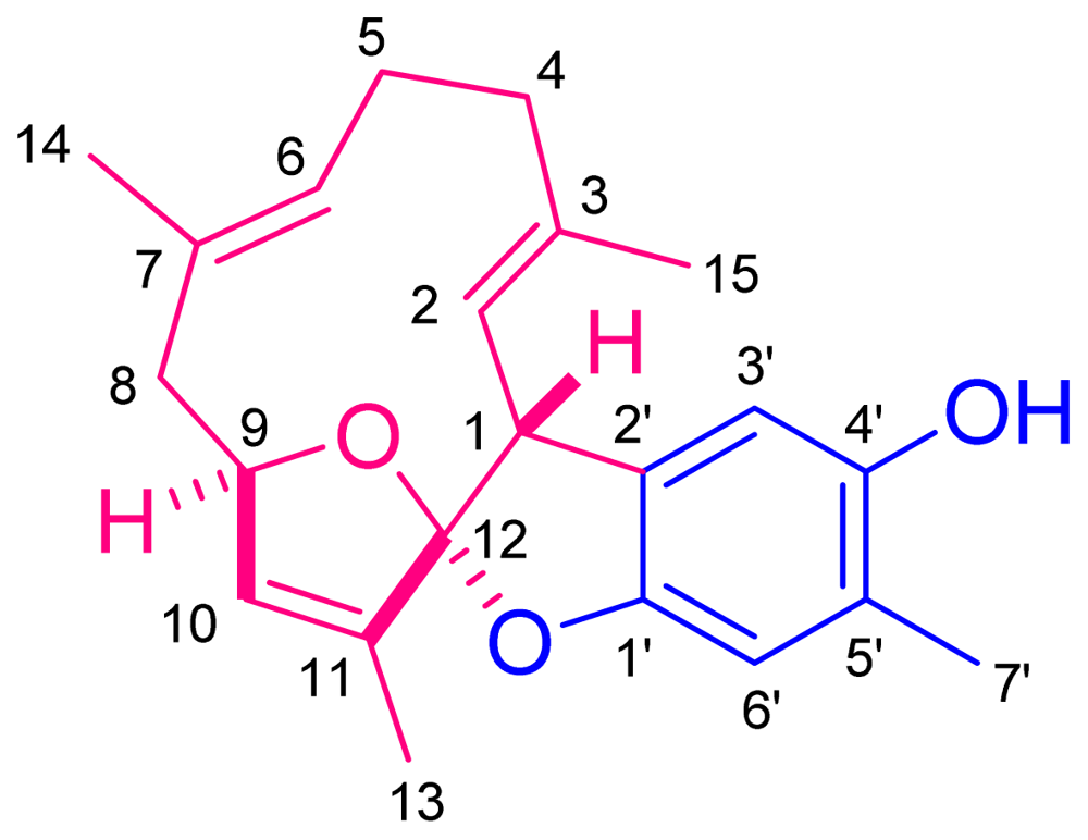
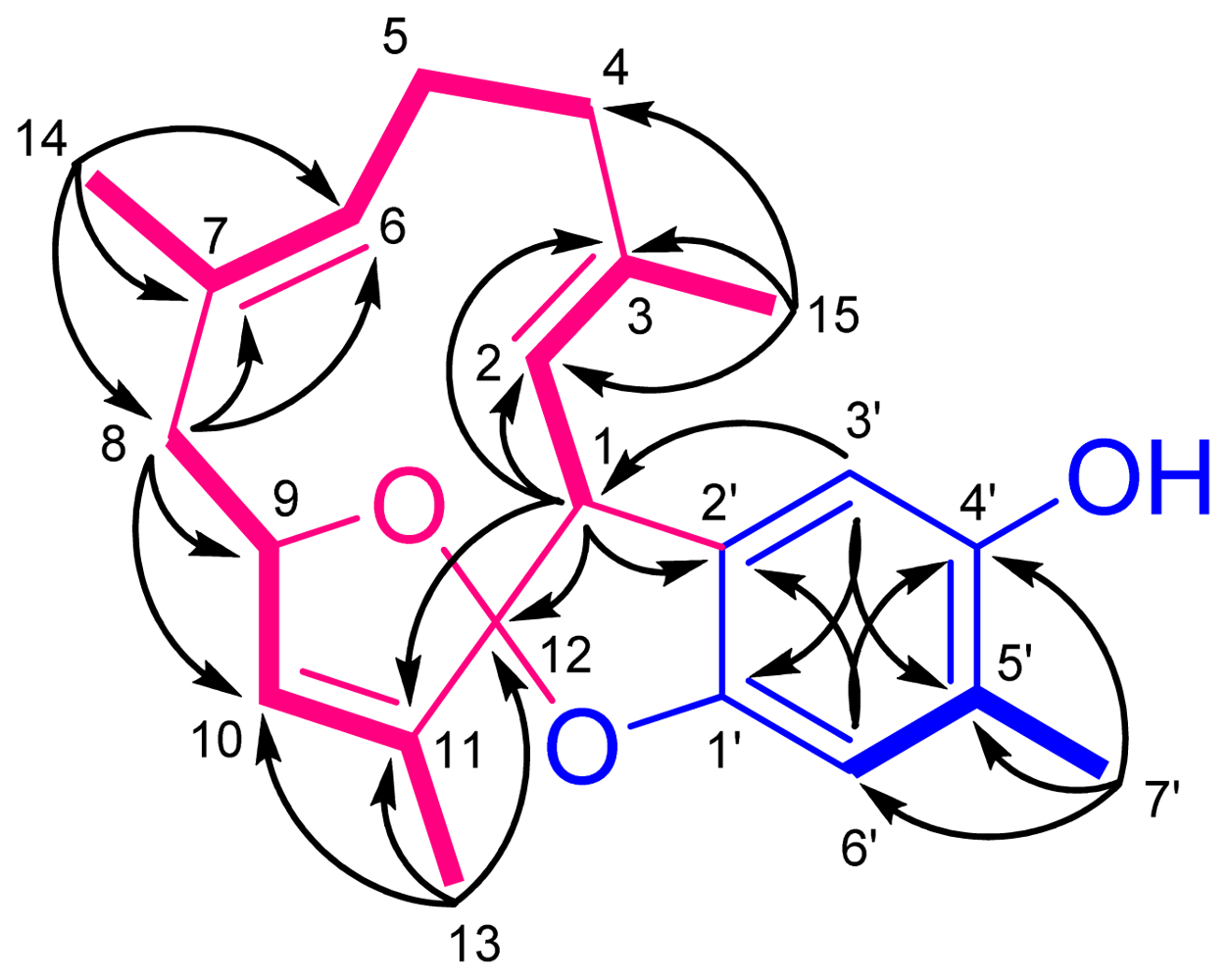
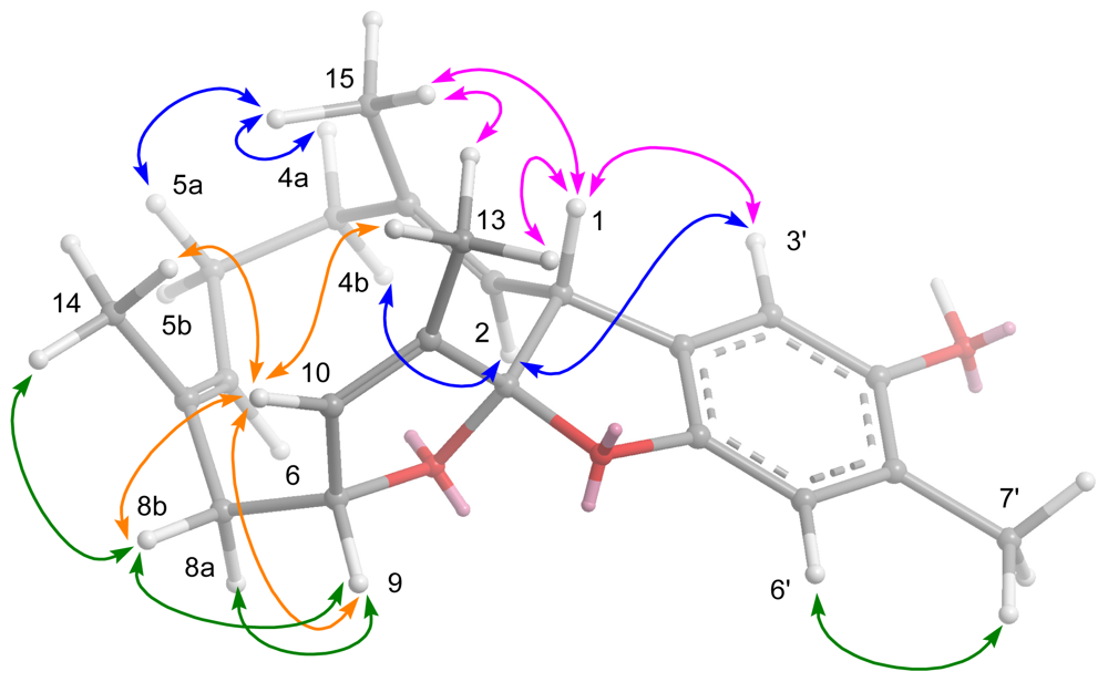
2. Results and Discussion

The acetone extract of S. capillosa was concentrated to a brown gum, which was partitioned with EtOAc and H2O. The EtOAc-soluble residue (60 g) was subjected to Si 60 CC using n-hexane–EtOAc mixtures of increasing polarity for elution, to yield 40 fractions. Fraction 14 (0.8 g), which eluted with n-hexane–EtOAc (2:1), was applied to a RP-18 gravity column and flushed with MeOH–H2O (9:1). The ensuing fraction (72 mg) was further purified by RP-18 High-performance liquid chromatography (HPLC) eluted with MeOH–H2O (9:1) to yield 1 (2.0 mg, 0.003%).
Capilloquinol (1) was obtained as a colorless, viscous oil. The HRESIMS of 1 exhibited a [M + Na]+ peak at m/z 361.1778, consistent with the molecular formula of C22H26O3, implying ten degrees of unsaturation. Its IR spectrum absorption at 3406 cm−1 revealed the presence of hydroxyl group(s). The NMR spectroscopic data (Table 1) of 1 contained resonances for five trisubstituted double bonds [δH 5.01 (d, J = 10.0 Hz, 1H); δC 137.5 (qC) and 125.7 (CH); δH 4.86 (br d, J = 10.5 Hz, 1H); δC 130.3 (qC) and 132.2 (CH); δH 5.97 (s, 1H); δC 135.9 (qC) and 131.8 (CH); δH 6.46 (s, 1H); δC 129.7 (qC) and 112.0 (CH); δH 6.45 (s, 1H); δC 152.2 (qC) and 111.7 (CH)] and a tetrasubstituted double bond [δC 150.8 (qC) and 125.0 (qC)]. The above functionalities accounted for six of the ten degrees of unsaturation, implying a tetracyclic structure for 1.
By interpretation of 1H–1H and long range COSY correlations (Figure 3), it was possible to establish three partial structures of consecutive proton systems extending from H-1 to Me-15 through H-2, from H2-4 to Me-14 through H2-5 and H-6, from H2-8 to Me-13 through H-9 and H-10. The linkages between C-3 and C-4; C-7 and C-8; C-11 and C-12; were elucidated on the basis of the HMBC correlations (Figure 3) from Me-15 to C-2, C-3, and C-4, from Me-14 to C-6, C-7, and C-8, and from Me-13 to C-10, C-11, and C-12. The HMBC spectrum showed correlations from H-1 to C-2, C-3, C-11, and C-12, proving the linkages from C-1 to C-11 through C-12. Additionally, the crucial HMBC correlations from H-3′ to C-1′ and C-5′, from H-6′ to C-2′ and C-4′, and from Me-7′ to C-4′, C-5′, and C-6′, demonstrated the presence of a 1,2,4,5-tetrasubstituted benzene ring. These HMBC correlations also confirmed the positioning of the oxygen-bearing quaternary carbons at C-1′ and C-4′ [δC 152.2 (qC) and 150.8 (qC)], and the methyl group at C-5′. Although there were no direct HMBC correlations available, the remaining two degrees of unsaturation indicated that the two oxygen bridges must be present between C-9/C-12 and C-12/C-1′. This assumption was further supported by its NMR spectroscopic data [δH 5.00 (1H, br s, H-9); δC 85.3 (CH, C-9) and 125.6 (qC, C-12)] [16] and revealed the presences of a 2,3-dihydrobenzofuran-5-ol moiety and an 13-oxa-bicyclo[8,2,1]tridecane ring. Moreover, the crucial HMBC correlation from H-3′ to C-1 established ring fusion at C-1 and C-12. Therefore, the planar structure of 1 was proposed as shown in Figure 2.
The geometries of the trisubstituted double bonds were assigned as 2E and 6E based on the crucial NOESY correlations (Figure 4) between H-1/Me-15, H-4b/H-2, and H-8b/H-6. The key NOE correlations between H-1/Me-13, H-1/Me-15, Me-13/Me-15, Me-15/H-4a, Me-15/H-5a, Me-13/H-10, H-10/Me-14, Me-14/H-8b, and H-8b/H-10 suggested that these protons were oriented on the same side of the macrocyclic ring, while H-2, H-4b, H-5b, H-6, H-8a, and H-9 were oriented on the opposite side. The above-mentioned findings indicated the 1R*, 9R*, and 12R* configurations as depicted in Figure 4. The results, together with other detailed NOESY correlations (Figure 4) of 1, determined the structure of capillosanol as shown in Figure 1.
It is worthwhile to mention that the framework of 1 may be involved in the possible biosynthesis of a farnesyl quinol [13] through oxidation, lactonization, cyclization, and etherization to result in the formation of capilloquinol (1) as depicted in Scheme 1.
Capilloquinol (1) was evaluated for cytotoxicity against P-388, A-459, and HT-29 cancer cell lines as well as antiviral activity against human cytomegalovirus. Metabolite 1 displayed cytotoxicity against P-388, with an ED50 of 3.8 μg/mL. With the exception of the above finding, 1 did not show cytotoxic against A-459, and HT-29 cancer cell lines, nor was it active against human cytomegalovirus (HCMV).

3. Experimental Section

3.1. General Experimental Procedures

Optical rotations were determined with a JASCO P1020 digital polarimeter. Ultraviolet (UV) and infrared (IR) spectra were obtained on a JASCO V-650 and JASCO FT/IR-4100 spectrophotometers, respectively. The NMR spectra were recorded on a Varian Unity INOVA 500 FT-NMR spectrometer at 500 MHz for 1H and 125 MHz for 13C, respectively. Chemical shifts are expressed in δ (ppm) referring to the solvent peaks δH 3.30 and δC 49.0 for CD3OD, respectively, and coupling constants are expressed in Hz. ESI-MS were recorded by ESI FT-MS on a Bruker APEX II mass spectrometer. Silica gel 60 (Merck, Germany, 230–400 mesh) and LiChroprep RP-18 (Merck, 40–63 μm) were used for column chromatography. Precoated silica gel plates (Merck, Kieselgel 60 F254, 0.25 mm) and precoated RP-18 F254s plates (Merck) were used for thin-layer chromatography (TLC) analysis. High-performance liquid chromatography (HPLC) was carried out using a Hitachi L-7100 pump equipped with a Hitachi L-7400 UV detector at 220 nm together with a semi-preparative reversed-phase column (Merck, Hibar LiChrospher RP-18e, 5 μm, 250 × 25 mm).

3.2. Animal Material

The soft coral S. capillosa was collected by hand using SCUBA along the coast reefs offshore from the Dongsha Atoll in April 2007, at a depth of 8–10 m, and was stored in a freezer at −20 °C for two months until extraction. Identification was kindly verified by Prof. Chang-Feng Dai, Institute of Oceanography, National Taiwan University, Taiwan. A voucher specimen (TS-06) was deposited in the Department of Marine Biotechnology and Resources, National Sun Yat-sen University, Taiwan.

3.3. Extraction and Isolation

The acetone extract of S. capillosa was concentrated to a brown gum, which was partitioned with EtOAc and H2O. The EtOAc-soluble residue (60 g) was subjected to Si 60 CC using n-hexane–EtOAc mixtures of increasing polarity for elution, to yield 40 fractions. Fraction 14 (0.8 g) was applied to a RP-18 gravity column to obtain a mixture (72 mg) that was further purified by RP-18 HPLC eluted with MeOH–H2O (9:1) to yield 1 (2.0 mg).
The frozen soft coral (2 kg) was chopped into small pieces and extracted exhaustively by maceration with fresh acetone for 24 h at room temperature. The quantity of solvent used for each extraction (2 L) was at least three times the amount of the soft coral material used. The acetone extracts were filtered and concentrated under vacuum to yield a brownish oily residue, which was subsequently partitioned between EtOAc and H2O. The resulting EtOAc-soluble residue (60 g) was subjected to column chromatography on silica gel using n-hexane with increasing amounts of EtOAc, and finally 100% MeOH as elution, to fractionate roughly 40 fractions on the basis of the 1H NMR spectroscopic data and TLC analyses. Fraction 14 (0.8 g) eluted with n-hexane–EtOAc (2:1) was subjected to was applied to a RP-18 gravity column to obtain a mixture (72 mg) that was further purified by RP-18 HPLC eluted with MeOH–H2O (9:1) to yield 1 (2.0 mg).
Capilloquinol (1): colorless, viscous oil; [α]D 25 +31 (c 0.1, CHCl3); IR (KBr) νmax 3406, 3038, 2973, 2928, 17498, 1457, 1422, 1737 cm−1; 1H NMR and 13C NMR data, see Table 1; ESIMS m/z 361 [M + Na]+; HRESIMS m/z 361.1778 [M + Na]+ (calcd. for C22H26O3Na, 361.1780).

3.4. Cytotoxicity Assay

Cytotoxicity was determined against P-388 (mouse lymphocytic leukemia), HT-29 (human colon adenocarcinoma), and A-549 (human lung epithelial carcinoma) tumor cells using a modification of the MTT [3-(4,5-dimethylthiazo-2-yl)-2,5-diphenyltetrazolium bromide] colorimetric method. The provision of the P-388 cell line was provided by J. M. Pezzuto, formerly of the Department of Medicinal Chemistry and Pharmacognosy, University of Illinois at Chicago. HT-29 and A-549 cell lines were purchased from the American Type Culture Collection. The experimental details of this assay were carried out according to a previously described procedure [1719].

3.5. Anti-HCMV Assay

To determine the effects of natural product upon HCMV cytopathic effect (CPE), confluent human embryonic lung (HEL) cells grown in 24-well plates were incubated for 1 h in the presence or absence of various concentrations of tested natural product. Then, cells were infected with HCMV at an input of 1000 pfu (plaque forming units) per well of 24-well dish. Antiviral activity was expressed as IC50 (50% inhibitory concentration), or compound concentration required to reduce virus induced CPE by 50% after 7 days as compared with the untreated control. To monitor the cell growth upon treating with natural products, an MTT-colorimetric assay was employed [20].

Acknowledgments

This research was financially supported by grants from the National Science Council (NSC99-2628-B-110-002-MY3) of Taiwan awarded to C.-Y.D.
  • Samples Availability: Not available.

References

  1. Capon, RJ. Marine sesquiterpene/quinones. In Studies in Natural Products Chemistry; Atta-ur-Raman, Ed.; Elsevier Science: New York, NY, USA, 1995; Volume 5, pp. 289–326. [Google Scholar]
  2. Schmitz, FJ; Lakashmi, V; Powell, DR; van der Helm, D. Arenarol and arenarone: Sesquiterpenoids with rearranged drimane skeletons from the marine sponge Dysidea arenaria. J. Org. Chem 1984, 49, 241–244. [Google Scholar]
  3. Venkateswarlu, Y; Faulkner, DJ; Steiner, JLR; Corcoran, E; Clardy, J. Smenochromenes, unusual macrocyclic sesquiterpene hydroquinone derivatives from a Seychelles sponge of the genus Smenospongia. J. Org. Chem 1991, 56, 6271–6274. [Google Scholar]
  4. Hirsch, S; Rudi, A; Kashman, Y; Loya, L. New avarone and avarol derivatives from the marine sponge Dysidea cinerea. J. Nat. Prod 1991, 54, 92–97. [Google Scholar]
  5. Rudi, A; Benayahu, Y; Kashman, Y. Likonides A and B: New ansa farnesyl quinols from the marine sponge Hyatella sp. Org. Lett 2004, 6, 4013–4016. [Google Scholar]
  6. Takahashi, Y; Kubota, T; Ito, J; Mikami, Y; Fromont, J; Kobayashi, J. Nakijiquinones G–I, new sesquiterpenoid quinones from marine sponge. Bioorg. Med. Chem 2008, 16, 7561–7564. [Google Scholar]
  7. Aoki, S; Kong, D; Matsui, K; Rachmat, R; Kobayashi, M. Sesquiterpene aminoquinones, from a marine sponge, induce erythroid differentiation in human chronic myelogenous leukemia, K562 cells. Chem. Pharm. Bull 2004, 52, 935–937. [Google Scholar]
  8. Talpir, R; Rudi, A; Kashman, Y. Three new sesquiterpene hydroquinones from marine origin. Tetrahedron 1994, 50, 4179–4184. [Google Scholar]
  9. Rochfort, SJ; Capon, RJ. A new sesquiterpene/phenol from the australian marine brown alga Perithalia caudate. J. Nat. Prod 1994, 57, 849–851. [Google Scholar]
  10. Fu, X; Hossain, MB; Schmitz, FJ; van der Helm, D. Longithorones, unique prenylated para- and metacyclophane type quinones from the tunicate Aplidium longithorax. J. Org. Chem 1997, 62, 3810–3819. [Google Scholar]
  11. Davis, RA; Carroll, AR; Quinn, RJ. Longithorols C–E, three new macrocyclic sesquiterpene hydroquinone metabolites from the Australian ascidian, Aplidium longithorax. J. Nat. Prod 1999, 62, 1405–1409. [Google Scholar]
  12. Issa, HH; Tanaka, J; Rachmat, R; Higa, T. Floresolides, new metacyclophane hydroquinone lactones from an ascidian, Aplidium sp. Tetrahedron Lett 2003, 44, 1243–1245. [Google Scholar]
  13. Coll, JC; Liyanage, N; Stokie, GJ; van Altena, IA; Nemorin, JNE; Sternhell, S; Kazlauska, R. Studies of Australian soft corals. III. A novel furanoquinol from Sinularia lochmodes. Aust. J. Chem 1978, 31, 157–162. [Google Scholar]
  14. Bowden, BF; Coll, JC; de Silva, ED; de Costa, MSL; Djura, PJ; Mahendran, M; Tapiolas, DM. Novel furanosesquiterpenes from several sinularian soft corals. Aust. J. Chem 1983, 36, 371–376. [Google Scholar]
  15. Loya, S; Tal, R; Kashman, Y; Hizi, A. Illimaquinone, a selective inhibitor of the RNase H activity of human immunodeficiency virus type 1 reverse transcriptase. Antimicrob. Agents Chemother 1990, 34, 2009–2012. [Google Scholar]
  16. Chao, C-H; Huang, L-F; Yang, Y-L; Su, J-H; Wang, G-H; Chiang, MY; Wu, Y-C; Dai, C-F; Sheu, J-H. Polyoxygenated steroids from the gorgonian Isis hippuris. J. Nat. Prod 2005, 68, 880–885. [Google Scholar]
  17. Hou, RS; Duh, CY; Chiang, MY; Lin, CN. Sinugibberol, a new cytotoxic cembranoid diterpene from the soft coral Sinularia gibberosa. J. Nat. Prod 1995, 58, 1126–1130. [Google Scholar]
  18. Geran, RI; Greenberg, NH; MacDonald, MM; Schumacher, AM; Abbott, BJ. Protocols for screening chemical agents and natural products against animal tumors and other biological syatems. Cancer Chemother. Rep 1972, 3, 1–91. [Google Scholar]
  19. Cheng, S-Y; Wen, Z-H; Wang, S-K; Chiou, S-F; Hsu, C-H; Dai, C-F; Duh, C-Y. Anti-inflammatory cembranolides from the soft coral Lobophytum durum. Bioorg. Med. Chem 2009, 17, 3763–3769. [Google Scholar]
  20. Stevens, M; Balzarini, J; Tabarrini, O; Andrei, G; Snoeck, R; Cecchetti, V; Fravolini, A; de Clercq, E; Pannecouque, C. Cell-dependent interference of a series of new 6-aminoquinolone derivatives with viral (HIV/CMV) transactivation. J. Antimicrob. Chemother 2005, 56, 847–855. [Google Scholar]
Figure 1. Soft coral Sinularia capillosa.
Marinedrugs 09 01469f1
Figure 2. The structure of capilloquinol (1).
Marinedrugs 09 01469f2
Figure 3. Key 1H–1H COSY () and HMBC (→) correlations of 1.
Marinedrugs 09 01469f3
Figure 4. Selected NOESY correlations of 1.
Marinedrugs 09 01469f4
Scheme 1. Possible biogenetic pathway for 1.
Marinedrugs 09 01469f5
Table 1. 1H and 13C NMR Spectroscopic Data of 1 a.
13C1H
146.5 (CH) b4.31 d (10.0)
2125.7 (CH)5.01 d (10.0)
3137.5 (qC)
440.3 (CH2)a: 2.23 dt (12.0, 3.5) c
b: 2.13 dt (12.0, 4.5)
526.5 (CH2)a: 2.31 dddd (13.0, 12.0, 10.5, 3.5)
b: 2.01 br d (13.0)
6132.2 (CH)4.86 br d (10.5)
7130.3 (qC)
845.1 (CH2)a: 2.51 dd (14.0, 4.5)
b: 2.28 br d (14.0)
985.3 (CH)5.00 s
10131.8 (CH)5.97 s
11135.9 (qC)
12125.6 (qC)
1312.0 (CH3)1.85 br s
1418.9 (CH3)1.47 s
1516.0 (CH3)1.57 s
1′152.2 (qC)
2′129.7 (qC)
3′112.0 (CH)6.45 s
4′150.8 (qC)
5′125.0 (qC)
6′111.7 (CH)6.46 s
7′16.7 (CH3)2.13 s
aSpectra were measured in CD3OD (1H, 500 MHz and 13C, 125 MHz);
bMultiplicities are deduced by HSQC and DEPT experiments;
cJ values (in Hz) are in parentheses.

