Article

Green Synthesis of Silver Nanoparticles, Their Characterization, Application and Antibacterial Activity †

1
Department of Biological and Environmental Sciences, Alabama A&M University, P.O. Box 1672, Normal, AL 35762, USA
2
Department of Physics, Chemistry and Mathematics, Alabama A&M University, 4900 Meridian Street N., Normal, AL 35762, USA
*
Author to whom correspondence should be addressed.
This paper is an extended version of paper presented at the Ninth International Symposium on Recent Advances in Environmental Health Research, Jackson, MS, USA; 16–19 September 2012.
Int. J. Environ. Res. Public Health 2013, 10(10), 5221-5238; https://doi.org/10.3390/ijerph10105221
Submission received: 10 November 2012 / Revised: 20 December 2012 / Accepted: 25 December 2012 / Published: 21 October 2013

Abstract

:
Our research focused on the production, characterization and application of silver nanoparticles (AgNPs), which can be utilized in biomedical research and environmental cleaning applications. We used an environmentally friendly extracellular biosynthetic technique for the production of the AgNPs. The reducing agents used to produce the nanoparticles were from aqueous extracts made from the leaves of various plants. Synthesis of colloidal AgNPs was monitored by UV-Visible spectroscopy. The UV-Visible spectrum showed a peak between 417 and 425 nm corresponding to the Plasmon absorbance of the AgNPs. The characterization of the AgNPs such as their size and shape was performed by Atom Force Microscopy (AFM), and Transmission Electron Microscopy (TEM) techniques which indicated a size range of 3 to 15 nm. The anti-bacterial activity of AgNPs was investigated at concentrations between 2 and 15 ppm for Gram-negative and Gram-positive bacteria. Staphylococcus aureus and Kocuria rhizophila, Bacillus thuringiensis (Gram-positive organisms); Escherichia coli, Pseudomonas aeruginosa, and Salmonella typhimurium (Gram-negative organisms) were exposed to AgNPs using Bioscreen C. The results indicated that AgNPs at a concentration of 2 and 4 ppm, inhibited bacterial growth. Preliminary evaluation of cytotoxicity of biosynthesized silver nanoparticles was accomplished using the InQ™ Cell Research System instrument with HEK 293 cells. This investigation demonstrated that silver nanoparticles with a concentration of 2 ppm and 4 ppm were not toxic for human healthy cells, but inhibit bacterial growth.

1. Introduction

The application of nanoscale materials structures, usually ranging from 1 to 100 nanometers (nm), is an emerging area of nanoscience and nanotechnology. Nanomaterials may provide solutions to technological and environmental challenges in the areas of solar energy conversion, catalysis, biology, biomedical science, and water treatment. Nanoparticles possess very high surface to volume ratios. This property can be utilized in the scientific fields, where high surface area is needed. As an example, in the catalytic industry, some nanoparticles have actually proven to be good catalysts [1]. Moreover, the nanoparticles show bactericidal effects. These essential qualities have been attributed to silver nanoparticles (AgNPs). Therefore, in nano-biotechnological research, AgNPs have received significant attention because of their unique physical, chemical, biological properties, and because of their applicability in electronics, optics and medicine [2,3,4,5,6,7,8,9].
Moreover, silver nanoparticles have different important applications: for example, they might be used as spectrally selective coatings for solar energy absorption, as an intercalation material for electrical batteries, and as optical receptors for biolabeling [10,11,12,13]. AgNPs are well known as inhibitory and antibacterial materials. However, resistance to antimicrobial agents by pathogenic bacteria has emerged in recent years. This is a major challenge for the health care industry, and has been widely studied [14,15,16,17,18,19,20,21,22,23,24].
Chemical and physical methods may successfully produce pure, well-defined nanoparticles, but these techniques are more expensive, energy consuming and potentially toxic to the environment. In this paper, we are considering an environmentally friendly technique to produce AgNPs using extracellular biosynthesis. Biosynthetic methods can employ either microorganism cells or plant extract for nanoparticles production. An exciting branch of biosynthesis of nanoparticles is the application of plant extracts to the biosynthesis reaction. Recently, the green processes for the synthesis of nanoparticles are evolving into an important branch of nanotechnology [25,26,27,28,29,30,31,32,33]. The choice of plants in the present paper has been made based on their medical applications [34,35,36].
In this paper we present a rapid method for nanoparticles production using plant leaves extracts, their characterization and their inhibitory effect against Gram-negative and Gram-positive bacteria. In addition, we are submitting the preliminary results of cytotoxicity of biosynthesized nanoparticles for Human Embryonic Kidney 293 cells (HEK 293).

2. Materials and Methods

The plants selected for silver nanoparticles production are listed in Table 1:
Table 1. Selected plants used for silver nanoparticles synthesis.
Scientific nameCommon name
Actaea racemosa Black cohosh
Magnolia grandifloraMagnolia
Aloe sp.“Tingtinkie”
EucalyptusangophoroidesEucalyptus
Sansevieria trifasciataMother-in-laws’ tongue
Impatiens balsamina Rose Balsam
Pelargonium graveolensGeranium
For nanoparticles production we used plant leaves extracts as reducing agents for silver nitrate salt AgNO3 (Carolina Biological Supplies, Burlington, NC, USA) to reduce nanoparticles. Ten grams of leaves were washed and finely cut, boiled in 50 mL of sterile distilled water for 5 min, 3 mL of the filtrated plant water extract was added to a heated 75 °C AgNO3 solution (50 mL of 103 M of AgNO3) [37,38]. The solution was then tested using a UV-Visible spectrophotometer (Varian, CARY 3E). The plant leaves extracts were also prepared using 10 g of finely cut plant leaves placed in 100 mL ethyl alcohol (C2H5OH) for 24 h.
Transmission Electron Microscopy analysis (TEM) was performed using a FEI Tecnai G2 Spirit Transmission Electron Microscope (Ohio State University, Dayton, OH, USA). Atomic Force Microscopy (AFM) analysis was completed using a Solver Scanning Probe Microscope (Solver P47H, Moscow, Russia) by using full contact and semi-contact methods.
The antibacterial activity of silver nanoparticles was evaluated against three Gram negative bacteria (Escherichia coli ATCC 25922, Salmonella typhimurium ATCC 14028, and Pseudomonas aeruginosa ATCC 15442) and three Gram positive bacteria (Staphylococcus aureus ATCC 6538, Kocuria rhizophila ATTC 9341, and Bacillus thuringiensis ATCC 10792). The microbiological media used in the study was supplied by Becton, Dickinson and Company (Franklin Lakes, NJ, USA). Growth and cytotoxicity analysis was performed using Bioscreen C (Automated Microbiology Growth Analysis System) manufactured in Finland by OY Growth Curves CE.
To study growth of bacteria in broth culture, 5 mL of bacterial culture were placed in a 250 mL flask containing the 200 mL of Bacto Nutrient broth. The culture was then placed in a Lab-Line® Incubator-Shaker by Orbit at a temperature of 37 °C, shaking at 175 rpm for 24 h. A 1:10 mixture of the 24 h bacterial growth and sterile physiological saline water was prepared. An aliquot of 100 µL from each solution was placed into different wells of the Bioscreen C. The first column wells served as controls and had no silver nanoparticles. Aliquots of 10 µL and 20 µL of different concentrations of silver nanoparticles were added to the samples. Growth rate was determined by measuring turbidity using the Bioscreen C for a total of 24 h at regular intervals (15 min).
In order to determine the dose response effect of the silver nanoparticles, both Gram-positive and Gram-negative bacteria were exposed to two different concentrations of silver nanoparticles (2 ppm and 4 ppm). An optical density (OD) curve was generated based on the turbidity measurement over a period of 24 h. During the experiment, data were exported to a PC in MS Excel. Microbiological calculations were generated directly to MS Excel sheets automatically. Measurements were then processed to generate microbiological growth curves, plotting turbidity vs. time [39].
The cytotoxicity evaluation was conducted using the InQ Plus bench top cell research system using HEK 293 cell. HEK 293 cells were divided and plated into InQ Cell Growth Cassettes at the density of 3 × 10⁵cells/mL in a total volume of 8 mL per well (16 mL) of 10% FBS DMEM. Two mL of AgNPs solution with various concentrations for several experiments in one cassette, and control solution (leave extract, pure solution of AgNO3) in second cassette has been injected. Three data points were selected and saved per side. Images were collected at 600 s intervals during the 48 h experiments. The untreated cells and plant extract solutions without AgNPs were used as negative control, while DMSO was used as positive control.

3. Results and Discussion

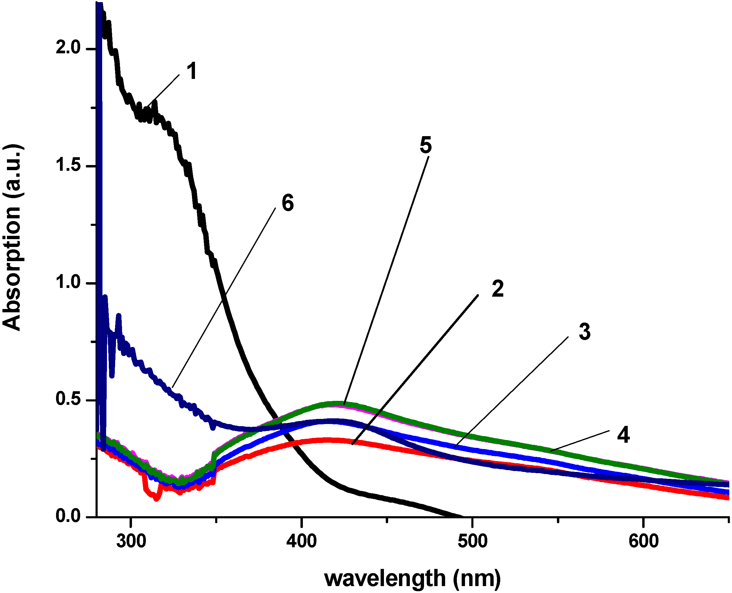
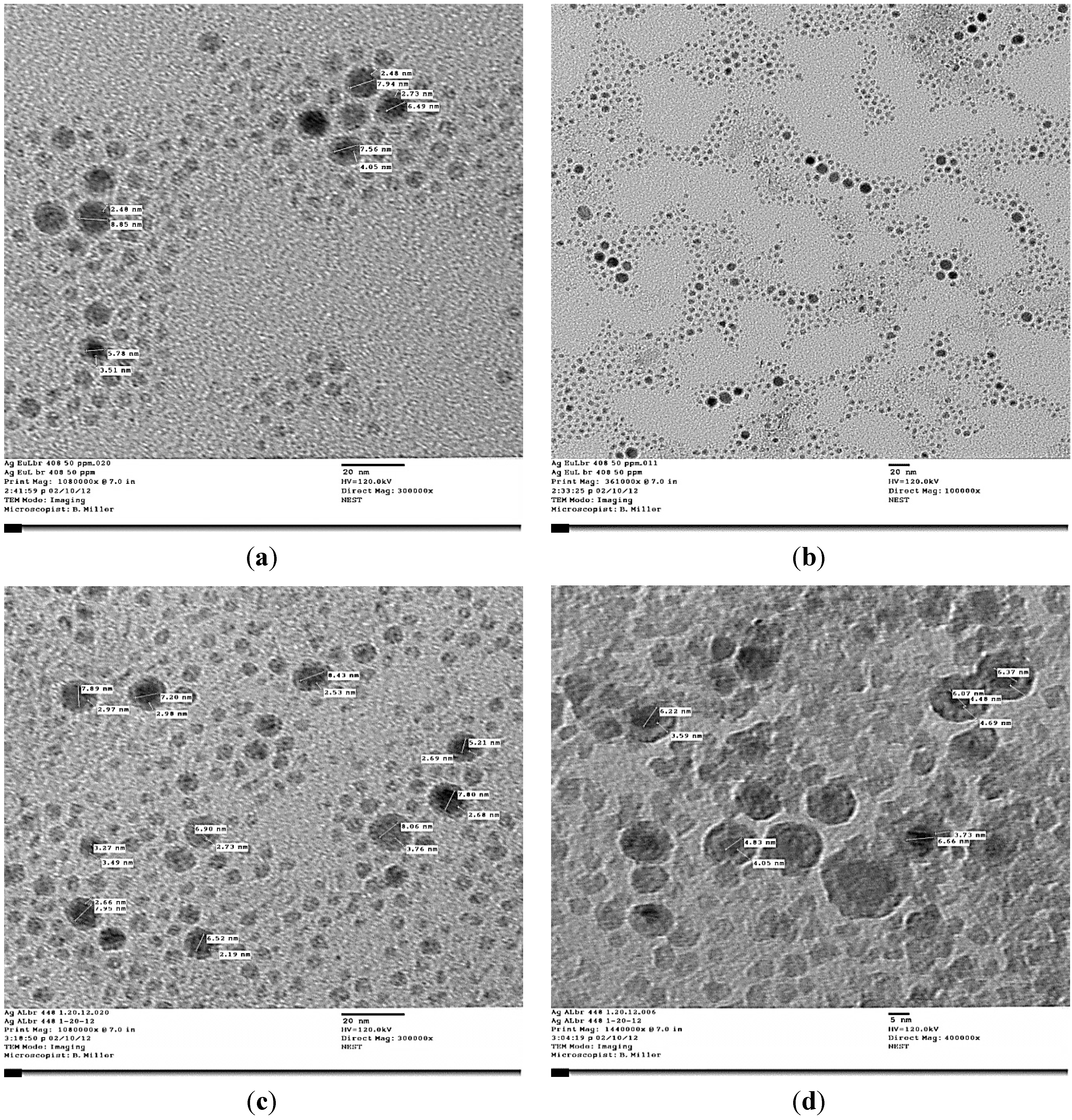
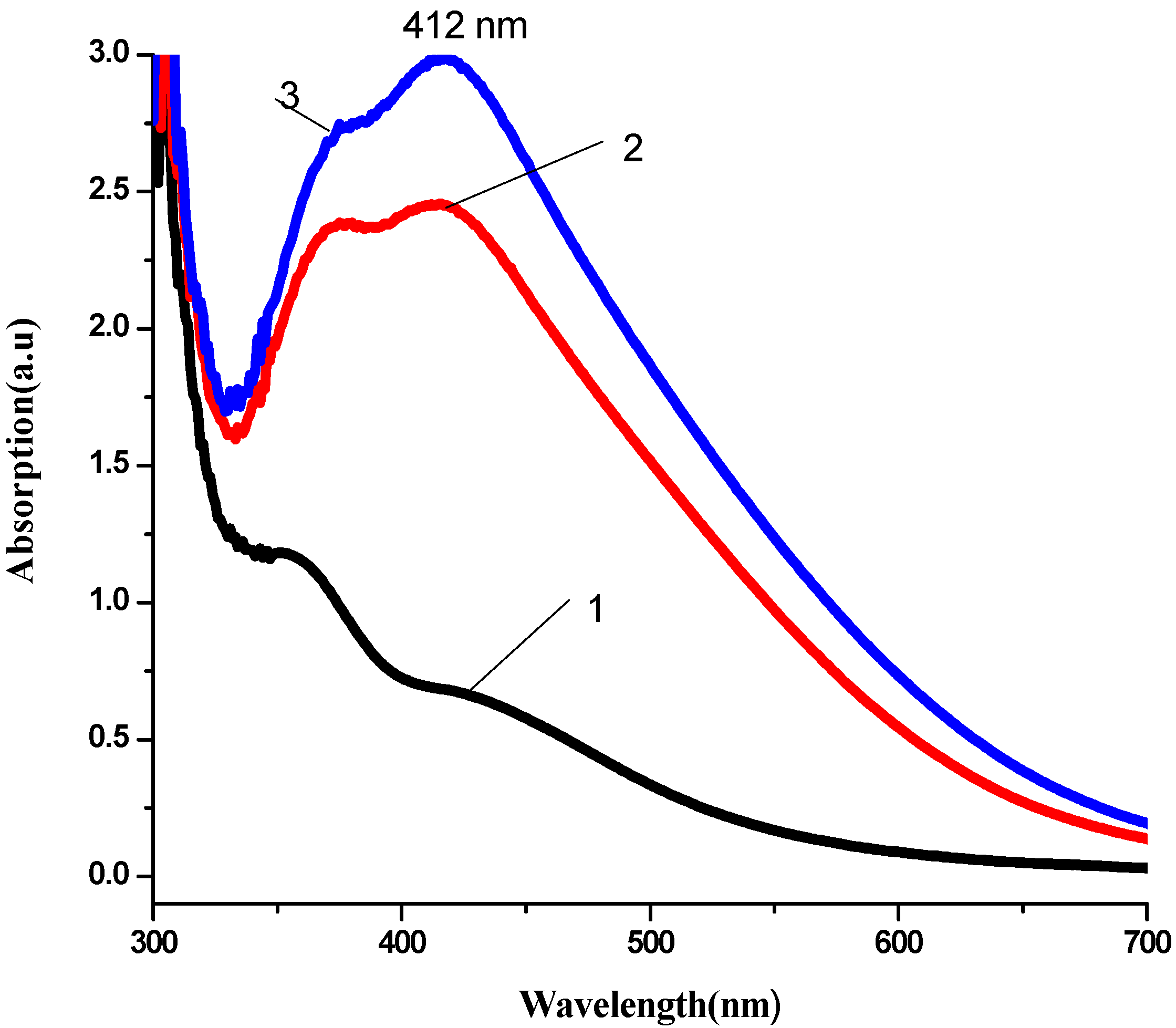
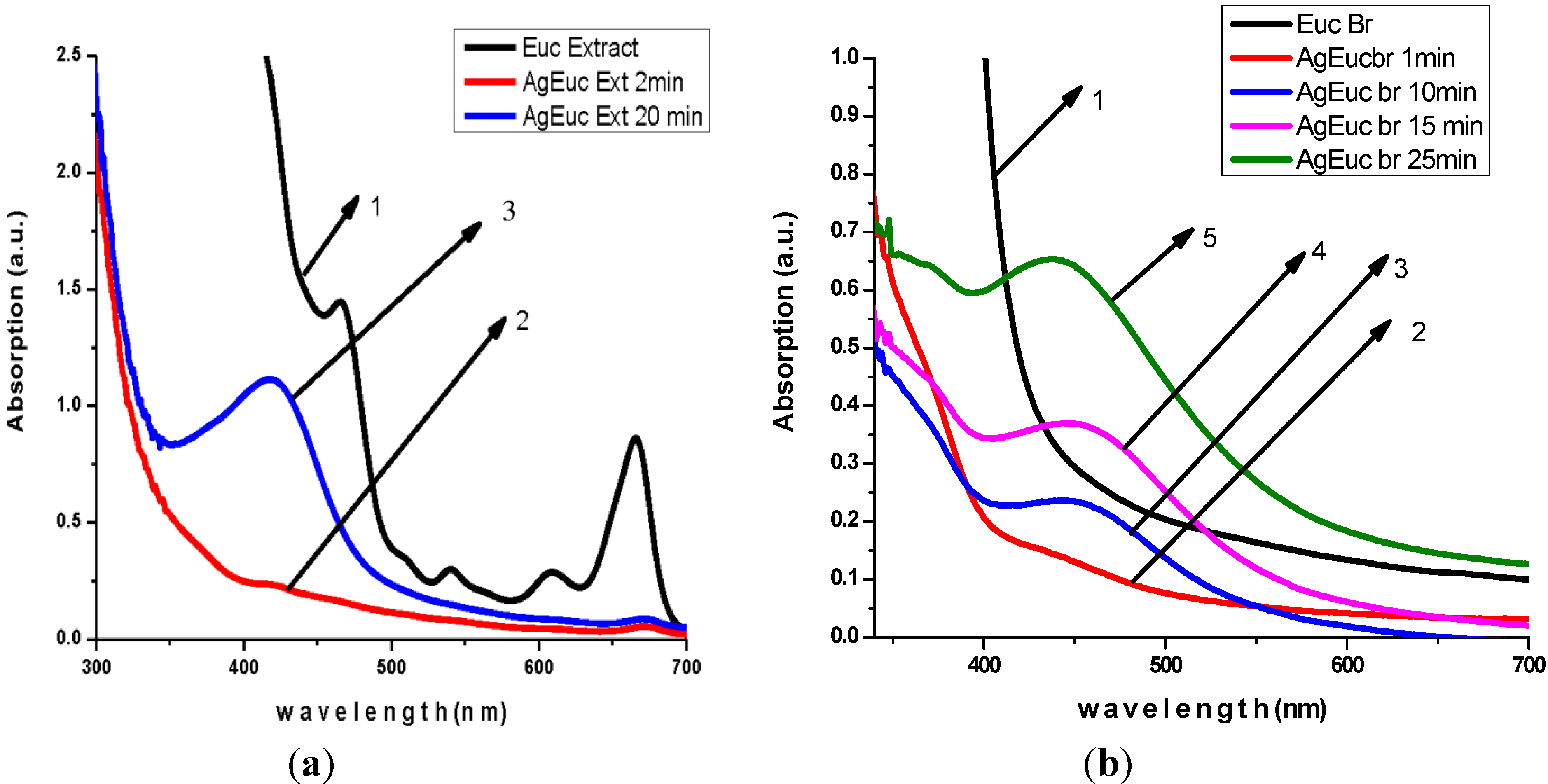
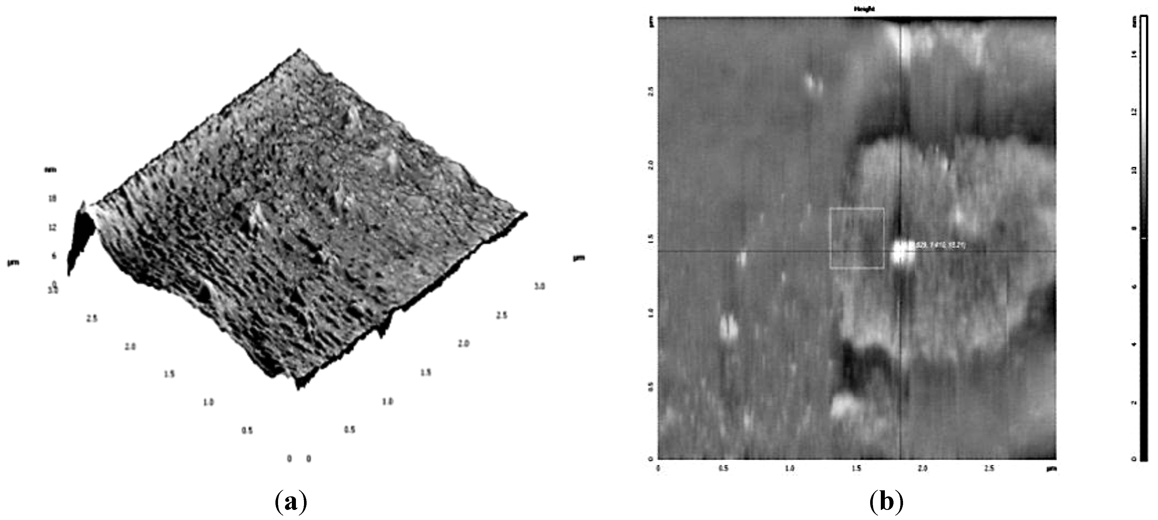
3.1. Nanoparticles Production and Characterization

The production of AgNPs was monitored using UV-Visible spectrometer. Kinetics of the reaction AgNPs production process are presented in Figure 1, Figure 2 and Figure 3.
Figure 1. Kinetic of reaction process of AgNPs production using Magnolia grandiflora water extract as reduction agent. (1) Magnolia leaves water extract in sterile distilled water. (2) AgNO3(10−3 M) + Magnolia aqueous extract (3 mL) after 3 min of reaction. (3) AgNO3 (10−3 M) + Magnolia water extract (3 mL) after 5 min of reaction. (4) AgNO3 (10−3 M) + Magnolia water extract (3 mL) after 10 min of reaction. (5) AgNO3 (10−3 M) + Magnolia water extract (3 mL) after 20 min of reaction. (6) Commercial Ag Colloidal solution—10 ppm concentration.
Ijerph 10 05221 g001
Figure 2. Kinetic of reaction process of AgNPs production using Geranium leaf water extract as reduction agent (1) 1 min after reaction, (2) 3 min after reaction, (3) 5 min after reaction.
Ijerph 10 05221 g002
Figure 3. Kinetic of reaction process of AgNPs production using Eucalyptus ethanol extract (a) and water extract (b) as reduction agent.
Ijerph 10 05221 g003
(a) 1: 2 min after reaction; 2: 1 min after reaction; 20 min after reaction; 3: 10 min after reaction;
(b) 1: 1 min after reaction; 2: 10min after reaction; 3: 15 min after reaction; 4: 25min after reaction; 5: 25 min after reaction.
The production of AgNPs by Aloe “Tingtinkie” leaves aqueous extract, Actaea racemosa (black cohosh), Sansevieria trifasciata and Impatiens scapiflora leaves aqueous extract and ethanol extracts demonstrated similar UV-Visible spectra.
The analysis of UV-Visible spectroscopy data showed an appearance of surface plasmon resonance peak (SPR) at the 417–430 nm wavelength range, which corresponds to AgNPs production. AgNPs absorb radiation intensely at a wavelength of 400 nm due to the transition of electrons. The exact mechanism of the extracellular biosynthesis of metal NP in not well understood. It was hypothesized that NADH coenzyme was working as an electron shuttle to neutralize Ag+ ion [40,41] .
The AgNPs was characterized by Transmission Electron Microscopy (TEM) and Atomic Force Microscopy (AFM) (Figure 4 and Figure 5). Transmission Electron Microscopy (TEM) results showed particles with spherical shape surrounded by biological molecules, which prevent AgNPs from aggregation. As shown in Figure 4 the average size of AgNPs is in the 3–9 nm range.
Figure 4. TEM Images of AgNPs produced by Eucalyptus (a); (b) and Aloe plant (c), (d).
Ijerph 10 05221 g004
Figure 5. AFM images of AgNPs produces by Geranium water extract. (a) 3-D image of Ag nanoparticles produced by Geranium water extract; (b) Estimation of nanoparticles size by decreasing the scanning range (3 µm), the size of AgNPs as shown on the image is ~15.2 nm.
Ijerph 10 05221 g005

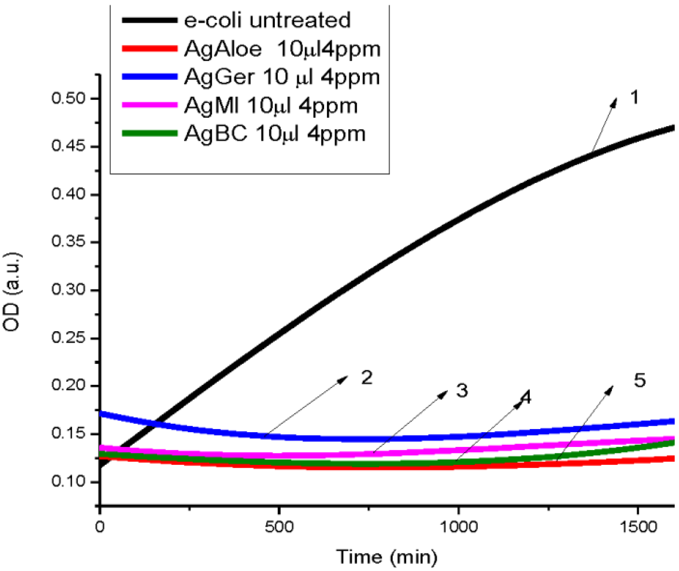
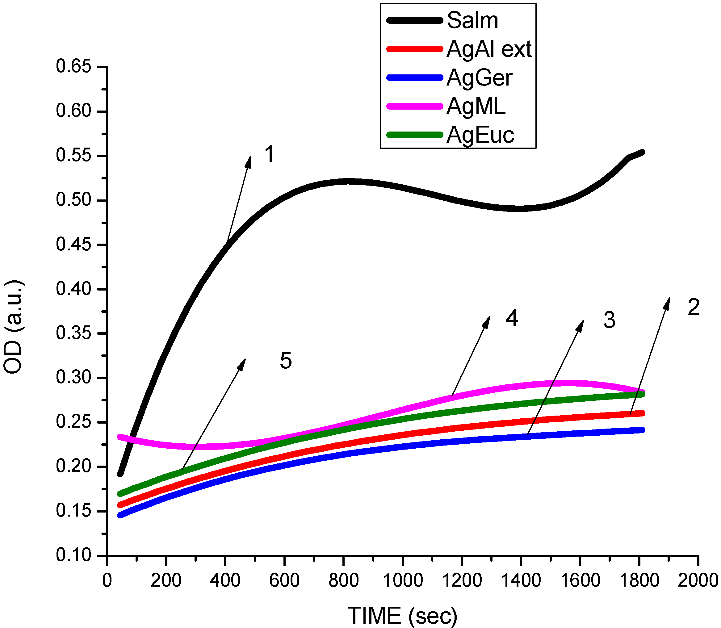
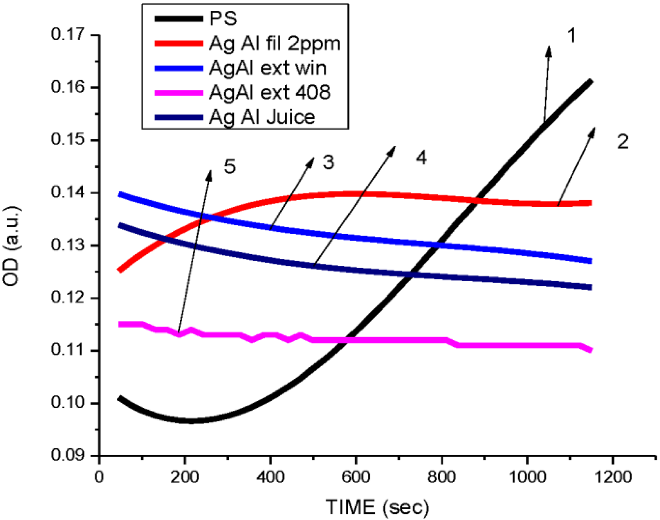
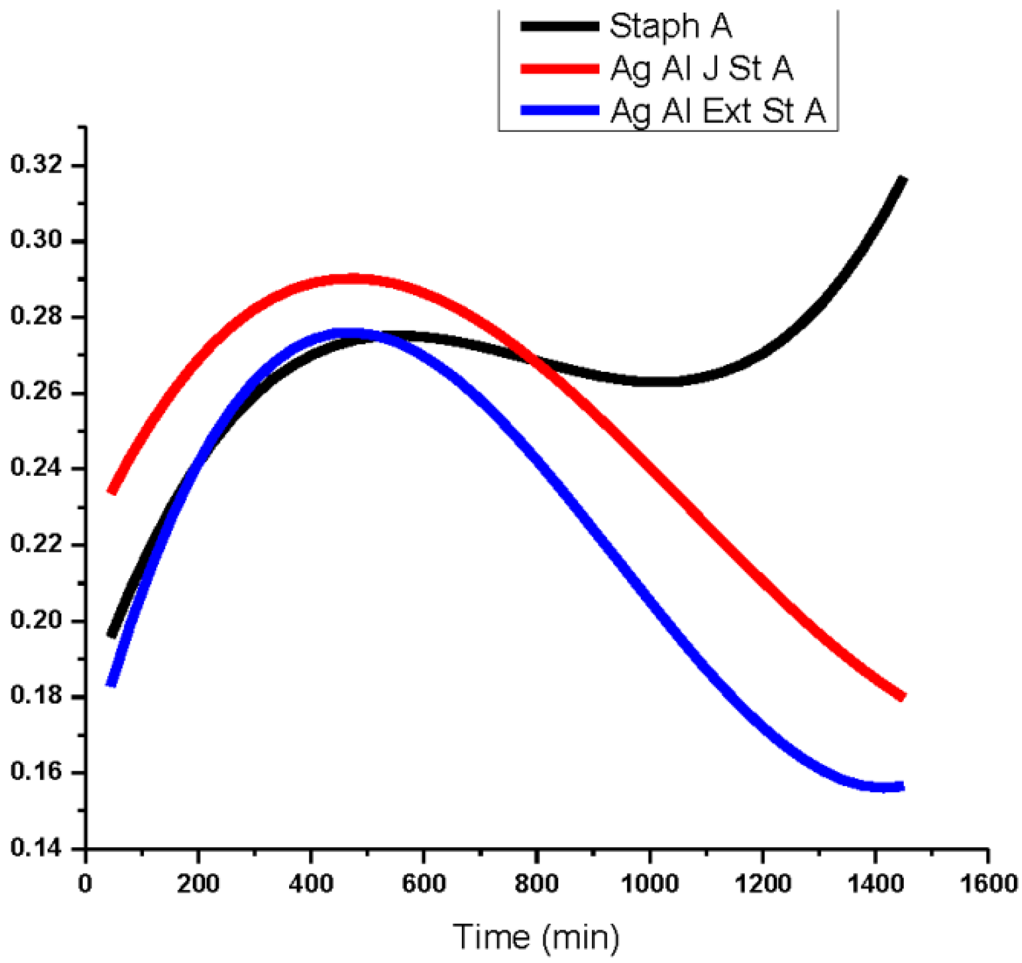
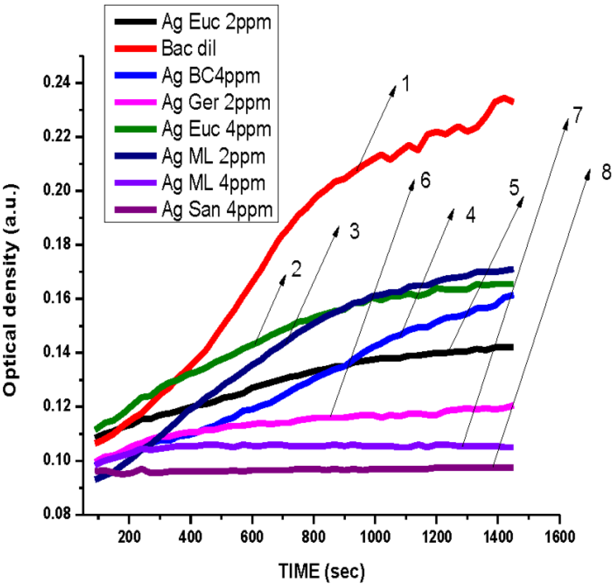
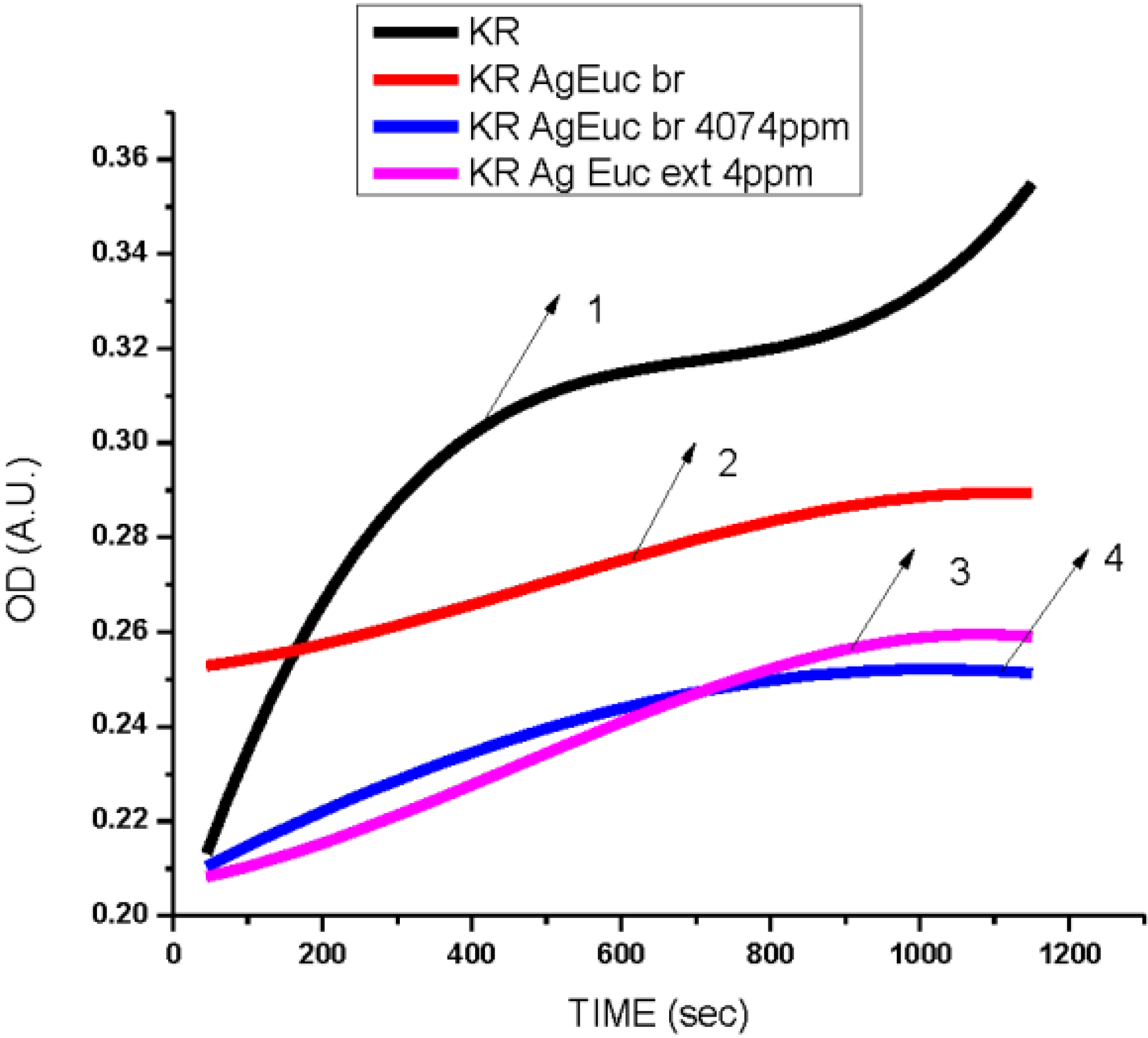
3.2. Antibacterial Activity of Silver Nanoparticles

The antibacterial activity of silver nanoparticles using select plants was found to exert inhibitory effects on different species of bacteria: three Gram negative bacteria and three Gram positive bacteria. The silver nanoparticles synthesized from aloe, geranium, magnolia and black cohosh extracts at a concentration of 4 ppm, all inhibited the growth of E. coli based on the OD readings as compared to the control. The control wells without AgNPs showed OD absorption units at 0.46, while the test wells showed OD units between 0.12 and 0.17 indicating little or no growth (Figure 6). Aloe extract nanoparticles showed the highest antibacterial activity followed by black cohosh and geranium nanoparticles with the lowest inhibition. These results are in agreement with previous studies [42] indicating that aloe-produced nanoparticles have a high inhibitory growth on E. coli at low concentrations.
According to Gogoi et al. [43], the cell surface of E. coli is negatively charged and thus Ag+ easily interacts with cell membranes, thus disabling their function. The silver nanoparticles from aloe extracts, at 4 ppm, had a bacteriostatic effect on Pseudomonas (Figure 7) compared to the untreated species. Similar results were obtained in the case of Salmonella where aloe extract—produced nanoparticles showed the highest antibacterial activity at a concentration of 2 ppm compared to the geranium, eucalyptus, and magnolia at 4 ppm. Aloe-produced silver nanoparticles inhibited the growth of Salmonella typhimurium (Figure 8) more than the growth of E. coli. Antibacterial activity of aloe silver nanoparticles on Staphylococcus (Figure 9) was less pronounced and antibacterial activity was delayed in comparison. Researchers [44,45] have reported similar results, in that DNA of Staphylococcus aureus cells loses its replication ability after 6 h of exposure to 50 µg/mL AgNPs. The delay may also be due to the presence of thick peptidoglycan and teichoic acids in the Gram-positive bacterial cell wall.
Figure 6. Dependence optical density on time of E. coli (1) Untreated E. coli. (2) Treated with 10 µL, 4 ppm AgNPs produced by Geranium. (3) Treated with 10 µL, 4 ppm AgNPs produced by Magnolia. (4) Treated with 10 µL, 4 ppm AgNPs produced by black cohosh. (5) Treated with 10 µL, 4 ppm AgNPs produced by Aloe.
Ijerph 10 05221 g006
Figure 7. Dependence optical density on time of Pseudomonas—Gram-negative bacteria (1) Untreated Pseudomonas. (2) Treated with 10 µL, 2 ppm AgNPs reduced by Aloe leaves water extract. (3) Treated with 10 µL, 4 ppm AgNPs reduced by Aloe leaves ethanol extract. (4) Treated with 10 µL, 4 ppm AgNPs reduced by Aloe juice. (5) Treated with 10 µL, 4 ppm AgNPs reduced by Aloe leaves extract (AgNP size 2–8 nm, Figure 5(c,d)).
Ijerph 10 05221 g007
Figure 8. Dependence optical density on time of Salmonella—Gram-negative bacteria treated with AgNPs reduced by various plants leaves. (1) Untreated Salmonella. (2) Treated with 10 µL, 4 ppm AgNPs produced by aloe leaves ethanol extract. (3) Treated with 10 µL, 4 ppm AgNPs produced by Geranium leaves water extract. (4) Treated with 10 µL, 4 ppm AgNPs produced by Magnolia leaves water extract. (5) Treated with 10 µL, 4 ppm AgNPs produced by Eucalyptus leaves water extract.
Ijerph 10 05221 g008
Figure 9. Dependence optical density on time of Staphylococcus aureus Gram-positive bacteria treated with AgNps reduced by aloe plant juice (red) and ethanol extract (blue).
Ijerph 10 05221 g009
The antibacterial effect of the silver nanoparticles from the test plant extracts was evaluated against Bacillus (Figure 10) at concentrations of 2 and 4 ppm. The AgNPs reduced by Magnolia and by Geranium did not show a direct dose-response relationship with Bacillus thuringensis, since growth inhibition appeared to decrease at 4 ppm. Also, as shown in Figure 10, nanoparticles reduced by Sansevieria demonstrated a considerable growth inhibition of Bacillus. The silver nanoparticles prepared by Geranium inhibited the growth of Bacillus at a concentration of 2 ppm. Although growth of Kocuria rhizophila was inhibited on exposure to AgNPs from Eucalyptus extracts, inhibition was more pronounced at 4 ppm.
Figure 10. Dependence optical density on time of Bacillus—Gram-positive bacteria treated with AgNPs reduced by various plants leaves, (1) Bacillus—no treatment, (2) Treated with 10 µL, 4 ppm AgNPs produced by Eucalyptus leaves water extract. (3) Treated with 10 µL, 2 ppm AgNPs produced by Magnolia leaves water extract. (4) Treated with 10 µL, 4 ppm AgNPs produced by black cohosh extract. (5) Treated with 10 µL, 2 ppm AgNPs produced by Eucalyptus leaves water extract. (6) Treated with 10 µL, 2 ppm AgNPs produced by Geranium leaves water extract. (7) Treated with 10 µL, 4 ppm AgNPs produced by Magnolia leaves water extract. (8) Treated with 10 µL, 4 ppm AgNPs produced by Sansevieria leaves extract.
Ijerph 10 05221 g010
The overall results indicated that AgNPs reduced using different plants extracts showed antibacterial activity at doses of 2 and 4 ppm towards the Gram-positive and Gram-negative test bacteria (Figure 6, Figure 7, Figure 8, Figure 9, Figure 10 and Figure 11) Also, the results of our antibacterial study revealed that AgNps reduced by aloe extract showed the best inhibitory activity against the tested bacteria. Our speculation is that the aloe-produced AgNPs may contain some bioactive molecules (quinones, other aromatic compounds) from aloe and this combination enhanced the inactivation or growth inhibition of the bacteria species [42]. In effect, the high antimicrobial effects of the aloe-produced silver nanoparticles may be due to a combination of the AgNPs and aloe materials. We are currently studying this.
The plant extract-produced AgNPs exhibited high inhibitory effects against E. coli and Salmonella while moderate activity was observed for Pseudomonas aeruginosa, Bacillus subtilis and Kocura rhizophila. Silver nanoparticles were found to be comparatively less active in killing Staphylococcus aureus. This implies that the antibacterial sensitivity of the gram-positive Staphylococcus aureus was greatly lower than that of the gram-negative E. coli. This is possibly due to the thickness of the peptidoglycan layer of Staphylococcus aureus which protects against toxins and chemicals [46].
Despite their extensive use, the antibacterial mechanism of the silver nanoparticles is still unclear. However it has been reported [47] that when silver nanoparticles are attached to the surface of the cell membrane, the respiratory function and permeability of the bacterial cells become unstable. Other studies suggest that when bacteria are treated with silver ions, DNA tends to lose its ability to replicate [48]. Also the cell wall structure of Gram negative bacteria facilitates Ag+ access to the cytoplasmic membrane compared to the Gram positive bacteria.
Figure 11. Dependence optical density on time of Kocuria rhizophila—Gram-positive bacteria treated with AgNPs reduced by Eucalyptus plants leaves. (1) untreated Kocuria rhizophila. (2) Treated with 10 µL, 4 ppm AgNPs produced by Eucalyptus leaves water extract. (3) Treated with 10 µL, 4 ppm AgNPs produced by Eucalyptus leaves water extract (AgNPs size 2–8 nm, Figure 5(a,b)) (4) Treated with 10 µL, 4 ppm AgNPs produced by Eucalyptus leaves ethanol extract.
Ijerph 10 05221 g011

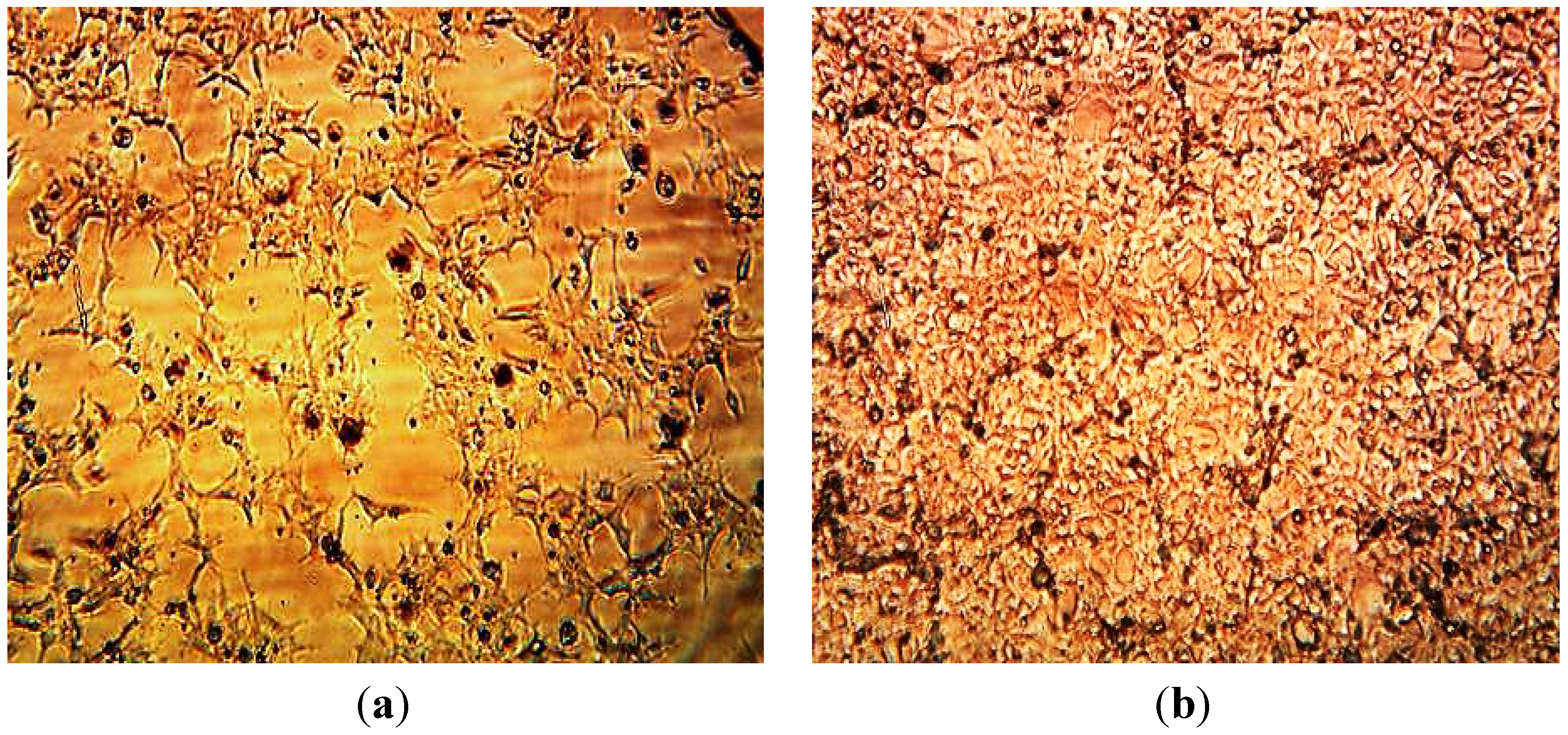
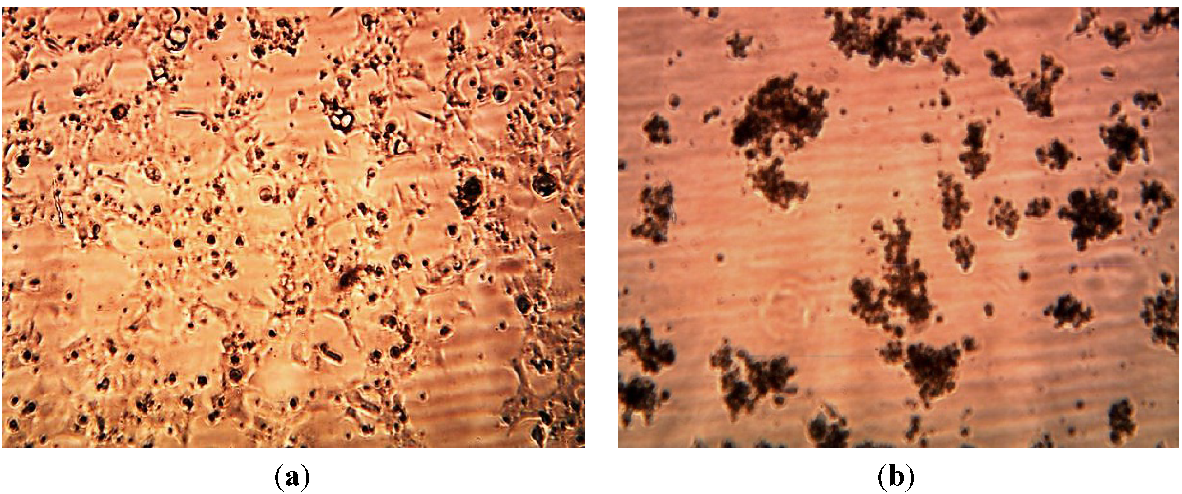
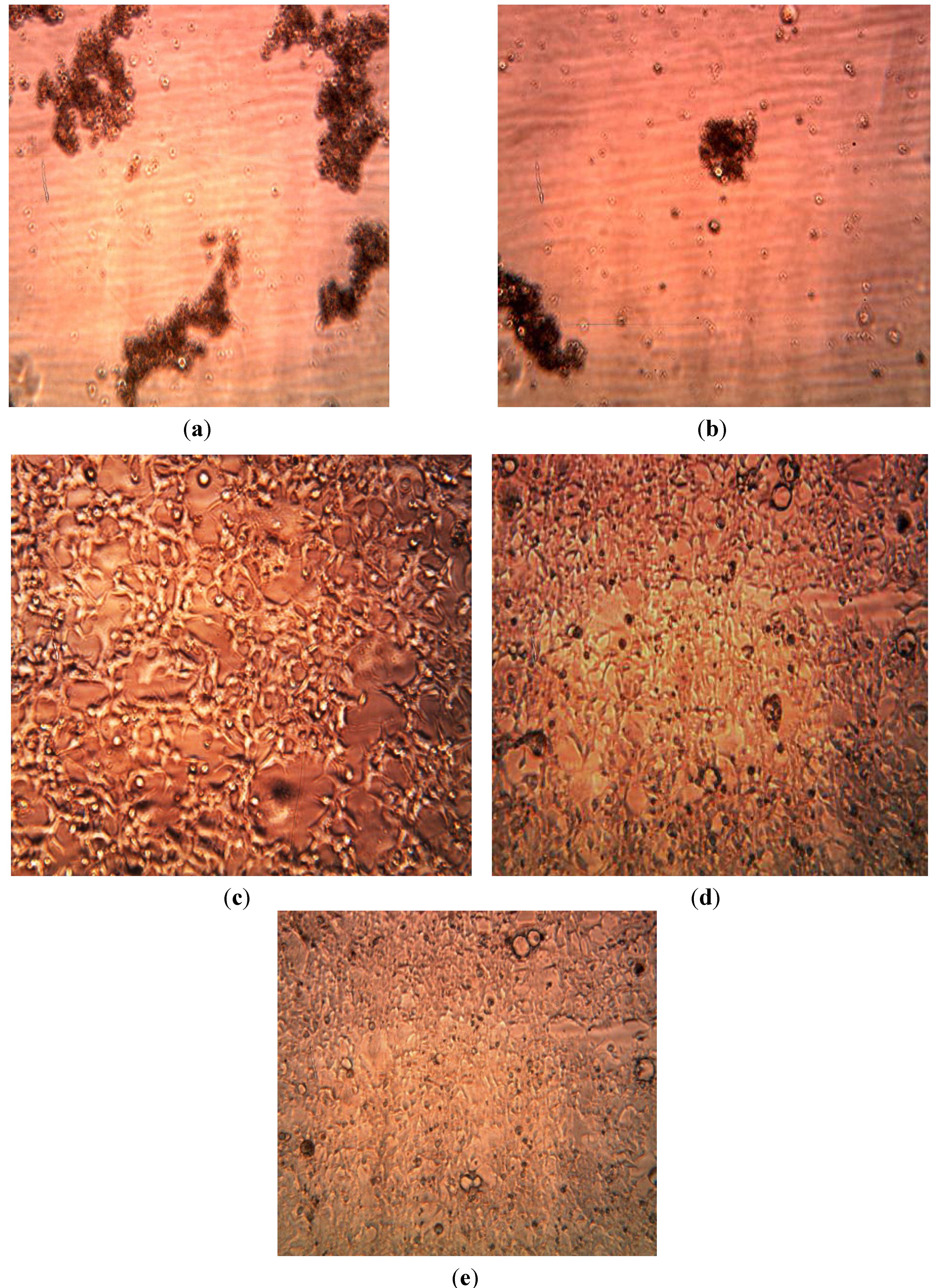
3.3. Cytotoxicity of Biosynthesized Noble Nanoparticles on Healthy Human Cells

We tested the cytotoxicity of Ag Nanoparticles produced using extracts of Aloe, Magnolia leaves and Eucalyptus leaves at concentrations of 2 ppm, 4 ppm, 15 ppm on Human Embryonic Kidney 293cells—HEK293 using the automated InQ Plus equipment.
The cassette containing the growing cells was divided in two parts. In real time we were able to compare the influence of the biosynthesized nanoparticles on human cell growth and control solutions. Figure 12, Figure 13 and Figure 14 below show our observations indicating that bio-produced AgNPs at concentrations of 2–4 ppm were not cytotoxic to human embryonic kidney cells (HEK 293). From available literature, silver nanoparticles (AgNPs) may exhibit toxic or nontoxic effects on various cells depending on their size, concentration and surface properties [49,50,51,52]. Bhakat et al. [53] found that their silver nanoparticles were toxic to HEK 293 cells. In the present study we found that biosynthesized AgNPs appeared to be non-toxic at 2–4 ppm concentrations to cultured human cells (HEK 293 cells) but were toxic to both Gram positive and Gram negative microorganisms. The non-toxicity of the biosynthesized nanoparticles to HEK 293 cells was probably due to the low concentration used in this study; this is similar to the report of Kawata et al. [51] They evaluated the in vitro toxicity of AgNPs at non-cytotoxic doses (<0.5 mg/L) in human hepatoma cell line, HepG2 and observed that the AgNPs actually accelerated cell proliferation at low doses due to induction of genes associated with cell cycle progression. We did not observe cell proliferation in the present study. Detailed and comprehensive dose-response studies need to be conducted in order to fully understand the effects of silver nanoparticles on human cells before these particles can be used in disease treatment and diagnosis.
Figure 12. Images of HEK 293 cells. (a) 6 h after Aloe ethanol extract injection. (b) 48 h after Aloe ethanolic extract injection. (c) 6 h after Aloe extract-produced AgNPs, (d) after 36 h, (e) after 48 h.
Ijerph 10 05221 g012
Figure 13. Images of HEK 293 cells: (a) 6 h after injection of 4 ppm AgNPs reduced using Eucalyptus extract. (b) 48 h after injection of 4 ppm AgNPs.
Ijerph 10 05221 g013
Figure 14. Images of HEK 293 cells: (a) 6 h after injection of 4 ppm AgNPs reduced by Eucalyptus extracts (b) Images of HEK 293 cells: 48 h after injection of 15 ppm AgNPs reducing by Eucalyptus ethanol extract.
Ijerph 10 05221 g014

4. Conclusions

The results showed that substantial amounts of silver nanoparticles (AgNPs) were produced and that this was verified using UV-visible Spectroscopy. We observed appearance of a Surface Plasmon Resonance (SPR) peak, which corresponds to the silver nanoparticles—417 nm in 1 min. In conclusion and based on our results, extracts of plant leaves produce usable silver nanoparticles. These nanoparticles inhibit the growth of both Gram-negative and Gram-positive bacteria, while based on preliminary evaluation of cytotoxicity of biosynthesized silver nanoparticles on HEK 293 cells healthy human cells were not adversely affected. This investigation demonstrated that bio-produced silver nanoparticles at concentrations of 2 ppm and 4 ppm are not cytotoxic for human healthy cells but inhibit bacteria growth. Further studies will be carried out to determine the minimum inhibitory concentration of AgNPs for bacterial growth, and to investigate the cytotoxicity of biosynthesized AgNPs on cancerous and other healthy cells.

Acknowledgments

We would like to acknowledge Alabama A & M University Interdisciplinary Center for Health Sciences and Health Disparities for funding provided through the Evans-Allen grant, administered by the College of Agriculture, Life and Natural Sciences, Title III Program of AAMU, HBCU-UP NSF Grant. In addition, we want to thank, our friends from The Ohio State University Columbus, Ohio 43210, for help with the TEM image of biosynthesis nanoparticles, Ed Jaye, VP Growth Curves Piscataway, NJ USA; Bioscreen Analyzer, for his tremendous help and assistance with the Bioscreen C. We thank Stephanie Jeffrey from InQ Biosciences Corporation, Huntsville, AL USA for help with the cytotoxicity evaluation and supply of the HEK 293 cells.

Conflicts of Interest

The authors declare no conflict of interest.

References

  1. Jiang, Z.J.; Liu, C.Y.; Sun, L.W. Catalytic properties of silver nanoparticles supported on silica spheres. J. Am. Chem. Soc. 2004, 71, 2341–2343. [Google Scholar]
  2. Wijnhoven, S.W.P.; Peijnenburg, W.J.G.M.; Herberts, C.A.; Hagens, W.I.; Oomen, A.G.; Heugens, E.H.W.; Roszek, B.; Bisschops, J.; Gosens, I.; van De Meent, D.; et al. Nano-silver—A review ofavailable data and knowledge gaps in human and environmental risk assessment. Nanotoxicology 2009, 3, 109–138. [Google Scholar] [CrossRef]
  3. Scheringer, M.; MacLeod, M.; Behra, T.; Sigg, L.; Hungerbuhler, K. Environmental risks associated with nanoparticulate silver used as biocide. Household Pers. Care Today 2010, 1, 34–37. [Google Scholar]
  4. Nowack, B. Nanosilver revisited downstream. Science 2010, 330, 1054–1055. [Google Scholar] [CrossRef]
  5. Benn, T.M.; Westerhoff, P. Nanoparticle silver released into water from commercially available sock fabrics. Environ. Sci. Technol. 2008, 42, 4133–4139. [Google Scholar] [CrossRef]
  6. Geranio, L.; Heuberger, M.; Nowack, B. Behavior of silver nanotextiles during washing. Environ. Sci. Technol. 2009, 43, 8113–8118. [Google Scholar] [CrossRef]
  7. Navarro, E.; Piccapietra, F.; Wagner, B.; Marconi, F.; Kaegi, R.; Odzak, N.; Sigg, L.; Behra, R. Toxicity of silver nanoparticles to chlamydomonas reinhardtii. Environ. Sci. Technol. 2008, 42, 8959–8964. [Google Scholar] [CrossRef]
  8. Kaegi, R.; Sinnet, B.; Zuleeg, S.; Hagendorfer, H.; Mueller, E.; Vonbank, R.; Boller, M.; Burkhardt, M. Release of silver nanoparticles from outdoor facades. Environ. Pollut. 2010, 158, 2900–2905. [Google Scholar] [CrossRef]
  9. Rai, M.; Duran, N. Metal Nanoparticles in Microbiology; Springer: Berlin, Germany, 2011. [Google Scholar]
  10. Bonsak, J.; Mayandi, J.; Thøgersen, A.; Marstein, E.S.; Mahalingam, U. Chemical synthesis of silver nanoparticles for solar cell applications. Phys. Status Solidi C 2011, 8, 924–927. [Google Scholar] [CrossRef]
  11. Nanoparticles Inspire Plasmonic Solar Cell. Available online: http://images.iop.org/objects/ntw/news/8/3/48/pdf.pdf (accessed on 21 March 2009).
  12. Technical Articles & Reports on Plastic Industry: Silver Nanoparticles Can Increase Electrical Power Generation of Polymer Solar Cells. Available online: http://www.plastemart.com/Plastic-TechnicalArticle.asp?LiteratureID=1436&Paper=silver-nanoparticles-increase-electrical-power-generation-polymer-solar-cells (accessed on 30 June 2010).
  13. McFarland, A.D.; van Duyne, R.P. Single silver nanoparticles as real-time optical sensors with zeptomole sensitivity. Nano Lett. 2003, 3, 1057–1062. [Google Scholar] [CrossRef]
  14. Dwivedi, P.; Narvi, S.S.; Tewari, R.P. Green route to a novel Ag/PLGA bionanocomposite: A self-sterilizing surgical suture biomaterial. Int. J. Adv. Eng. Sci. Technol. 2012, 2, 236–243. [Google Scholar]
  15. Shahverdi, A.R.; Fakhimi, A.; Shahverdi, H.R.; Minaian, S. Synthesis and effect of silver nanoparticles on the antibacterial activity of different antibiotics against Staphylococcus aureus and Escherichia coli. Nanomed. Nanotechnol. Biol. Med. 2007, 3, 168–171. [Google Scholar] [CrossRef]
  16. Fayaz, A.M.; Balaji, K.; Girilal, M.; Yadav, R.; Kalaichelvan, P.T.; Venketesan, R. Biogenic synthesis of silver nanoparticles and their synergistic effect with antibiotics: A study against gram-positive and gram-negative bacteria. Nanomed. Nanotechnol. Biol. Med. 2010, 6, 103–109. [Google Scholar] [CrossRef]
  17. Lara, H.H.; Garza-Treviño, E.N.; Ixtepan-Turrent, L.; Singh, K.D. Silver nanoparticles are broad-spectrum bactericidal and virucidal compounds. J. Nanobiotechnol. 2011. [Google Scholar] [CrossRef]
  18. Illingworth, B.; Bianco, R.W.; Weisberg, S. In vivo efficacy of silver-coated fabric against Staphylococcus epidermidis. J. Heart Valve Dis. 2000, 9, 135–141. [Google Scholar]
  19. Hoffmann, S. Silver sulfadiazine: An antibacterial agent for topical use in burns: A review of the literature. J. Plast. Surg. Hand Surg. 1984, 18, 119–126. [Google Scholar] [CrossRef]
  20. Lara, H.H.; Ayala-Nunez, N.V.; Ixtepan-Turrent, L.; Rodriguez-Padilla, C. Mode of antiviral action of silver nanoparticles against HIV-1. J. Nanobiotechnol. 2010, 8. [Google Scholar] [CrossRef]
  21. Petrus, E.M.; Tinakumari, S.; Chai, L.C.; Ubong, A.; Tunung, R.; Elexson, N.; Chai, L.F.; Son, R. A study on the minimum inhibitory concentration and minimum bactericidal concentration of nano colloidal silver on food-borne pathogens. Int. Food Res. J. 2011, 18, 55–66. [Google Scholar]
  22. Ansari, M.A.; Khan, H.M.; Khan, A.A.; Malik, A.; Sultan, A.; Shahid, M.; Shujatullah, F.; Azam, A. Evaluation of antibacterial activity of silver nanoparticles against MSSA and MRSA on isolates from skin infections. Biol. Med. 2011, 3, 141–146. [Google Scholar]
  23. Prakash, A.; Sharma, S.; Ahmad, N.; Ghosh, A.; Sinha, P. Synthesis of AgNPs by Bacillus Cereus bacteria and their antimicrobial potential. J.Biomater.Nanobiotechnol. 2011, 2, 156–162. [Google Scholar]
  24. Egorova, E.M. Interaction of silver nanoparticles with biological objects: Antimicrobial properties and toxicity for the other living organisms. J. Phys. 2011, 291. [Google Scholar] [CrossRef]
  25. Sadowski, Z. Biosynthesis and Application of Silver and Gold Nanoparticles. Available online: www.intechopen.com/books/silver-nanoparticles/biosynthesis-and-application-of-silver-and-gold-nanoparticles (accessed on 18 December 2012).
  26. Sathyavathi1, R.; Krishna, M.B.; Rao, S.V.; Saritha, R.; Rao, D.N. Biosynthesis of silver nanoparticles using coriandrum sativum leaf extract and their application in nonlinear optics. Adv. Sci. Lett. 2010, 3, 1–6. [Google Scholar] [CrossRef]
  27. Virender, K.S.; Yngard, R.A.; Lin, Y. Silver nanoparticles: Green synthesis and their antimicrobial activities. Adv. Colloid Interface Sci. 2009, 145, 83–96. [Google Scholar] [CrossRef]
  28. Gardea-Torresdey, J.L.; Gomez, E.; Peralta-Videa, J.R.; Parsons, J.G.; Troiani, H.Y. Process variables in biomimetic synthesis of silver nanoparticles by aqueous extract of Azadirachtaindica (Neem) leaves MJ. Langmuir 2003, 19, 237–246. [Google Scholar]
  29. Umashankari, J.; Inbakandan, D.; Ajithkumar, T.T.; Balasubramanian, T. Mangrove plant, Rhizophora mucronata (Lamk, 1804) mediated one pot green synthesis of silver nanoparticles and its antibacterial activity against aquatic pathogens. Aquat. Biosy. 2012. [Google Scholar] [CrossRef]
  30. Shankar, S.S.; Rai, A.; Ahmad, A.; Sastry, M. Controlling the optical properties of lemongrass extract synthesized gold nanotriangles and potential application in infrared-absorbing optical coatings. Chem. Mater. 2005, 17, 566–572. [Google Scholar] [CrossRef]
  31. Richardson, A.; Chan, B.C.; Crouch, R.D.; Janiec, A.; Chan, B.C.; Crouch, R.D. Synthesis of silver nanoparticles: An undergraduate laboratory using green approach. Chem. Educ. 2006, 11, 331–333. [Google Scholar]
  32. Vaidyanathan, R.; Kalishwaralal, K.; Gopalram, S.; Gurunathan, S. Nanosilver—The burgeoning therapeutic molecule and its green synthesis. Biotechnol. Adv. 2009, 27, 924–937. [Google Scholar] [CrossRef]
  33. Sharma, V.K.; Yngard, R.A.; Lin, Y. Silver nanoparticles: Green synthesis and their antimicrobial activities. Adv. Colloid Interface Sci. 2009, 145, 83–96. [Google Scholar] [CrossRef]
  34. Satyajit, D.; Sarker, Y. Maruyama Magnolia: The Genus Magnolia (Medicinal and Aromatic Plants—Industrial Profiles); Taylor& Francis: New York, NY, USA, 2003. [Google Scholar]
  35. Wolverton, B.C.; Johnson, A.; Bounds, K. Interior Landscape Plants for Indoor Air Pollution Abatement NASA. Available online: http://ntrs.nasa.gov/archive/nasa/casi.ntrs.nasa.gov/19930073077_1993073077.pdf (accessed on 18 September 2012).
  36. Albert, V.A.; Williams, S.E.; Chase, M.W. Carnivorous plants: Phylogeny and structural evolution. Science 1992, 257, 1491–1495. [Google Scholar]
  37. Shankar, S.S.; Ahmad, A.; Sastry, M. Geranium leaf assisted biosynthesis of silver nanoparticles. Biotechnol. Prog. 2003, 19, 1627–1631. [Google Scholar] [CrossRef]
  38. Kumar, V.; Yadav, S.K. Plant-mediated synthesis of silver and gold nanoparticles and their applications. J. Chem. Technol. Biotechnol. 2009, 84, 151–157. [Google Scholar] [CrossRef]
  39. Growth Curves USA. Available online: www.growthcurvesusa.com (accessed on 28 December 2008).
  40. Labrenz, M.; Druschel, G.K.; Thomsen, E.T.; Gilbert, B.; Welch, S.A.; Kemner, K.M. Formation of sphalerite (ZnS) deposits in natural biofilms of sulfate-reducing bacteria. Science 2000, 1, 744–747. [Google Scholar]
  41. Roh, Y.; Bai, J.; Lauf, R.J.; Mcmillan, A.D.; Phelps, T.J.; Rawn, C.J. Microbial synthesis of metal-substituted magnetites. Solid State Commun. 2001, 11, 529–534. [Google Scholar]
  42. Zhang, Y.; Yang, D.; Kong, Y.; Wang, X.; Pandoli, O.; Gao, G. Synergetic antibacterial effects of silver nanoparticles@Aloe Vera prepared via a green method. Nano Biomed. Eng. 2010, 2, 252–257. [Google Scholar]
  43. Gogoi, S.; Gopinath, P.; Paul, A.; Ramesh, A.; Ghosh, S.; Chattopadhyay, A. Green fluorescent protein-expressing Escherichia coli as a model system for investigating the antimicrobial activities of silver nanoparticles. Langmuir 2006, 22, 9322–9328. [Google Scholar] [CrossRef]
  44. Kim, S.H.; Lee, H.S.; Ryu, D.S.; Choi, S.J.; Lee, D.S. Antibacterial activity of silver-nanoparticles against Staphylococcus aureus and Escherichia coli Korean. J. Microbiol. Biotechnol. 2011, 39, 77–85. [Google Scholar]
  45. Li, W.R.; Xie, X.B.; Shi, Q.S.; Duan, S.S.; Ouyang, Y.S.; Chen, Y.B. Antibacterial effect of silver nanoparticles on Staphylococcus aureus. Biometals 2011, 24, 135–141. [Google Scholar] [CrossRef]
  46. Soo-Hwan, J.; Jung, W.L.; Dengteng, G.; Kai, S.; Takuya, N.; Seong, II Y.; Ashish, A.; Yao, L.; Kotov, N.A. Reversible nanoparticle gels with colour switching. J. Mater. Chem. 2011, 21, 11639–11643. [Google Scholar] [CrossRef]
  47. Kvitek, L.; Panacek, A.; Soukupova, J.; Kolar, M.; Vecerova, R.; Prucek, R. Effect of surfactant and polymers on stability and antibacterial activity of silver nanoparticles (NPs). J. Phys.Chem. 2008, 112, 5825–5834. [Google Scholar]
  48. Pal, S.; Tak, Y.K.; Song, J.M. Does the antibacterial activity of silver nanoparticles depend on the shape of the nanoparticle? A study of the gram-negative bacterium Escherichia coli. Appl. Environ. Microbiol. 2007, 73, 1712–1720. [Google Scholar] [CrossRef]
  49. Sur, I.; Altunbek, M.; Kahraman, M.; Culha, M. The influence of the surface chemistry of silver nanoparticles on cell death. Nanotechnology 2012. [Google Scholar] [CrossRef]
  50. Kim, T.-H.; Kim, M.; Park, H.-S.; Shin, U.S.; Gong, M.-S.; Kim, H.-W. Size-dependent cellular toxicity of silver nanoparticles. J. Biomed. Mater. Res. A 2012, 100, 1033–1043. [Google Scholar]
  51. Kawata, K.; Osawa, M.; Okabe, S. In vitro toxicity of silver nanoparticles at noncytotoxic doses to HepG2 human hepatoma cells. Environ. Sci. Technol. 2009, 43, 6046–6051. [Google Scholar] [CrossRef]
  52. Carlson, C.; Hussain, S.M.; Schrand, A.M.; Braydich-Stolle, L.K.; Hess, K.L.; Jones, R.L.; Schlager, J.J. Unique cellular interaction of silver nanoparticles: Size-dependent generation of reactive oxygen species. J. Phys. Chem. 2008, 112, 13608–13619. [Google Scholar]
  53. Bhakat, C.; Chetal, G.; Sarkar, P.; Singh, P.; Babu, S.; Reddy, A. Effects of silver nanoparticles synthesize from ficus benjamina on normal cells and cancer cells. IOSR J. Pharm. Biol. Sci. 2012, 1, 33–36. [Google Scholar]

