Article

Association among Complement Factor H Autoantibodies, Deletions of CFHR, and the Risk of Atypical Hemolytic Uremic Syndrome

1
The First Affiliated Hospital, Xi’an Jiaotong University College of Medicine, 277 Yanta West Road, Xi’an 710061, China
2
Key Laboratory of Shaanxi Province for Craniofacial Precision Medicine Research, College of Stomatology, Xi’an Jiaotong University, Xi’an 710004, China
3
School of Public Health, Xi’an Jiaotong University Health Science Center, 76 Yanta West Road, Xi’an 710061, China
4
Xi’an Honghui Hospital, 555 Friendship Road, Xi’an 710054, China
5
Key Laboratory of Environment and Genes Related to Diseases (Xi’an Jiaotong University), Ministry of Education of China, Xi’an 710061, China
*
Authors to whom correspondence should be addressed.
These authors contributed equally to this work.
Int. J. Environ. Res. Public Health 2016, 13(12), 1209; https://doi.org/10.3390/ijerph13121209
Submission received: 19 September 2016 / Revised: 22 November 2016 / Accepted: 25 November 2016 / Published: 5 December 2016

Abstract

:
To evaluate the association among complement factor H-related (CFHRs) gene deficiency, complement factor H (CFH) autoantibodies, and atypical hemolytic uremic syndrome (aHUS) susceptibility. EMBASE, PubMed, and the ISI Web of Science databases were searched for all eligible studies on the relationship among CFHRs deficiency, anti-FH autoantibodies, and aHUS risk. Eight case-control studies with 927 cases and 1182 controls were included in this study. CFHR1 deficiency was significantly associated with an increased risk of aHUS (odds ratio (OR) = 3.61, 95% confidence interval (95% CI), 1.96, 6.63, p < 0.001), while no association was demonstrated in individuals with only CFHR1/R3 deficiency (OR = 1.32, 95% CI, 0.50, 3.50, p = 0.56). Moreover, a more significant correlation was observed in people with both FH-anti autoantibodies and CFHR1 deficiency (OR = 11.75, 95% CI, 4.53, 30.44, p < 0.001) in contrast to those with only CFHR1 deficiency. In addition, the results were essentially consistent among subgroups stratified by study quality, ethnicity, and gene detection methods. The present meta-analysis indicated that CFHR1 deletion was significantly associated with the risk of aHUS, particularly when combined with anti-FH autoantibodies, indicating that potential interactions among CFHR1 deficiency and anti-FH autoantibodies might impact the risk of aHUS.

1. Introduction

Atypical, non-diarrhea-associated hemolytic uremic syndrome (aHUS) is an aggressive, fatal systemic disease, which is characterized by thrombocytopenia, microangiopathic hemolysis, and acute renal injury [1]. The main populations affected are children and young adults. In comparison with typical hemolytic uremic syndrome, aHUS has a higher risk of permanent renal failure and poorer prognosis [2]. It is reported that 25% of patients died in the acute phase, and up to 50% of patients who progress to end-stage renal disease (ESRD) need renal transplantation or dialysis for the remainder of their life [3,4]. Therefore, investigating the associated relevant risk factors may contribute to designing new diagnostic approaches and therapeutic guidelines.
The dysregulation of the alternative pathway of the complement cascade has been confirmed to play a pivotal role in the development of aHUS [5]. Mutations, polymorphisms, deficiency, or rearrangements in gene coding for various complement proteins have been reported in aHUS patients [6]. As the main complement protein, complement factor H (CFH) mediates the elimination of central complement activation component C3b [7]. The available data indicated that the mutations in CFH were linked to uncontrolled activation of the complement alternative pathway (CAP). Cell surface lack of control of the CAP could lead to CAP over-activation, which promotes microthrombi formation within small blood vessels, thereby resulting in tissue ischemia of the kidney [8,9]. Previous studies have revealed that CFH activity was partly modulated by factor H-related (FHR) proteins [10]. CFHRs (CFHR1-5) encoding FHR proteins and manifesting high degrees of sequence identity with CFH indicated that mutations in CFHRs may be partly responsible for the function abnormalities of CFH [11]. Several studies have investigated the association between CFHR deficiency and the pathogenesis of aHUS [12,13]. Although some studies have found an increased risk with CFHR1 deficiency, others have failed to find this correlation [14,15]. Furthermore, it has been proposed that anti-FH autoantibodies together with CFHR1 deficiency took part in the formation of CFH dysfunction, indicating that both autoantibodies and CFHR1 absence might participate in the occurrence of aHUS. However, there still remains a need to attach importance to the effect of interactions among autoantibodies of the complement factors and CAP regulators on the development of aHUS [16,17].
Therefore, we conducted a meta-analysis of all eligible studies to estimate the association among CFHRs deficiency, anti-FH autoantibodies, and aHUS risk.

2. Materials and Methods

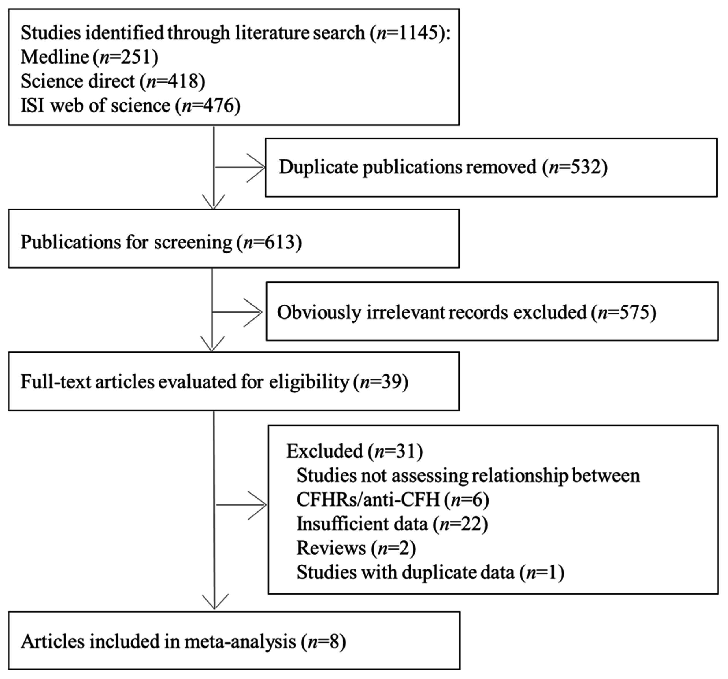
2.1. Search Strategy

A systematic literature search of PubMed, EMBASE, and the ISI Web of Science databases was performed up to March 2016. The following search terms and key words were used: (“CFHRs” or “complement factor H-related protein” or “FH-related proteins” or “anti-FH” or “factor H antibodies” or “anti-complement factor H autoantibody”) and (“aHUS ”or “atypical hemolytic uremic syndrome or atypical HUS”). There were no language restrictions. In addition, we manually searched the reference lists and pertinent reviews of retrieved articles for published or unpublished data. When datasets were incomplete for the required data, ongoing investigations or related experts were contacted for more information.

2.2. Study Selection

An initial screen of abstracts and titles was performed to identify all potential retrieved articles. Thereafter the full text was reviewed to determine whether the articles were suitable for this systematic review. Articles were eligible for inclusion in this study if they fulfilled the following criteria: (1) the study objective was to evaluate the relationship among CFHRs deficiency, anti-FH autoantibodies, and aHUS; (2) the original article was published as a cohort, case-control, or cross-sectional study; (3) the article provided the odds ratio (OR), relative risk (RR), or hazard ratio (HR) with a 95% confidence interval (95% CI) or sufficient data was provided to calculate it. If the articles reported on the same or overlapping data, only the article with the most updated data was included in this study. Three investigators (Hong Jiang, Meng-Nan Fan, and Min Yang) identified the eligible studies independently. Any differences in opinion were resolved through consensus with a third author (Le Ma) for adjudication.

2.3. Data Extraction and Quality Assessment

The following data were extracted using a standardized data extraction form that included: first author’s name, publication year, country of origin, study design, characteristics of the study population (number of aHUS cases and controls, origin of comparison group), gene and protein detection methods. If a study provided several risk estimates, only the most completely adjusted estimate was extracted. The quality of studies was evaluated based on the Newcastle-Ottawa Quality Assessment Scale (NOS) [18]. Each article was estimated with three domains (eight aspects): four criteria regarding the selection of participants (case definition, representativeness of the cases, selection of controls, and definition of controls), one item relating to comparability (comparability of cases and controls on the basis of the design or analysis), three items relating to outcomes (ascertainment of exposure, same method of ascertainment for cases and controls, and comparison of nonresponse rate between cases and controls). For each study, a maximum of one star can be given for each numbered criterion within the selection and outcome categories, and a maximum of two stars can be given for comparability. The quality scores of studies range from zero (worst) to nine (best). When a study’s score was ≥5, it was considered high quality, otherwise, it was considered low quality. Three authors (Hong Jiang, Meng-Nan Fan, and Min Yang) extracted data and assessed study quality independently. Discrepancies were resolved through discussion with a third author (Le Ma).

2.4. Statistical Analysis

The relative risk (RR) was considered as a measure to estimate the strength of the association across studies in principle. For case-control studies, relative odds ratios (OR) were considered as a surrogate estimate of the corresponding RR. Because the absolute risk of aHUS is low, the OR could approximate the RR. If articles did not report RR, we used the number of events in each group to calculate the RR for each study and used them for pooling analyses. In this meta-analysis, pooled RR and 95% CI were used to estimate the strength of the association among CFHRs, anti-FH autoantibodies, and aHUS, using a fixed effects model or a random effects model according to the heterogeneity. Heterogeneity between studies was examined by calculating Cochran’s Q statistic and the I2 test (p value less than 0.10 was considered to indicate heterogeneity and I2 > 50% indicated a statistical significance). When significant statistical heterogeneity was detected, a stratified analysis was conducted to explore the potential source of heterogeneity. The variables included in the stratified analyses were study quality, ethnicity, and gene detection methods. In addition, sensitivity analyses were performed to evaluate the effect of each individual study by excluding each study from the meta-analysis. Egger’s weighted regression method, Begg’s rank correlation method, and visual inspection of funnel plots were used to assess the publication bias [19,20]. All analyses were performed using Stata version 11.0 (Stata Corp LP, College Station, TX, USA).

3. Results

A total of 1145 articles were retrieved through comprehensive searching. After excluding duplicates and irrelevant references, 39 records were left for full text and data effectiveness review, and eight articles (nine studies) were included in this meta-analysis [10,13,14,15,17,21,22,23] (Figure 1).

3.1. Characteristics of the Studies

The characteristics of the included studies are presented in Table 1. All eligible studies were case-control designed, comprising a total of 1065 cases and 1266 controls. Seven studies were carried out in Western countries and two in Asian countries. Six of the included studies used population-based (PB) controls, one investigation used hospital-based (HB) controls, and the remaining one used PB + HB controls. Six of nine studies detected CFHR deficiency through multiplex ligation-dependent probe amplification. The other three studies used polymerase chain reaction, comparative genomic hybridization, or Western blot, respectively. Six of the eight studies detected anti-FH autoantibodies in serum samples using enzyme-linked immunosorbent assay. Furthermore, quality assessment revealed that six studies scored ≥5 and were classified as high quality.

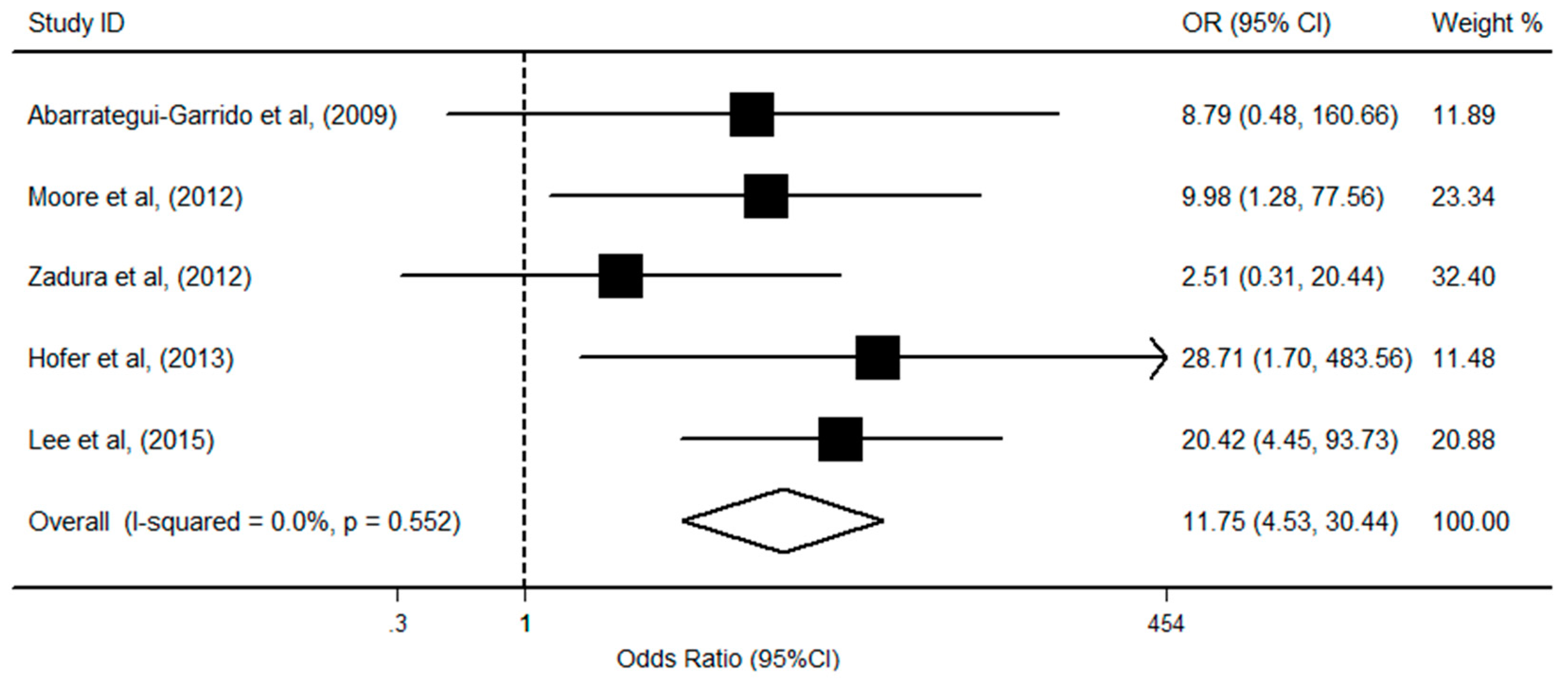
3.2. CFHR1 Deficiency and aHUS Risk

The relationship between CFHR1 deficiency and the risk of aHUS was evaluated in seven studies, including 832 cases and 1028 controls. Among the studies, five reported a statistically significant relationship. There was significant heterogeneity (I2 = 63.4%; p = 0.01) between the data sets, and the random-effects model was used to calculate the pooled OR. The pooled results showed that CFHR1 deficient individuals had a higher susceptibility to aHUS by approximately 2.6-fold in comparison with no-deficiency patients (OR = 3.61; 95% CI, 1.96, 6.63; p < 0.001; Figure 2). The result of stratified analyses by study quality and ethnicity showed that these factors did not alter the association direction and magnitude of the overall effect. The meta-regression analysis showed that gene detection methods were associated with an increased risk of aHUS (Table 2). The sensitivity analysis showed that the significant association among the pooled OR remained stable between CFHR1 deficiency and aHUS. In addition, there was no obvious asymmetry of funnel plots, and no evidence of publication bias was detected among studies (Begg’s test p = 0.76; Egger’s test p = 0.16).

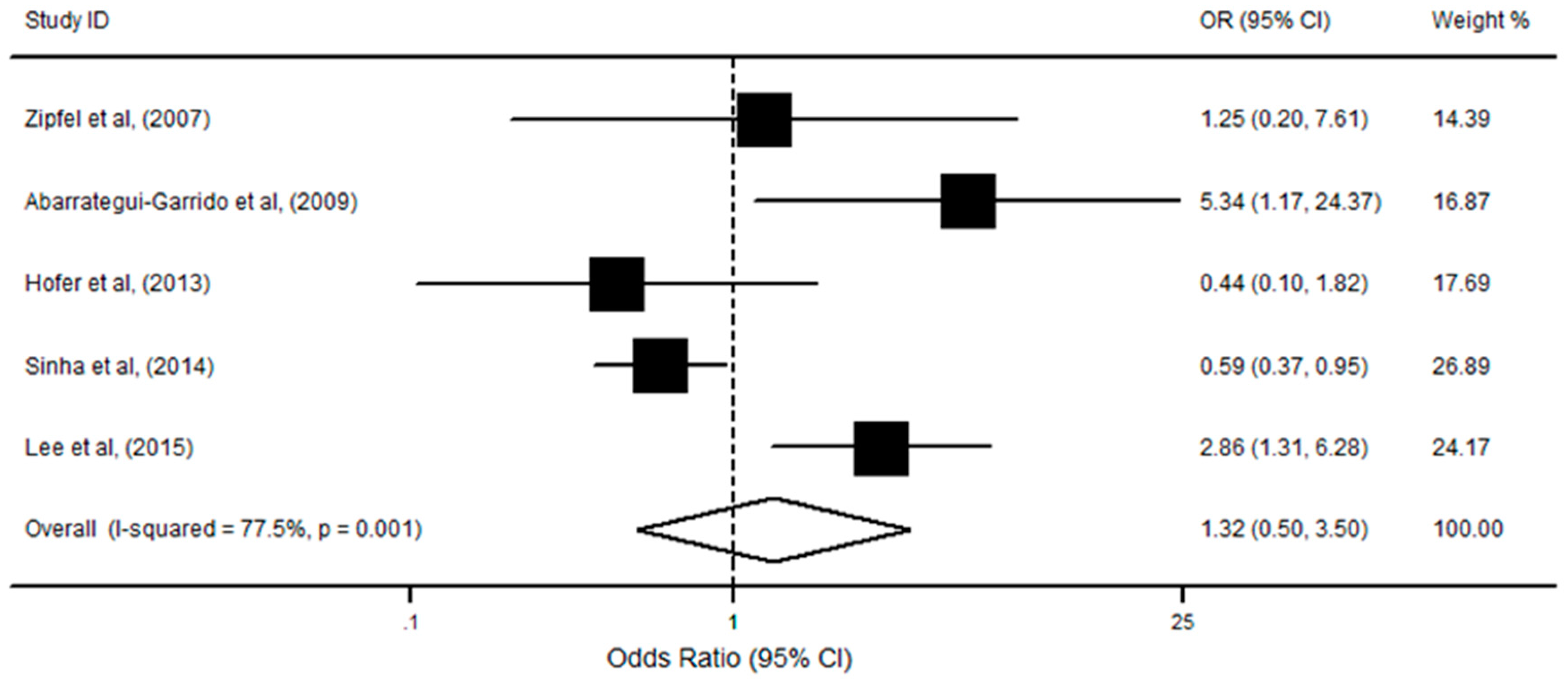
3.3. CFHR1/R3 Deficiency with aHUS Risk

Five studies reported data on the association between only CFHR1/R3 deficiency (without anti-FH autoantibodies) and aHUS risk, with a total of 477 cases and 436 controls. Three reported a slight association of such deficiency with aHUS incidence. Findings from the current analysis showed that there was no significant association among CFHR1/R3 absence and the risk of aHUS (OR = 1.32; 95% CI, 0.50, 3.50; p = 0.56; Figure 3), using the random-effects model (I2 = 77.5%; p = 0.001). Subsequently, we also performed stratified analysis by study quality, ethnicity, and gene detection methods. The result showed that these factors did not alter the association direction and magnitude of the overall effect. Moreover, no publication bias was detected and the funnel plots were symmetric upon visual inspection (p = 0.46), which is confirmed by the Egger’s test (p = 0.35).

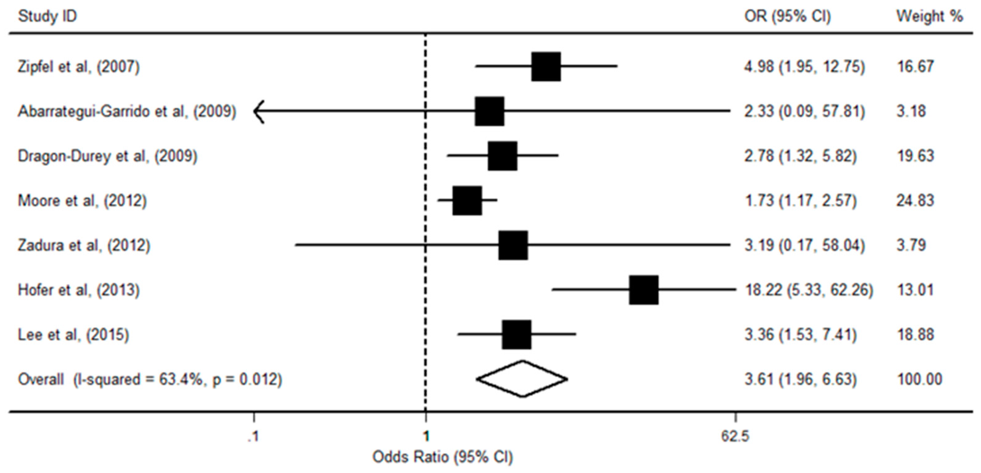
3.4. CFHR1 Deficiency, Anti-CFH, and aHUS Risk

We subsequently evaluated the effect of anti-FH autoantibodies with CFHR1 deficiency on the risk of aHUS in five studies, which included 545 cases and 377 controls. Of the included studies, three showed that both CFHR1 deficiency and autoantibody had a positive relationship with aHUS risk. There was no evidence of statistical heterogeneity among the eligible studies (I2 = 0.0%, p = 0.55), and the fixed-effects model showed that individuals with CFHR1 deletion and anti-FH autoantibodies had an obviously increased risk of aHUS (OR = 11.75; 95% CI, 4.53, 30.44; p < 0.001; Figure 4). In addition, neither the Egger’s test (p = 0.77) nor the Begg’s test (p = 0.46) revealed significant publication bias.

4. Discussion

The results from this comprehensive meta-analysis demonstrated that the deletion of CFHR1 was significantly associated with aHUS. Additionally, further analysis found a stronger association in patients with both CFHR1 deletion and anti-FH autoantibodies in comparison with only CFHR1 deficiency, indicating that CFHR1 deficiency might integrate with anti-FH autoantibodies, which contributes to the development of aHUS.
Insufficient inhibition of complement is associated with a quantity of autoimmune and inflammatory diseases’ pathological processes such as aHUS [23,24]. Several studies have suggested that CFH plays a key role in maintaining complement homeostasis through inhibiting the formation and accelerating the decay of C3-convertase or acting as a cofactor to complement factor I (CFI) in the degradation of C3b in the alternative pathway [25,26]. Moreover, as the major components of a complement system, CFH activities were modulated by other factors consisting of FHRs. Both CFH and FHRs (FHR1-5) belong to human chromosome 1q32 and reside in the regulators of complement activation (RCA) gene cluster [11,27]. CFHRs do encode proteins (FHR1-5) that are likely to modify the function of CFH and regulate complement activity, which would maintain the balance of the complement system and prevent humans from damaging aHUS. In concordance with previous studies that reported CFHR1 deletion was relevant to aHUS susceptibility, the results of present meta-analysis showed a significant association between CFHR1 deficiency and an increased risk of aHUS. The potential mechanism of FHR1 on complement activation and regulation might be via its ability to mediate CFH activity [28]. Under normal circumstances, alternative pathway discrimination between activator and non-activator surfaces is mediated by CFH, CFH can use the heparin/gycosaminoglycan site on short consensus repeats (SCRs) 20 for binding to endothelial cells and to C3b and its cleavage fragment C3d [29,30,31], which has high homology with FHR4-5. Nevertheless, the auto-antigenic epitope of CFH and its homologous site in FHR1 are structurally different [30]. Therefore, CFHR1 deficiency may induce auto-antigenic neoepitope which is related to the formation of autoantibodies, and reduces the function of CFH. Meanwhile, subtotal CFH insufficiency can give rise to aHUS after a complement-activating trigger within the kidney and that the latter is C5 dependent [32,33]. Moreover, FHRs could influence complement activation and regulation through efficiently competing with CFH for binding to the specific surface. The abnormal multimeric structure of FHR1 proteins likely contributes to the cell surface dysregulation [29]. Cell surface lacking control of the CAP would lead to its over-activation and promote microthrombi formation within small blood vessels. Such microthrombi will injure the red blood cells and cause kidney tissue ischemia [8,33]. Moreover, CFHRs and CFH have several independent large genomic duplications, such repeats tend to occur in nonallelic homologous recombinations, which results in the loss of CFHR1 [34,35]. FHR1 deficiency has a potential impact on CFH binding to the CAP convertase or the regulatory function of the terminal pathway, which is related to the occurrence of aHUS [8,35]. In addition, a regulatory function of FHR1 on the C5 convertase or terminal pathway has recently been reported, which might also contribute to aHUS pathogenesis [36]. In contrast with the significant association between CFHR1 deficiency and the increased risk of aHUS, the present studies showed that individuals carrying CFHR1/R3 deficiency (without autoantibodies) had no significant association with aHUS. It is possibly influenced by the small number of included studies. Thus, further multi-centric studies investigating the relationship between aHUS and different combinations of CFHRs deficiency need to be conducted before definitive conclusions can be made.
Evidence from a previous study found that autoantibodies to CFH were connected with the development of aHUS [37]. The epitope of the autoantibody is located on the C-terminal SCRs 19–20 of CFH, which is responsible for recognition and binding to C3b and to cellular surfaces. Blocking the SCRs 19–20, anti-FH autoantibodies could impair the binding of factor H to C3b and be associated with uncontrolled complement activation at the cell surface and with hemolysis [31]. Results from the present study indicated that both anti-FH autoantibodies and CFHR1 deficiency were significantly associated with the increased risk of aHUS, which suggests that this autoantibody might cross-react with CFHR1 deficiency to promote the progress of aHUS. The underlying mechanism for such an observed relationship might be that such a deficiency may favor development of these specific autoantibodies. CFH and FHR1 have high degrees of sequence duplications, especially in SCRs 19–20 and SCRs 4–5 (autoantibody epitope site) [35,38]. Such a special gene structure would cause a failure of central and/or peripheral tolerance and induce the anti-FH autoantibodies, resulting in increased progression of kidney injury [39,40].
Findings from this meta-analysis have important public health significance. Cases of aHUS generally have a poor outcome [2,41]. Therefore, early specific diagnosis and treatment might lead to favorable outcomes. The central pathophysiology of aHUS is uncontrolled activation of the complement pathway. The finding of a significant relationship between aHUS and CFHRs deficiency and/or autoantibodies potentially has important clinical implications for investigating these risk factors in the early stage of aHUS and improving prognosis. Though the result from Holmes et al. suggests that CFHR1/R3 deletion in the normal population is different worldwide. Depending on ethnic origin, the highest frequency for the deletion (54.7%) was seen in Nigeria [42]. CFHRs deficiency and autoantibodies could also be considered as a surrogate biomarker for some clinically relevant incidents or aHUS susceptibility, especially in designing new diagnostic approaches, identifying susceptible people, and monitoring the treatment effects.
Several potential limitations should be also considered in interpreting the results of this meta-analysis. First, as no available cohort studies were found, this meta-analysis only extracted data from case-control studies. Retrospective study is subjected to internal methodological deficiencies, which may limit the power of this meta-analysis. Second, our results were based on unadjusted estimates, potential confounding components such as pregnancy, family history, drugs, other complement factor genes, and other genetic abnormalities [43] likely affect the risk of aHUS, and studies in this meta-analysis did not control for these factors or provide sufficient data to analyze the association among CFHRs deficiency, anti-FH autoantibodies, and aHUS adjusted for these covariates. Thereby, other complement factors, genetic abnormalities, or other confounding factors might affect the results of the present analysis. Nevertheless, the qualities of most of the eligible studies were high, which adds to the strength of our analysis. Third, the present results were also likely to be affected by different separate examination methods of CFHR1. The way to examine the CFHR1 status of the patients and controls, varies from one study to another, and that might affect the present results as a potential confounding factor. Furthermore, one study used western blot to identify CFHR1 deficiency. As aHUS could be treated by administration of blood derived products, the approach by western blotting might be at high risk of false results in aHUS. Although this study should not substantially affect the overall qualitative inference, we cannot exclude the potential influence which might have led to underestimation or overestimation of the association. Fourth, the pooled results were mainly conducted in Western countries, which limited the generalization of findings. Last, although statistical tests did not suggest the presence of publication bias for the present study, we could not exclude all publication bias.

5. Conclusions

The present meta-analysis provides evidence of the association between CFHR1 deletion, anti-FH autoantibodies and aHUS. CFHR1 deficiency was significantly associated with increased risk of aHUS. Moreover, this study also identified potential interactions between anti-FH autoantibodies and CFHR1 deletion in aHUS patients, indicating that the development of aHUS could be exacerbated via interaction between CFHR1 deficiency and anti-FH autoantibodies.

Acknowledgments

This study was partially supported by grants from the National Natural Science Foundation of China (NSFC-81202198, NSFC-81473059); the Natural Science Foundation of Shaanxi Province of China (2013JQ4008); New-star Plan of Science and Technology of Shaanxi Province (2015LJXX-07); the China Postdoctoral Science Special Foundation (2015T81036); the Fundamental Research Funds for the Central Universities (qngz2016004); and the China Postdoctoral Science Foundation Funded Project (2014M560790).

Author Contributions

Le Ma and Xiao-Hong Liu designed the study; Hong Jiang, Meng-Nan Fan, and Min Yang extracted and collected the data; Hong Jiang, Meng-Nan Fan, and Min Yang assessed the quality of the included studies; Hong Jiang, Meng-Nan Fan, Min Yang, and Chao Lu analyzed data; All authors were involved in writing the paper; Le Ma, Xiao-Hong Liu, and Ming Zhang gave a great deal of advice and direction in both study design and preparing the manuscript; All the authors have read and approved the final submitted version.

Conflicts of Interest

None of the authors have any conflicts of interest to disclose. All authors had access to all the study data, take responsibility for the accuracy of the analysis, and had authority over manuscript preparation and the decision to submit the manuscript for publication. All authors approve the manuscript and agree to adhere to all terms outlined in Annals of Internal Medicine information for authors including terms for copyright.

References

  1. Noris, M.; Remuzzi, G. Atypical hemolytic-uremic syndrome. N. Engl. J. Med. 2009, 361, 1676–1687. [Google Scholar] [CrossRef] [PubMed]
  2. Bresin, E.; Rurali, E.; Caprioli, J.; Sanchezcorral, P.; Fremeauxbacchi, V.; De Cordoba, S.R.; Pinto, S.; Albert, M.; Ribes, D.; Valoti, E.; et al. Combined complement gene mutations in atypical hemolytic uremic syndrome influence clinical phenotype. JASN 2013, 24, 475–486. [Google Scholar] [CrossRef] [PubMed]
  3. Loirat, C.; Fremeauxbacchi, V. Atypical hemolytic uremic syndrome. Orphanet. J. Rare Dis. 2011, 6, 1–30. [Google Scholar] [CrossRef] [PubMed]
  4. Westra, D.; Wetzels, J.F.; Volokhina, E.B.; van den Heuvel, L.P.; van de Kar, N.C. A new era in the diagnosis and treatment of atypical hemolytic uremic syndrome. Neth. J. Med. 2012, 70, 121–129. [Google Scholar] [PubMed]
  5. Warwicker, P.; Goodship, T.H.; Donne, R.L.; Pirson, Y.; Nicholls, A.; Ward, R.; Turnpenny, P.D.; Goodship, J.A. Genetic studies into inherited and sporadic hemolytic uremic syndrome. Kidney Int. 1998, 53, 836–844. [Google Scholar] [CrossRef] [PubMed]
  6. Skerka, C.; Qian, C.; Fremeauxbacchi, V.; Roumenina, L.T. Complement factor H related proteins (CFHRs). Mol. Immunol. 2013, 56, 170–180. [Google Scholar] [CrossRef] [PubMed]
  7. Harboe, M.; Thorgersen, E.B.; Mollnes, T.E. Advances in assay of complement function and activation. Adv. Drug Deliv. Rev. 2011, 63, 976–987. [Google Scholar] [CrossRef] [PubMed]
  8. Schramm, E.C.; Roumenina, L.T.; Rybkine, T.; Chauvet, S.; Vieiramartins, P.; Hue, C.; Maga, T.; Valoti, E.; Wilson, V.; Jokiranta, T.S.; et al. Mapping interactions between complement C3 and regulators using mutations in atypical haemolytic uraemic syndrome. Blood 2015, 125, 2359–2369. [Google Scholar] [CrossRef] [PubMed]
  9. Noris, M.; Remuzzi, G. Glomerular diseases dependent on complement activation, including atypical hemolytic uremic syndrome, membranoproliferative glomerulonephritis, and C3 glomerulopathy: Core curriculum 2015. Am. J. Kidney Dis. 2015, 66, 359–375. [Google Scholar] [CrossRef] [PubMed]
  10. Abarrategui-Garrido, C.; Martínez-Barricarte, R.; López-Trascasa, M.; de Córdoba, S.R.; Sánchez-Corral, P. Characterization of complement factor H-related (CFHR) proteins in plasma reveals novel genetic variations of CFHR1 associated with atypical hemolytic uremic syndrome. Blood 2009, 114, 4261–4271. [Google Scholar] [CrossRef] [PubMed] [Green Version]
  11. Valoti, E.; Alberti, M.; Tortajada, A.; Garciafernandez, J.; Gastoldi, S.; Besso, L.; Bresin, E.; Remuzzi, G.; De Cordoba, S.R.; Noris, M. A novel atypical hemolytic uremic syndrome–associated hybrid CFHR1/CFH gene encoding a fusion protein that antagonizes factor H–dependent complement regulation. JASN 2015, 26, 209–219. [Google Scholar] [CrossRef] [PubMed]
  12. Bu, F.; Maga, T.; Meyer, N.C.; Wang, K.; Thomas, C.P.; Nester, C.M.; Smith, R.J. Comprehensive genetic analysis of complement and coagulation genes in atypical hemolytic uremic syndrome. JASN 2014, 5, 55–64. [Google Scholar] [CrossRef] [PubMed]
  13. Zipfel, P.F.; Edey, M.; Heinen, S.; Jozsi, M.; Richter, H.; Misselwitz, J.; Hoppe, B.; Routledge, D.; Strain, L.; Hughes, A.E.; et al. Deletion of complement factor H–related genes CFHR1 and CFHR3 is associated with atypical hemolytic uremic syndrome. PLoS Genet. 2007, 3, 387–392. [Google Scholar] [CrossRef] [PubMed]
  14. Hofer, J.; Janecke, A.; Zimmerhackl, L.B.; Riedl, M.; Rosales, A.; Giner, T.; Cortina, G.; Haindl, C.J.; Petzelberger, B.; Pawlik, P.; et al. Complement factor H-related protein 1 deficiency and factor H antibodies in pediatric patients with atypical hemolytic uremic syndrome. Clin. J. Am. Soc. Nephrol. 2013, 8, 407–415. [Google Scholar] [CrossRef] [PubMed]
  15. Dragon-Durey, M.; Blanc, C.; Marliot, F. The high frequency of complement factor H related CFHR1 gene deletion is restricted to specific subgroups of patients with atypical hemolytic uremic syndrome. J. Med. Genet. 2009, 46, 447–450. [Google Scholar] [CrossRef] [PubMed] [Green Version]
  16. Noris, M.; Remuzzi, G. Overview of complement activation and regulation. Semin. Nephrol. 2013, 33, 479–492. [Google Scholar] [CrossRef] [PubMed]
  17. Zadura, A.F.; Zipfel, P.F.; Bokarewa, M.; Sturfelt, G.; Jonsen, A.; Nilsson, S.; Hillarp, A.; Saxne, T.; Trouw, L.A.; Blom, A.M. Factor H autoantibodies and deletion of complement factor H-related protein-1 in rheumatic diseases in comparison to atypical hemolytic uremic syndrome. Arthritis Res. Ther. 2012, 14, R185. [Google Scholar] [CrossRef] [PubMed]
  18. Wells, G.; Shea, B.; O’Connell, D.; Roberton, J.; Peterson, J.; Welch, V.; Losos, M.; Tugwell, P. The Newcastle-Ottawa Scale (NOS) for Assessing the Quality of Nonrandomized Studies in the Meta-Analysis. Available online: http://www.ohri.ca/programs/clinical_epidemiology/oxford.asp (accessed on 20 September 2016).
  19. Egger, M.; Smith, G.D.; Schneider, M.; Minder, C.E. Bias in meta-analysis detected by a simple, graphical test. BMJ 1997, 315, 629–634. [Google Scholar] [CrossRef] [PubMed]
  20. Begg, C.B.; Mazumdar, M. Operating characteristics of a rank correlation test for publication bias. Biometrics 1994, 50, 1088–1101. [Google Scholar] [CrossRef] [PubMed]
  21. Moore, I.; Strain, L.; Pappworth, I.Y.; Kavanagh, D.J.; Barlow, P.N.; Herbert, A.P.; Schmidt, C.Q.; Staniforth, S.J.; Holmes, L.V.V.; Ward, R.; et al. Association of factor H autoantibodies with deletions of CFHR1, CFHR3, CFHR4, and with mutations in CFH, CFI, CD46, and C3 in patients with atypical hemolytic uremic syndrome. Blood 2010, 115, 379–387. [Google Scholar] [CrossRef] [PubMed]
  22. Lee, J.; Park, Y.S.; Lee, J.H.; Park, S.J.; Shin, J.I.; Park, Y.; Yoo, K.H.; Cho, M.H.; Kim, S.; Kim, S.H.; et al. Atypical hemolytic uremic syndrome: Korean pediatric series. Pediatr. Int. 2015, 57, 431–438. [Google Scholar] [CrossRef] [PubMed]
  23. Sinha, A.; Gulati, A.; Saini, S.; Blanc, C.; Gupta, A.; Gurjar, B.S.; Saini, H.; Kotresh, S.; Ali, U.S.; Bhatia, D.; et al. Prompt plasma exchanges and immunosuppressive treatment improves the outcomes of anti-factor H autoantibody-associated hemolytic uremic syndrome in children. Kidney Int. 2014, 85, 1151–1160. [Google Scholar] [CrossRef] [PubMed]
  24. Carneirosampaio, M.; Liphaus, B.L.; Jesus, A.A.; Silva, C.A.; Oliveria, J.B.; Kiss, M.H. Understanding systemic lupus erythematosus physiopathology in the light of primary immunodeficiencies. J. Clin. Immunol. 2008, 28, 34–41. [Google Scholar] [CrossRef] [PubMed]
  25. Hyvärinen, S.; Uchida, K.; Varjosalo, M.; Jokela, R.; Jokiranta, T.S. Recognition of malondialdehyde-modified proteins by the C terminus of complement factor H is mediated via the polyanion binding site and impaired by mutations found in atypical hemolytic uremic syndrome. J. Biol. Chem. 2014, 289, 4295–4306. [Google Scholar] [CrossRef] [PubMed]
  26. Mele, C.; Remuzzi, G.; Noris, M. Hemolytic uremic syndrome. Semin. Immunopathol. 2014, 36, 399–420. [Google Scholar] [CrossRef] [PubMed]
  27. Venables, J.P.; Strain, L.; Routledge, D.; Bourn, D.; Powell, H.M.; Warwicker, P.; Diaztorres, M.L.; Sampson, A.; Mead, P.A.; Webb, M.; et al. Atypical hemolytic uremic syndrome associated with a hybrid complement gene. PLoS Med. 2006, 3, 1957–1967. [Google Scholar] [CrossRef] [PubMed]
  28. Zipfel, P.F.; Skerka, C. Complement regulators and inhibitory proteins. Nat. Rev. Immunol. 2009, 9, 729–740. [Google Scholar] [CrossRef] [PubMed]
  29. Tortajada, A.; Yebenes, H.; Abarrateguigarrido, C.; Anter, J.; Garciafernandez, J.; Martinezbarricarte, R.; Albadominguez, M.; Malik, T.H.; Bedoya, R.; Perez, R.C.; et al. C3 glomerulopathy–associated CFHR1 mutation alters FHR oligomerization and complement regulation. J. Clin. Investig. 2013, 123, 2434–2446. [Google Scholar] [CrossRef] [PubMed]
  30. Bhattacharjee, A.; Reuter, S.; Trojnar, E.; Kolodziejczyk, R.; Seeberger, H.; Hyvarinen, S.; Uzonyi, B.; Szilagyi, A.; Prohaszka, Z.; Goldman, A.; et al. The major autoantibody epitope on Factor H in atypical Hemolytic Uremic Syndrome is structurally different from its homologous site in Factor H related protein 1 supporting a novel model for induction of autoimmunity in this disease. J. Biol. Chem. 2015, 290, 9500–9510. [Google Scholar] [CrossRef] [PubMed]
  31. Blanc, C.; Roumenina, L.T.; Ashraf, Y.; Hyvarinen, S.; Sethi, S.K.; Ranchin, B.; Niaudet, P.; Loirat, C.; Gulati, A.; Bagga, A.; et al. Overall Neutralization of Complement Factor H by autoantibodies in the acute phase of the autoimmune form of atypical hemolytic uremic syndrome. J. Immunol. 2012, 189, 3528–3537. [Google Scholar] [CrossRef] [PubMed]
  32. Vernon, K.A.; Ruseva, M.M.; Cook, H.T.; Botto, M.; Malik, T.H.; Pickering, M.C. Partial complement factor H deficiency associates with C3 glomerulopathy and thrombotic microangiopathy. J. Am. Soc. Nephrol. 2015, 27, 1334–1342. [Google Scholar] [CrossRef] [PubMed]
  33. Pickering, M.C.; De Jorge, E.G.; Martinezbarricarte, R.; Recalde, S.; Garcialayana, A.; Rose, K.L.; Moss, J.; Walport, M.J.; Cook, H.T.; De Cordoba, S.R.; et al. Spontaneous hemolytic uremic syndrome triggered by complement factor H lacking surface recognition domains. J. Exp. Med. 2007, 204, 1249–1256. [Google Scholar] [CrossRef] [PubMed]
  34. Francis, N.J.; Mcnicholas, B.; Awan, A.; Waldron, M.; Reddan, D.N.; Sadlier, D.M.; Kavanagh, D.J.; Strain, L.; Marchbank, K.J.; Harris, C.L.; et al. A novel hybrid CFH/CFHR3 gene generated by a microhomology-mediated deletion in familial atypical hemolytic uremic syndrome. Blood 2012, 119, 591–601. [Google Scholar] [CrossRef] [PubMed]
  35. Strobel, S.; Abarrategui-Garrido, C.; Fariza-Requejo, E.; Seeberger, H.; Sánchez-Corral, P.; Józsi, M. Factor H-related protein 1 neutralizes anti-factor H autoantibodies in autoimmune hemolytic uremic syndrome. Kidney Int. 2011, 80, 397–404. [Google Scholar] [CrossRef] [PubMed]
  36. Heinen, S.; Hartmann, A.; Lauer, N.; Wiehl, U.; Dahse, H.; Schirmer, S.; Gropp, K.; Enghardt, T.; Wallich, R.; Halbich, S.; et al. Factor H-related protein 1 (CFHR-1) inhibits complement C5 convertase activity and terminal complex formation. Blood 2009, 114, 2439–2447. [Google Scholar] [CrossRef] [PubMed]
  37. Jozsi, M.; Licht, C.; Strobel, S.; Zipfel, S.L.H.; Richter, H.; Heinen, S.; Zipfel, P.F.; Skerka, C. Factor H autoantibodies in atypical hemolytic uremic syndrome correlate with CFHR1/CFHR3 deficiency. Blood 2008, 111, 1512–1514. [Google Scholar] [CrossRef] [PubMed]
  38. Dragondurey, M.A.; Loirat, C.; Cloarec, S.; Macher, M.; Blouin, J.; Nivet, H.; Weiss, L.; Fridman, W.H.; Fremeauxbacchi, V. Anti-Factor H autoantibodies associated with atypical hemolytic uremic syndrome. J. Am. Soc. Nephrol. 2005, 16, 555–563. [Google Scholar] [CrossRef] [PubMed]
  39. Strobel, S.; Hoyer, P.F.; Mache, C.J.; Sulyok, E.; Liu, W.; Richter, H.; Oppermann, M.; Zipfel, P.F.; Jozsi, M. Functional analyses indicate a pathogenic role of factor H autoantibodies in atypical haemolytic uraemic syndrome. Nephrol. Dial. Trans. 2010, 25, 136–144. [Google Scholar] [CrossRef] [PubMed]
  40. Legendre, C.; Licht, C.; Muus, P.; Greenbaum, L.A.; Babu, S.; Bedrosian, C.L.; Bingham, C.; Cohen, D.J.; Delmas, Y.; Douglas, K.; et al. Terminal complement inhibitor eculizumab in atypical hemolytic-uremic syndrome. N. Engl. J. Med. 2013, 368, 2169–2181. [Google Scholar] [CrossRef] [PubMed]
  41. Nozal, P.; Bernabeuherrero, M.E.; Uzonyi, B.; Szilagyi, A.; Hyvarinen, S.; Prohaszka, Z.; Jokiranta, T.S.; Sanchezcorral, P.; Lopeztrascasa, M.; Jozsi, M. Heterogeneity but individual constancy of epitopes, isotypes and avidity of factor H autoantibodies in atypical hemolytic uremic syndrome. Mol. Immunol. 2016, 70, 47–55. [Google Scholar] [CrossRef] [PubMed]
  42. Holmes, L.V.; Strain, L.; Staniforth, S.J.; Moore, I.; Marchbank, K.J.; Kavanagh, D.J.; Goodship, J.A.; Cordell, H.J.; Goodship, T.H.J. Determining the population frequency of the CFHR3/CFHR1 deletion at 1q32. PLoS ONE 2013, 8, e60352. [Google Scholar] [CrossRef] [PubMed]
  43. Dragondurey, M.; Sethi, S.K.; Bagga, A.; Blanc, C.; Blouin, J.; Ranchin, B.; Andre, J.; Takagi, N.; Cheong, H.I.; Hari, P.; et al. Clinical features of anti-factor H autoantibody-associated hemolytic uremic syndrome. J. Am. Soc. Nephrol. 2010, 21, 2180–2187. [Google Scholar] [CrossRef] [PubMed]
Figure 1. Flowchart for the selection of eligible studies.
Ijerph 13 01209 g001
Figure 2. Forest plot on the association between CFHR1 deficiency and aHUS risk. For each study, the estimation of OR and its 95% confidence interval (95% CI) are plotted with a box and a horizontal line. The pooled odds ratio is represented by a diamond. The area of the black squares reflects the weight of the study in the meta-analysis.
Ijerph 13 01209 g002
Figure 3. Forest plot on the association between CFHR1/R3 deficiency and aHUS risk. For each study, the estimation of OR and its 95% CI are plotted with a box and a horizontal line. The pooled odds ratio is represented by a diamond. The area of the black squares reflects the weight of the study in the meta-analysis.
Ijerph 13 01209 g003
Figure 4. Forest plot on the association among CFHR1 deficiency, anti-FH autoantibodies, and aHUS risk. For each study, the estimation of OR and its 95% CI are plotted with a box and a horizontal line. The pooled odds ratio is represented by a diamond. The area of the black squares reflects the weight of the study in the meta-analysis.
Ijerph 13 01209 g004
Table 1. Characteristics of studies included in this meta-analysis.
First Author (y)Country of OriginStudy TypeSample Size (Cases)Sample Size (Controls)Source of ControlsGene Detection MethodsProtein Detection MethodsStudy Quality *
Lee et al., 2015KoreaCase-Control51100HBMLPAELISAHigh
Sinha et al., 2014IndianCase-Control13884PBMLPAWestern blotHigh
Hofer et al., 2013EuropeanCase-Control116118PBCGHELISALow
Moore et al., 2012UKCase-Control142100PBMLPAELISAHigh
Zadura et al., 2012GermanCase-Control103345PB-ELISAHigh
Abarrategui-Garrid et al., 2009SpainCase-Control151230HB + PBMLPAELISAHigh
Dragon-Durey et al., 2009FranceCase-Control17770PBMLPAELISAHigh
Zipfel et al., 2007GermanyCase-Control121100PBPCRNRLow
Zipfel et al., 2007UKCase-Control66119PBMLPAWestern BlotLow
aHUS, Atypical hemolytic uremic syndrome; HB, Hospital-based; PB, Population-based; MLPA, Multiplex ligation-dependent probe amplification; CGH, Comparative genomic hybridization; ELISA, Enzyme linked immunosorbent assay; PCR, polymerase chain reaction; NR, Not reported; * Study quality was judged based on Newcastle-Ottawa Scale.
Table 2. Meta-analysis of the association between the CFHR1 deficiency/CFHR1/R3 deficiency and atypical hemolytic uremic syndrome risk.
SubgroupNPooled OR95% CIp
HeterogeneityMeta-Regression
CFHR1 Deficiency-----
Study Quality7---0.059
High52.121.55, 2.900.572
Low23.611.96, 6.630.095
Ethnicity----0.703
Caucasians61.881.22, 2.540.005
Asians13.360.42, 6.30NA
Gene Detection Methods6----
MLPA52.581.70, 3.91<0.0010.001
CGH118.225.33, 62.26NA
CFHR1/R3 Deficiency-----
Study Quality5---0.763
High41.350.44, 4.150.001
Low11.250.20, 7.61NA
Ethnicity5---0.694
Caucasians31.410.31, 6.520.06
Asians21.260.27, 5.090.001
Gene Detection Methods5----
MLPA31.860.47, 7.350.3760.461
CGH10.440.10, 1.82NA
PCR11.250.20, 761NA
OR, odds ratio; 95% CI, 95% confidence interval; MLPA, Multiplex ligation-dependent probe amplification; CGH, Comparative genomic hybridization; PCR, polymerase chain reaction. NA, Not Available.

