Population-Based Study on the Effect of a Forest Environment on Salivary Cortisol Concentration

 ,
,  , , ,
, , , 
Abstract
:1. Introduction
2. Materials and Methods
2.1. Participants
2.2. Experimental Procedures and Salivary Cortisol Measurement
2.3. Outlier Processing
- Q1: quartile 1 (25th percentile)
- Q3: quartile 3 (75th percentile)
- IQR: interquartile range (Q3–Q1)
2.4. Statistical Analysis
3. Results
4. Discussion
5. Conclusions
Acknowledgments
Author Contributions
Conflicts of Interest
References
- Bowler, D.E.; Buyung-Ali, L.M.; Knight, T.M.; Pullin, A.S. A systematic review of evidence for the added benefits to health of exposure to natural environments. BMC Public Health 2010, 10, 456. [Google Scholar] [CrossRef] [PubMed]
- Sonntag-Öström, E.; Nordin, M.; Lundell, Y.; Dolling, A.; Wiklund, U.; Karlsson, M.; Carlberge, B.; Järvholm, L.S. Restorative effects of visits to urban and forest environments in patients with exhaustion disorder. Urban For. Urban Green. 2014, 13, 344–354. [Google Scholar] [CrossRef]
- Hartig, T.; Mitchell, R.; De Vries, S.; Frumkin, H. Nature and health. Annu. Rev. Public Health 2014, 35, 207–228. [Google Scholar] [CrossRef] [PubMed]
- Matsunaga, K.; Park, B.J.; Kobayashi, H.; Miyazaki, Y. Physiologically relaxing effect of a hospital rooftop forest on older women requiring care. J. Am. Geriatr. Soc. 2011, 59, 2162–2163. [Google Scholar] [CrossRef] [PubMed]
- Mitchell, R.; Popham, F. Effect of exposure to natural environment on health inequalities: An observational population study. Lancet 2008, 372, 1655–1660. [Google Scholar] [CrossRef]
- Kobayashi, H.; Song, C.; Ikei, H.; Kagawa, T.; Miyazaki, Y. Analysis of individual variations in autonomic responses to urban and forest environments. Evid. Based Complement. Altern. Med. 2015, 2015, 671094. [Google Scholar] [CrossRef] [PubMed]
- Richardson, M.; McEwan, K.; Maratos, F.; Sheffield, D. Joy and calm: How an evolutionary functional model of affect regulation informs positive emotions in nature. Evolut. Psychol. Sci. 2016, 2, 308–320. [Google Scholar] [CrossRef] [Green Version]
- Lee, J.; Park, B.J.; Ohira, T.; Kagawa, T.; Miyazaki, Y. Acute effects of exposure to a traditional rural environment on urban dwellers: A crossover field study in terraced farmland. Int. J. Environ. Res. Public Health 2015, 12, 1874–1893. [Google Scholar] [CrossRef] [PubMed]
- Ochiai, H.; Ikei, H.; Song, C.; Kobayashi, M.; Takamatsu, A.; Kagawa, T.; Li, Q.; Kumeda, S.; Imai, M.; Miyazaki, Y. Physiological and psychological effects of a forest therapy program on middle-aged females. Int. J. Environ. Res. Public Health 2015, 12, 15222–15232. [Google Scholar] [CrossRef] [PubMed]
- Song, C.; Ikei, H.; Miyazaki, Y. Physiological effects of nature therapy: A review of the research in Japan. Int. J. Environ. Res. Public Health 2016, 13, 781. [Google Scholar] [CrossRef] [PubMed]
- Tsunetsugu, Y.; Park, B.J.; Miyazaki, Y. Trends in research related to “Shinrin-yoku” (taking in the forest atmosphere or forest bathing) in Japan. Environ. Health Prev. Med. 2010, 15, 27–37. [Google Scholar] [CrossRef] [PubMed]
- Mao, G.X.; Lan, X.G.; Cao, Y.B.; Chen, Z.M.; He, Z.H.; Lv, Y.D.; Wang, Y.Z.; Hu, X.L.; Wang, G.F.; Yan, J. Effects of short-term forest bathing on human health in a broad-leaved evergreen forest in Zhejiang Province. China Biomed. Environ. Sci. 2012, 25, 317–324. [Google Scholar] [PubMed]
- Thompson, C.W.; Roe, J.; Aspinall, P.; Mitchell, R.; Clow, A.; Miller, D. More green space is linked to less stress in deprived communities: Evidence from salivary cortisol patterns. Landsc. Urban Plan. 2012, 105, 221–229. [Google Scholar] [CrossRef] [Green Version]
- Sung, J.; Woo, J.M.; Kim, W.; Lim, S.K.; Chung, E.J. The effect of cognitive behavior therapy-based “forest therapy” program on blood pressure, salivary cortisol level, and quality of life in elderly hypertensive patients. Clin. Exp. Hypertens. 2012, 34, 1–7. [Google Scholar] [CrossRef] [PubMed]
- Tyrväinen, L.; Ojala, A.; Korpela, K.; Lanki, T.; Tsunetsugu, Y.; Kagawa, T. The influence of urban green environments on stress relief measures: A field experiment. J. Environ. Psychol. 2014, 38, 1–9. [Google Scholar] [CrossRef]
- Gidlow, C.J.; Jones, M.V.; Hurst, G.; Masterson, D.; Clark-Carter, D.; Tarvainen, M.P.; Smith, G.; Nieuwenhuijsen, M. Where to put your best foot forward: Psycho-physiological responses to walking in natural and urban environments. J. Environ. Psychol. 2016, 45, 22–29. [Google Scholar] [CrossRef]
- Beil, K.; Hanes, D. The influence of urban natural and built environments on physiological and psychological measures of stress—A pilot study. Int. J. Environ. Res. Public Health 2013, 10, 1250–1267. [Google Scholar] [CrossRef] [PubMed]
- Rose, G. Sick individuals and sick populations. Int. J. Epidemiol. 2001, 30, 427–432. [Google Scholar] [CrossRef] [PubMed]
- Park, B.J.; Tsunetsugu, Y.; Kasetani, T.; Kagawa, T.; Miyazaki, Y. The physiological effects of Shinrin-yoku (taking in the forest atmosphere or forest bathing): Evidence from field experiments in 24 forests across Japan. Environ. Health Prev. Med. 2010, 15, 18. [Google Scholar] [CrossRef] [PubMed]
- Park, B.J.; Tsunetsugu, Y.; Lee, J.; Kagawa, T.; Miyazaki, Y. Effect of the forest environment on physiological relaxation—The results of field tests at 35 sites throughout Japan. In Forest Medicine; Li, Q., Ed.; Nova Science Publishers: New York, NY, USA, 2012; pp. 55–65. [Google Scholar]
- Kim, T.H.; White, H. On more robust estimation of skewness and kurtosis. Financ. Res. Lett. 2004, 1, 56–73. [Google Scholar] [CrossRef]
- Schwertman, N.C.; Owens, M.A.; Adnan, R. A simple more general boxplot method for identifying outliers. Comput. Stat. Data Anal. 2004, 47, 165–174. [Google Scholar] [CrossRef]
- Phipson, B.; Smyth, G.K. Permutation p-values should never be zero: Calculating exact p-values when permutation are randomly drawn. Stat. Appl. Genet. Mol. Biol. 2010, 9, 39. [Google Scholar] [CrossRef] [PubMed]
- Kirschbaum, C.; Hellhammer, D.H. Salivary cortisol. Encycl. Stress 2000, 3, 379–383. [Google Scholar]
- Power, C.; Li, L.; Hertzman, C. Associations of early growth and adult adiposity with patterns of salivary cortisol in adulthood. J. Clin. Endocrinol. Metab. 2006, 91, 4264–4270. [Google Scholar] [CrossRef] [PubMed]
- Hek, K.; Direk, N.; Newson, R.S.; Hofman, A.; Hoogendijk, W.J.; Mulder, C.L.; Tiemeier, H. Anxiety disorders and salivary cortisol levels in older adults: A population-based study. Psychoneuroendocrinology 2013, 38, 300–305. [Google Scholar] [CrossRef] [PubMed]
- Kobayashi, H.; Miyazaki, Y. Distribution characteristics of salivary cortisol measurements in a healthy young male population. J. Physiol. Anthropol. 2015, 34, 30. [Google Scholar] [CrossRef] [PubMed]
- Smyth, J.; Ockenfels, M.; Gorin, A.; Catley, D.; Porter, L.S.; Kirschbaum, C.; Hellhammer, D.H.; Stone, A.A. Individual differences in the diurnal cycle of cortisol. Psychoneuroendocrinology 1997, 22, 89–105. [Google Scholar] [CrossRef]
- Kirschbaum, C.; Kudielka, B.M.; Gaab, J.; Schommer, N.C.; Hellhammer, D.H. Impact of gender, menstrual cycle phase, and oral contraceptives on the activity of the hypothalamus-pituitary-adrenal axis. Psychosom. Med. 1999, 61, 154–162. [Google Scholar] [CrossRef] [PubMed]
- Brandtstädter, J.; Baltes-Götz, B.; Kirschbaum, C.; Hellhammer, D. Developmental and personality correlates of adrenocortical activity as indexed by salivary cortisol: Observations in the age range of 35 to 65 years. J. Psychosom. Res. 1991, 35, 173–185. [Google Scholar] [CrossRef]
- Ising, H.; Braun, C. Acute and chronic endocrine effects of noise: Review of the research conducted at the Institute for Water, Soil and Air Hygiene. Noise Health 2000, 7, 39–56. [Google Scholar]
- Evans, G.W.; Wener, R.E. Crowding and personal space invasion on the train: Please don’t make me sit in the middle. J. Environ. Psychol. 2007, 27, 90–94. [Google Scholar] [CrossRef]
- Manenschijn, L.; van Kruysbergen, R.G.; de Jong, F.H.; Koper, J.W.; van Rossum, E.F. Shift work at young age is associated with elevated long-term cortisol levels and body mass index. J. Clin. Endocrinol. Metab. 2011, 96, E1862–E1865. [Google Scholar] [CrossRef] [PubMed]
- Dowd, J.B.; Simanek, A.M.; Aiello, A.E. Socio-economic status, cortisol and allostatic load: A review of the literature. Int. J. Epidemiol. 2009, 38, 1297–1309. [Google Scholar] [CrossRef] [PubMed]


| Variable | Age (Year) | Height (m) | Body Mass (kg) |
|---|---|---|---|
| max | 28 | 1.87 | 104 |
| min | 20 | 1.58 | 42 |
| mean | 21.7 | 1.72 | 64.2 |
| SD | 1.6 | 0.05 | 9.7 |
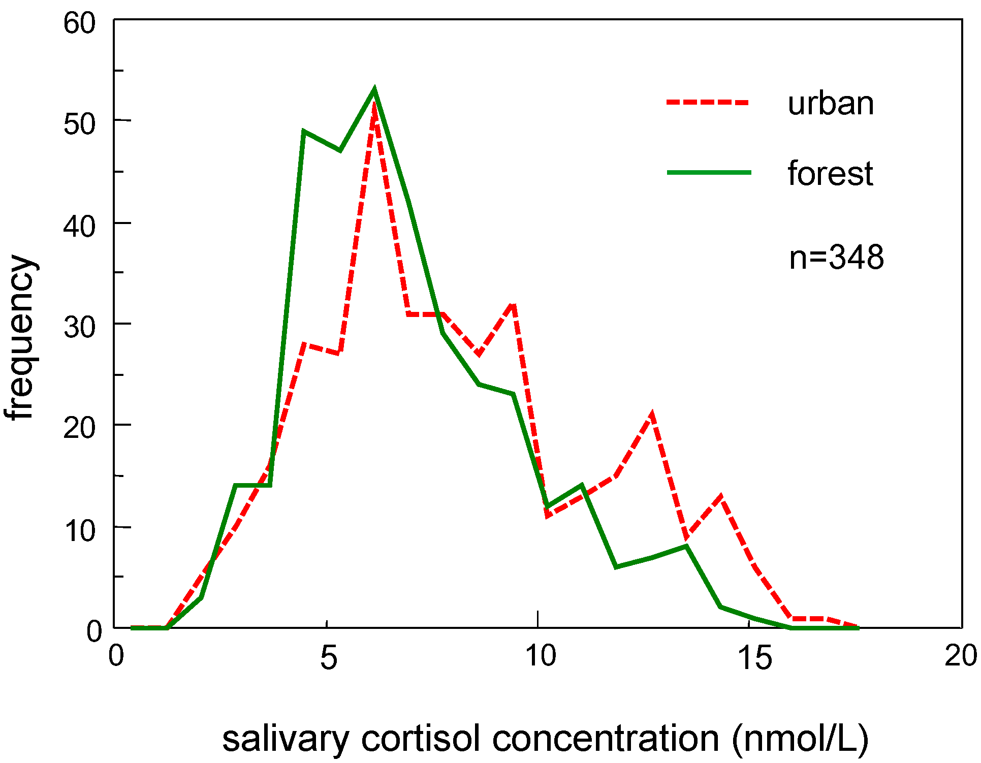
| Variable | Urban Environment | Forest Environment | Difference |
|---|---|---|---|
| n = 348 | n = 348 | ||
| mean (nmol/L) | 7.98 | 6.88 | p < 0.001 |
| median (nmol/L) | 7.45 | 6.35 | p < 0.001 |
| SD (nmol/L) | 3.33 | 2.75 | p < 0.001 |
| CV (%) | 40.8 | 38.5 | p = 0.206 |
| Q1 | 5.79 | 4.97 | p = 0.001 |
| Q3 | 9.93 | 8.55 | p < 0.001 |
| IQR (nmol/L) | 4.14 | 3.59 | p = 0.123 |
| skewness | 0.49 | 0.76 | p = 0.037 |
| kurtosis | 2.47 | 3.23 | p = 0.017 |
© 2017 by the authors. Licensee MDPI, Basel, Switzerland. This article is an open access article distributed under the terms and conditions of the Creative Commons Attribution (CC BY) license (http://creativecommons.org/licenses/by/4.0/).
Share and Cite
Kobayashi, H.; Song, C.; Ikei, H.; Park, B.-J.; Lee, J.; Kagawa, T.; Miyazaki, Y. Population-Based Study on the Effect of a Forest Environment on Salivary Cortisol Concentration. Int. J. Environ. Res. Public Health 2017, 14, 931. https://doi.org/10.3390/ijerph14080931
Kobayashi H, Song C, Ikei H, Park B-J, Lee J, Kagawa T, Miyazaki Y. Population-Based Study on the Effect of a Forest Environment on Salivary Cortisol Concentration. International Journal of Environmental Research and Public Health. 2017; 14(8):931. https://doi.org/10.3390/ijerph14080931
Chicago/Turabian StyleKobayashi, Hiromitsu, Chorong Song, Harumi Ikei, Bum-Jin Park, Juyoung Lee, Takahide Kagawa, and Yoshifumi Miyazaki. 2017. "Population-Based Study on the Effect of a Forest Environment on Salivary Cortisol Concentration" International Journal of Environmental Research and Public Health 14, no. 8: 931. https://doi.org/10.3390/ijerph14080931