Article

Photochemical Reaction of 7,12-Dimethylbenz[a]anthracene (DMBA) and Formation of DNA Covalent Adducts

1
Department of Chemistry, Jackson State University, Jackson, MS 39217, USA
2
National Center for Toxicological Research, Jefferson, AR 72079, USA
3
Department of Chemistry, Central University for Nationalities, Beijing 100081, China
*
Author to whom correspondence should be addressed.
Int. J. Environ. Res. Public Health 2005, 2(1), 114-122; https://doi.org/10.3390/ijerph2005010114
Submission received: 15 November 2004 / Accepted: 6 February 2005 / Published: 30 April 2005

Abstract

:
DMBA, 7,12-dimethylbenz[a]anthracene, is a widely studied polycyclic aromatic hydrocarbon that has long been recognized as a probable human carcinogen. It has been found that DMBA is phototoxic in bacteria as well as in animal or human cells and photomutagenic in Salmonella typhimurium strain TA102. This article tempts to explain the photochemistry and photomutagenicity mechanism. Light irradiation converts DMBA into several photoproducts including benz[a]anthracene-7,12-dione, 7-hydroxy-12-keto-7-methylbenz[a]anthracene, 7,12-epidioxy-7,12-dihydro-DMBA, 7-hydroxymethyl-12-methylbenz[a]anthracene and 12-hydroxymethyl-7-methylbenz[a]anthracene. Structures of these photoproducts have been identified by either comparison with authentic samples or by NMR/MS. At least four other photoproducts need to be assigned. Photo-irradiation of DMBA in the presence of calf thymus DNA was similarly conducted and light-induced DMBA-DNA adducts were analyzed by 32P-postlabeling/TLC, which indicates that multiple DNA adducts were formed. This indicates that formation of DNA adducts might be the source of photomutagenicity of DMBA. Metabolites obtained from the metabolism of DMBA by rat liver microsomes were reacted with calf thymus DNA and the resulting DNA adducts were analyzed by 32P-postlabeling/TLC under identical conditions. Comparison of the DNA adduct profiles indicates that the DNA adducts formed from photo-irradiation are different from the DNA adducts formed due to the reaction of DMBA metabolites with DNA. These results suggest that photo-irradiation of DMBA can lead to genotoxicity through activation pathways different from those by microsomal metabolism of DMBA.

Introduction

Polycyclic aromatic hydrocarbons (PAHs) are widespread genotoxic and tumorigenic environmental pollutants. It has long been known that PAHs require metabolic activation in order to exert their biological activities, including carcinogenicity [14]. Upon metabolism, PAHs are either metabolized into biologically active metabolites, including diol epoxides and free radical intermediates, which bind to cellular DNA forming covalent DNA adducts responsible for mutagenicity and carcinogenicity [14]. However, recent studies have demonstrated that activation of PAHs can also be achieved by photo-irradiation [57]. Studies have shown that upon photo-irradiation, some PAHs are more toxic to microorganisms, plants, and other organisms than PAHs themselves without light irradiation [7, 811]. Since skin is the largest organ in human and when concomitantly exposed to environmental chemicals and sunlight, these chemicals may be activated by photo-irradiation and exert adverse health effects [12, 13].
We have been interested in the photochemistry and phototoxicity of PAHs, including DNA single strand cleavage [1418], photoreaction [18, 19], DNA damage [21, 22], and DNA covalent adduct formation [23]. 7,12-Dimethylbenz[a]anthracene (DMBA), is a widely studied PAH that has long been recognized as a probable human carcinogen [14]. This study reports that light irradiation of DMBA results in the formation of photodecomposition products including benz[a]anthracene-7,12-dione, 7,12-epidioxy-7,12-dihydro-DMBA, 7-hydroxymethyl-12-methylbenz[a]anthracene (7-HOCH2-12-MBA) and 12-hydroxymethyl-7-methylbenz[a]anthracene (12-HOCH2-7-MBA). Photo-irradiation of DMBA, 7-HOCH2-12-MBA and 12-HOCH2-7-MBA in the presence of calf thymus DNA followed by 32P-postlabeling/TLC analysis indicated that multiple DNA adducts are formed, and these adducts are different from the DNA adducts formed from reaction of DMBA metabolites with DNA. These results suggest that photo-irradiation of DMBA can lead to genotoxicity through activation pathways different from those by microsomal metabolism of DMBA.

Materials and Methods

Materials

DMBA, lead tetraacetate, 2,3-dichloro-5,6-dicyanobenzoquinone (DDQ), glucose-6-phosphate dehydrogenase (type XII, Sigma), NADP+, and glucose-6-phosphate were purchased from Sigma Chemical Co. (St. Louis, MO). Cloned T4 polynucleotide kinase (PNK) was obtained from U.S. Biochemical Corp. (Cleveland, OH). [γ-32P]Adenosine 5′-triphosphate ([γ-32P]ATP) (sp. act. >7,000 Ci/mmol) was purchased from ICN Biomedicals, Inc. (Costa Mesa, CA). All other reagents were obtained through commercial sources and were the highest quality available. All solvents used were HPLC grade.
According to the method published by Boyland and Sims [24], 7-HOCH2-12-MBA and 12-HOCH2-7-MBA were prepared by reaction of DMBA with lead tetraacetate, separation of the resulting 7-acetoxymethyl-12-MBA and 12-acetoxymethyl-7-MBA by column chromatography, and hydrolysis to the hydroxyl DMBA. 7-Formyl-12-methyllbenz[a]anthracene (7-CHO-12-MBA) and 12-Formyl-7-methyllbenz[a]anthracene (12-CHO-7-MBA) were synthesized by oxidation of 7-HOCH2-12-MBA and 12- HOCH2-7-MBA, respectively with DDQ [25]. 7,12-Epidioxy-7,12-dihydro-DMBA was prepared according to Wood et al. [26].
3-Methylcholanthene-induced female Sprague-Dawley rat liver microsomes were purchased from In Vitro Technologies (Baltimore, MA). Protein concentrations were determined using a protein assay based on the Bradford method using a Bio-Rad protein detection kit (Bio-Rad Laboratories, Hercules, CA).

Light Source

The UVA light box was custom made with a 4-lamp unit using UVA lamps (National Biologics). The irradiance of light was determined using an Optronics OL754 Spectroradiometer (Optronics Laboratories, Orlando, FL), and the light dose was routinely measured using a Solar Light PMA-2110 UVA detector (Solar Light Inc., Philadelphia, PA). The maximum emission of the UVA is between 340 – 355 nm. The light intensities at wavelengths below 320 nm (UVB light) and above 400 nm (visible light) are about two orders of magnitude lower than the maximum at 340–355 nm.

Photo-Irradiation of DMBA

A solution (2–3 mL) of 0.4 mM DMBA dissolved in 90% ethanol was placed in a UV-transparent cuvette and photo-irradiated under UVA light to receive a light dose of 2.6 J/cm2/min for a period of 40, 90, and 360 min, respectively. The reaction mixture was then concentrated to about 200 μL under reduced pressure. Reversed-phase HPLC separation of the resulting photodecomposition products was accomplished using a Prodigy 5 μ ODS column (4.6 × 250 mm, Phenomenex, Torrance, CA) eluted isocratically with 90% methanol in water (v/v) at 1 mL/min. For isolation of photodecomposition products in larger amounts, a Prodigy 5 μ ODS column (10 × 250 mm, Phenomenex, Torrance, CA) eluted isocratically with 90% methanol in water (v/v) at 5 mL/min was used.

Photo-Irradiation of DMBA, 7-HOCH2-12-MBA, 12-HOCH2-7-MBA, 7-CHO-12-MBA, and 12-CHO-7-MBA in the Presence of Calf thymus DNA

A solution (0.4 mM dissolved in 2 mL of 90% ethanol) placed in a UV-transparent cuvette was added with 20 mg calf thymus DNA in 1 mL tetrahydrofuran. The resulting solution was photo-irradiated under UVA light with a total light dose of 14 J/cm2. After incubation, DNA was isolated and the DNA adducts were analyzed by 32P-postlabeling/TLC.
Following a published procedure [27], approximately 10 μg of DNA was 32P-postlabeled using nuclease P1 enrichment. Adducts were separated by thin layer chromatography performed on 0.1 mm Machery Nagel 300 polyethylene imine cellulose plates (Alltech, Deerfield, IL) using the following solvent directions, D1: 0.9 M sodium phosphate, pH 6.8; D2: 3.6 M lithium formate, 8.5 M urea, pH 3.5; D3: 1.2 M lithium chloride, 0.5 M Tris HCl, 8 M urea, pH 8.0. A final wash was conducted in D3 with solvent used in D1. Areas of radioactivity were measured with a Storm 860 phosphor imaging system (Molecular Dynamics, Sunnyvale, CA).

Reaction of DMBA Metabolites with Calf Thymus DNA and Analysis of DNA Adducts by 32P-postlabeling/TLC

In vitro metabolism was carried out by incubation of DMBA solution (0.8 mM dissolved in 200 μl of acetone) with shaking at 37° for 30 min in a 10 ml reaction mixture containing 0.5 mM of Tris-HCl (pH 7.5), 30 μM of MgCl2, 1 unit of glucose-6-phosphate dehydrogenase (type XII, Sigma), 1 mg NADP+, 6 mg of glucose-6-phosphate, 10 mg of microsomal protein, and 10 mg of purified calf thymus DNA. After incubation, the reaction was cooled with ice-water, then sequentially extracted with 5.0 mL phenol, 5.0 mL phenol/chloroform/isoamyl alcohol (v/v/v, 25/24/1), and 5.0 mL chloroform/isoamyl alcohol (v/v, 24/1). The DNA in the aqueous phase was precipitated by adding 1 mL 5 M sodium chloride followed by equal volume of cold ethanol and washed three times with 70% ethanol. After redissolving in 3 mL distilled water, the DNA concentration and purity were determined spectrophotometrically, and DNA adducts were analyzed by 32P-postlabeling/TLC analysis with the method described above.

Instrumentation

A Waters HPLC system consisting of a Model 600 controller, a Model 996 photodiode array detector, and pump was used for separation and purification of DMBA photodecomposition products. Direct exposure probe (DEP) mass spectrometry (MS) was performed on a ThermoFinnigan TSQ 700 triple quadrupole mass spectrometer operated in the electron ionization (EI) mode.

Results

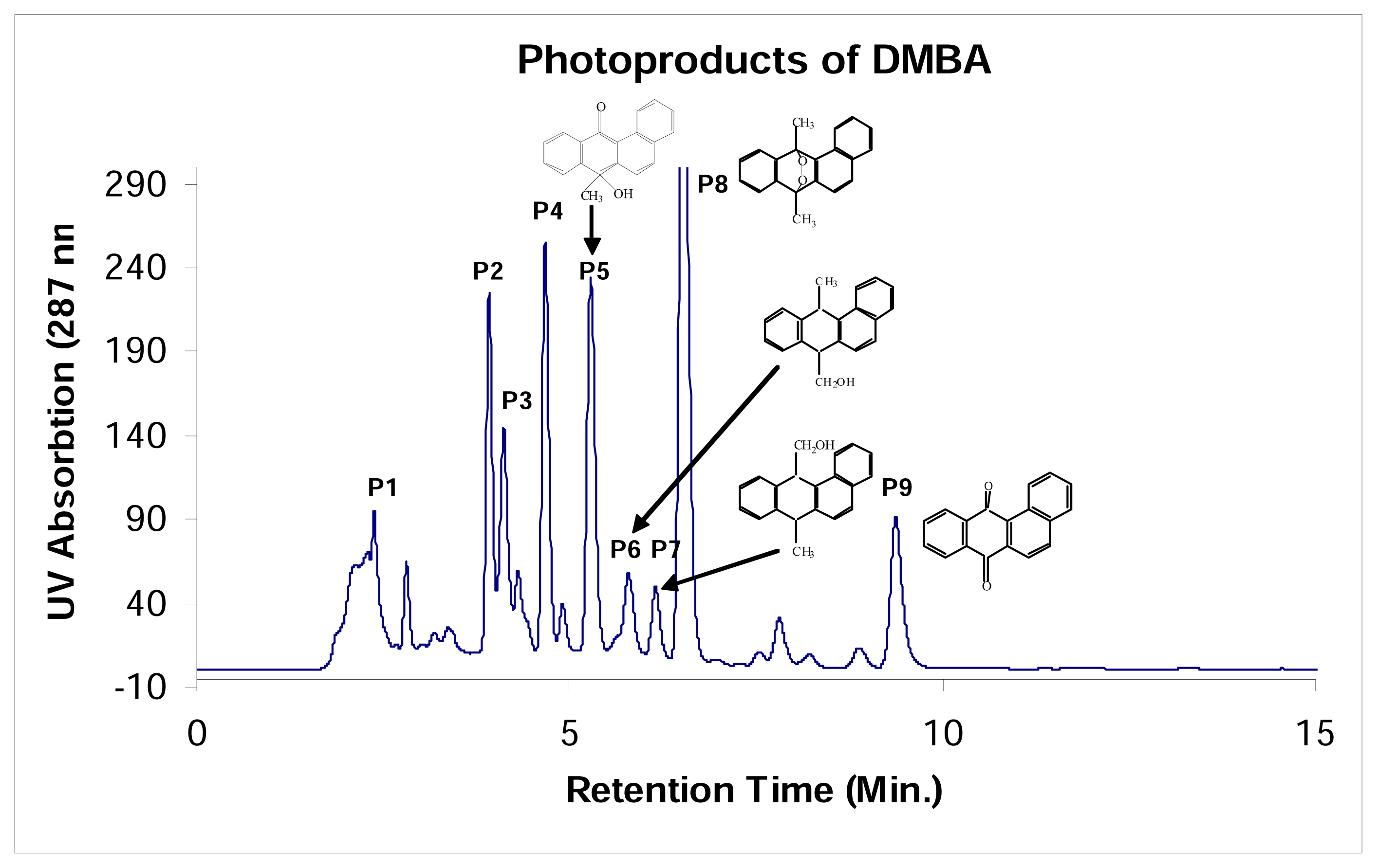
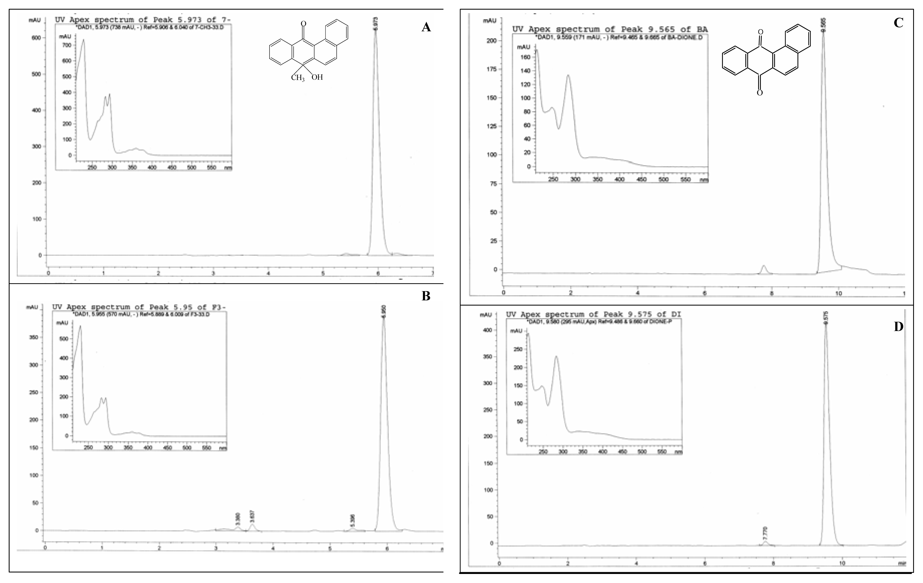
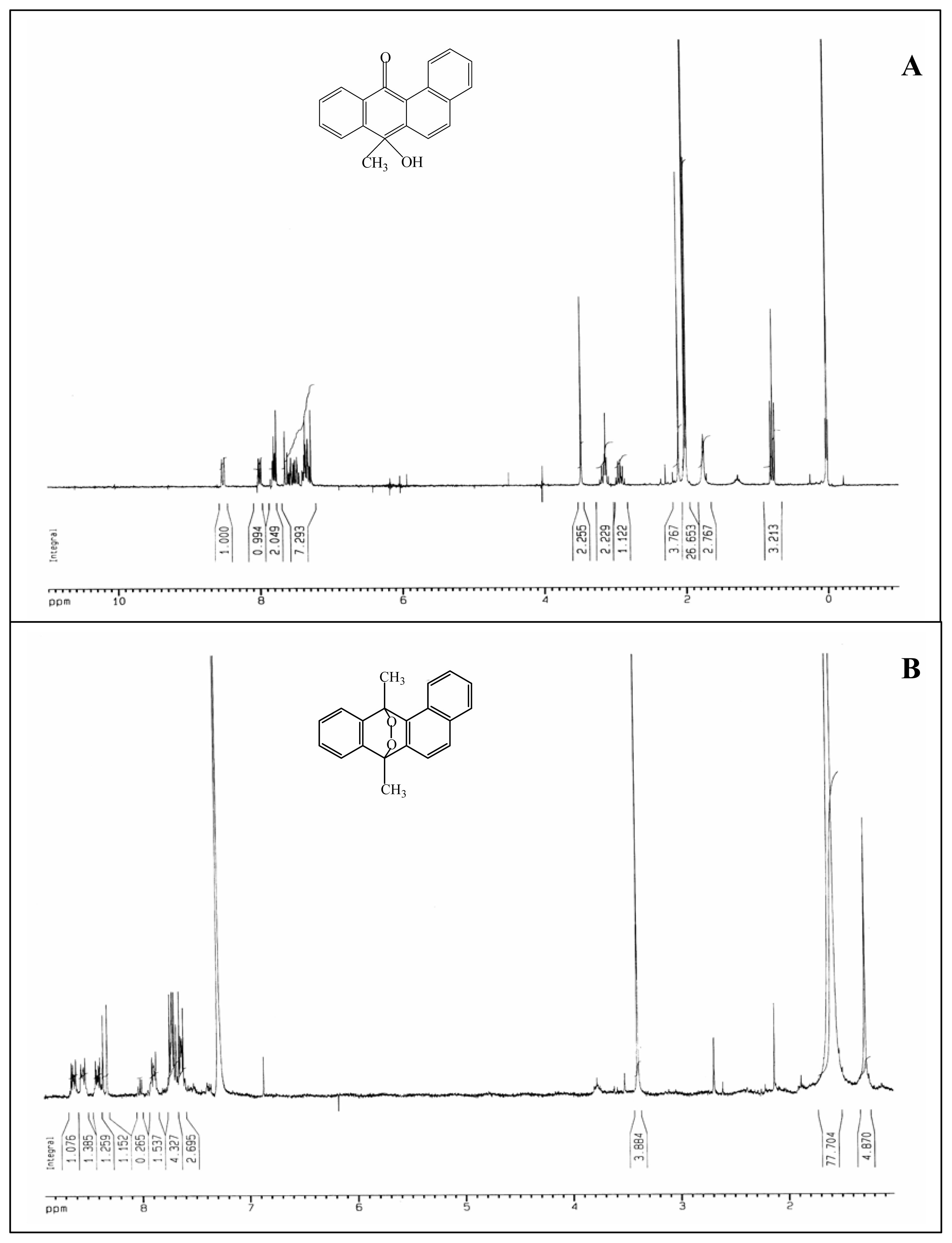
DMBA Photoproduct Analysis

Photo-irradiation of DMBA in ethanol/water (v/v, 90/10) by UVA light at a light dose of 2.6 J/cm2/min for a period of 40, 90, and 360 min, respectively was conducted and the reaction mixture was separated by reversed phase HPLC (Figure 1). Based on comparison of the HPLC retention time, UV-absorption spectrum, and mass spectrum with those of DMBA, the material contained in the chromatographic peak eluting at 19.0 min was identified as the recovered DMBA. As shown in Figure 1A, the amount of DMBA decreased and the amounts of photodecomposition products increased rapidly. For collection of sufficient amount of the photodecomposition products for structural identification, the products formed after 360 min of photo-irradiation were separated by repeated preparative HPLC (Figure 2). Based on mass (Figure 3A) and NMR (Figure 4A) spectral analysis, the material in the chromatographic peak eluting at 5.3 min (P5 in Figure 1C) was tentatively identified as 7-hydroxy-12-keto-7-methylbenz[a]anthracene (7-OH-12-keto-7-MBA). The chromatographic peak eluting at 6.6 min (P8) was identified as 7,12-epidioxy-7,12-dihydro-DMBA. This is based on the comparison of its UV-visible absorption spectrum, HPLC retention time, mass spectrum (Figure 3B), and NMR spectrum (Figure 4B) with those of the authentic sample (data not shown) [26]. The material in chromatographic peak eluting at 5.8 min (P6) (Figure 2) had a mass spectrum with a molecular ion M+ at m/z 272 (data not shown), suggesting this is an oxygenated DMBA. This compound has the mass spectrum, UV-visible absorption spectrum (Figure 5A insert) and HPLC retention time (Figure 5A) identical to those of the synthetic standard for 7-HOCH2-12-MBA (Figure 5B). Thus, it confirms that this photodecomposition product is 7-HOCH2-12-MBA. The material in chromatographic peak (P7) eluting at 6.2 min was similarly identified as 12-HOCH2-7-MBA using a synthetic standard. Based on comparison of HPLC retention time and UV-visible absorption spectrum (Figure 5C and insert) with those of the authentic BA-7,12-dione (Figure 5D and insert), the chromatographic peak (P9) eluting at 9.4 min was identified as BA-7,12-dione.

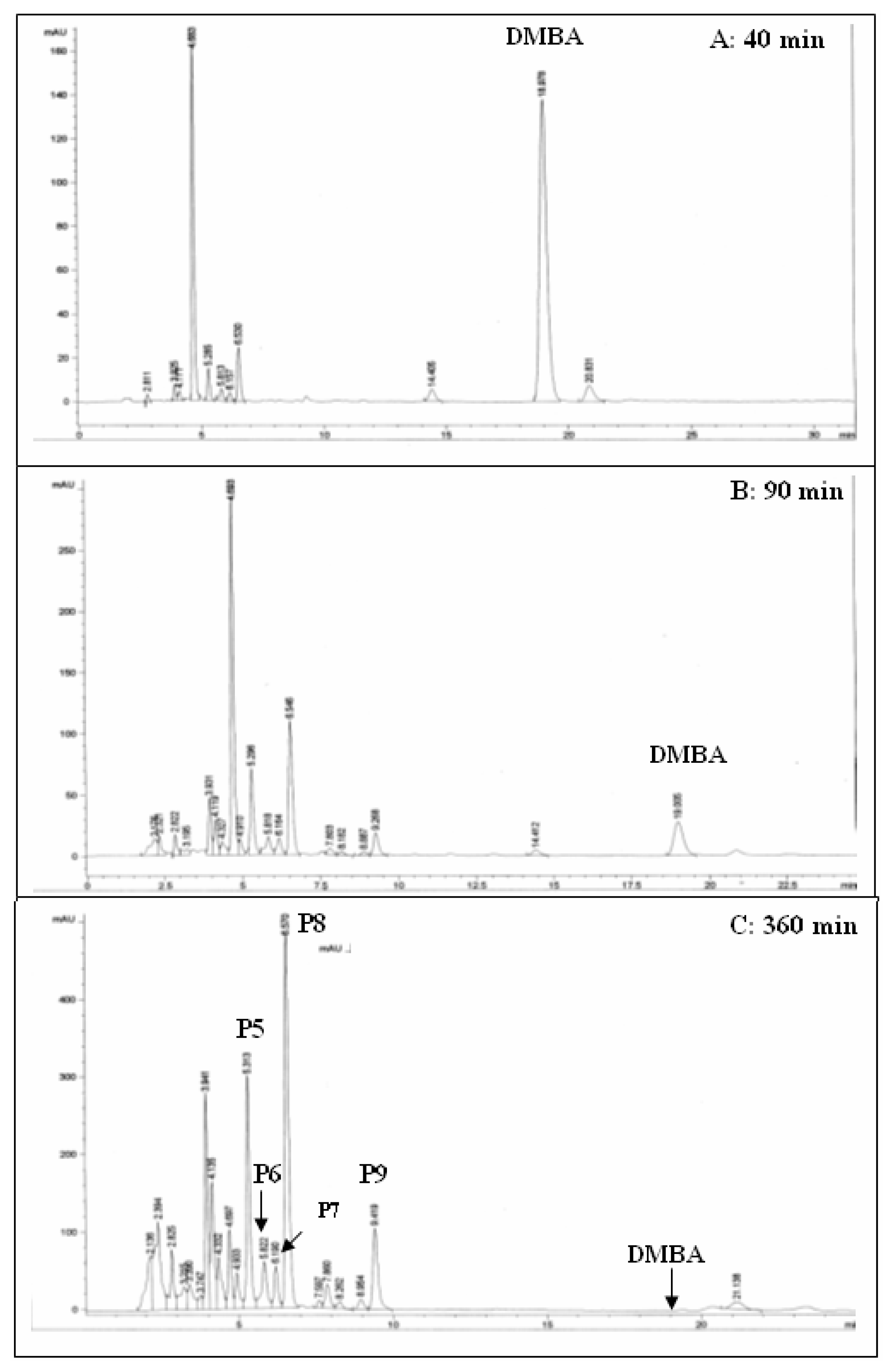
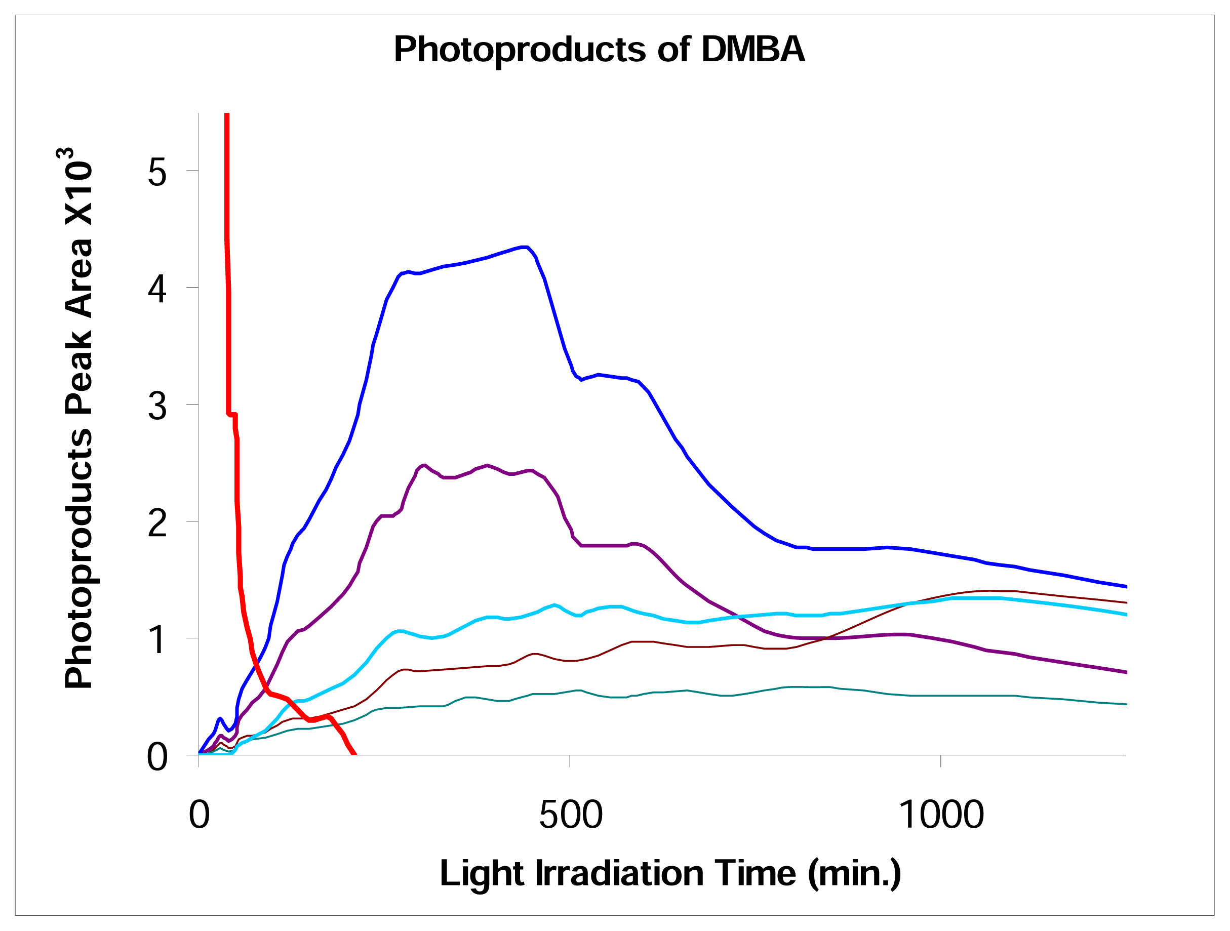
Kinetics for the Photodecomposition of DMBA and Photoproduct Formation

The formation and decomposition of the five identified photodecomposition products, P5-P9, were studied. As shown in Figure 6, while DMBA completely decomposed at about 260 min under light irradiation, the photodecomposition products P5 and P8 reached the highest yield at about 400 min of irradiation time. Compound P9 kept increasing, suggesting that the other decomposition products gradually converted into BA-7,12-dione (P9). Compounds P6 and P7 also increased during the whole course of irradiation.

Photo-Irradiation of DMBA, 7-HOCH2-12-MBA, 12-HOCH2-7-MBA, 7-CHO-12-MBA, and 12-CHO-7-MBA in the Presence of Calf thymus DNA

Photo-irradiation of DMBA, 7-HOCH2-12-MBA, 12-HOCH2-7-MBA, 7-CHO-12-MBA, and 12-CHO-7-MBA in the presence of calf thymus DNA was carried out, the resulting DNA was isolated, and the DNA adducts were analyzed by 32P-postlabeling/TLC. Although both 7-CHO-12-MBA and 12-CHO-7-MBA were not formed as photodecomposition products in our study, these compounds were formed as reported by Wood et al. (26). Therefore, for facilitating in mechanistic understanding, the DNA adduct formation from these two compounds was also pursued.
As shown in Figure 7, the 3′,5′-bisphosphate deoxyribonucleosides of DMBA (Figure 7A) and the four oxidized derivatives (Figure 7C–7F) were separated by thin-layer chromatography (TLC) into multiple spots. Analysis of these resulting TLC spots indicated that the spot profiles are nearly identical.
Figure 8 shows the autoradiogram of 32P-postlabeled nuclease P1-treated calf thymus DNA from (A) photo-irradiation of the DNA in the presence of DMBA by UVA light (14 J/cm2) and (B) the metabolite mixture from metabolism of DMBA by rat liver microsomes in the presence of calf thymus DNA.

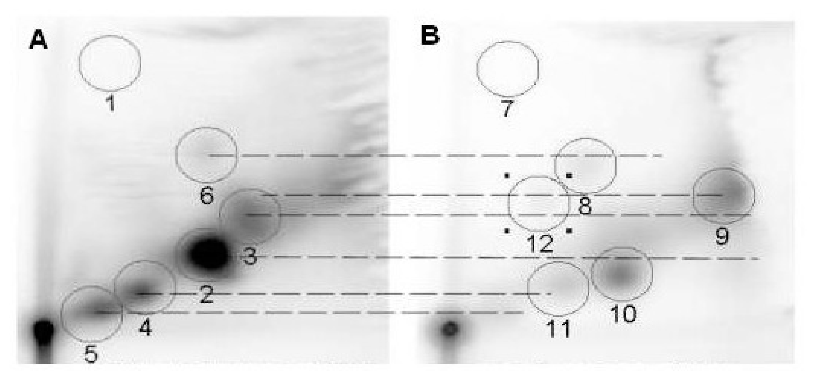
Reaction of Metabolites of DMBA with Calf thymus DNA and DNA Adducts Analyzed by 32P-postlabeling/TLC

For comparison of DNA adduct profile, the metabolites obtained from metabolism of DMBA by rat liver microsome in the presence of calf thymus DNA were also analyzed by 32P-postlabeling/TLC under identical conditions. Comparison of the DNA adduct profiles indicates that the DNA adducts formed from photo-irradiation of DMBA are different from those from reaction of DMBA metabolites (Figure 8).

Discussion

In this study, photo-irradiation of DMBA under UVA light resulted in the formation of multiple photodecomposition products, of which four products were identified, including benz[a]anthracene-7,12-dione, 7,12-epidioxy-7,12-dihydro-DMBA, 7-HOCH2-12-MBA, and 12-HOCH2-7-MBA, and one product tentatively assigned as 7-hydroxy-12-keto-7-MBA (Figure 1). Although Wood et al. [26] reported that 7-CHO-12-MBA and 12-CHO-7-MBA were produced from photo-oxidation of DMBA, these two compounds were not formed under our experimental conditions. This discrepancy illustrates that the formation of photo-oxidation products from DMBA is highly dependent on experimental conditions, particularly the light wavelength.
The results of 32P-postlabeling/TLC analysis indicate that photo-irradiation of DMBA and the four oxidized derivatives (7-HOCH2-12-MBA, 12-HOCH2-7-MBA, 7-CHO-12-MBA, and 12-CHO-7-MBA) by UVA light in the presence of calf thymus DNA all generated multiple DNA adducts and the DNA adduct profiles are nearly identical.
These results highly suggest that the photo-induced DNA adduct formation from DMBA is mediated through these oxidized derivatives by two distinct pathways: (i) oxidation of DMBA at the 7-methyl group to 7-HOCH2-12-MBA, then 7-CHO-12-MBA, then to the reactive species; and (ii) oxidation of DMBA at the 12-methyl group to 12-HOCH2-7-MBA, then 12-CHO-7-MBA, then to the reactive species. Also the kinetic study of photo-irradiation of DMBA, as shown in Figure 6, indicates that BA-7,12-dione is the final and stable product. Consequently, we conclude that the reactive photodecomposition product that can bind to DNA and form DNA adducts is from 7-CHO-12-MBA or 12-CHO-7-MBA, and is further oxidized to the inert BA-7,12-dione. Comparison of the DNA adduct profiles indicates that the DNA adducts formed from photo-irradiation of DMBA and from metabolism of DMBA by 3-methylcholanthrene-induced rat liver microsomes are different. These results suggest that photo-irradiation of DMBA can lead to genotoxicity through activation pathways different from those by microsomal metabolism of DMBA.
Thus, our study indicates that photo-irradiation of DMBA generates genotoxic photo-oxidation products that can lead to DNA adduct formation. Besides, Boyland et al. reported that 7-HOCH2-12-MBA is able to cause adrenal apoplexy and mammary cancer in rats [27]. 7,12-Epidioxy-7,12-dihydro-DMBA has been shown to be toxic to chicken fibroblast cells [28]. Consequently, these results suggest that photo-irradiation of PAHs can generate genotoxic products and can be highly harmful to human health. This warrants further investigation.
Figure 1. HPLC profiles of photoproducts of DMBA after irradiation with UVA light (2.6 J/cm2/min) in ethanol for (A) 40 min; (B) 90 min; (C) 360 min.
Ijerph 02 00114f1
Figure 2. Identification of some of the photoproducts of DMBA in ethanol after 360 min of irradiation using UVA light (2.6 J/cm2/min).
Ijerph 02 00114f2
Figure 3. Mass spectrum profiles of purified P5 (A) and P8 (B) of DMBA photoproducts.
Ijerph 02 00114f3
Figure 4. 1H-NMR spectra of purified P5 (A) and P8 (B) of DMBA photoproducts.
Ijerph 02 00114f4
Figure 5. HPLC and UV spectrum profiles of 7-hydroxymethyl-12-methylbenz[a]anthracene standard (A), purified P6 from DMBA photoproducts (B); benz[a]anthracen-7,12-dione standard (C), and purified P9 from DMBA photoproducts (D).
Ijerph 02 00114f5
Figure 6. Time course of photodecomposition of DMBA and formation and photodecomposition of the identified DMBA photodecomposition products.
Ijerph 02 00114f6
Figure 7. Autoradiogram of 32P-postlabeled nuclease P1-treated DNA from photo-irradiation in the presence of (A) DMBA, (B) blank, (C) 7-HOCH2-12-MBA, (D) 12-HOCH2-7-MBA, (E) 7-CHO-12-MBA, and (F) 12-CHO-7-MBA in THF and calf thymus DNA by UVA light at a total dose of 14 J/cm2.
Ijerph 02 00114f7
Figure 8. Autoradiogram of 32P-postlabeled nuclease P1-treated calf thymus DNA from (A) photo-irradiation of the DNA in the presence of DMBA by UVA light (14 J/cm2) and (B) the metabolite mixture from metabolism of DMBA by rat liver microsomes in the presence of calf thymus DNA.
Ijerph 02 00114f8

Acknowledgements

This research was in part supported by the US Army Research Office DAAD 1901-1-0733 to JSU and the National Institutes of Health: NIH-SCORE S06 GM08047. We thank the NIH-RCMI for core Molecular and Cellular Biology and Analytical Chemistry facilities at JSU.

References

  1. Dipple, A.; Moschel, R. C.; Bigger, C. A. H. Polynuclear aromatic carcinogens. American Chemical Society: Washington, DC; Searle, C. E., Ed.; 1984; Volume 1, pp. 41–163. [Google Scholar]
  2. Yang, S. K.; Silverman, B. D. (Eds.) Polycyclic Aromatic Hydrocarbon Carcinogenesis: Structure-Activity Relationships; CRC Press: Boca Raton, FL, 1988; Volume I and II.
  3. Harvey, R. G. Polycyclic Aromatic Hydrocarbons; Wiley-VCH: New York, 1997. [Google Scholar]
  4. Conney, A. H. Induction of Microsomal Enzymes by Foreign Chemicals and Carcinogenesis by Polycyclic Aromatic Hydrocarbons. Cancer Res 1982, 42, 4875–4917. [Google Scholar]
  5. Arfsten, D. P.; Schaeffer, D. J.; Mulveny, D. C. The effects of near ultraviolet radiation on the toxic effects of polycyclic aromatic hydrocarbons in animals and plants: A review. Ecotoxicol. Environ. Safety 1996, 33, 1–24. [Google Scholar]
  6. Dabestani, R.; Ivanov, I. N. A comparison of physical, spectroscopic and photophysical properties of polycyclic aromatic hydrocarbons. Photochem. Photobiol 1999, 70, 10–34. [Google Scholar]
  7. Yu, H. Environmental carcinogenic polycyclic aromatic hydrocarbons: Photochemistry and phototoxicity. J. Environ. Sci. Health C-Environ. Carcinog. & Ecotoxic. Rev 2002, 20, 149–183. [Google Scholar]
  8. Pelletier, M. C.; Burgess, R. M.; Ho, K. T.; Kuhn, A.; McKinney, R. A.; Ryba, S. A. Phototoxicity of individual polycyclic aromatic hydrocarbons and petroleum to marine invertebrate larvae and juveniles. Environ. Toxicol. Chem 1997, 16, 2190–2199. [Google Scholar]
  9. Swartz, R. C.; Ferraro, S. P.; Lamberson, J. O.; Cole, F. A.; Ozretich, R. J.; Boese, B. L.; Schults, D. W.; Behrenfeld, M.; Ankley, G. T. Photoactivation and toxicity of mixtures of polycyclic aromatic hydrocarbon compounds in marine sediment. Environ. Toxicol. Chem 1997, 16, 2151–2157. [Google Scholar]
  10. Kagan, J.; Tuveson, R. W.; Gong, H. H. The light-dependent cytotoxicity of benzo[a]pyrene: effect on human erythrocytes, Escherichia coli cells, and Haemophilus influenzae transforming DNA. Mutat. Res 1989, 216, 231–242. [Google Scholar]
  11. Fernandez, M.; L’Haridon, J. Influence of lighting conditions on toxicity and genotoxicity of various PAH in the newt in vivo. Mutat. Res 1992, 298, 31–41. [Google Scholar]
  12. Mukhtar, H. (Ed.) Skin Cancer: Mechanisms and Human Relevance; CRC Press: Boca Raton, 1995.
  13. Ahmad, H.; Mukhtar, H. Toxicology of the skin: new and emerging concepts. Toxicol Applied Pharm 2004, 195, 265–266. [Google Scholar]
  14. Dong, S.; Hwang, H.-M.; Harrison, C.; Holloway, L.; Shi, X.; Yu, H. UVA light-induced DNA single strand cleavage by selected polycyclic aromatic hydrocarbons. Bull. Environ. Contam. Toxicol 2000, 64, 467–474. [Google Scholar]
  15. Dong, S.; Wang, S.; Stewart, G.; Hwang, H.-M.; Fu, P. P.; Yu, H. Effect of organic solvents and biologically relevant ions on the light-induced DNA cleavage by pyrene and its amino and hydroxy derivatives. Intl. J. Mol. Sci 2002, 3, 937–947. [Google Scholar]
  16. Dong, S.; Fu, P. P.; Hwang, H.-M.; Yu, H. Effect of histidine on light-induced DNA single strand cleavage by selected polycyclic aromatic hydrocarbons. Polycycl. Arom. Compd 2002, 22, 451–458. [Google Scholar]
  17. Yu, H.; Dong, S.; Fu, P. P.; Hwang, H.-M. UVA light-induced DNA single strand cleavage by hydroxybenzo[a]pyrenes. Polycycl. Arom. Compd 2002, 22, 861–870. [Google Scholar]
  18. Dong, S.; Fu, P. P.; Shirsat, R. N.; Hwang, H.-M.; Leszczynski, J.; Yu, H. UVA Light-Induced DNA cleavage by isomeric methylbenz[a]anthracenes. Chem. Res. Toxicol 2002, 15, 400–407. [Google Scholar]
  19. Zeng, K.; Hwang, H.-M.; Fu, P. P.; Yu, H. Identification of 1-hydroxypyrene photoproducts and study of the effect of humic substances on its photolysis. Polycycl. Arom. Compd 2002, 22, 459–467. [Google Scholar]
  20. Wang, L.; Yan, J.; Cohly, H.; Hwang, H.-M.; Wang, S.; Fu, P. P.; Yu, H. Photo-Acute toxicity and genotoxicity of azulene on human Jurkat T-cells. Mutat. Res 2004, 562(1–2), 143–150. [Google Scholar]
  21. Wang, L.; Yan, J.; Fu, P. P.; Parekh, K. A.; Yu, H. Photomutagenicity of cosmetic ingredient chemicals azulene and guaiazulene. Mutat.. Res 2003, 530, 19–26. [Google Scholar]
  22. Yan, J.; Wang, L.; Fu, P. P.; Yu, H. Photomutagenicity of sixteen polycyclic aromatic hydrocarbons from the US EPA’s priority pollutants. Mutat. Res 2004, 557, 99–108. [Google Scholar]
  23. Dong, S.; Hwang, H.-M.; Shi, X.; Holloway, L.; Yu, H. UVA-induced DNA single strand cleavage by 1-hydroxypyrene and formation of covalent adducts between DNA and 1-hydroxypyrene. Chem. Res. Toxicol 2002, 13, 585–593. [Google Scholar]
  24. Boyland, E.; Sims, P. Metabolism of polycyclic compounds. The metabolism of 7,12-dimethylbenz(alpha) anthracene by rat-liver homogenates. Biochem. J 1965, 95, 780–787. [Google Scholar]
  25. Pataki, J.; Wlos, R.; Cho, Y.-C. Adrenocorticolytic derivatives of benz[.alpha.] anthracene. J. Med. Chem 1968, 11, 1083–1090. [Google Scholar]
  26. Wood, J.; Barker, C. L.; Grubbs, C. J. Photooxidation products of 7,12-dimethylbenz[a]anthracene. Chem.-Biol. Interact 1979, 26, 339–347. [Google Scholar]
  27. Gupta, R. C. Enhanced sensitivity of 32P-postlabeling analysis of aromatic carcinogen:DNA adducts. Cancer Res 1985, 45, 5656–5662. [Google Scholar]
  28. Boyland, E.; Sims, P.; Huggins, C. Induction of adrenal damage and cancer with metabolites of 7,12-dimethylbenz[a]anthracene. Nature 1969, 207, 816–817. [Google Scholar]
  29. Warshawsky, D.; Kerns, E.; Bissell, M. J.; Calvin, M. Characterization of a photoproduct of 7,12-dimethylbenz[a]anthracene and its effects on chick embryo cells in culture. Biochem. J 1977, 164, 481–48. [Google Scholar]

