Blood Lead Levels Among Pregnant Women: Historical Versus Contemporaneous Exposures

Abstract
:1. Introduction
2. Data and Methods
3. Results
4. Discussion
5. Conclusions
Acknowledgments
References
- Lanphear, BP; Dietrich, K; Auinger, P; Cox, C. Cognitive deficits associated with blood lead concentrations <10 μg/dL in US children and adolescents. Public Health Rep 2000, 115, 521–529. [Google Scholar]
- Schnaas, L; Rothenberg, S; Flores, MF; Martinez, S; Hernandez, C; Osorio, E; Velasco, SR; Perroni, E. Reduced intellectual development in children with prenatal lead exposure. Environ. Health Perspect 2006, 114, 791–797. [Google Scholar]
- Chiodo, LM; Jacobson, SW; Jacobson, JL. Neurodevelopmental effects of postnatal lead exposure at very low levels. Neurotoxicol. Teratol 2004, 26, 359–371. [Google Scholar]
- Canfield, RL; Henderson, CR; Cory-Slechta, DA; Cox, C; Jusko, TA; Lanphear, BP. Intellectual impairment in children with blood lead concentrations below 10 μg per deciliter. N. Engl. J. Med 2003, 348, 1517–1526. [Google Scholar]
- Miranda, ML; Kim, D; Overstreet Galeano, MA; Paul, C; Hull, A; Morgan, SP. The relationship between early childhood blood lead levels and performance on End of Grade Tests. Environ. Health Perspect 2007, 115, 1242–1247. [Google Scholar]
- Tong, S; Baghurst, P; McMichael, A; Sawyer, M; Mudge, J. Lifetime exposure to environmental lead and children's intelligence at 11–13 years, the Port Pirie cohort study. Br. Med. J 1996, 312, 1569–1575. [Google Scholar]
- Bellinger, DC; Stiles, KM; Needleman, HL. Low-level lead exposure, intelligence and academic achievement, a long-term follow-up study. Pediatrics 1992, 90, 855–861. [Google Scholar]
- Dietrich, KN; Berger, OG; Succop, PA; Hammond, PB; Bornschein, RL. The developmental consequences of low to moderate prenatal and postnatal lead exposure, intellectual attainment in the Cincinnati Lead Study Cohort following school entry. Neurotoxicol. Teratol 1993, 15, 37–44. [Google Scholar]
- Miranda, ML; Kim, D; Reiter, J; Overstreet Galeano, MA; Maxson, P. Environmental contributors to the achievement gap. Neurotoxicology 2009, 30, 1019–1024. [Google Scholar]
- Miranda, ML; Maxson, P; Kim, D. Early childhood lead exposure and exceptionality designations for students. Int J Child Health Hum Dev 2010, 3. [Google Scholar]
- Lanphear, BP; Hornung, R; Khoury, J; Yolton, K; Baghurst, P; Bellinger, DC; Canfield, RL; Dietrich, KN; Bornschein, R; Greene, T; Rothenberg, SJ; Needleman, HL; Schnaas, L; Wasserman, G; Graziano, J; Roberts, R. Low-level environmental lead exposure and children's intellectual function, an international pooled analysis. Environ. Health Perspect 2005, 113, 894–899. [Google Scholar]
- Schwartz, J. Low-level lead exposure and children's IQ, a meta-analysis and search for a threshold. Environ. Res 1994, 65, 42–55. [Google Scholar]
- Gatsonis, CA; Needleman, HL. Recent epidemiologic studies of low-level lead exposure and the IQ of children, A meta-analytic review. In Human Lead Exposure; Needleman, HL, Ed.; CRC Press: Boca Raton, FL, USA, 1992; pp. 244–255. [Google Scholar]
- Schwartz, J. Beyond LOEL's, p values, and vote counting, methods for looking at the shapes and strengths of associations. Neurotoxicology 1993, 14, 237–246. [Google Scholar]
- Needleman, HL; Landrigan, PJ. Letter, What level of lead in blood is toxic for a child? Am. J. Public Health 2004, 94, 8. [Google Scholar]
- Schwartz, J; Angle, C; Pitcher, H. Relationship between childhood blood lead levels and stature. Pediatrics 1986, 77, 281–288. [Google Scholar]
- Huseman, CA; Varma, MM; Angle, CR. Neuroendocrine effects of toxic and low blood lead levels in children. Pediatrics 1992, 90, 186–189. [Google Scholar]
- Gomaa, A; Hu, H; Bellinger, D; Schwartz, J; Tsaih, SW; Gonzalez-Cossio, T; Schnaas, L; Peterson, K; Aro, A; Hernandez-Avila, M. Maternal bone lead as an independent risk factor for fetal neurotoxicity, a prospective study. Pediatrics 2002, 110, 110–118. [Google Scholar]
- Jelliffe-Pawlowski, LL; Miles, SQ; Courtney, JG; Materna, B; Charlton, V. Effect of magnitude and timing of maternal pregnancy blood lead (Pb) levels on birth outcomes. J. Perinatol 2006, 26, 154–162. [Google Scholar]
- Ronchetti, R; van den Hazel, P; Schoeters, G; Hanke, W; Rennezova, Z; Barreto, M; Villa, MP. Lead neurotoxicity in children: Is prenatal exposure more important than postnatal exposure? Acta Paediatr 2006, 95, 45–49. [Google Scholar]
- Wright, JP; Dietrich, KN; Ris, MD; Hornung, RW; Wessel, SD; Lanphear, BP; Ho, M; Rae, MN. Association of prenatal and childhood blood lead concentrations with criminal arrests in early adulthood. PLoS Med 2008, 5, e101. [Google Scholar]
- Mushak, P; Davis, JM; Crocetti, AF; Grant, LD. Prenatal and postnatal effects of low-level lead exposure, integrated summary of a report to the U.S. Congress on childhood lead poisoning. Environ. Res 1989, 50, 11–36. [Google Scholar]
- Sowers, M; Jannausch, M; Scholl, T; Li, W; Kemp, FW; Bogden, JD. Blood lead concentrations and pregnancy outcomes. Arch. Environ. Health 2002, 57, 489–495. [Google Scholar]
- Hauth, JC; Ewell, MG; Levine, RJ; Esterlitz, JR; Sibai, B; Curet, LB; Catalano, PM; Morris, CD. Pregnancy outcomes in healthy nulliparas who developed hypertension. Calcium for Preeclampsia Prevention Study Group. Obstet. Gynecol 2000, 95, 24–28. [Google Scholar]
- Allen, V; Joseph, KS; Murphy, K; Magee, L; Ohlsson, A. The effect of hypertensive disorders in pregnancy on small for gestational age and stillbirth, a population based study. BMC Pregnancy Childbirth 2004, 4, 17. [Google Scholar]
- Centers for Disease Control and Prevention. Blood Lead Levels—United States, 1999–2002. MMWR 2005, 54, 513–516. [Google Scholar]
- Lee, MG; Chun, OK; Song, WO. Determinants of the blood lead level of US women of reproductive age. J. Am. Coll. Nutr 2005, 24, 1–9. [Google Scholar]
- Rothenberg, SJ; Karchmer, S; Schnaas, L; Perroni, E; Zea, F; Fernandez, AJ. Changes in serial blood lead levels during pregnancy. Environ. Health Perspect 1994, 102, 876–880. [Google Scholar]
- Hertz-Picciotto, I; Schramm, M; Watt-Morse, M; Chantala, K; Anderson, J; Osterloh, J. Patterns and determinants of blood lead during pregnancy. Am. J. Epidemiol 2000, 152, 829–837. [Google Scholar]
- Ettinger, AS; Lamadrid-Figueroa, H; Tellez-Rojo, MM; Mercado-Garcia, A; Peterson, KE; Schwartz, J; Hu, H; Hernandez-Avila, M. Effect of calcium supplementation on blood lead levels in pregnancy, a randomized placebo-controlled trial. Environ. Health Perspect 2009, 117, 26–31. [Google Scholar]
- Lamadrid-Figueroa, H; Tellez-Rojo, MM; Hernandez-Cadena, L; Mercado-Garcia, A; Smith, D; Solano-Gonzalez, M; Hernandez-Avila, M; Hu, H. Biological markers of fetal lead exposure at each stage of pregnancy. J. Toxicol. Environ. Health A 2006, 69, 1781–1796. [Google Scholar]
- Miranda, ML; Dolinoy, DC; Overstreet, MA. Mapping for prevention, GIS models for directing childhood lead poisoning prevention programs. Environ. Health Perspect 2002, 110, 947–953. [Google Scholar]
- Kim, D; Overstreet Galeano, MA; Hull, A; Miranda, ML. A Framework for widespread replication of a highly spatially resolved childhood lead exposure risk model. Environ. Health Perspect 2008, 116, 1735–1739. [Google Scholar]
- Mayo Medical Laboratories, Test Catalog; Mayo Clinic: Rochester, MN, USA, 2008.
- National Center for Health Statistics, VitalStats; Department of Health and Human Services, Centers for Disease Control and Prevention: Hyattsville, MD, USA, 1 September 2009.
- Detailed Birth Record; North Carolina State Center for Health Statistics: Raleigh, NC, USA, 2007; (Confidential Patient Database).
- Agency for Toxic Substances and Disease Registry, The Nature and Extent of Childhood Lead Poisoning in Children in the United States; Department of Health and Human Services/Public Health Service: Atlanta, GA, USA, 1988.
- Anderson, AC; Pueschel, SM; Linakis, JG. Pathophysiology of lead poisoning. In Lead Poisoning in Childhood; Pueschel, SM, Linakis, JG, Anderson, AC, Eds.; Paul H. Brookes Publishing Co: Baltimore, MD, USA, 1996. [Google Scholar]
- Theppeang, K; Glass, TA; Bandeen-Roche, K; Todd, AC; Rohde, CA; Schwartz, BS. Gender and race/ethnicity differences in lead dose biomarkers. Am. J. Public Health 2008, 98, 1248–1255. [Google Scholar]
- Pueschel, SM; Linakis, JG; Anderson, A. A historical perspective. In Lead Poisoning in Childhood; Pueschel, SM, Linakis, JG, Anderson, AC, Eds.; Paul H. Brookes Publishing Co: Baltimore, MD, USA, 1996. [Google Scholar]
- Sowers, MF; Scholl, T; Harris, L; Jannausch, M. Bone loss in adolescent and adult pregnant women. Obstet. Gynecol 2000, 96, 189–193. [Google Scholar]
- Ilich, JZ; Kerstetter, JE. Nutrition in bone health revisited, A story beyond calcium. J. Am. Coll. Nutr 2000, 19, 715–737. [Google Scholar]
- Park, SK; Mukherjee, B; Xia, X; Sparrow, D; Weisskopf, MG; Nie, HL; Hu, H. Bone lead level prediction models and their application to examine the relationship of lead exposure and hypertension in the third national health and nutrition examination Survey. J. Occup. Environ. Med 2009, 51, 1422–1436. [Google Scholar]
- National Center for Health Statistics, National Health and Nutrition Examination Survey Data 2005–2006; Department of Health and Human Services, Centers for Disease Control and Prevention: Hyattsville, MD, USA, 2008.
- Census 2000, Summary File 3; USA Census Bureau: Washington, DC, USA, 2006. Available online: http://factfinder.census.gov (accessed on October 5, 2009).


| Basic model | Basic model + age of housing | Basic model + modeled exposure risk | |||||||
|---|---|---|---|---|---|---|---|---|---|
| n | % | n | % | n | % | ||||
| Race | |||||||||
| Non-Hispanic white | 179 | 20.7% | 159 | 20.7% | 130 | 18.5% | |||
| Non-Hispanic black | 627 | 72.6% | 563 | 73.1% | 525 | 74.9% | |||
| Hispanic | 58 | 6.7% | 48 | 6.2% | 46 | 6.6% | |||
| Age (yrs) | |||||||||
| 18–19 | 70 | 8.1% | 61 | 7.9% | 57 | 8.1% | |||
| 20–24 | 301 | 34.8% | 268 | 34.8% | 251 | 35.8% | |||
| 25–29 | 191 | 22.1% | 170 | 22.1% | 159 | 22.7% | |||
| 30–34 | 166 | 19.2% | 148 | 19.2% | 132 | 18.8% | |||
| 35–39 | 107 | 12.4% | 97 | 12.6% | 81 | 11.6% | |||
| 40–44 | 29 | 3.4% | 26 | 3.4% | 21 | 3.0% | |||
| Education | |||||||||
| Less than high school | 111 | 12.9% | 97 | 12.6% | 91 | 13.0% | |||
| Parity | |||||||||
| Mean | 2.14 | 2.17 | 2.16 | ||||||
| SD | 1.27 | 1.29 | 1.25 | ||||||
| Tobacco use | 150 | 17.4% | 128 | 16.6% | 117 | 16.7% | |||
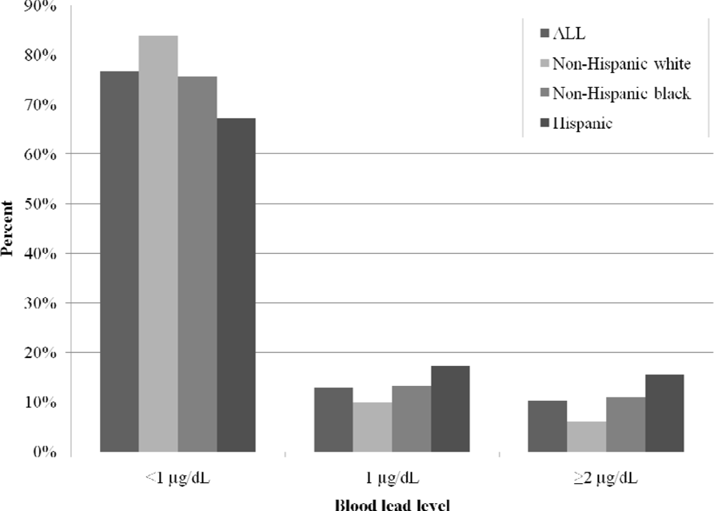
| Blood lead level | |||||||||
| < 1 μg/dL | 663 | 76.7% | 583 | 75.7% | 531 | 75.8% | |||
| 1 μg/dL | 112 | 13.0% | 105 | 13.6% | 95 | 13.6% | |||
| ≥ 2 μg/dL | 89 | 10.3% | 82 | 10.7% | 75 | 10.7% | |||
| Basic model (n = 864) | Basic model + age of housing (n = 770) | Basic model + modeled exposure risk (n = 701) | ||||
|---|---|---|---|---|---|---|
| aOR (95% CI) | P-value | aOR (95% CI) | P-value | aOR (95% CI) | P-value | |
| Race | ||||||
| Non-Hispanic white | 1.0 — | — | 1.0 — | — | 1.0 — | — |
| Non-Hispanic black | 2.90 (1.79–4.70) | < 0.001 | 3.49 (2.09–5.85) | < 0.001 | 3.11 (1.75–5.52) | 0.002 |
| Hispanic | 4.92 (2.39–10.09) | < 0.001 | 4.54 (2.04–10.13) | < 0.001 | 3.84 (1.65–8.89) | < 0.001 |
| Age (yrs) | ||||||
| 18–19 | 0.60 (0.28–1.27) | 0.179 | 0.57 (0.26–1.25) | 0.160 | 0.65 (0.29–1.47) | 0.296 |
| 20–24 | 0.54 (0.33–0.89) | 0.015 | 0.51 (0.31–0.87) | 0.012 | 0.60 (0.35–1.02) | 0.058 |
| 25–29 | 1.0 — | — | 1.0 — | — | 1.0 — | — |
| 30–34 | 2.39 (1.47–3.91) | < 0.001 | 2.47 (1.48–4.12) | < 0.001 | 2.50 (1.46–4.25) | < 0.001 |
| 35–39 | 2.98 (1.71–5.18) | < 0.001 | 3.32 (1.85–5.96) | < 0.001 | 3.25 (1.74–6.09) | < 0.001 |
| 40–44 | 7.69 (3.49–16.93) | < 0.001 | 6.27 (2.71–14.55) | < 0.001 | 6.83 (2.72–17.11) | < 0.001 |
| Education | ||||||
| Less than high school | 2.17 (1.34–3.51) | 0.002 | 1.99 (1.19–3.31) | 0.008 | 2.07 (1.22–3.49) | 0.007 |
| Parity | 0.90 (0.78–1.03) | 0.118 | 0.863 (0.75–0.99) | 0.045 | 0.91 (0.78–1.07) | 0.265 |
| Tobacco use | 1.64 (1.07–2.50) | 0.022 | 1.70 (1.08–2.66) | 0.021 | 1.54 (0.96–2.48) | 0.073 |
| Exposure measures | ||||||
| Age of housing (year built) | 0.99 (0.98–1.00) | 0.249 | ||||
| Modeled lead exposure risk | 0.99 (0.29–3.40) | 0.986 | ||||
© 2010 by the authors; licensee Molecular Diversity Preservation International, Basel, Switzerland. This article is an open-access article distributed under the terms and conditions of the Creative Commons Attribution license (http://creativecommons.org/licenses/by/3.0/).
Share and Cite
Miranda, M.L.; Edwards, S.E.; Swamy, G.K.; Paul, C.J.; Neelon, B. Blood Lead Levels Among Pregnant Women: Historical Versus Contemporaneous Exposures. Int. J. Environ. Res. Public Health 2010, 7, 1508-1519. https://doi.org/10.3390/ijerph7041508
Miranda ML, Edwards SE, Swamy GK, Paul CJ, Neelon B. Blood Lead Levels Among Pregnant Women: Historical Versus Contemporaneous Exposures. International Journal of Environmental Research and Public Health. 2010; 7(4):1508-1519. https://doi.org/10.3390/ijerph7041508
Chicago/Turabian StyleMiranda, Marie Lynn, Sharon E. Edwards, Geeta K. Swamy, Christopher J. Paul, and Brian Neelon. 2010. "Blood Lead Levels Among Pregnant Women: Historical Versus Contemporaneous Exposures" International Journal of Environmental Research and Public Health 7, no. 4: 1508-1519. https://doi.org/10.3390/ijerph7041508