Article

Gamma Irradiation Induced Degradation of Orange Peels

by
Raymundo Sánchez Orozco
1,2,*,
Patricia Balderas Hernández
1,*,
Nelly Flores Ramírez
3,
Gabriela Roa Morales
1,
Jaime Saucedo Luna
4 and
Agustín Jaime Castro Montoya
4
1
Faculty of Chemistry, Autonomous University of the State of Mexico, Paseo Colón esq., Paseo Tollocan s/n, Col. Residencial Colón, C.P. 50120 Toluca, Estado de México, Mexico
2
Division of Industrial Engineering, Technological of Superior Studies of Jocotitlan, Carretera Toluca-Atlacomulco Km 44.8, Ejido San Juan y San Agustín, C.P. 50700 Jocotitlán, Estado de México, Mexico
3
Faculty of Wood Engineering and Technology, University Michoacana of San Nicolas of Hidalgo, Edif. D, Cd. Universitaria, C.P. 58060 Morelia, Michoacán, Mexico
4
Faculty of Chemical Engineering, University Michoacana of San Nicolas of Hidalgo, Edif. V-1, Cd. Universitaria, C.P. 58060 Morelia, Michoacán, Mexico
*
Authors to whom correspondence should be addressed.
Energies 2012, 5(8), 3051-3063; https://doi.org/10.3390/en5083051
Submission received: 8 June 2012 / Revised: 21 July 2012 / Accepted: 1 August 2012 / Published: 14 August 2012
(This article belongs to the Special Issue Biomass and Biofuels 2012)

Abstract

:
In this study, gamma irradiation induced degradation of orange peels (OP) was investigated. The lignocellulosic biomass degradation was carried out at doses of 0 (control), 600, 1800 and 3500 kGy using a Co-60 gamma radiation source. The samples were tested for total and reducing sugars. The concentrations of total sugars ranged from 0.530 g∙g−1 in control sample to 0.382 g∙g−1 of dry weight in the sample which received the highest radiation dose. The reducing sugars content varying from 0.018 to 0.184 g∙g−1 of dry weight with the largest rise occurring in the sample irradiated at 3500 kGy. The concentrations of sucrose, glucose and fructose were determined. The changes generated in physico-chemical properties were determined by Fourier Transform Infrared Spectroscopy (FTIR) and termogravimetric analysis (TG-DTG). The results show that OP was affected, but not significantly, which suggests that lignocellulose and sugars profiles were partially degraded after gamma irradiation.

1. Introduction

Energy consumption has increased gradually over the last century as the planet’s population has grown and more countries are becoming industrialized. The use of biofuels for transport is becoming of increasing importance for a number of reasons, such as environmental concerns relating to climate change, depletion of fossil fuel reserves, and reduction of reliance on imports [1,2,3,4]. A potential source for low-cost biofuel (i.e., bio-ethanol) production is to utilize lignocellulosic materials such as citrus residues of orange peel as feedstocks.
In Mexico, the production of oranges has significant economic importance and this product is essentially processed to make juice. The production of orange juice generates mainly peel, bagasse and seed wastes, which represent about 50% of the raw processed fruit. These citrus wastes do not find any commercial use and are largely disposed of in municipal dumps or as underutilized cattle feed [5,6,7]. Orange peels are rich in fermentable sugars, that is, glucose, fructose, and sucrose, along with insoluble polysaccharides, cellulose and pectin [8,9]. Citrus fruit residues thus represent an abundant, inexpensive, and readily available source of renewable lignocellulosic biomass, and their utilization is attracting increased interest all over the World, mainly for the production of novel materials for environmentally friendly industrial utilization after chemical modification.
Current technologies for bioethanol production mostly rely on starch and sugars, and have been considerably debated because of poor sustainability [10,11]. In this context, bioethanol produced from lignocellulosic biomass is an interesting alternative because these raw materials do not compete with food crops and they are also less expensive than conventional agricultural feedstocks [12,13,14]. Their hydrolysis yields fermentable sugars which can serve as chemical feedstocks and energy sources. Initially, the major technical problem for ethanol production from lignocellulosic biomass is how to hydrolyze lignocelluloses into fermentable monosaccharides. Cellulose and hemicellulose pretreatment is an essential step for obtaining potentially fermentable sugars in the hydrolysis step. In this regard, only pretreatments such as dilute-acid and steam explosion, prior to enzymatic hydrolysis and fermentation, have been studied for orange peel wastes [8,15,16].
Few reports exist on the use of gamma irradiation on lignocellulosic biomass [17,18,19,20]. Gamma irradiation, if is used on lignocellulosics in high dosage, causes a decrease in cell wall constituents or depolymerizes and delignifies the fiber [21]. In addition, ionizing radiation leads to the degradation of polysaccharides such as starch, cellulose, and pectin by the cleavage of the glycosidic bonds. The basic advantages of degradation of polymers by radiation include the ability to promote changes reproducibly and quantitatively, without the introduction of chemical reagents or the need for special equipment/setup to control for temperature, environment, and additives. Therefore, this technology is simpler and more environmentally friendly than other conventional methods. However, no information is yet available about the effect of 60Co-γ irradiation treatment on the structural properties of orange peel. The objective of this work was thus to evaluate the effects of gamma irradiation at higher doses on the physico-chemical structure of orange peels.

2. Results and Discussion

2.1. Proximate Analysis

Orange peels were evaluated for proximate composition. The moisture content of the peel in this study was approximately 68% (wet basis). Hemicellulose is present in a significant quantity (14.4%). The cellulose content was found to be 11.97%. The total protein content in the substrate was 5.93%. Relatively low levels of lignin (2.15%) were evident. Fractions of neutral detergent fiber and acid detergent fiber were 28.53% and 14.12%, respectively. The above values indicate that orange peels are good sources of carbohydrates and fiber. The findings of the present study concerning proximate composition of wastes are corroborated by the results of other studies [22,23]. However, these studies have reported results that diverge from the findings in the present investigation. In fact, these differences are expected owing to varietal, cultivar, environmental and soil condition differences. One of the main obstacles for using orange peel waste for fermentation is its content of peel oil. Extraction of essential oil produced an approximate yield of 0.53% (w/w) so from 500 g of fresh orange peel about 2.66 g of oil was obtained. The extraction yield is acceptable as extraction yields between 0.5% and 0.8%, have been reported, which depends on the variety and ripeness, as well as equipment and extraction methods used [5]. Orange peel oil contains more than 90% (w/w) d-limonene, a monoterpene that inhibits yeasts and other microorganism [8].

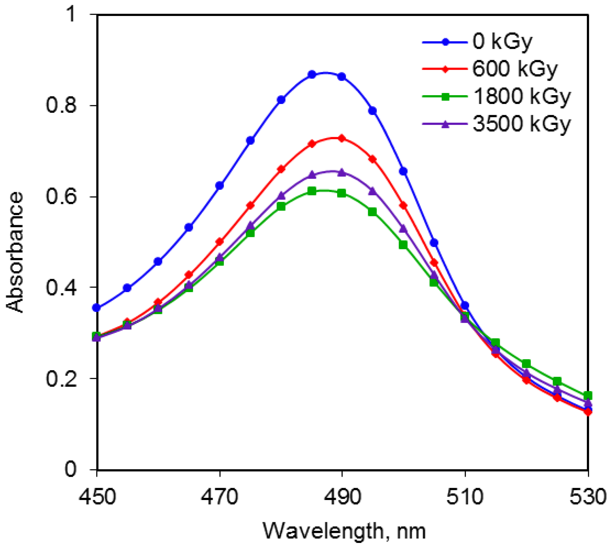
2.2. Comparison of Radiation Doses and Absorbance for Total Sugars

Oligosaccharides present in the non-irradiated and irradiated substrates were determined by the phenol–sulfuric acid assay [24] Therefore, in the analysis of mono- and oligosaccharides in the substrate, the intermediate product after treatment is mostly a mixture of sucrose, glucose and fructose, which are thus the main compounds measured in the sugar assay. The developed complex during the reaction was a chromophore that absorbs electromagnetic radiation in the UV-Vis region and the absorbance was proportional to the sugar concentration.
A previous report indicated that at 490 nm most sugars can be measured at or near their maximum absorption and that the absorption spectra of mannose, xylose, fructose, galactose, and glucose have peaks at 491, 486, 482, 491, and 493 nm, respectively [25]. In general, the maximum absorbance can be observed at 490 nm for hexoses and 480 nm for pentoses. In this way the measurement of total sugars by phenol–sulfuric colorimetric reagent was highly reproducible in the irradiated substrate. The levels of total sugars were calculated from the equation derived from a standard curve; for the control and irradiated samples of cobalt-60 source. The values ranged between 0.530 and 0.382 g∙g−1 of dry weight. Since the total sugar content of the OP varied in relation to control samples, with the application of irradiation. This can be observed in the Figure 1 that, from the wavelength of 450 to 530 nm, the responses of the material to gamma radiation have similar behavior pattern for the irradiation doses up to 3500 kGy. Starting from the irradiation dose of 600 kGy, the absorption intensity of the irradiated samples, was higher than the intensity of original absorption (non-irradiated substrate), in all cases was observed a maximum absorption value of 490 nm.
Figure 1 shows that as the irradiation doses increase, the absorbance tends to decrease, possibly owing to chain scission processes and the alteration of the structure of the irradiated samples as well as the breakage of the glycosidic linkages of the polysaccharide. This result is supported by the reports of depolymerization by radiolysis of polysaccharides such as starch [26,27], chitosan [28] and β-glucan [21]. The levels of total sugars present in the irradiated OP according to measured absorbance showed no significant difference between the sample control indicating that the impact of gamma radiation at the doses analyzed, were not sufficient to modify the sugars present in OP.
Figure 1. Absorbance of total sugars for control and irradiated OP.
Energies 05 03051 g001

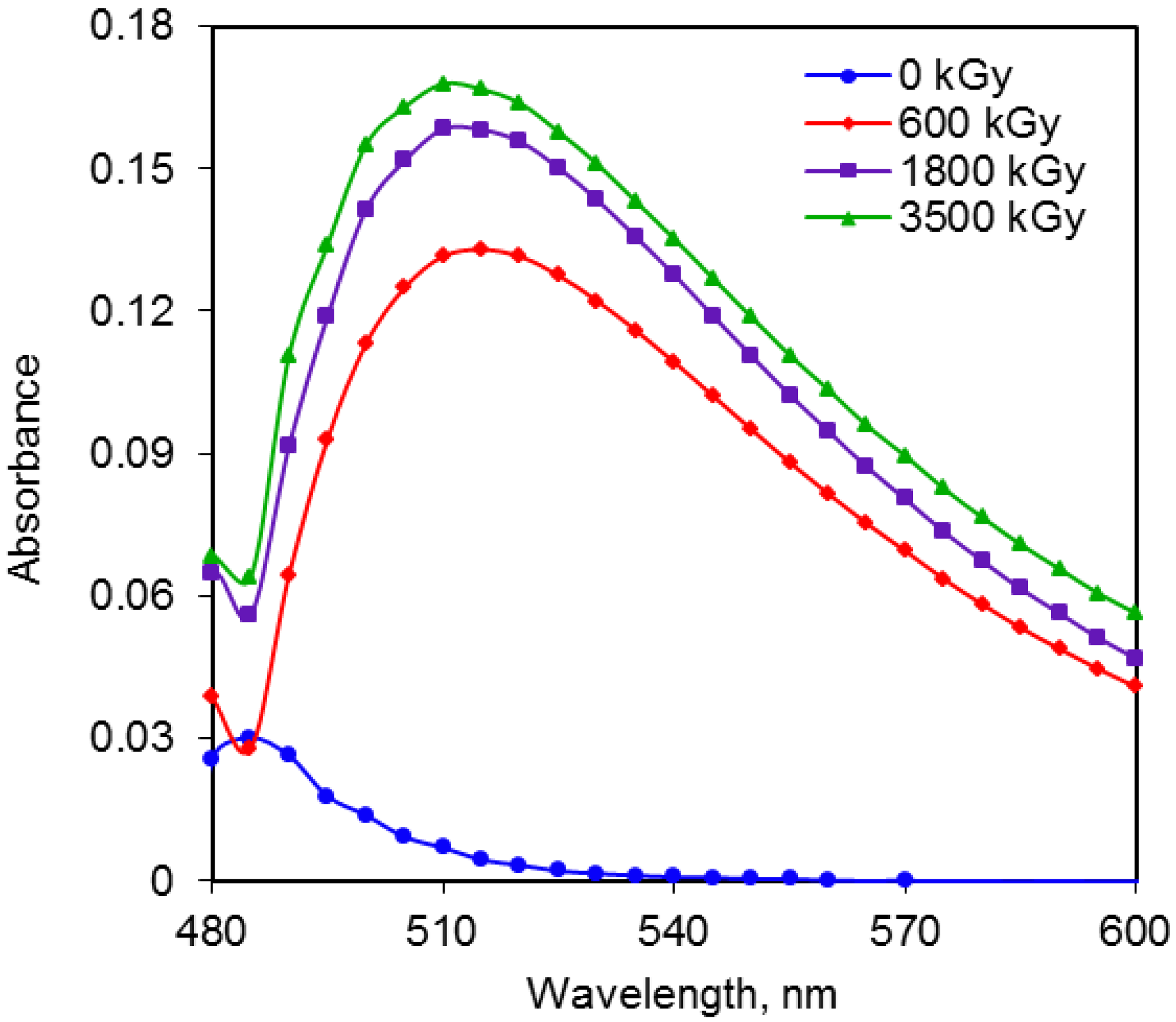
2.3. Comparison of Radiation Doses and Absorbance for Reducing Sugars

The absorbance of reducing sugars released in the substrate was assayed after radiation treatment at different doses by the DNS method [29] which detects the presence of the free carbonyl groups in the sugar chemical structure. Color changes from yellow to brown were observed due to the concentration of reducing sugars in the sample. The UV-Vis absorbance measurements of reducing sugars were reported to understand the impact of gamma irradiation pretreatments on cellulose and hemicellulose conversion. The samples irradiated in a source of cobalt-60 when compared to the sample control showed low levels of variation for reducing sugars, the contents varying from 0.018 to 0.184 g∙g−1 of dry weight, with the largest rise occurring in the sample irradiated at 3500 kGy. Figure 2 shows that there tended to be less release of reducing sugars in the non-irradiated substrate compared to irradiated substrates. The relationship between irradiation dose and the absorbance indicates that substrate degradation is not significant when using low doses (<600 kGy) are used compared to the effects observed at high absorbed doses of gamma irradiation. For easier comparison, the maximum release of reducing sugars at 3500 kGy, indicates that the irradiation induces degradation of lignocelluloses. The total sugars content increased slowly with increasing irradiation dose from 1800 to 3500 kGy. This process might be attributed to the loss of potential reducing sugar by radiolysis into low molecular weight products or the accumulation of inhibited byproducts. During the DNS assay, it was found that the UV-Vis absorbance measurements increased as a function of irradiation dose due to degradation of the substrate that improves the release of reducing sugars. This result is consistent with other studies on wheat straw [18] and potatoes [26]. The changes that occurred can be explained by the fact that the breakdown and depolymerization of the polysaccharide by radiolysis might induce the production of low molecular weight fragments and this leads to an increase in the solubility [30]. Despite these results, the increase in absorbance between 0.03 and 0.17 involves a limited release of oligosaccharides. Therefore, the total sugar conversion from irradiated biomass becomes an increasingly important parameter for bioethanol research.
Figure 2. Absorbance of reducing sugars for control and irradiated OP.
Energies 05 03051 g002

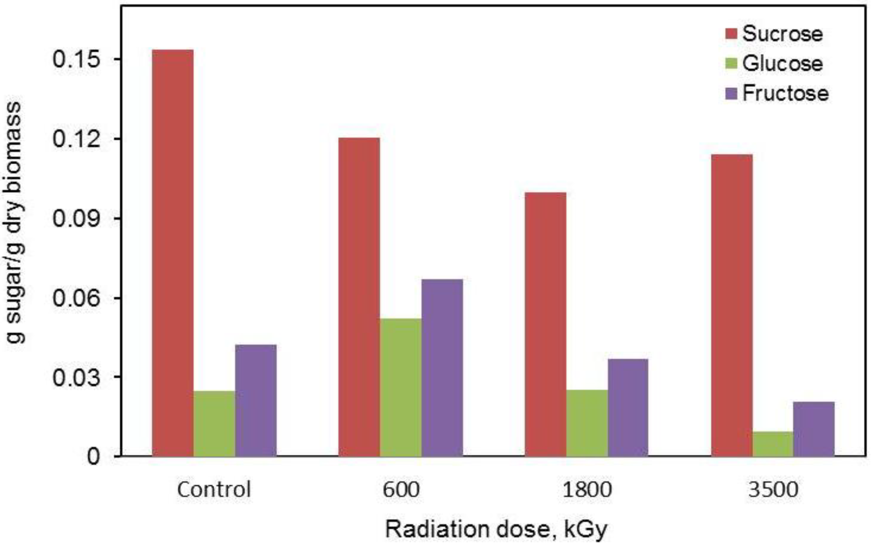
2.4. Liquid Chromatographic Analysis of Sugars

By HPLC analysis, a few sugars such as sucrose, glucose and fructose were found in the control and irradiated substrates, but the sucrose level was significantly higher than the levels of glucose and fructose. The purity of the three kinds of sugar content (control sample), was 3.06, 0.49 and 0.84 g∙L−1 for sucrose, glucose and fructose, respectively. Variations of the fructose content showed similar patterns to that observed with glucose (Figure 3). After the first radiation dose, a slight increase in the level of glucose and fructose was observed. However, a further increase in the dose radiation level (1800 kGy) resulted a decline in both glucose and fructose in the sugar concentration that can be due to the glycolytic degradation. The sucrose content showed a different pattern of variation but does not induce quantitative significant changes. After treatment and compared to the initial content, sucrose decreased significantly with respect to increasing radiation dose; however, this decrease was not significant between control and both irradiated OP. The sugar degradation pattern depends upon the structure of polysaccharides, intra- and intermolecular interactions between soluble polysaccharides, and the kind and concentrations the sugars present in cellulose and hemicellulose. Thus, it can be noted that the impact of gamma radiation emitted to established dose rate by Cobalt-60, to those doses analyzed, were not sufficient to modify significantly the sugars aspects present in OP.
Figure 3. Sugars content in control and irradiated OP.
Energies 05 03051 g003

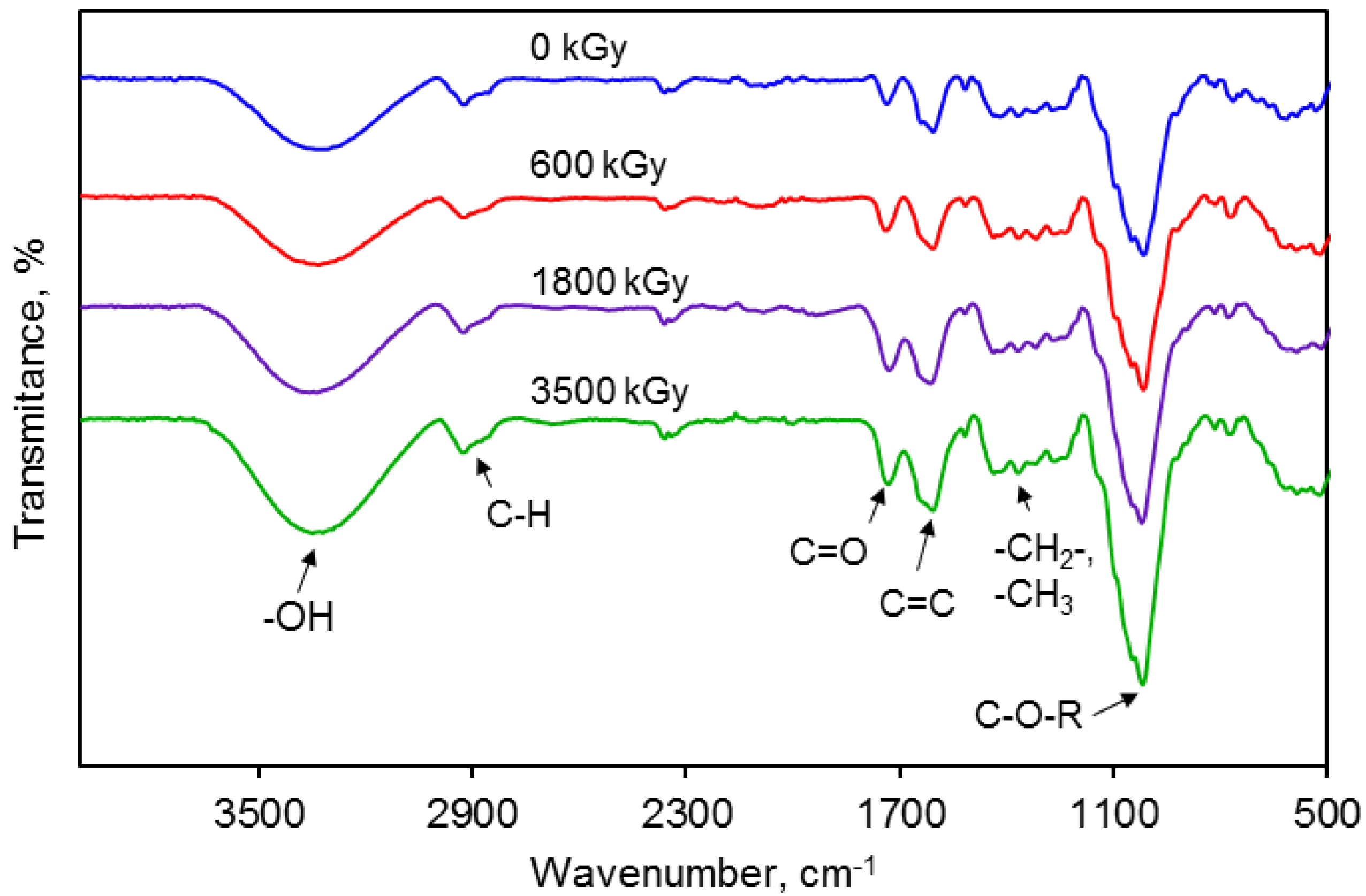
2.5. FTIR Spectra of the Irradiated Substrates at Different Doses

Fourier transform infrared spectroscopy (FTIR) was used to assess differences in the functional groups of the OP before and after irradiation. The FTIR spectra of OP treated with different doses of gamma irradiation are shown in Figure 4.
Figure 4. FTIR spectrum of the irradiated orange peel sample at different dose.
Energies 05 03051 g004
The most intense band at 3322 cm−1 was assigned to the stretching of –OH groups of the carbohydrates and those of lignin [31]. The signals at 2921 and 2851 cm−1 are caused by asymmetrical and symmetrical stretching vibrations of C–H groups. The band at 1732 cm−1 increases at rising the irradiation dose and was assigned to the carbonyl (C=O) stretching. The intense band at 1015 cm−1 corresponds to the link C–O–R while the distinctive band around 1265 cm−1 was attributed to aliphatic chains (–CH2– and –CH3) forming the basic structure of this lignocellulosic materials [32]. The relative absorbance of studied peaks increases with the rising gamma irradiation. The increase in the peaks’ intensity occurred particularly when the dose irradiation surpassed 600 kGy. The above result shows that the non-irradiated and irradiated OP both have similar FTIR spectra patterns, with small changes in the functional groups status. These changes mean that the chemical linkages of OP were partially altered by gamma irradiation. The deformation of lignocellulose by irradiation might occur because of the splitting of glycosidic bonds. Most of the bands have contributions from both carbohydrates (cellulose and hemicellulose) and lignin.

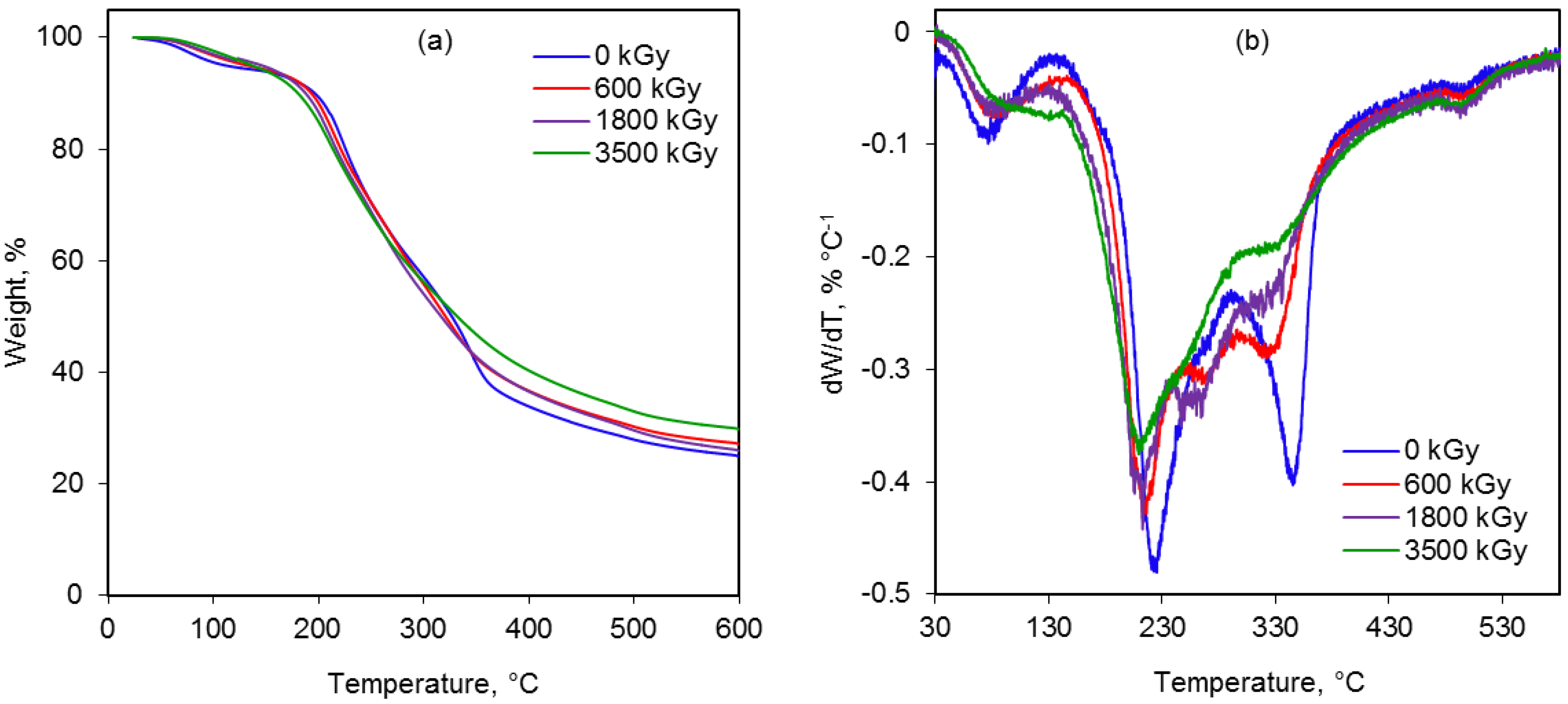
2.6. Thermal Behavior

The occurrence of chain scission is clearly demonstrated by TGA. If the polymer undergoes degradation, its weight will decrease. TG and DTG curves of the non-irradiated and irradiated OP are shown in Figure 5 and Table 1.
Figure 5. (a) TG and (b) DTG curves of orange peel at different irradiation dose.
Energies 05 03051 g005
Corresponding to the weight loss on TG curves at least four main thermal events can be clearly distinguished up to 600 °C. In all TG curves (Figure 5a), the first weight loss step below 105 °C refers to volatile components and physically adsorbed water molecules in the samples. It has been reported that the dried biomass is stable up 140 °C, and then begins to decompose [31]. The main mass losses are associated to the biomass decomposition, essentially, to its three main components (hemicellulose, cellulose and lignin). The second step from 150 to 265 °C can be attributed to decomposition of hemicelluloses. The third decomposition process between 265 and 372 °C was associated to the degradation of cellulose. Finally, the carbon-carbon linkage between lignin structural units was cleaved in the temperature range from 372 to 570 °C. The cellulose thermal degradation occurs in many steps, starting by its depolymerization [17]. This takes place when the cellulose structure has absorbed enough energy to activate the cleavage of the glycosidic linkage to produce glucose and oligosaccharides. Aguiar et al. [33] have reported that hemicellulose decomposes between 200 and 260 °C, most of the decomposition happens to 180 °C. Decomposition of cellulose is complete at around 360 °C. Cellulose forms long chains that are bonded to each other by a long network of hydrogen bonds. Up to 300 °C, there is the breaking of bonds α-β-aryl-alkyl-ether. This process is followed by aromatic ring bonds breakage and finally a rupture of carbon-carbon bonds between the structural units of lignin at temperatures from 370 to 400 °C [34]. In this way, when the irradiation dose increases between 600 and 3500 kGy then the hemicellulose and cellulose content falls more rapidly as compared with the non-irradiated substrate.
Table 1. Temperature intervals, weight losses and residues of samples.
Radiation dose, kGyPeak temperature (Tmax), °CWeight loss, %Residue, %
Stage 2Stage 3Stage 4Stage 2Stage 3Stage 4
03462234962927923
6003242184982826925
18003222124952724726
35003212104952624728
The data from Table 1 allow observing that after irradiation the sample components in the form of hemicellulose and cellulose gradually undergo chain breakage when the absorbed radiation dose reaches 600 kGy or higher, while the lignin structure is less affected by gamma radiation. It is clear that in the DTG curves (Figure 5b) there is a slight shift to the left of the maximum temperatures of degradation as the radiation energy increases. Therefore; it is assumed that the lower temperature range would be due to the scission of chemical bond such as a glycosidic bonds. DTG curves of lignocellulosic biomass show a main peak at high temperatures mainly due to the degradation of cellulose, and a shoulder or a peak at lower temperatures that can be attributed to the partial degradation of hemicelluloses.

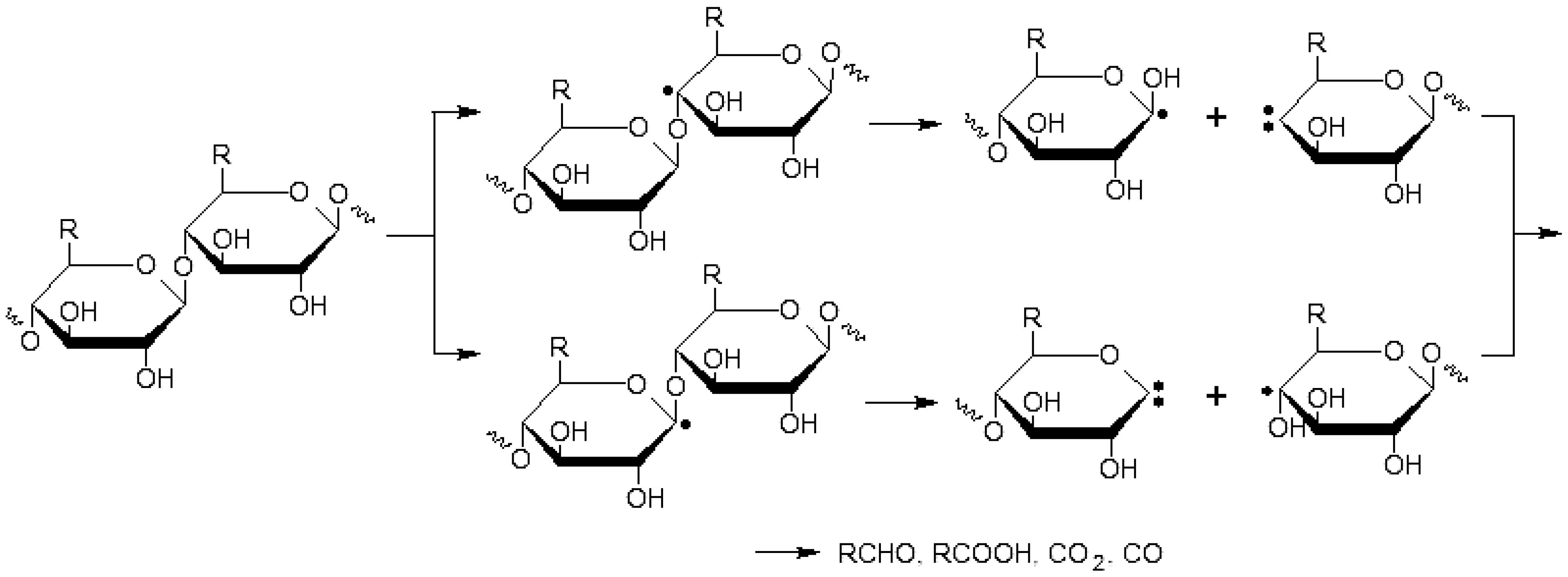
2.7. Cleavage of the Glycosidic Bonds

The breakdown of lignocellulosic biomass involves the formation of long-chain polysaccharides, mainly cellulose and hemicellulose, and the subsequent hydrolysis of these polysaccharides into their component 5- and 6-carbon chain sugars. Cell wall polysaccharides were partially degraded, particularly cellulose and hemicellulose. Lignin, according to Figure 5, is more radiation resistant. It has been shown that the lignin plays a protective role in the cell wall by absorbing and scattering radiation energy. The main irradiation effect in hemicellulose and cellulose causes degradation by splitting of the glycosidic bond with subsequent formation of reducing groups, such as reducing sugars (e.g., glucose). Each glucose residue of cellulose has two inter- and intramolecular hydrogen bonds. These bonds stabilize the long and parallel chains of cellulose. Gamma irradiation affects these bonds and causes the van der Walls power to weaken, which results in the degradation of cellulose and increasing degradability of the cell wall constituents [28]. Additionally, with the breaking of hydrogen bonds, free radicals are produced and then the concentration of free radicals and also, the number of separated chains from cellulose (Figure 6), increases with the increasing irradiation dose [19,21]. The irradiation causes the formation of carbonyl groups of cellulose at the presence of oxygen that helps cellulose breakdown. Gamma rays leads to the hydrolysis of the glycoside bonds. As proposed in Figure 6, cellulose mainly decomposes into large fragments and then to the ultimate radiolysis products which include RCHO, RCOOH, CO2, CO, etc. and the loss of bound water in the processes of chain scission and the structure alteration of irradiated sample [18,35]. The predominant reaction of hemicellulose and cellulose polymers upon radiation is main-chain scission that is a direct consequence of interactions of gamma rays with the polymers leading to the breakage in the polymer backbone.
Figure 6. Mechanisms degradation of cellulose by gamma irradiation, adapted from Ershov [35].
Energies 05 03051 g006

3. Experimental Section

3.1. Materials

Oranges used in this study were Citrus sinensis L. supplied by a local market. The peels were separated from the endocarp by cutting with a hand knife into small pieces and washed thoroughly with de-ionized water to remove physically adsorbed contamination. The biomass were dried in a vacuum oven at 60 °C for 24 h and then crushed and sieved through an 80 mesh screen. The substrate was placed in hermetically sealed bags and stored in a cold chamber until treatment.

3.2. Proximate Analysis

The peel samples were analyzed for dry matter, moisture, protein (N × 6.25), neutral detergent fiber (NDF), acid detergent fiber (ADF), and acid detergent lignin (ADL). The proportion of hemicellulose was calculated from the difference between NDF and ADF and that of lignin from the difference between ADF and cellulose. The analytical methods and extraction of essential oil from OP were performed by following their respective procedures [36].

3.3. Gamma Irradiation

The solid substrate was placed in sealed glass bottles and was irradiated at a National Nuclear Research Institute, Mexico using a 60Co-γ irradiation device (Transelektro LGI-01). Gamma irradiation pretreatment experiments were performed using specific levels of 60Co-γ and the irradiation doses used were 600, 1800 and 3500 kGy at a dose rate of 10 kGy h−1. The control (0 kGy) and irradiated samples were stored for further tests.

3.4. Total Sugars

The phenol–sulfuric colorimetric method [24] was used to determine the total concentration of carbohydrates present in the substrate. Two mL of clear aqueous solutions (0.5 g irradiated biomass/10 mL water) of the carbohydrates to be analyzed were placed in a test-tube and then 1.0 mL of phenol (5% w/v) and 5.0 mL of sulfuric acid (96% v/v) were added and immediately vortexed. Samples were allowed to stand for 10 min and then placed in a thermal bath at 30 °C for 20 min and measured at 480 nm. This method is non-stoichiometric and so it was necessary to prepare a calibration curve using D-glucose standards on a UV-Vis spectrophotometer (Lambda 25, PerkinElmer Inc., Waltham, MA, USA).

3.5. Reducing Sugars

Concentration of reducing sugars was measured by 3,5-dinitrosalicylic acid reagent (DNS) [29]. The DNS reagent is nonspecific and reacts with both five and six carbon reducing sugars. Although this assay does not allow discrimination among specific carbohydrates, it can be used to quantify hydrolysis of a wide range of polysaccharide substrates. Initially 1.0 mL of liquid sample (0.5 g irradiated biomass/10 mL water) was mixed with 1.0 mL of DNS reagent. The mixture was heated at 99 °C for 5 min to develop red-brown color. After cooling to room temperature, 8.0 mL of distilled water was added to reagent mixture and absorbance with a spectrophotometer at 575 nm was measured. A standard curve was prepared using different glucose concentrations. All experiments were carried out in the same square quartz UV-Vis cells (path length, 1 cm).

3.6. Analysis of Sugars by HPLC

The sucrose, glucose and fructose content were determined by HPLC. Samples were homogenized (0.2 g dried substrate/10 mL water) and filtered (Phenomenex 0.45 μm RC membranes) into HPLC vials. The sugars were separated by HPLC using a Metacarb column 87C (300 × 7.8 mm) set at 70 °C with an appropriate guard column (Varian Inc., Palo Alto, CA, USA) and a differential refractometer detector (Prostar 350, Varian Inc., Palo Alto, CA, USA). The mobile phase was DDI water at a flow rate of 0.6 mL∙min–1. The sugars were identified and quantified by comparison with standards (Sigma-Aldrich Co., St. Louis, MO, USA) and each determination was run in triplicate.

3.7. Instrumentation and Characterization Methods

Fourier transform infrared spectroscopy (FTIR) was used to assess differences in the general functional groups of the irradiated and control samples. FTIR spectra were obtained using a Vertex 70 spectrometer in the spectral range 4000–500 cm−1 in ATR mode. Thermogravimetric analysis (TGA) was carried out using a TA Instrument TGA-DSC, with a volume of 10 mg, and a purge flow of 50 mL/min, at a scan rate of 20 °C/min under nitrogen flow.

4. Conclusions

In this study, slight changes in the physico-chemical properties were observed when OP was irradiated by 0–3500 kGy gamma rays. The results showed that the levels of total and reducing sugars present in the control and irradiated OP was affected, but not significantly, which suggests that lignocellulose and sugars profiles were partially degraded after gamma irradiation. The sample irradiated at highest dose presented the most intense reduction in the concentration of glucose and fructose in comparison to the control. Sucrose seems to be less affected by radiation because it does not induce significant quantitative changes in the content. IR analysis showed that the non-irradiated and irradiated OP both have a similar spectral patterns, with little changes in the functional group profile. These changes mean that the chemical linkages of OP were partially altered by gamma irradiation. The TG and DTG characterization suggests that degradation process occurs in at least three main steps associated to the OP decomposition, essentially, to its three main components (hemicellulose, cellulose and lignin). The gamma irradiation could affect partially the lignocellulose structure of the OP, sequentially influencing its microstructure, and finally brought about changes of its physicochemical properties due to partial degradation of the substrate. Against these results, it can be concluded that the impact of gamma radiation emitted to specified dose rate by Cobalt-60, to those doses analyzed, were not sufficient to modify the sugars profiles found in OP.

Acknowledgments

This research was supported by a doctoral fellowship from Consejo Nacional de Ciencia y Tecnología (CONACYT), México. The authors would like to thank Araceli Salazar Peralta for the style correction of the manuscript.

Conflict of Interest

The authors declare no conflict of interest.

References

  1. Haberl, H.; Erb, K.H.; Krausmann, F.; Bondeau, A.; Lauk, C.; Muller, C.; Plutzar, C.; Steinberger, J.K. Global bioenergy potentials from agricultural land in 2050: Sensitivity to climate change diets and yields. Biomass Bioenergy 2011, 35, 4753–4769. [Google Scholar] [CrossRef] [PubMed]
  2. Champagne, P.; Li, C. Enzymatic hydrolysis of cellulosic municipal wastewater treatment process residuals as feedstocks for the recovery of simple sugars. Bioresour. Technol. 2009, 100, 5700–5706. [Google Scholar] [CrossRef] [PubMed]
  3. Matías, J.; González, J.; Royano, L.; Barrera, R.A. Analysis of sugars by liquid chromatography-mass spectrometry in Jerusalem artichoke tubers for bioethanol production optimization. Biomass Bioenergy 2011, 35, 2006–2012. [Google Scholar] [CrossRef]
  4. Peng, L.; Chen, Y. Conversion of paper sludge to ethanol by separate hydrolysis and fermentation (SHF) using Saccharomyces cerevisiae. Biomass Bioenergy 2011, 35, 1600–1606. [Google Scholar]
  5. Mamma, D.; Kourtoglau, E.; Christakopoulos, P. Fungal multienzyme production on industrial by-products of the citrus-processing industry. Bioresour. Technol. 2008, 99, 2373–2383. [Google Scholar] [CrossRef] [PubMed]
  6. Ververis, C.; Georghiou, K.; Danielidis, D.; Hatzinikolaou, D.G.; Santas, P.; Santas, R.; Corleti, V. Cellulose, hemicelluloses, lignin and ash content of some organic materials and their suitability for use as paper pulp supplements. Bioresour. Technol. 2007, 98, 296–301. [Google Scholar] [CrossRef] [PubMed]
  7. Boluda-Aguilar, M.; García-Vidal, L.; González-Castañeda, F.P.; López-Gómez, A. Mandarin peel wastes pretreatment with steam explosion for bioethanol production. Bioresour. Technol. 2010, 101, 3506–3513. [Google Scholar] [CrossRef] [PubMed]
  8. Wilkins, M.R.; Widmer, W.W.; Grohmann, K. Simultaneous saccharification and fermentation of citrus peel waste by Saccharomyces cerevisiae to produce ethanol. Process Biochem. 2007, 42, 1614–1619. [Google Scholar] [CrossRef]
  9. Oberoi, H.S.; Vadlani, P.V.; Madl, R.L.; Saida, L.; Abeykon, J.P. Ethanol production from orange peels: Two-stage hydrolysis and fermentation studies using optimized parameters though experimental design. J. Agric. Food Chem. 2010, 58, 3422–3429. [Google Scholar] [CrossRef] [PubMed]
  10. Cardona, C.A.; Sánchez, O.J. Fuel ethanol production: Process design trends and integration opportunities. Bioresour. Technol. 2007, 98, 2415–2457. [Google Scholar] [CrossRef] [PubMed]
  11. Sánchez, O.J.; Cardona, C.A. Trends in biotechnological production of fuel ethanol from different feedstocks. Bioresour. Technol. 2008, 99, 5270–5295. [Google Scholar] [CrossRef] [PubMed]
  12. Hamelinck, C.N.; Hooijdonk, G.V.; Faaij, A.P. Ethanol from lignocellulosic biomass: Techno-economic performance in short-, middle- and long-term. Biomass Bioenergy 2005, 28, 384–410. [Google Scholar] [CrossRef]
  13. Cunha, J.A.; Pereira, M.M.; Valente, L.M.M.; de la Piscina, P.R.; Homs, N.; Santos, M.R. Waste biomass to liquids: Low temperature conversion of sugarcane bagasse to bio-oil. The effect of combined hydrolysis treatments. Biomass Bioenergy 2011, 35, 2106–2116. [Google Scholar] [CrossRef]
  14. Moxley, G.; Zhu, Z.; Percival, Y.H. Efficient sugar release by the cellulose solvent-based lignocellulose fractionation technology and enzymatic cellulose hydrolysis. J. Agric. Food Chem. 2008, 56, 7885–7890. [Google Scholar] [CrossRef] [PubMed]
  15. Widmer, W.; Zhou, W.; Grohmann, K. Pretreatment effects on orange processing waste for making ethanol by simultaneous saccharification and fermentation. Bioresour. Technol. 2010, 101, 5242–5249. [Google Scholar] [CrossRef] [PubMed]
  16. Talebnia, F.; Pourbafrani, M.; Lundin, M.; Taherzadeh, M.J. Optimization study of citrus wastes saccharification by dilute-acid hydrolysis. Bioresources 2008, 3, 108–122. [Google Scholar]
  17. Severiano, L.C.; Lahr, F.A.R.; Bardi, M.A.G.; Santos, A.C.; Machado, L.D.B. Influence of gamma radiation on properties of common Brazilian wood species used in artwork. Prog. Nucl. Energy 2010, 52, 730–734. [Google Scholar] [CrossRef]
  18. Chunping, Y.; Zhiqiang, S.; Guoce, Y.; Jianlong, W. Effect and aftereffect of γ radiation pretreatment on enzymatic hydrolysis of wheat straw. Bioresour. Technol. 2008, 99, 6240–6245. [Google Scholar] [CrossRef] [PubMed]
  19. Khan, F.; Ahmad, S.R.; Kronfli, E. γ-Radiation induced changes in the physical and chemical properties of lignocellulose. Biomacromolecules 2006, 7, 2303–2309. [Google Scholar] [CrossRef] [PubMed]
  20. Li, B.; Li, J.; Xia, J.; Kennedy, J.F.; Yie, X.; Liu, T.G. Effect of gamma irradiation on the condensed state structure and mechanical properties of konjac glucomannan/chitosan blend films. Carbohydr. Polym. 2011, 83, 44–51. [Google Scholar] [CrossRef]
  21. Byun, E.H.; Kim, J.H.; Sung, N.Y.; Choi, J.I.; Lim, S.T.; Kim, K.H.; Yook, H.S.; Byun, M.W.; Lee, J.W. Effects of gamma irradiation on the physical and structural properties of β-glucan. Radiat. Phys. Chem. 2008, 77, 781–786. [Google Scholar] [CrossRef]
  22. Ajila, C.M.; Bhat, S.G.; Prasada, U.J.S. Valuable components of raw and ripe peels from two Indian mango varieties. Food Chem. 2007, 102, 1006–1011. [Google Scholar] [CrossRef]
  23. Emaga, T.H.; Robert, C.; Ronkart, S.N.; Wathelet, B.; Paquot, M. Dietary fiber components and pectin chemical features of peels during ripening in banana and plantain varieties. Bioresour. Technol. 2008, 10, 4346–4354. [Google Scholar] [CrossRef]
  24. Dubois, M.; Gilles, K.; Hamilton, J.; Reberts, P.; Smithd, F. Colorimetric method for determination of sugars and related substances. Anal. Chem. 1956, 28, 350–356. [Google Scholar] [CrossRef]
  25. Masuko, T.; Minami, A.; Iwasakib, N.; Majima, T.; Nishimura, S.I.; Lee, Y.C. Carbohydrate analysis by a phenol–sulfuric acid method in microplate format. Anal. Biochem. 2005, 339, 69–72. [Google Scholar] [CrossRef] [PubMed]
  26. Ezekiel, R.; Rana, G.; Singh, N.; Singh, S. Physicochemical, thermal and pasting properties of starch separated from γ-irradiated and stored potatoes. Food Chem. 2007, 105, 1420–1429. [Google Scholar] [CrossRef]
  27. Wu, D.; Shu, Q.; Wang, Z.; Xia, Y. Effect of gamma irradiation on starch viscosity and physicochemical properties of different rice. Radiat. Phys. Chem. 2002, 65, 79–86. [Google Scholar] [CrossRef]
  28. Choi, J.; Kim, H.J.; Kim, J.H.; Byun, M.W.; Chun, B.S.; Ahn, D.H.; Hwang, Y.J.; Kim, D.J.; Kim, G.H.; Lee, J.W. Application of gamma irradiation for the enhanced physiological properties of polysaccharides from seaweeds. Appl. Radiat. Isot. 2009, 67, 1277–1281. [Google Scholar] [CrossRef] [PubMed]
  29. Miller, G.L. Use of dinitrosalicylic acid reagent for determination of reducing sugar. Anal. Chem. 1959, 31, 426–428. [Google Scholar] [CrossRef]
  30. Choi, J.; Kim, J.K.; Srinivasan, P.; Kim, J.H.; Park, H.J.; Byun, M.W.; Lee, J.W. Comparison of gamma ray and electron beam irradiation on extraction yield, morphological and antioxidant properties of polysaccharides from tamarind seed. Radiat. Phys. Chem. 2009, 78, 605–609. [Google Scholar] [CrossRef]
  31. Zapata, B.; Balmaseda, J.; Fregoso-Israel, E.; Torres-García, E. Thermo-kinetics study of orange peel in air. J. Therm. Anal. Calorimetry. 2009, 98, 309–315. [Google Scholar] [CrossRef]
  32. Hernández-Montoya, V.; Montes-Mora, M.A.; Elizalde-González, M.P. Study of the thermal degradation of citrus seeds. Biomass Bioenergy 2009, 33, 1295–1299. [Google Scholar] [CrossRef]
  33. Aguiar, L.; Márquez-Montesinos, F.; Gonzalo, A.; Sánchez, J.L.; Arauzo, J.L. Influence of temperature and particle size on the fixed bed pyrolysis of orange peel residues. J. Anal. Appl. Pyrolysis 2008, 83, 124–130. [Google Scholar] [CrossRef]
  34. Zeng, J.; Singh, D.; Chen, S. Thermal decomposition kinetics of wheat straw treated by Phanerochaete chrysosporium. Int. Biodeterior. Biodegrad. 2011, 65, 410–414. [Google Scholar] [CrossRef]
  35. Ershov, B.G. Radiation-chemical degradation of cellulose and other polysaccharides. Russ. Chem. Rev. 1998, 67, 314–334. [Google Scholar] [CrossRef]
  36. Association of Official Analytical Chemists (AOAC). Official Methods of Analysis, 16th ed.; AOAC: Washington, DC, USA, 1998. [Google Scholar]

