Review

Effects of Environmental Factors and Nutrient Availability on the Biochemical Composition of Algae for Biofuels Production: A Review

by
Ankita Juneja
1,
Ruben Michael Ceballos
2 and
Ganti S. Murthy
1,*
1
Biological and Ecological Engineering, Oregon State University, Corvallis, OR 97331, USA
2
Native American Research Laboratory, Division of Science and Mathematics, University of Minnesota-Morris, Morris, MN 56267, USA
*
Author to whom correspondence should be addressed.
Energies 2013, 6(9), 4607-4638; https://doi.org/10.3390/en6094607
Submission received: 24 June 2013 / Revised: 15 August 2013 / Accepted: 16 August 2013 / Published: 3 September 2013
(This article belongs to the Special Issue Algae Fuel 2013)

Abstract

:
Due to significant lipid and carbohydrate production as well as other useful properties such as high production of useful biomolecular substrates (e.g., lipids) and the ability to grow using non-potable water sources, algae are being explored as a potential high-yield feedstock for biofuels production. In both natural and engineered systems, algae can be exposed to a variety of environmental conditions that affect growth rate and cellular composition. With respect to the latter, the amount of carbon fixed in lipids and carbohydrates (e.g., starch) is highly influenced by environmental factors and nutrient availability. Understanding synergistic interactions between multiple environmental variables and nutritional factors is required to develop sustainable high productivity bioalgae systems, which are essential for commercial biofuel production. This article reviews the effects of environmental factors (i.e., temperature, light and pH) and nutrient availability (e.g., carbon, nitrogen, phosphorus, potassium, and trace metals) as well as cross-interactions on the biochemical composition of algae with a special focus on carbon fixation and partitioning of carbon from a biofuels perspective.

1. Introduction

Increasing demand for energy and global warming are two major challenges facing modern society. Dependence on fossil fuels for meeting increasing energy demands is unsustainable due to increasing levels of consumption and a dearth in discovery of new sources for these non-renewables. This concern has motivated researchers to focus on the development of alternative energy sources including solar, wind, water, and biomass. Biofuels are alternatives to liquid fossil fuels and are produced from sugar, starch, cellulosic or lipid-rich substrates. These substrates can be derived from feedstocks such as cereal crops, including corn and wheat [1]; sugar crops, including sorghum and sugarcane [2]; energy crops, such as switchgrass [3,4]; agricultural wastes, including straws and corn stover [5,6,7,8]; municipal wastes; and, several aquatic species. Currently, ethanol is produced from corn and sugarcane in significant volumes as a supplemental fuel and as a partial substitute for gasoline. Production and use of ethanol as a transportation fuel results in a net reduction of greenhouse gas (GHG) emissions. This claim is based on the idea that carbon dioxide emissions produced during the processing of biomass and from the use of biofuels is readily sequestered.
Biofuels produced from cereal crops and sugarcane are commonly called first-generation biofuels (FGB). The large-scale production (and use) of FGB products as competitive substitutes for fossil fuels is hindered by several limitations including: intensive agricultural inputs, land requirements, and trade-offs between food crop and fuel crop production (i.e., the food vs. fuel debate) [9]. Therefore, although they are renewable, FGBs alone are not a viable solution for solving global liquid fuel demands. Second-generation biofuels (SGB) are fuels derived from lignocellulosic biomass. Production of SGBs circumvents several of the negative outcomes associated with FGBs [10]. At present, the development of competitive SGB products is at various stages of research and pilot demonstrations. A few products have been commercialized. However, SGB feedstock production still requires agricultural inputs, land, and freshwater that could be used for food crops. Thus, SGBs are also subject to the food-versus-fuel dilemma in the long term. Producing liquid fuels from aquatic organisms, such as algae, is considered to be the third-generation in biofuels (TGB). The use of TGB feedstocks, which contain significant amounts of lipids and carbohydrates, from which biodiesel and bioethanol products may be produced, avoids most of the limitations noted for FGBs and SGBs. Arguably, this includes overcoming the food vs. fuel dilemma. Most aquatic plant feedstocks are championed as a viable source of lipids for the production of bio-oil [11]. Specifically, via thermochemical conversions or biochemical conversions, algae can be used to produce: biofuel oil and gas; or, bioethanol, biodiesel, and biohydrogen, respectively [12].
A recent resurgence of interest in algal-based TGBs is attributed to significant benefits associated with TGB production. These benefits include: year-round production; higher productivity compared to terrestrial crops; the potential for off-shore production (and, thus, non-competition with food crops); reduced need for arable land; reduced need for water treatment; and potential advantages in nutrient cycling [13,14,15].
Earlier research demonstrated that under select conditions, algae have the potential to produce 40 times the amount of oil for biodiesel production compared to oilseed crops (i.e., soy and canola) per unit land area [16]. However, such advantages over FGBs and SGBs have only been established in academic/research facilities. Presently, in the absence of publicly available data, it is unknown whether such gains can be realized on a commercial scale. Therefore, the economic potential of algal-based biofuels to significantly impact current and future fuels needs remains in question. Nonetheless, algal-based TGBs are an intense focus within the alternative fuels research community and production companies are paying close attention to advances. Of particular interest are microalgae-based TGB products. Technical and economic aspects of high-yield production of biodiesel from microalgae are currently being studied by many groups. A few review papers have discussed the processes and challenges in detail [17,18,19,20]. The potential of ethanol production from algae has also been investigated [21,22,23,24]. In both cases, it is generally agreed that there is significant potential for algal-based products; however, progress toward commercial-scale biofuels production from algae has been slow due to multiple challenges in production and processing.
Whether in open ponds or in closed photobioreactors, culturing algae requires consideration of numerous environmental conditions. Environmental factors such as temperature, light, pH, and nutrients not only affect photosynthesis and growth rate of the algae, but also influence the activity of cellular metabolism and composition. These effects have been identified individually by researchers [25,26,27,28,29,30,31], however, no consolidated review on the effect of these factors on algae is available. Most of the recent reviews have focused on production and processing techniques [11,16,32] with few data on the impacts of environmental and nutritional factors on algal growth rates. One recent, highly informative review, does consider microalgae and multiple environmental factors including temperature, light, pH, and salinity [33,34,35], however, it is almost exclusively focused on impacts of environmental factors on lipid production. Understanding how these factors influence algal growth and broader metabolic functions (not only lipid induction) is critical for successful scale-up of algae cultures in commercial systems for algal biofuels and bioproducts production. This review directly addresses this issue by focusing on the impacts of environmental and nutritional factors on algae biochemistry specifically related to biofuels production processes. The paper is organized as follows: This introductory section is followed by a brief overview of different production and processing technologies. The subsequent section provides a detailed review of the environmental and nutritional factors that affect algal growth. The third section addresses interaction effects between environmental factors and nutrient availability. This is followed by a discussion of sustainable production of algal biofuels. Finally, we offer a discussion of future outlook and some concluding remarks.

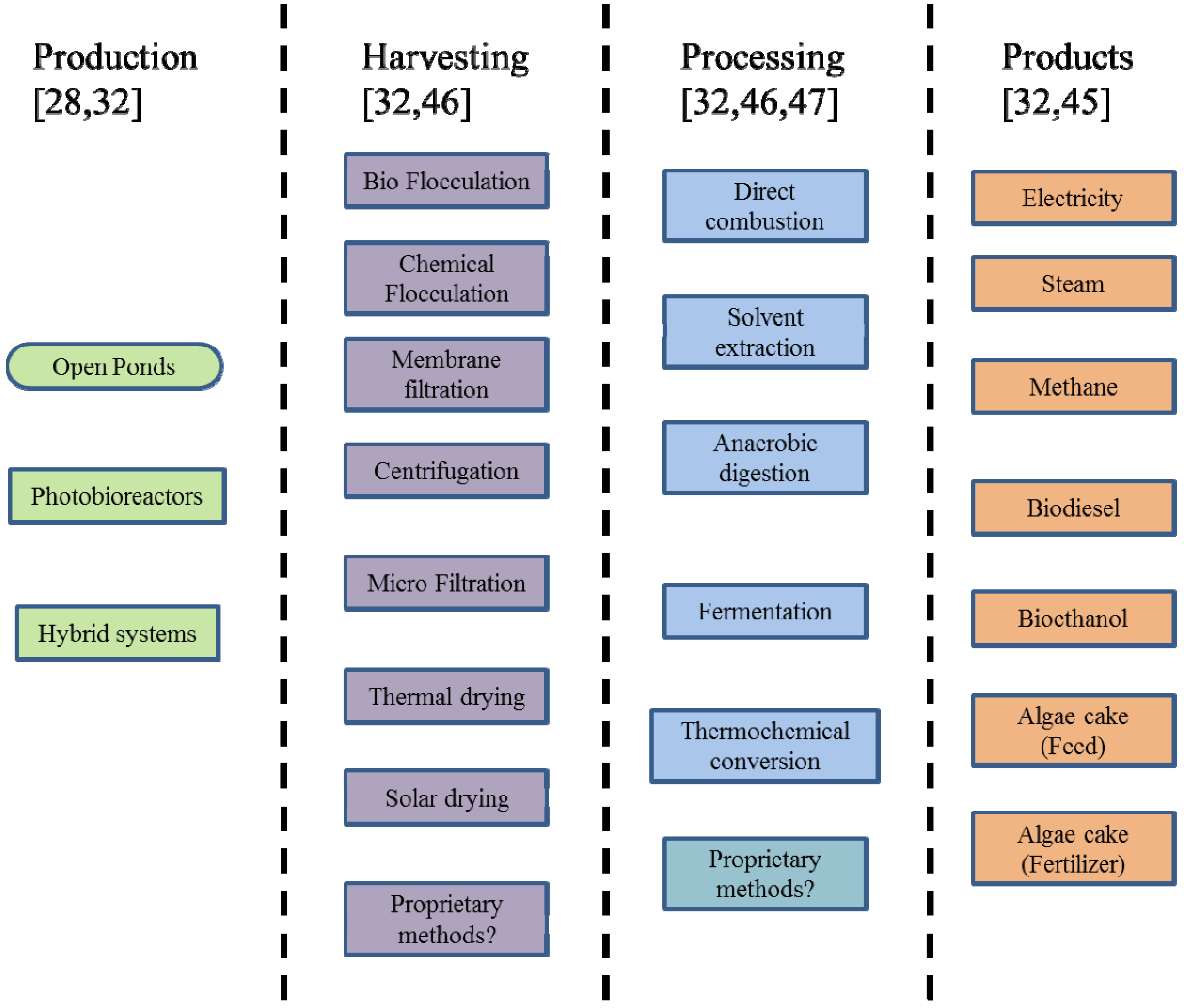
1.1. Algae Production, Processing and Use

Algal production processes can be categorized into three general classes of growth regimes based on the energy source and mode of utilization; these include: photoautotrophic, heterotrophic and mixotrophic. These growth processes can occur in open raceway ponds or closed bioreactor systems [36]. Different production schemes involving combinations of different growth regimes in various reactor configurations have been proposed in an effort to maximize biofuel productivity. Substrates for algal feedstock production can range from industrial effluents and municipal waste water to synthetic media consisting of sugars from molasses, starch or lignocellulosic feedstocks depending on the growth regime and bioreactor configuration. Although open raceway ponds require low energy inputs and lower capital costs, several issues such as contamination (i.e., by unwanted algal species as well as viral, bacterial and fungal pathogens) and low final biomass concentrations (often less than 1.0 g/L) increase production costs to levels that are still economically unviable for large-scale production of biofuels [32]. Closed photobioreactors (CPBR) in different configurations have been proposed to address the contamination issues associated with open raceway systems. CPBRs also permit more stringent control of growth conditions and harvesting [13]. However, CPBRs often require extensive upfront capital investments compared to open raceway ponds and therefore face similar commercialization challenges.
In addition to system setup, there are challenges associated with specific stages within the production process. Lipid and starch fractions of algae can be processed into renewable fuel by several different processing methods, including: pyrolysis [37], thermochemical liquefaction [38], fermentation [39], and transesterification [40]. Most thermochemical conversion methods such as direct combustion, gasification, and pyrolysis require low-moisture-content biomass. This poses a challenge due to high energy requirements for drying algal feedstock. However, hydrothermal liquefaction process can use wet slurry for algal-oil production [41] thus reducing costs associated with drying. Biochemical conversion processes such as anaerobic digestion produce methane (60%–70%) and carbon dioxide (30%–40%) [42]. Fermentation of sugars produced from the starch fraction can be used to produce ethanol from algal biomass [39]. Advantages and limitations of these processes are tabulated in Table 1.
Table 1. Advantages and limitations of biofuel production processes from microalgae.
ProcessAdvantagesLimitations
PyrolysisHigh bio-oil yields possible(up to 57.5% w/w for fast and flash pyrolysis [43])Low-moisture-content biomass required
High-energy-content required to dry feedstock
Thermochemical liquefactionAlgal wet slurry can be used Energy (and cost) reduction
High yields possible (up to 60% w/w [44])
Reactors are complex and expensive
FermentationCo-products can be utilized Conversion of sugar to bioethanol possibleLong processing times required Biomass has to be preprocessed to be converted to sugars
TransesterificationEnhanced physical properties of renewable fuels
Biodiesel has a current market that simplifies commercialization
Limited to conversion of lipids and does not utilize carbohydrate and protein fractions of feedstock
Details concerning algae biomass production processes, different processing technologies for conversion into biofuels and bioproducts, and challenges associated with commercialization of algal biofuels have been discussed thoroughly in recent reviews [11,32,35,45]. A summary of various algae production, harvesting and processing alternatives are presented in Figure 1 [36,46,47].

1.2. Relevance of Individual Algal Cellular Components for Biofuels Production

The exploitation of microalgae as a protein source has led to increased interest in the use of microalgae (e.g., Spirulina, Chlorella and Scenedesmus) in health food production [48]. Algae such as Dunaliella and Spirulina are also used for pigment production [49,50]. However, it is the high density lipid content that renders algae (e.g., Chlamydomonas, Nannochloropsis, Schizochytrium, Chlorella, Nitzschia) attractive for biofuels production [51]. Lipid content in algae can be as high as 50% (dry-weight) with higher percentages of total dry weight (~60%) found as protein and starch [11]. These fractions of microalgae can be used to produce biodiesel [20,52], bioethanol [22,23], biohydrogen [53,54], bioplastics [55], and other products, while simultaneously contributing to CO2 mitigation [56,57]. Still, it is the lipid fraction that has been the major focus for biodiesel production and renewable jet fuel. Depending on the species and growth conditions, dry algae weight can contain 20%–50% lipid [52], which is the major substrate for biofuels production. Total lipid composition includes (but is not limited to) neutral lipids, polar lipids, wax esters, sterols, hydrocarbons, and prenyl derivatives such as tocopherols, carotenoids, terpenes, quinones and phytylated pyrrole derivatives, including chlorophylls [51].
Figure 1. Production, harvesting and processing alternatives for algae.
Energies 06 04607 g001
Carbohydrates (primarily starch) are another valuable component of the algal cell. Typical dry weight content of carbohydrates in algae range from 20% to 40% of total cell mass [58]. Certain components, such as starch, can be easily converted to ethanol by hydrolysis and fermentation. Currently, the starch used for ethanol production is obtained from food crops such as corn, wheat, sorghum, and others [1]. For algae, high starch strains including Chlorella vulgaris (with 37% dry weight starch) [59], are being studied for potential use has high-yield feedstocks. Algal starch is known to be readily fermentable by yeast [60] and, therefore, is being intensely studied for use in ethanol production [22,23].
Upon utilization of lipids and starch fractions for bio-oil production, the residual algae cake, which is rich in proteins, is important for producing valuable co-products. Protein (and starch) content can constitute up to 60% of dry weight of algae [48]. This residual protein from the biomass can be used for livestock, poultry, and fish feed additives [11]. It has been reported that algae can replace about 5%–10% of conventional protein sources in poultry feed [61]. Recently residual algae cake after lipid extraction has been used in large animal feeding trials [62]. However high concentrations of nucleic acids in algae can pose challenges for the utilization in animal feed applications [63].

2. Effect of Environmental Factors

During photosynthesis, using only light and nutrients, algae produce lipids, proteins, and carbohydrates. The relative amounts of these metabolic products are tightly linked to environmental and nutrient conditions including: the amount and intensity of sunlight; CO2 levels; pH; temperature; available nutrients; and, the presence (or absence) of other organisms. Carbon, hydrogen, and oxygen are required non-mineral nutrients for algal growth. Macronutrients include nitrogen, phosphorus, sulfur, potassium and magnesium. Micronutrients such as iron and manganese are also required in small amounts (30–2.5 ppm) while other elements such as cobalt, zinc, boron, copper and molybdenum are essential trace elements (4.5–2.5 ppm) [64]. Collectively, environmental conditions (especially light and temperature) and the availability of non-mineral nutrients, macronutrients, and micronutrients, greatly influence the biochemical composition of microalgae [25,30,65,66,67]. Other factors such as pH and the present toxic metals are also important factors impacting algal growth and metabolism. In general, all of these factors can affect photosynthesis, thus altering carbon fixation and the allocation of carbon into different types of macromolecules. In turn, the cell’s macromolecular composition determines its usefulness in biofuels production.

2.1. Temperature

Temperature is perhaps one of the most important environmental factors that influences algal growth rate, cell size, biochemical composition and nutrient requirements. In the United States, algae grow under a broad range of temperatures (from 15 to 40 °C), depending upon strain, region, and season. Below optimal growth temperatures, growth rate (μ) increases with increasing temperature but declines markedly above the species- or strain- specific optimum [68]. Growth at temperature optima results in minimal cell size [69,70] and the efficiency of carbon and nitrogen utilization decreases at non-optimal temperatures [71]. It has been suggested that changes in cytoplasmic viscosity under sub-optimal temperature conditions is responsible for less efficient carbon and nitrogen utilization [72,73]. Temperature may also play a key role in photoinhibition, which is known to impact algal growth rate. Several mechanisms of temperature-dependent photoinhibition have been postulated. These include mechanisms under which: (i) low temperature results in reduced electron transport at a given photon flux rate due to slower rate of CO2 fixation; (ii) low temperature inhibits the active oxygen species, which results in reducing photoinhibition by protecting PSII; and (iii) low temperature inhibits the synthesis of the D1 protein degraded during photoinhibition, consequently impeding the PSII repair cycle [74].
One of the most commonly observed changes with temperature shift is the alteration in the level of unsaturation of fatty acids in the lipid membrane [75,76,77]. In a study on eight marine plankton species, fatty acids (14:0) increased from ~4% at 10 °C to >20% at 25 °C while PUFA (polyunsaturated fatty acids) were consistently higher at lower temperature (10 °C) [27]. Dunaliella salina has shown a considerable increase in fatty acid unsaturation in response to decrease in temperature from 30 to 12 °C [78]. Lower temperatures decrease the fluidity in the cell membrane. Cells then compensate by increasing levels of unsaturated fatty acids to increase fluidity. However, it also makes the membranes more susceptible to damage by free radicals [73,79]. Along with greater fluidity, increased levels of unsaturated fatty acids tend to enhance the stability of the cellular membranes (particularly the thylakoid membrane). This, in turn, protects the photosynthetic machinery from photoinhibition at low temperatures [79]. For example, in a study involving Botryococcus braunii, a green alga that secretes extracellular lipids, differences in lipid composition were observed at three different growth temperatures (18 °C, 25 °C, and 32 °C). Intracellular lipid synthesis was found to be inhibited at supra-optimal temperature (32 °C); consequently, lipid content decreased to 5% dry weight at 32 °C in comparison with 22% at 25 °C. The decrease in lipid content led to an accumulation of polysaccharides. However, temperature did not affect the secretion of extracellular lipids [66]. Similar effects were observed in Nannochloropsis oculata and Chlorella vulgaris, both of which have an optimum growth temperature of 25 °C. Increasing the growth temperature from 20 to 25 °C doubled the lipid content (from 7.90% to 14.92%) in N. oculata. Increase of temperature from 25 to 30 °C decreased the lipid content in C. vulgaris from 14.71% to 5.90% [80].
Increasing temperature beyond the optimum reduces protein synthesis and consequently results in decreased growth rates [81]. Morris et al. [25] studied the growth of alga, Phaeodactylum tricornutum, a marine diatom, and reported a considerable increase in protein synthesis rates at night with lower the temperatures, presumably due to the fact that protein synthesis is a significant component of nighttime algal metabolism [82]. Similarly, Rhee and Gotham [83] observed an increase in protein concentration in Scenedesmus sp. with decreasing temperature. However, in this study, the efficiency of protein synthesis (in terms of rate of protein synthesis per unit RNA) was reduced upon increasing temperatures beyond the optimum. An increase in temperature from 20 to 30 °C in cultures of Ulva pertusa resulted in higher intercellular free amino acid concentrations from approximately 840 to 1810 mg/100 g dry weight [84]. An increase in free amino acid concentration is an indicator of lower protein content.
Temperature is also reported to impact starch content in the algal cell. Starches are synthesized by phosphorylated metabolites in the dark reactions of the photosynthesis cycle using energy-rich phosphate bonds (i.e., ATP) formed in the light reactions [85]. Increased temperature leads to degradation of the starch produced [65,86]. Enzymes that have been suggested to play a critical role in the temperature dependent degradation of starch are α-amylase and α-glucan phosphorylase [87]. Nakamura and Miyachi studied the effect of temperature on the starch degradation in Chlorella vulgaris grown autotrophically at 20 °C [65]. This study reported a significant reduction in starch (17%) with concomitant increase in sucrose (57%), when culture was exposed to 38 °C for 10 min after 30 min at 20 °C. However, another study with Chlorella vulgaris grown autotrophically at 38 °C, reported a reduced degradation of starch at 38 °C [88]. This effect could be explained by noting that the starch produced at 38 °C was not subjected to any degradation since there was no change in temperature, but the cultures grown at 20 °C degraded the starch into sugars in response to increasing culture temperatures. Consistent with this reasoning, Nakamura and Imamura observed a reversible transformation of high-molecular-weight L starch (amylopectin-like molecule fraction with MW > 2 × 10⁶) to low-molecular-weight S starch (amylose-like molecule fraction with MW <104) at high temperatures (38 °C) [89].
Temperature has a significant effect on the formation of carotenoids. Carotenoids absorb light energy for use in photosynthesis. They also protect chlorophyll from photodamage [90]. Furthermore, they play a vital role in the photosynthetic reaction center by either participating in the energy-transfer process or protecting the reaction center from auto-oxidation. Carotenoid accumulation in algal species increases with temperature because of the increased oxidative and photodamaging effects noted at elevated temperatures [91,92,93]. Tjahjono et al. [91] reported a three-fold increase in astaxanthin formation in the green alga Haematococcus pluvialis with an increase in cultivation temperature from 20 to 30 °C. These results were confirmed by another study on a different green alga, Chlorococcum sp., in which a two fold increase in total carotenoid content was observed by raising the temperature from 20 to 35 °C under conditions of nitrogen deprivation [92]. Increase in carotenoid formation with increasing temperature is generally attributed to cellular response to enhanced active free oxygen radical formation [91] or increased biosynthetic enzyme activity [92].

2.2. Light

Light is the energy source during photoautotrophic growth phase and organisms use light energy to convert carbon dioxide to organic compounds—especially, sugars. The range of light intensity in USA varies from 1500 to 8500 W℘h/m2/day with strong regional and seasonal dependence [94]. Light intensity effects growth of algae through its impact on photosynthesis [95]. Although rate of growth under increasing light intensity is a function of strain and culture temperature, the growth rate of algae is maximal at saturation intensity and decreases with both increase or decrease in light intensity [96]. The photoadaptation/photoacclimation process in algae leads to changes in cell properties according to the availability of light and an increase in photosynthetic efficiency [97]. Adaptation can occur through multiple mechanisms such as changes in types and quantities of pigments, growth rate, dark respiration rate or the availability of essential fatty acids [67]. Morphological photoacclimation is accompanied by changes in cell volume and the number and density of thylakoid membranes [98]. Algae overcome light limitation by desaturation of chloroplast membranes [99]. Light intensity increase above saturating limits causes photoinhibition [30,100]. This is due to the disruption of the chloroplast lamellae caused by high light intensity [101] and inactivation of enzymes involved in carbon dioxide fixation [102]. For example, growth rate of Dunaliella viridis decreased to 63% with increase in light intensity from 700 to 1500 µmol·m−2·s−1 [30].
Light intensity also affects the cellular composition of algae. Dunaliela tertiolecta exhibits a decrease in protein content and an increase in the lipid fraction with increasing light intensities up to saturation [82]. Similar results were reported by Morris et al. [25], in a study on the marine diatom, Phaeodactylum tricornutum, in which low light (400 lux at the culture surface) led to an increase in the rate of protein synthesis. Low light intensity has been observed to result in higher protein content while high photon flux density (PFD) results in increased extracellular polysaccharide content [102]. Absence of light was observed to increase the total lipid content of the D. virdis but reduce triglycerides, free fatty acids, free alcohols and sterols [103]. In Nannochloropsis sp., grown under low light conditions (35 µE·m−2·s−1), 40% of the total lipids were found to be galactolipids and 26% were found to be triacylglycerols. In the same system, high light (550 µE·m−2·s−1) conditions resulted in an increased synthesis of triacylglycerol with a reduction in galactolipid synthesis [26]. High light, in general, leads to oxidative damage of PUFA. Numerous studies have suggested that the cellular lipid content and PUFA decrease with increase in light intensity [104,105,106]. Conversely, Nannochloropsis cells under low light conditions were characterized by high lipid content and high proportions of eicosapentaenoic acid (EPA; 5,8,11,14,17-icosapentanoic acid) [26]. Confirming this observed trend, another study on the same species reported an increase in unsaturated fatty acids mainly due to an increase in EPA (from 44.3% to 60.7% of the organic content) and a decrease in protein content, with decreasing irradiance [67]. Increase in PUFA under light-limited growth conditions are coupled with an increase in total thylakoid membrane in the cell [98]. However, there are some contradictory studies in which PUFA levels were observed to be increasing with higher light intensity [107]. This difference in response to environmental conditions by different alga may be related to difference in their metabolic pathways. Increase in oxygen-mediated lipid desaturation could be one potential reason for the observed increase in PUFA levels under conditions of higher light intensity [107].
In addition to total light intensity, light cycles and the spectral composition of incident light impact algae. For example, Wu and Merchuk [108] investigated the effect of light and dark cycles on the growth of algae and observed that with increasing photon flux density (PFD), specific growth rate increases up to a certain threshold PFD value after which a decline in growth rate was observed. Sustained high light intensities have also been reported to cause photoinhibition and reduce light utilization efficiency. Light utilization efficiency may be optimized by prolonging the dark period under conditions of high light intensity. This allows the photosynthesis machinery in the cell to fully utilize captured photons and convert them into chemical energy thus avoiding the effects of photoinhibiton [109].
Since the energy content of near-ultraviolet (300–400 nm) and blue light (400–480 nm) is greater than that of red light (620–750 nm), fewer photons of blue light are required to achieve an equivalent magnitude of energy intensity using red light. In addition to differences in the energy intensity, specific components of light are known to impact the cellular regulatory processes including: chlorophyll synthesis, photodamage repair, and cell division. For example, blue light was shown to be essential for the division of Chlamydomonas reinhardtii cells [110]. Evidence for the influence of blue light on short-term growth rate is equivocal [111]. It has been observed that blue and red light can help to increase growth and polysaccharide production [100]. Emerson and Lewis also reported blue and red light to be the most effective for photosynthesis of Chlorella [112]. Miyachi and Kamiya studied the starch formation in Chlorella vulgaris under blue (456 nm) and red (660 nm) light. They reported that the carbon pathway in photosynthesis is regulated by short wavelength light (blue), even under low intensity [113]. Red light of high intensity was observed to incorporate carbon from CO2 into sucrose and starch synthesis pathways. However, superimposition of monochromatic blue light even at low intensities resulted in a significant decrease in sucrose and starch formation along with increasing levels of alanine, aspartate, glutamate, glutamine, malate, citrate, lipids and the alcohol-water-insoluble non-carbohydrate fraction [113].
Ultraviolet light (UV; 215–400 nm) adversely affects the algal primarily due to the damage to the photosynthetic machinery in the cells. UV-B (215–380 nm) causes more damage to the cells compared to UV-A radiation (380–400 nm) even at similar intensities [114]. The UV-B radiation causes direct damage to cellular DNA, UV-A damage is limited to indirect damage through enhance production of reactive oxygen and hydroxyl radicals. At moderate levels, UV-A may stimulate photosynthesis while UV-B has a negative effect of photosynthesis irrespective of the intensity. Some of the response of the algae to minimize the damage caused by UV radiation includes migration, development of protective cell walls, increased synthesis of carotenoids and other pigments [114,115,116].

2.3. pH

One of the most important factors in algal cultivation is pH since it determines the solubility and availability of CO2 and essential nutrients, and because it can have a significant impact on algal metabolism [28,117]. Due to uptake of inorganic carbon by algae, pH can rise significantly in algal cultures [118]. Maximum algal growth occurs around neutral pH, although optimum pH is the initial culture pH at which an alga is adapted to grow. Changing pH in media may limit algal growth via metabolic inhibition [119]. Pruder and Bolton observed that T. pseudonana cells adapted to low pH (6.5) had lower growth rate at sub-optimal pH (8.8) [120]. Normal growth rate was restored after the pH was lowered by addition of HCl. Similar results were reported by Chen and Durbin, where photosynthetic rate and algal growth was minimal at pH 9.0, but carbon uptake rates were enhanced when the pH was lowered to 8.3 [28].
Notably, pH is the major determining factor of relative concentrations of the carbonaceous species in water [121]. Higher pH limits the availability of carbon from CO2, which, in turn, suppresses algal growth [28,121]. At higher pH, the carbon for algae is available in form of carbonates [122]. Higher pH also lowers the affinity of algae to free CO2 [121,123]. In photoautotrophic cultures, replacement of CO2 taken up for photosynthesis is slower resulting in a decrease of CO2 partial pressure and thus leading to an increase in pH [120]. Alkaline pH increases the flexibility of the cell wall of mother cells, which prevents its rupture and inhibits autospore release, thus increasing the time for cell cycle completion [124]. Alkaline pH indirectly results in an increase in triglyceride accumulation but a decrease in membrane-associated polar lipids because of cell cycle inhibition. Membrane lipids in Chlorella were observed to be less unsaturated under conditions of alkaline pH [124].
Similar to alkaline pH, acidic conditions can alter nutrient uptake [125] or induce metal toxicity [126,127] and thus affect algal growth. As previously stated, most species of algae grow maximally around neutral pH (7.0–7.6). This has been observed in studies of Ceratium lineatum, Heterocapsa triquetra and Prorocentrum minimum [118] and Chlamydomonas applanata [31]. Visviki and Santikul [31] studied the growth of Chlamydomonas applanata within a pH range 1.4 to 8.4 with 1 point increments. No growth was observed from pH 1.4 to 3.4, above which tolerance of pH in C. applanata was observed (with optimum growth observed at 7.4). Exponential growth was observed for up to five days at pH 5.4 to 8.4, but maximum growth was achieved at pH 7.4. In a study on Chlamydomonas acidophila at pH 4.4, it was observed that hydrogen ions denature V-lysin, a proteolytic enzyme that facilitates releasing of daughter cells from within the parental wall [128]. Hargreaves and Whitton [129] studied the effects of low pH on the morphology of five algal species. Acidic conditions (pH 1.3–1.5) were observed to limit the motility of cells in Chlamydomonas applanata var. acidophila and Euglena mutabilis. Coleman and Colman studied the effect of external pH on photosynthesis of Coccochloris peniocystis and found a significant decrease in total accumulated carbon and oxygen evolution at pH 5.0 and 6.0, which suggested the reduction in photosynthesis in this cyanobacteria (blue-green alga) at these pH ranges [130]. Maintenance of neutral intracellular pH in an acidic pH external environment would require an expenditure of energy to pump protons out of the cell [131]. On the other hand, acid-tolerant algae such as Chlorella saccharophila [132] and Euglena mutabilis [133] can change intracellular pH in response to changing external pH. In Chlorella saccharophila, an internal pH of 7.3 was maintained for an external pH range of 5.0–7.5; however, decreasing the pH further to 3.0 caused a decrease in cellular pH to 6.4 [132]. Similarly, Euglena mutabilis exhibited an internal pH range from 5.0 (at low external pH < 3.0) to 8.0 (at high external pH > 9.0) [133]. The energy required to maintain internal pH in these acid-tolerant algae is conserved as the internal pH goes down. This may be a mechanism for maintaining cellular metabolism such that algal growth is not drastically affected under acidic conditions [132]. Such a mechanism would endow acid-tolerant algae with the ability to adjust internal pH in response to external pH fluctuations, thereby, maintaining an energy advantage over acid-intolerant species at low external pH.
Some algae such as Dunaleilla acidophila adapt to acidic conditions in growth media by accumulating glycerol to prevent the osmotic imbalance caused by high concentrations of H2SO4 [134] while other species such as Chlamydomonas sp. and Pinnilaria braunii var. amplicephala (an acidophilic diatom) accumulate storage lipids such as triacylglycerides under highly acidic conditions (pH 1) [135]. Another adaptation observed under acidic conditions is an increase in saturated fatty acid content, which reduces membrane fluidity and inhibits high proton concentrations [135]. Such adaptation was reported in a Chlamydomonas sp., in which total fatty acid content increased from 2% at pH 7 to 2.4% at pH 2.7, a modest but statistically significant increase [136].
Under alkaline conditions whereby the extracellular pH is higher than intracellular pH, the cell must rely on active transport of HCO3 and not on passive flux of CO2 for inorganic carbon accumulation [121,137]. Affinity of algae for CO2 increases at lower pH [121,123]. Moroney and Tolbert studied the effects of pH on carbon uptake of Chlamydomonas reinhardtii [138]. They reported an efficient utilization of CO2 for photosynthesis at lower pH (<6.95). However, at high external pH (6.95–9.5), where HCO3 dominates, algae cannot efficiently accumulate carbon and require high supply of carbonates for maintaining photosynthetic activity. In more acidic environments, where the internal pH exceeds that of the surrounding medium and where CO2 comprises a major portion of the total external inorganic carbon, carbon accumulation is thought to be accomplished by the passive movement of CO2 along a pH gradient into the cell or chloroplasts [139,140].

2.4. Salinity

Salinity is another important factor that alters the biochemical composition of algal cells (salinity refers primarily to sodium chloride concentration unless otherwise specified). Exposing algae to lower or higher salinity levels than their natural (or adapted) levels can change growth rate and alter composition. For example, higher salinity increases the algae lipid content [141,142,143,144]. Dunaliella, a marine alga, exhibited an increase in saturated and monounsaturated fatty acids with an increase in NaCl concentration from 0.4 to 4 M [145]. In another study with Dunaliella tertiolecta, an increase in intracellular lipids (60% to 67%) and triglyceride concentration (40% to 56%) with an increase in NaCl concentration from 0.5 (freshwater concentration) to 1.0 M was observed [146]. Increasing the NaCl level in cultures of Botryococcus braunii, a fresh water alga, showed an increase in growth rate, carbohydrate content, and lipid content; however, the greatest biomass concentration was achieved at the lowest salinity level [147]. These results are supported by another study in which lipid content of Botryococcus braunii grown in 0.50 M NaCl was higher compared to media without NaCl addition, but protein, carbohydrates, and pigments levels were lower [143]. Another study with the same alga reported a decrease in protein content with unchanged carbohydrate and lipid content with an increase in salinity [148]. This study also reported reduced growth at higher salinities, which may have been due to an inability of the alga to adapt to high salinity. A study with Tetraselmis suecica also reported reduction in protein content per cell of up to 20% with increasing salinity [144].

2.5. Nutrients

Considerable variation in the biochemical composition under conditions of nutrient limitation can be observed in algae depending upon which nutrient is limited and to what degree. In general, the growth rate of algae is proportional to the uptake rate of the most limiting nutrient under optimal conditions of temperature and pH and is generally described by Michaelis-Menten equation [149].
Nitrogen and phosphate are two important macronutrients for growth and metabolism of algal cells. Nitrogen is a fundamental element for the formation of proteins and nucleic acids. Being an integral part of essential molecules such as ATP, the energy carrier in cells, phosphate is another very important nutrient. Phosphate is also a part of the backbone of DNA and RNA, which are essential macromolecules for all living cells. Phosphorus is also a key component of phospholipids. It is not unusual for algae to become nutrient-limited (i.e., nitrogen- and phosphorus-limited) in the natural environment [70]. Limitation of these key nutrients shifts the metabolic pathway of the organism. For example, nitrogen and phosphorus starvation shifts the lipid metabolism from membrane lipid synthesis to neutral lipid storage. This, in turn, increases the total lipid content of green algae [58]. Specific effects of major nutrients are discussed below.
Carbon. Carbon, hydrogen, and oxygen are three essential non-mineral nutrients. Abundance of hydrogen and oxygen in the media for algae cultures means that their availability is not a challenge to cellular growth or metabolism. Carbon is one of the other major nutrients that must be supplied. It is essential for photosynthesis and hence algal growth and reproduction. Carbon fixed by the algae can end up in three destinations; it will either be used: (a) for respiration; (b) as an energy source; or, (c) as a raw material in the formation of additional cells [150]. Reduced carbon fixation rate implies a reduction in algal growth rate. Algae require an inorganic carbon source to perform photosynthesis. Carbon can be utilized in the form of CO2, carbonate, or bicarbonate for autotrophic growth and in form of acetate or glucose for heterotrophic growth. CO2 in water may be present in any of these forms depending upon pH, temperature and nutrient content:
CO2 + H2 ↔ H2CO3 ↔ H+ + HCO3 ↔ 2H+ + CO32−
With an increase in pH, carbonate increases while molecular CO2 and bicarbonate decrease [28]. At the average pH of seawater (8.2), 90% of the total CO2 is present in the form of HCO3; only 1% exists as molecular CO2 and the rest is bicarbonate [122,151].
Riebesell et al. [152] studied the effect of CO2 concentration on lipid distribution in Emiliania huxleyi. A significant effect of CO2 on the composition of the polyunsaturated fatty acids and alkenones was reported. Specifically, lower CO2 concentrations led to an increase in 22:6 (n−3) PUFA, whereas 14:0 fatty acids were found to be predominant at higher CO2 concentrations. Along with the change in composition, increased CO2 was also observed to increase the amount of fatty acid accumulation in Dunaliella salina [153]. Similar increase in fatty acid content and unsaturation with increase in CO2 concentrations were reported by Tsuzuki et al. [154]. Another study with the cyanobacterium Spirulina platensis reported that elevated CO2 concentrations decrease relative concentrations of proteins and pigments in the cells but increase carbohydrate content. This change in the cell composition was accompanied by reduction in the maximum biomass yield [155].
Nitrogen. Nitrogen is an essential constituent of all structural and functional proteins in the algal cells and accounts for 7%–20% of cell dry weight [58]. Inorganic nitrogen taken up by algae is rapidly assimilated into biochemically active compounds and recycled within cells to meet changing physiological needs [156,157]. Major effects of nitrogen deficiency in algal culture include the enhanced biosynthesis and accumulation of lipids [75,80,158,159,160] and triglycerides [161,162] with a concomitant reduction in protein content [25,29,163,164,165,166]. This, in turn, results in a higher lipid/protein ratio [80] at the expense of growth rate [167]. Thus, attempts to increase lipid concentration via nitrogen limitation must be carefully evaluated to ensure high lipid productivity [168]. Algae grown in nitrogen-depleted cultures also tend to divert their photosynthetically fixed carbon to carbohydrate synthesis [58]; however, the physical significance of this is not clear. Other effects of nitrogen reduction include decrease in oxygen evolution, carbon dioxide fixation, chlorophyll content, and tissue production [169,170]. Holm-Hansen et al. [164] reported an increase in amino acid content of Chlorella pyrenoidosa at the expense of sugar phosphates (such as glucose-6-phosphate, fructose-6-phosphate) with addition of ammonium (nitrogen source) to the growing culture.
Degradation of phycolbillisomes with nitrogen limitation has been demonstrated in the case of cyanobacteria and red alga [171]. Phycolbillisomes are the light harvesting antennae of photosystem II in these algae. Photosynthesis continues at a reduced rate, until cell nitrogen falls below a particular species-dependent threshold value. Under nitrogen deficient conditions, Spiriluna platensis cells exhibit reduced carbon fixation capacity even under normal to high available CO2 concentrations [155]. Nitrogen starvation also alters the enzyme balance of cells, resulting in the synthesis of lipids and a decrease in chlorophyll synthesis leading to excess carotenoids in the cells [172]. Dunaliella sp. and Haematococcus pluvialis are observed to accumulate high amounts of carotenoids, astaxanthin and its acylesters (up to 13% w/w), when grown under nitrogen-depleting conditions [173,174,175]. Zhekisheva et al. [176] reported that under nitrogen depleting conditions, Haematococcus pluvialis produced fatty acids and astaxanthin in a 5:1 ratio. It was suggested that the production of the oleic acid-rich triacyl-glycerols and the esterification of the astaxanthin, maintain a high content of astaxanthin esters by enabling the oil globules.
Phosphorus. Phosphorus is an important component required for normal growth and development of algal cells [58]. It has been shown that phosphorus, rather than nitrogen, is the primary limiting nutrient for microalgae in many natural environments [177]. Phosphorus typically constitutes 1% of dry weight of algae [178], but it may be required in significant excess since not all added phosphate is bioavailable due to formation of complexes with metal ions [52]. Immediate effects of phosphorus limitation include a reduction in the synthesis and regeneration of substrates in the Calvin-Benson cycle and a consequential reduction in the rate of light utilization required for carbon fixation [179].
Phosphorus limitation also leads to accumulation of lipids. Total lipid content in Scenedesmus sp. was observed to increase from 23% to 53% with a reduction in initial total phosphorus (as phosphate) concentration of 0.1 from 2.0 mg L−1 [180]. Phosphatidylglycerol (PG), which is one of four major glycerolipids constituting membrane lipids in chloroplasts, was observed to decrease with phosphorus limitation in Chlamydomonas reinhartdtii [181]. PG is essential for cell growth, the maintenance of chlorophyll-protein complex levels, and normal structure-function of the PSII complex. Total acidic lipid (such as sulphoquinovosyldiacylglycerol and PG) content of the chloroplast did not change significantly since a decrease in one acidic lipid was accompanied by an increase in another acidic lipid [181]. Phosphate limitation also reduces the synthesis of n-3 PUFA [182].
Similar to the effects of nitrogen deficiency, phosphorus starvation reduces chlorophyll a and protein content thereby increasing the relative carbohydrate content in algal cells [29,183,184]. Phosphate deficiency has been demonstrated to result in accumulation of astaxanthin and an overall reduction in cell growth [185]. A decrease in cellular phycobilisome under conditions of phosphorus deficiency (due to cell division and the cessation of phycobilisomes synthesis) has also be shown [171]. Theodorou et al. [186] observed that phosphorus starvation in Selenastrum minutum reduces respiration rate.
Trace Metals. Trace metals are metals present in algal cells in extremely small quantities (<4 ppm) but that are an essential component of phycophysiology. Iron (Fe), manganese (Mn), cobalt (Co), zinc (Zn), copper (Cu) and nickel (Ni) are the six most important trace metals required by algae for various metabolic functions [187]. As the aqueous concentration of trace metals is not an indicator of the bioavailability of metals, trace metal availability to algae is highly dependent on speciation (free ion concentration) [188]. Deficiencies in trace metals can limit algal growth, whereas excesses or high metal concentrations (above the toxicity threshold) may inhibit growth, impair photosynthesis, deplete antioxidants, and damage the cell membrane.
Iron is an important trace metal for normal growth and functioning of photosynthesis and respiration in algae. It acts as redox catalyst in photosynthesis and nitrogen assimilation and mediates electron transport reactions in photosynthetic organisms [131]. Iron limitation significantly depresses photosynthetic electron transfer, resulting in a reduction in NADPH formation. Reduction in iron decreases the cellular abundance of ferredoxin, which contains Fe, and forces the substitution of flavodoxin, a non-iron functional equivalent, in the cell [189,190,191,192]. Since the catalytic capacity of ferredoxin is much higher than flavodoxin, this can be problematic [193]. Iron limitation also reduces cellular chlorophyll concentration [194]. High concentrations of iron in cultures of Chlorella vulgaris were observed to increase the lipid content [195]. Decrease in iron content reduces carotenoid composition [185,196].
While there are some non-essential metals (e.g., Cd, Pb and Cr), which can inhibit many metabolic processes even in small quantities [197], there are some essential elements (e.g., Zn and Cu) which when in excess can cause toxicity [198]. The cell surfaces of algae contain a number of functional groups with high affinity for metal ions and that carry a net negative charge mainly due to carboxylic, sulfhydryl, and phosphatic groups [199]. These groups are binding sites that transport metal ions across the cell membrane and into the cell. Cu, Ni and Fe are metals that are commonly observed to be toxic to the algae, if present in supra-threshold concentrations. Cu is one of the most toxic of these metals. Toxic metals can inhibit carbon fixation and delay nutrient uptake [200]. Copper ions were observed to inhibit both cell division and photosynthesis in Asgerionella glacialis (marine diatom) and Chlorella pyrenoidosa (freshwater alga) [201]. Metal toxicity in algae is observed to be a pH-dependent effect, possibly due to the “competition between H+ and free metal cations for cellular binding sites”. For example, copper toxicity was observed to increase 76-fold from pH 5.0–6.5 for the green alga Scenedesmus quadricauda [202]. Cadmium (Cd) is particularly toxic to algal cells. Although Cd has no biological significance in a living cell, it is taken up by marine cells in the form of complexes with organic matter and is absorbed onto organic matter and inorganic matter in ionic form [203]. Cd is inhibits phosphorus uptake, which is also a pH-dependent phenomena. This toxic effect increases in the pH range of 5.5–8.5 [202]. Zinc is also a toxic metal which is rapidly taken up by the algae and is incorporated primarily into polysaccharide and nucleic acid fractions [204].

3. Interaction among Environmental Factors

Environmental factors may influence other systems factors related to algal growth or cellular composition. For example, increase in temperature can lead to reductions in nutrient availability [205]. Such interaction effects have been observed in many studies and could underlie contradictory results between some studies. For example, in a study on phytoplankton in Antarctic Ocean, Smith and Morris [206] reported that at higher temperatures phytoplankton incorporate more carbon into the protein fraction with a concomitant reduction in lipids. To the contrary, Morris et al. [25] reported an increase in the protein fraction of P. tricornutum at low temperatures. This variability could be due to the species-specific effects, differences in light intensity, and/or differences in the primary growth conditions.
In another study, Kudo et al. [207] observed the effect of iron stress on growth rate and cellular composition of the marine diatom Phaeodactylum tricornutum, over the temperature range of 5–30 °C. (Note that the optimum temperature for growth of P. tricornutum was 20 °C). The growth rate of Fe-stressed cells was 50% of Fe-replete cell growth rate at the optimum growth temperature. Differences in growth rates diverged significantly at suboptimum temperatures. It was also reported that at optimal temperature, the C:N ratio in the cell decreased by about 5% in cells induced with an iron stress (2 µM to 2 nM Fe). However, an increase of about 4% was demonstrated with the same transition in iron concentration but at a lower temperature (10 °C).
Algal growth rates are also affected by light-by-nutrient interactions. Cloern et al. [208] developed a model of phytoplankton chlorophyll—specifically, carbon ratio as a function of light and nutrients. The model suggests an increase in growth efficiency with nutrient availability under low light conditions. Morgan and Kalff [209] studied the interaction of temperature and light on the growth of Cryptomonas erosa under nutrient saturated conditions. Results indicated that algal carbon uptake capacity is reduced by 85% with a 90% reduction in light at 23.5 °C compared to 71% with light reduction of 82% at 4 °C. At 4 °C and 1.792 µE·m−2·s−1 light intensity, or 1 °C and 0.32 µE·m−2·s−1, cell division was strongly inhibited. Another study on the interaction effects of light and temperature using Chlorella pyrenoidosa showed an increased saturation light intensity for algal growth at higher temperatures compared to lower temperatures. For higher temperatures, this alga exhibited a higher growth rate even at higher light intensities. The limitation on growth of algae under low temperature and high light intensity conditions is through effects on photosynthesis [210].
Converti et al. [80] studied the effects of temperature and nitrogen concentration on cell growth and lipid content in two strains of algae—Chlorella vulgaris and Nannochloropsis oculata. Reducing the nitrate concentrations in the growth media by 75% (1.5 to 0.375 g L−1 for Chlorella vulgaris and 0.3 to 0.075 L−1 for Nannochloropsis oculata), lipid accumulation tripled and doubled respectively, with only a small reduction in growth rate at optimal growth temperature. This result indicates that it may be possible to achieve higher lipid productivity for biofuels production by employing nitrogen limitation with fine temperature control. Interactions between salinity and nutrient concentrations (i.e., NaNO3) for a salt-tolerant alga, Tetraselmis suecica, were also reported [144]. Optimum growth conditions were found at high salinity (25%–35%) and low nutrient concentrations (2, 4, and 8 mM NaNO3). It was also reported that the total protein and protein content per cell increased with increase in salinity at constant nutrient concentration. Furthermore, a decrease in protein content was observed with increase in nitrate concentration at constant salinity. The transformation of nitrate to protein increased with the salinity and decreased with increasing nitrate concentrations.
High light (300 µE·m−2·s−1) with warm temperatures (22 °C) was observed to promote carbon fixation since this pathway acts as a sink for energy in conjunction with NO3 reduction pathways, which dissipate excess light energy. Confirming this hypothesis, a change in nitrogen source from NO3 to NH4+ controlling for light and temperature resulted in an increase of photorespiration, which is also a sink for excess energy. On the contrary, high light intensity (300 µE·m−2·s−1) and lower temperatures (12 °C) with NO3 as the nitrogen source resulted in increased energy dissipation by means of photorespiration and NO3 reduction (rather than via diversion to cellular carbohydrates). It was postulated that this result is due to lower carbon flows through the Calvin cycle at lower temperatures; in turn, this necessitates dissipation of the excess energy via photorespiration and NO3 reduction. Synergistic interaction effects of light, temperature, and nitrogen source on the transcription of five genes that are central to the carbon and nitrogen metabolism pathways in Thalassiosira pseudonana were also demonstrated [211].
The effects of environmental factors on biochemical composition of algae are summarized in Table 2. These results suggest that past growth conditions, interaction effects of various nutrients and environmental factors are all important to attain a thorough understanding of the behavior of algal cells in large-scale systems.
Table 2. Summary of general impact of environmental factors on biochemical composition of algae.
FactorOrganismConditionsBiochemical changes observedReferences
TemperatureBotryccoccus brauniiIncreased from 25 to 32 °CDecrease in intracellular lipid content from 22% to 5% wt. Accumulation of polysaccharides[66]
Chlorella vulgarisIncreased from 20 to 38 °CDecrease in starch resulting in increase in sucrose[65]
Increased from 10 to 38 °CTransformation of L starch (high molecular weight) to S starch (low molecular weight) Reversible with temperature[89]
Haematococcus pluvialisIncreased from 20 to 30 °C3-fold increase in astaxanthin formation[91]
Chlorococcum sp.Temperature increase from 20 to 35 °C under nitrogen deprivationTwo fold increase in total carotenoid content[92]
Nitella mucronata MiquelIncreased from 5 to 20 °CIncrease in velocity of cytoplasmic streaming[73]
LightDunaliella virdisDarkness (No light)Increase in total lipid content Decrease in free fatty acids, alcohol, sterol[30]
Nannochloropsis sp.Light limited conditionsIncrease in lipid content Increase in EPA * proportions[26]
Porphyridium cruentumRed lightEnhanced Photosystem II relative to Photosystem I and phycobilisome[212]
Chlorella vulgarisRed lightIncrease in sucrose and starch formation[113]
Blue lightIncrease in lipid fraction and alcohol-water insoluble non carbohydrate fraction
pHChlamydomonas acidophilapH 4.4Denaturation of V-lysin[128]
Coccochloris peniocystispH decreased from 7.0 to 5.0 and 6.0Decrease in total accumulated carbon and oxygen evolution[130]
NitrogenNannochloropsis oculata75% decrease in NitrogenIncrease in lipid synthesis from 7.90% to 15.31%[80]
Phaeodactylum tricornutumNitrogen limitationIncrease in lipid synthesis; Decrease in protein content[25]
Chlorella vulgaris75% decrease in NitrogenIncrease in lipid synthesis from 5.90% to 16.41%[80]
Haematococcus pluvialisNitrogen limitationIncrease in carotenoid formation (13% w/w)[174]
PhosphorusChlamydomonas reinhardtiiLimitationDecrease in phosphatidylglycerol[181]
Ankistrodesmus falcatusLimitationDecrease in chl a and protein; Increase in carbohydrate and lipids[29,183,184]
Selenastrum minutumStarvationReduced rate of respiration; Decreased photosynthetic CO2 fixation[186]
IronDunaliella tertiolectaLimitationDecrease in cellular chlorophyll concentration[194]
Chlorella vulgarisHigh concentration of ironIncrease in lipid content[195]
Haematococcus pluvialisHigh concentration of ironIncrease in carotenoid formation[185]
CarbonChlamydomonas reinhardtiipH exceeding 9.0Inefficient accumulation of carbon High supply of carbonates required to maintain photosynthetic activity[138]
Dunaliella salinaCO2 concentration increased from 2% to 10% for 1 day30% increase in amount of fatty acid (dry weight basis)[153]
CO2 concentration increased from 2% to 10% for 7 days2.7 fold increase in fatty acid
Spirulina platensisElevated CO2 concentrationsIncrease in carbohydrate content; Decrease in proteins and pigments[155]
Note: * EPA: Eicosapentanoic acid, 5,7,11,14,17-icosapentanoic acid.

4. Conclusions

4.1. Algae as a Sustainable Biofuel Source

For the success of any sustainable biofuel, there are three principal considerations: technical feasibility; economic viability; and, resource sustainability. Algal-based biofuel is technically feasible. However, to date, economic viability has not been achieved. Furthermore, resource sustainability, in terms of land, water, nutrient and energy utilization, must be meticulously quantified for each type of production system in order for the feedstock to be considered truly “sustainable”. With large-scale biofuel production processes, this water-energy-nutrient nexus is the subject of significant consideration and debate.
Sustainable use of wastewater and seawater resources poses one of the most significant challenges to large-scale production of algal-based biofuels. Algal biofuel production using waste water, brackish water, or sea water in open ponds or using closed photobioreactors, in which the evaporative water losses are negligible, are both potential solutions to this challenge.
Sustainable use of nutrients, such as nitrogen and phosphorous, also poses a serious challenge to large-scale production of algal-based biofuel. Energy intensive production methods for nitrogenous fertilizers and concerns about long-term availability of phosphorous accentuate these concerns. Phosphate is typically obtained by mining and some recent studies estimate that peak phosphorous could occur as early as 2030 [213]. Recycling nitrogen, phosphorous and other nutrients is a strategy to address some of these challenges while addressing other ecological issues such as eutrophication. Algae are capable of utilizing nutrients (including, nitrogen and phosphorus) from wastewater and thus could play a key role in nutrient recovery from waste waters [42,51,214,215,216,217,218,219,220,221]. Maximizing algae production and minimizing costs associated with harvesting are critical to cost-effective nutrient removal system development [222].

4.2. Future Outlook

In this review, we discussed the effects of various nutritional factors (i.e., carbon, nitrogen, phosphorous, iron and trace metals) environmental factors (i.e., light, temperature and pH) and their interactions on algae growth and composition. During the last four decades, significant progress has been made toward a better understanding of algae growth. This includes an enhanced understanding of the effects of the nutrient availability and environmental factors on algae cell division and composition. However, interaction effects induced by multiple factors acting in concert have been generally underexplored. With advances in biotechnology and bioinformatics, a large volume of genetic information is now available rendering studies of interaction effects tractable. For example, full genomes for Chalmydomonas reinhardtii [223], Chlorella vulgaris [224] and Dunaleilla salina [225] have been sequenced. Many genetic manipulation tools have been developed for Chlamydomonas reinhardtii and others are being developed for other algal species. Continued research will not only lead to an enhanced understanding of basic algal cell biology but it will also aid in development of more accurate predictive models for algae growth [226]. Predictive models, in turn, can be used for development of automated optimal control systems for managing algae growth in large-scale production systems. This knowledge will be critical for successful scale-up of algae production systems for sustainable production of biofuels and other algae-based bioproducts.

Conflicts of Interest

The authors declare no conflict of interest.

References

  1. Nichols, N.N.; Bothast, R.J. Production of Ethanol from Grain. In Genetic Improvement of Bioenergy Crops; Vermerris, W., Ed.; Springer: New York, NY, USA, 2008; pp. 75–88. [Google Scholar]
  2. Goldemberg, J.; Coelho, S.T.; Guardabassi, P. The sustainability of ethanol production from sugarcane. Energy Policy 2008, 36, 2086–2097. [Google Scholar] [CrossRef]
  3. Samson, R.A.; Omielan, J.A. Switchgrass: A Potential Biomass Energy Crop for Ethanol Production. In Proceedings of the Thirteenth North American Prarie Conference, Windsor, ON, Canada, 6–9 August 1992; pp. 253–258.
  4. Schmer, M.R.; Vogel, K.P.; Mitchell, R.B.; Perrin, R.K. Net energy of cellulosic ethanol from switchgrass. Proc. Natl. Acad. Sci. USA 2008, 105, 464–469. [Google Scholar] [CrossRef]
  5. Sheehan, J.; Aden, A.; Paustian, K.; Killian, K.; Brenner, J.; Walsh, M.; Nelson, R. Energy and environmental aspects of using corn stover for fuel ethanol. J. Ind. Ecol. 2003, 7, 117–146. [Google Scholar] [CrossRef]
  6. Ballesteros, I.; Negro, M.J.; Oliva, J.M.; Cabañas, A.; Manzanares, P.; Ballesteros, M. Ethanol production from steam-explosion pretreated wheat straw. Appl. Biochem. Biotechnol. 2006, 129–132, 496–508. [Google Scholar] [CrossRef] [PubMed]
  7. Kumar, D.; Murthy, G.S. Pretreatments and enzymatic hydrolysis of grass straws for ethanol production in the Pacific Northwest U.S. Biol. Eng. 2010, 3, 97–110. [Google Scholar] [CrossRef]
  8. Kumar, D.; Murthy, G.S. Impact of pretreatment and downstream processing technologies on economics and energy in cellulosic ethanol production. Biotechnol. Biofuels 2011, 4, 27. [Google Scholar] [CrossRef] [PubMed]
  9. Sanchez, O.J.; Cardona, C.A. Trends in biotechnological production of fuel ethanol from different feedstocks. Bioresour. Technol. 2008, 99, 5270–5295. [Google Scholar] [CrossRef] [PubMed]
  10. Gelfand, I.; Sahajpal, R.; Zhang, X.; Izaurralde, R.C.; Gross, K.L.; Robertson, G.P. Sustainable bioenergy production from marginal lands in the U.S. Midwest. Nature 2013, 493, 514–517. [Google Scholar] [CrossRef] [PubMed]
  11. Singh, J.; Gu, S. Commercialization potential of microalgae for biofuels production. Renew. Sustain. Energy Rev. 2010, 14, 2596–2610. [Google Scholar] [CrossRef]
  12. Amin, S. Review on biofuel oil and gas production processes from microalgae. Energy Convers. Manag. 2009, 50, 1834–1840. [Google Scholar] [CrossRef]
  13. Nakas, J.; Schaedle, M.; Parkinson, C.; Coonley, C.; Tanenbaum, S. System development for linked-fermentation production of solvents from algal biomass. Appl. Environ. Microbiol. 1983, 46, 1017–1023. [Google Scholar] [PubMed]
  14. Sander, K.; Murthy, G.S. Life cycle analysis of algae biodiesel. Int. J. Life Cycle Assess. 2010, 15, 704–714. [Google Scholar] [CrossRef]
  15. Georgianna, D.R.; Mayfield, S.P. Exploiting diversity and synthetic biology for the production of algal biofuels. Nature 2012, 488, 329–335. [Google Scholar] [CrossRef] [PubMed]
  16. Sheehan, J. A Look Back at the U.S. Department of Energy’s Aquatic Species Program: Biodiesel from Algae; National Renewable Energy Laboratory: Golden, CO, USA, 1998. [Google Scholar]
  17. Ahmad, A.; Yasin, N.; Derek, C.; Lim, J. Microalgae as a sustainable energy source for biodiesel production: A review. Renew. Sustain. Energy Rev. 2011, 15, 584–593. [Google Scholar] [CrossRef]
  18. Deng, X.; Li, Y.; Fei, X. Microalgae: A promising feedstock for biodiesel. Afr. J. Microbiol. Res. 2009, 3, 1008–1014. [Google Scholar]
  19. Khan, S.A.; Hussain, M.Z.; Prasad, S.; Banerjee, U. Prospects of biodiesel production from microalgae in India. Renew. Sustain. Energy Rev. 2009, 13, 2361–2372. [Google Scholar] [CrossRef]
  20. Mata, T.M.; Martins, A.A.; Caetano, N.S. Microalgae for biodiesel production and other applications: A review. Renew. Sustain. Energy Rev. 2010, 14, 217–232. [Google Scholar] [CrossRef]
  21. Eshaq, F.S.; Ali, M.N.; Mohd, M.K. Spirogyra biomass a renewable source for biofuel (bioethanol) production. Int. J. Eng. Sci. Technol. 2010, 2, 7045–7054. [Google Scholar]
  22. Harun, R.; Danquah, M.K.; Forde, G.M. Microalgal biomass as a fermentation feedstock for bioethanol production. J. Chem. Technol. Biotechnol. 2010, 85, 199–203. [Google Scholar]
  23. John, R.P.; Anisha, G.; Nampoothiri, K.M.; Pandey, A. Micro and macroalgal biomass: A renewable source for bioethanol. Bioresour. Technol. 2011, 102, 186–193. [Google Scholar] [CrossRef]
  24. Moen, E. Biological Degradation of Brown Seaweeds. Ph.D. Thesis, Norwegian University of Science and Technology, Trondheim, Norway, November 1997. [Google Scholar]
  25. Morris, I.; Glover, H.; Yentsch, C. Products of photosynthesis by marine phytoplankton: The effect of environmental factors on the relative rates of protein synthesis. Mar. Biol. 1974, 27, 1–9. [Google Scholar] [CrossRef]
  26. Sukenik, A.; Carmeli, Y.; Berner, T. Regulation of fatty acid composition by irradiance level in the eustigmatophyte Nannochloropsis sp. J. Phycol. 1989, 25, 686–692. [Google Scholar] [CrossRef]
  27. Thompson, P.A.; Guo, M.; Harrison, P.J. Effects of variation in temperature. I. On the biochemical composition of eight species of marine phytoplankton. J. Phycol. 1992, 28, 481–488. [Google Scholar] [CrossRef]
  28. Chen, C.Y.; Durbin, E.G. Effects of pH on the growth and carbon uptake of marine phytoplankton. Mar. Ecol.-Prog. Ser. 1994, 109, 83–94. [Google Scholar] [CrossRef]
  29. Kilham, S.; Kreeger, D.; Goulden, C.; Lynn, S. Effects of nutrient limitation on biochemical constituents of Ankistrodesmus falcatus. Freshw. Biol. 1997, 38, 591–596. [Google Scholar] [CrossRef]
  30. Gordillo, F.J.L.; Goutx, M.; Figueroa, F.L.; Niell, F.X. Effects of light intensity, CO2 and nitrogen supply on lipid class composition of Dunaliella viridis. J. Appl. Phycol. 1998, 10, 135–144. [Google Scholar] [CrossRef]
  31. Visviki, I.; Santikul, D. The pH tolerance of Chlamydomonas applanata (Volvocales, Chlorophyta). Arch. Environ. Contam. Toxicol. 2000, 38, 147–151. [Google Scholar] [CrossRef] [PubMed]
  32. Brennan, L.; Owende, P. Biofuels from microalgae—A review of technologies for production, processing, and extractions of biofuels and co-products. Renew. Sustain. Energy Rev. 2010, 14, 557–577. [Google Scholar] [CrossRef]
  33. Potter, A.L.; Hassid, W.Z. Starch. II. Molecular weights of amyloses and amylopectins from starches of various plant origins. J. Am. Chem. Soc. 1948, 70, 3774–3777. [Google Scholar] [CrossRef] [PubMed]
  34. Roessler, P.G. Environmental control of glycerolipid metabolism in microalgae: Commercial implications and future research directions. J. Phycol. 1990, 26, 393–399. [Google Scholar] [CrossRef]
  35. Sharma, K.K.; Schuhmann, H.; Schenk, P.M. High lipid induction in microalgae for biodiesel production. Energies 2012, 5, 1532–1553. [Google Scholar] [CrossRef]
  36. Chen, C.Y.; Yeh, K.L.; Aisyah, R.; Lee, D.J.; Chang, J.S. Cultivation, photobioreactor design and harvesting of microalgae for biodiesel production: A critical review. Bioresour. Technol. 2011, 102, 71–81. [Google Scholar] [CrossRef] [PubMed]
  37. Du, Z.; Li, Y.; Wang, X.; Wan, Y.; Chen, Q.; Wang, C.; Lin, X.; Liu, Y.; Chen, P.; Ruan, R. Microwave-assisted pyrolysis of microalgae for biofuel production. Bioresour. Technol. 2011, 102, 4890–4896. [Google Scholar] [CrossRef] [PubMed]
  38. Zou, S.P.; Wu, Y.L.; Yang, M.D.; Li, C.; Tong, J.M. Thermochemical catalytic liquefaction of the marine microalgae Dunaliella tertiolecta and characterization of bio-oils. Energy Fuels 2009, 23, 3753–3758. [Google Scholar] [CrossRef]
  39. Ueno, Y.; Kurano, N.; Miyachi, S. Ethanol production by dark fermentation in the marine green alga, Chlorococcum littorale. J. Ferment. Bioeng. 1998, 86, 38–43. [Google Scholar] [CrossRef]
  40. Sharma, Y.C.; Singh, B. Development of biodiesel: Current scenario. Renew. Sustain. Energy Rev. 2009, 13, 1646–1651. [Google Scholar] [CrossRef]
  41. Goyal, H.B.; Seal, D.; Saxena, R.C. Bio-fuels from thermochemical conversion of renewable resources: A review. Renew. Sustain. Energy Rev. 2008, 12, 504–517. [Google Scholar] [CrossRef]
  42. Cantrell, K.B.; Ducey, T.; Ro, K.S.; Hunt, P.G. Livestock waste-to-bioenergy generation opportunities. Bioresour. Technol. 2008, 99, 7941–7953. [Google Scholar] [CrossRef] [PubMed]
  43. Miao, X.; Wu, Q. High yield bio-oil production from fast pyrolysis by metabolic controlling of Chlorella protothecoides. J. Biotechnol. 2004, 110, 85–93. [Google Scholar] [CrossRef] [PubMed]
  44. Duan, P.; Jin, B.; Xu, Y.; Yang, Y.; Bai, X.; Wang, F.; Zhang, L.; Miao, J. Thermo-chemical conversion of Chlorella pyrenoidosa to liquid biofuels. Bioresour. Technol. 2013, 133, 197–205. [Google Scholar] [CrossRef] [PubMed]
  45. Chisti, Y. Constraints to commercialization of algal fuels. J. Biotechnol. 2013, 167, 201–214. [Google Scholar] [CrossRef] [PubMed]
  46. Pragya, N.; Pandey, K.K.; Sahoo, P. A review on harvesting, oil extraction and biofuels production technologies from microalgae. Renew. Sustain. Energy Rev. 2013, 24, 159–171. [Google Scholar] [CrossRef]
  47. Suali, E.; Sarbatly, R. Conversion of microalgae to biofuel. Renew. Sustain. Energy Rev. 2012, 16, 4316–4342. [Google Scholar] [CrossRef]
  48. Becker, E.W. Micro-algae as a source of protein. Biotechnol. Adv. 2007, 25, 207–210. [Google Scholar] [CrossRef] [PubMed]
  49. Borowitzka, L.J.; Borowitzka, M.A. Commercial production of-carotene by Dunaliella salina in open ponds. Bull. Mar. Sci. 1990, 47, 244–252. [Google Scholar]
  50. Chen, F.; Zhang, Y. High cell density mixotrophic culture of Spirulina platensis on glucose for phycocyanin production using a fed-batch system. Enzyme Microb. Technol. 1997, 20, 221–224. [Google Scholar] [CrossRef]
  51. Hu, Q.; Sommerfeld, M.; Jarvis, E.; Ghirardi, M.; Posewitz, M.; Seibert, M.; Darzins, A. Microalgal triacylglycerols as feedstocks for biofuel production: Perspectives and advances. Plant J. 2008, 54, 621–639. [Google Scholar] [CrossRef] [PubMed]
  52. Chisti, Y. Biodiesel from microalgae. Biotechnol. Adv. 2007, 25, 294–306. [Google Scholar] [CrossRef] [PubMed]
  53. Beer, L.L.; Boyd, E.S.; Peters, J.W.; Posewitz, M.C. Engineering algae for biohydrogen and biofuel production. Curr. Opin. Biotechnol. 2009, 20, 264–271. [Google Scholar] [CrossRef] [PubMed]
  54. Benemann, J. Feasibility analysis of photobiological hydrogen production. Int. J. Hydrog. Energy 1997, 22, 979–987. [Google Scholar] [CrossRef]
  55. Hempel, F.; Bozarth, A.S.; Lindenkamp, N.; Klingl, A.; Zauner, S.; Linne, U.; Steinbüchel, A.; Maier, U.G. Microalgae as bioreactors for bioplastic production. Microb. Cell Fact. 2011, 10, 81. [Google Scholar] [CrossRef] [PubMed]
  56. Jeong, M.L.; Gillis, J.M.; Hwang, J.Y. Carbon dioxide mitigation by microalgal photosynthesis. Bull. Korean Chem. Soc. 2003, 24, 1763–1766. [Google Scholar] [CrossRef]
  57. Keffer, J.E.; Kleinheinz, G.T. Use of Chlorella vulgaris for CO2 mitigation in a photobioreactor. J. Ind. Microbiol. Biotechnol. 2002, 29, 275–280. [Google Scholar] [CrossRef] [PubMed]
  58. Hu, Q. Environmental Effects on Cell Composition. In Handbook of Microalgal Culture: Biotechnology and Applied Phycology; Richmond, A., Ed.; Blackwell: Oxford, UK, 2004; pp. 83–93. [Google Scholar]
  59. Hirano, A.; Ueda, R.; Hirayama, S.; Ogushi, Y. CO2 fixation and ethanol production with microalgal photosynthesis and intracellular anaerobic fermentation. Energy 1997, 22, 137–142. [Google Scholar] [CrossRef]
  60. Nguyen, M.T.; Choi, S.P.; Lee, J.; Lee, J.H.; Sim, S.J. Hydrothermal acid pretreatment of Chlamydomonas reinhardtii biomass for ethanol production. J. Microbiol. Biotechnol. 2009, 19, 161–166. [Google Scholar] [CrossRef] [PubMed]
  61. Spolaore, P.; Joannis-Cassan, C.; Duran, E.; Isambert, A. Commercial applications of microalgae. J. Biosci. Bioeng. 2006, 101, 87–96. [Google Scholar] [CrossRef] [PubMed] [Green Version]
  62. Hasan, M.R.; Chakrabarti, R. Use of Algae and Aquatic Macrophytes as Feed in Small-Scale Aquaculture: A Review; Food and Agriculture Organization of the United Nations (FAO): Rome, Italy, 2009. [Google Scholar]
  63. Zepka, L.Q.; Jacob-Lopes, E.; Goldbeck, R.; Souza-Soares, L.A.; Queiroz, M.I. Nutritional evaluation of single-cell protein produced by Aphanothece microscopica Nägeli. Bioresour. Technol. 2010, 101, 7107–7111. [Google Scholar] [CrossRef]
  64. Walker, J.B. Inorganic micronutrient requirements of Chlorella. II. Quantitative requirements for iron, manganese, and zinc. Arch. Biochem. Biophys. 1954, 53, 1–8. [Google Scholar] [CrossRef] [PubMed]
  65. Nakamura, Y.; Miyachi, S. Effect of temperature on starch degradation in Chlorella vulgaris 11 h cells. Plant Cell Physiol. 1982, 23, 333–341. [Google Scholar]
  66. Kalacheva, G.; Zhila, N.; Volova, T.; Gladyshev, M. The effect of temperature on the lipid composition of the green alga Botryococcus. Microbiology 2002, 71, 286–293. [Google Scholar] [CrossRef]
  67. Fábregas, J.; Maseda, A.; Domínguez, A.; Otero, A. The cell composition of Nannochloropsis sp. changes under different irradiances in semicontinuous culture. World J. Microbiol. Biotechnol. 2004, 20, 31–35. [Google Scholar] [CrossRef]
  68. Renaud, S.M.; Thinh, L.V.; Lambrinidis, G.; Parry, D.L. Effect of temperature on growth, chemical composition and fatty acid composition of tropical Australian microalgae grown in batch cultures. Aquaculture 2002, 211, 195–214. [Google Scholar] [CrossRef]
  69. Rhee, G.Y. Effects of environmental factors and their interactions on phytoplankton growth. Adv. Microb. Ecol. 1982, 6, 33–74. [Google Scholar]
  70. Harris, G.P. Phytoplankton Ecology: Structure, Function and Fluctuation; Chapman and Hall: New York, NY, USA, 1986. [Google Scholar]
  71. Darley, W.M. Algal Biology: A Physiological Approach; Blackwell: Oxford, UK, 1982; Volume 9, pp. 1–168. [Google Scholar]
  72. Hope, A.B.; Walker, N.A. The Physiology of Giant Algal Cells; Cambridge University Press: London, UK, 1975. [Google Scholar]
  73. Raven, J.A.; Geider, R.J. Temperature and algal growth. New Phytol. 1988, 110, 441–461. [Google Scholar] [CrossRef]
  74. Vonshak, A.; Torzillo, G. Environmental Stress Physiology. In Handbook of Microalgal Culture; Richmond, A., Ed.; Blackwell: Oxford, UK, 2004; pp. 57–82. [Google Scholar]
  75. Thompson, G.A., Jr. Lipids and membrane function in green algae. Biochim. Biophys. Acta 1996, 1302, 17–45. [Google Scholar] [CrossRef] [PubMed]
  76. Harwood, J.L. Involvement of Chloroplast Lipids in the Reaction of Plants Submitted to Stress. In Lipids in Photosynthesis: Structure, Function and Genetics; Siegenthaler, P.A., Murata, N., Eds.; Springer: Berlin, Germany, 2004; Volume 6, pp. 287–302. [Google Scholar]
  77. Guschina, I.A.; Harwood, J.L. Algal Lipids and Effect of the Environment on Their Biochemistry. In Lipids in Aquatic Ecosystems; Springer: Berlin, Germany, 2009; pp. 1–24. [Google Scholar]
  78. Lynch, D.V.; Thompson, G.A., Jr. Low temperature-induced alterations in the chloroplast and microsomal membranes of Dunaliella salina. Plant Physiol. 1982, 69, 1369–1375. [Google Scholar] [CrossRef] [PubMed]
  79. Nishida, I.; Murata, N. Chilling sensitivity in plants and cyanobacteria: The crucial contribution of membrane lipids. Annu. Rev. Plant Biol. 1996, 47, 541–568. [Google Scholar] [CrossRef]
  80. Converti, A.; Casazza, A.A.; Ortiz, E.Y.; Perego, P.; del Borghi, M. Effect of temperature and nitrogen concentration on the growth and lipid content of Nannochloropsis oculata and Chlorella vulgaris for biodiesel production. Chem. Eng. Process. 2009, 48, 1146–1151. [Google Scholar] [CrossRef]
  81. Konopka, A.; Brock, T.D. Effect of temperature on blue-green algae (cyanobacteria) in Lake Mendota. Appl. Environ. Microbiol. 1978, 36, 572–576. [Google Scholar] [PubMed]
  82. Cuhel, R.L.; Ortner, P.B.; Lean, D.R.S. Night synthesis of protein by algae. Limnol. Oceanogr. 1984, 29, 731–744. [Google Scholar] [CrossRef]
  83. Rhee, G.Y.; Gotham, I.J. The effect of environmental factors on phytoplankton growth: Temperature and the interactions of temperature with nutrient limitation. Limnol. Oceanogr. 1981, 26, 635–648. [Google Scholar] [CrossRef]
  84. Kakinuma, M.; Coury, D.; Kuno, Y.; Itoh, S.; Kozawa, Y.; Inagaki, E.; Yoshiura, Y.; Amano, H. Physiological and biochemical responses to thermal and salinity stresses in a sterile mutant of Ulva pertusa (Ulvales, Chlorophyta). Mar. Biol. 2006, 149, 97–106. [Google Scholar] [CrossRef]
  85. Emerson, R.; Stauffer, J.; Umbreit, W. Relationships between phosphorylation and photosynthesis in Chlorella. Am. J. Bot. 1944, 31, 107–120. [Google Scholar] [CrossRef]
  86. Mitsui, A.; United States-Japan Cooperative Science Program; National Science Foundation (U.S.). Biological Solar Energy Conversion; Academic Press: New York, NY, USA, 1977. [Google Scholar]
  87. Nakamura, Y. Change in molecular weight distribution in starch when degraded at different temperatures in Chlorella vulgaris. Plant Sci. Lett. 1983, 30, 259–265. [Google Scholar] [CrossRef]
  88. Nakamura, Y.; Miyachi, S. Change in starch photosynthesized at different temperatures in Chlorella. Plant Sci. Lett. 1982, 27, 1–6. [Google Scholar] [CrossRef]
  89. Nakamura, Y.; Imamura, M. Change in properties of starch when photosynthesized at different temperatures in Chlorella vulgaris. Plant Sci. Lett. 1983, 31, 123–131. [Google Scholar] [CrossRef]
  90. Armstrong, G.A.; Hearst, J.E. Carotenoids 2: Genetics and molecular biology of carotenoid pigment biosynthesis. FASEB J. 1996, 10, 228–237. [Google Scholar] [PubMed]
  91. Tjahjono, A.E.; Hayama, Y.; Kakizono, T.; Terada, Y.; Nishio, N.; Nagai, S. Hyper-accumulation of astaxanthin in a green alga Haematococcus pluvialis at elevated temperatures. Biotechnol. Lett. 1994, 16, 133–138. [Google Scholar] [CrossRef]
  92. Liu, B.H.; Lee, Y.K. Secondary carotenoids formation by the green alga Chlorococcum sp. J. Appl. Phycol. 2000, 12, 301–307. [Google Scholar] [CrossRef]
  93. Tripathi, U.; Sarada, R.; Ravishankar, G. Effect of culture conditions on growth of green alga—Haematococcus pluvialis and astaxanthin production. Acta Physiol. Plant. 2002, 24, 323–329. [Google Scholar] [CrossRef]
  94. NREL Web Page. Dynamic Maps, GIS Data, and Analysis Tools—Solar Maps. Available online: http://www.nrel.gov/gis/solar.html (accessed on 9 May 2013).
  95. Stockenreiter, M.; Haupt, F.; Graber, A.K.; Seppälä, J.; Spilling, K.; Tamminen, T.; Stibor, H. Functional group richness: Implications of biodiversity for light use and lipid yield in microalgae. J. Phycol. 2013, in press. [Google Scholar]
  96. Sorokin, C.; Krauss, R.W. The Effects of light intensity on the growth rates of green algae. Plant Physiol. 1958, 33, 109–113. [Google Scholar] [CrossRef] [PubMed]
  97. Dubinsky, Z.; Matsukawa, R.; Karube, I. Photobiological aspects of algal mass culture. J. Mar. Biotechnol. 1995, 2, 61–65. [Google Scholar]
  98. Berner, T.; Dubinsky, Z.; Wyman, K.; Falkowski, P.G. Photoadaptation and the “package” effect in Dunaliella tertiolecta (chlorophycae). J. Phycol. 1989, 25, 70–78. [Google Scholar] [CrossRef]
  99. Mock, T.; Kroon, B.M.A. Photosynthetic energy conversion under extreme conditions—II: The significance of lipids under light limited growth in Antarctic sea ice diatoms. Phytochemistry 2002, 61, 53–60. [Google Scholar] [CrossRef] [PubMed]
  100. You, T.; Barnett, S.M. Effect of light quality on production of extracellular polysaccharides and growth rate of Porphyridium cruentum. Biochem. Eng. J. 2004, 19, 251–258. [Google Scholar] [CrossRef]
  101. Brody, M.; Vatter, A.E. Observations on cellular structures of Porphyridium cruentum. J. Biophys. Biochem. Cytol. 1959, 5, 289–294. [Google Scholar] [CrossRef] [PubMed]
  102. Iqbal, M.; Zafar, S. Effects of photon flux density, CO2, aeration rate, and inoculum density on growth and extracellular polysaccharide production by Porphyridium cruentum. Folia Microbiol. 1993, 38, 509–514. [Google Scholar] [CrossRef]
  103. Smith, R.; Cavaletto, J.; Eadie, B.; Gardner, W. Growth and lipid composition of high Arctic ice algae during the spring bloom at Resolute, Northwest Territories, Canada. Mar. Ecol. Prog. Ser. 1993, 97, 19–29. [Google Scholar] [CrossRef]
  104. Cohen, Z. Porphyridium Cruentum. In Chemicals from Microalgae; Cohen, Z., Ed.; CRC Press: Boca Raton, FL, USA, 1999; pp. 1–24. [Google Scholar]
  105. Renaud, S.; Parry, D.; Thinh, L.V.; Kuo, C.; Padovan, A.; Sammy, N. Effect of light intensity on the proximate biochemical and fatty acid composition of Isochrysis sp. and Nannochloropsis oculata for use in tropical aquaculture. J. Appl. Phycol. 1991, 3, 43–53. [Google Scholar] [CrossRef]
  106. Orcutt, D.M.; Patterson, G.W. Effect of light intensity upon lipid composition of Nitzschia closterium (Cylindrotheca fusiformis). Lipids 1974, 9, 1000–1003. [Google Scholar] [CrossRef]
  107. Molina Grima, E.; Garcia Camacho, F.; Acien Fernandez, F. Production of EPA from Phaeodactylum Tricornumtum. In Chemicals from Microalgae; Cohen, Z., Ed.; CRC Press: Boca Raton, FL, USA, 1999; pp. 57–92. [Google Scholar]
  108. Wu, X.; Merchuk, J.C. A model integrating fluid dynamics in photosynthesis and photoinhibition processes. Chem. Eng. Sci. 2001, 56, 3527–3538. [Google Scholar] [CrossRef]
  109. Long, S.; Humphries, S.; Falkowski, P.G. Photoinhibition of photosynthesis in nature. Annu. Rev. Plant Biol. 1994, 45, 633–662. [Google Scholar] [CrossRef]
  110. Voigt, J.; Münzner, P. Blue light-induced lethality of a cell wall-deficient mutant of the unicellular green alga Chlamydomonas reinhardtii. Plant Cell Physiol. 1994, 35, 99–106. [Google Scholar]
  111. Borodin, V.B. Effect of red and blue light on acclimation of Chlamydomonas reinhardtii to CO2-limiting conditions. Rus. J. Plant Physiol. 2008, 55, 441–448. [Google Scholar] [CrossRef]
  112. Emerson, R.; Lewis, C.M. The dependence of the quantum yield of Chlorella photosynthesis on wave lenghth of light. Am. J. Bot. 1943, 30, 165–178. [Google Scholar] [CrossRef]
  113. Miyachi, S.; Kamiya, A. Wavelength effects on photosynthetic carbon metabolism in Chlorella. Plant Cell Physiol. 1978, 19, 277–288. [Google Scholar]
  114. Fernanda Pessoa, M. Harmful effects of UV radiation in algae and aquatic macrophytes—A review. Emir. J. Food Agric. 2012, 24, 510–526. [Google Scholar] [CrossRef]
  115. Xue, L.; Zhang, Y.; Zhang, T.; An, L.; Wang, X. Effects of enhanced ultraviolet-B radiation on algae and cyanobacteria. Crit. Revi. Microbiol. 2005, 31, 79–89. [Google Scholar] [CrossRef]
  116. Rastogi, R.P.; Incharoensadki, A. UV radiation-induced accumulation of photoprotective compounds in the green alga Tetraspora sp. CU2551. Plant Physiol. Biochem. 2013, 70, 7–13. [Google Scholar] [CrossRef]
  117. Goldman, J.C. Letter: Carbon dioxide and pH: Effect on species succession of algae. Science 1973, 182, 306–307. [Google Scholar] [CrossRef] [PubMed]
  118. Hansen, P.J. Effect of high pH on the growth and survival of marine phytoplankton: Implications for species succession. Aquat. Microb. Ecol. 2002, 28, 279–288. [Google Scholar] [CrossRef]
  119. Goldman, J.C.; Azov, Y.; Riley, C.B.; Dennett, M.R. The effect of pH in intensive microalgal cultures. I. biomass regulation. J. Exp. Mar. Biol. Ecol. 1982, 57, 1–13. [Google Scholar] [CrossRef]
  120. Pruder, G.D.; Bolton, E.T. The role of CO2 enrichment of aerating gas in the growth of an estuarine diatom. Aquaculture 1979, 17, 1–15. [Google Scholar] [CrossRef]
  121. Azov, Y. Effect of pH on inorganic carbon uptake in algal cultures. Appl. Environ. Microbiol. 1982, 43, 1300–1306. [Google Scholar] [PubMed]
  122. Nielsen, E.S. Marine Photosynthesis: With Special Emphasis on the Ecological Aspects; Elsevier: Amsterdam, The Netherlands, 1975; Volume 13. [Google Scholar]
  123. Rotatore, C.; Colman, B. The acquisition and accumulation of inorganic carbon by the unicellular green alga Chlorella ellipsoidea. Plant Cell Environ. 1991, 14, 377–382. [Google Scholar] [CrossRef]
  124. Guckert, J.B.; Cooksey, K.E. Triglyceride accumulation and fatty acid profile changes in Chlorella (Chlorophyta) during high pH induced cell cycle inhibition. J. Phycol. 1990, 26, 72–79. [Google Scholar] [CrossRef]
  125. Gensemer, R.W.; Smith, R.E.H.; Duthie, H.C. Comparative effects of pH and aluminum on silica limited growth and nutrient uptake in Asterionella ralfsii var. Americana (Bacillariophyceae). J. Phycol. 1993, 29, 36–44. [Google Scholar] [CrossRef]
  126. Sunda, W. The Relationship between Cupric Ion Activity and the Toxicity of Copper to Phytoplankton. Ph.D. Thesis, Massachusetts Institute of Technology, Cambridge, MA, USA, April 1975. [Google Scholar]
  127. Anderson, D.M.; Morel, F.M.M. Copper sensitivity of Gonyaulax tamarensis. Limnol. Oceanogr. 1978, 23, 283–295. [Google Scholar] [CrossRef]
  128. Visviki, I.; Palladino, J. Growth and cytology of Chlamydomonas acidophila under acidic stress. Bull. Environ. Contam. Toxicol. 2001, 66, 623–630. [Google Scholar] [PubMed]
  129. Hargreaves, J.; Whitton, B. Effect of pH on growth of acid stream algae. Eur. J. Phycol. 1976, 11, 215–223. [Google Scholar] [CrossRef]
  130. Coleman, J.R.; Colman, B. Inorganic carbon accumulation and photosynthesis in a blue-green alga as a function of external pH. Plant Physiol. 1981, 67, 917–921. [Google Scholar] [CrossRef] [PubMed]
  131. Terry, N.; Abadía, J. Function of iron in chloroplasts. J. Plant Nutr. 1986, 9, 609–646. [Google Scholar] [CrossRef]
  132. Gehl, K.A.; Colman, B. Effect of external pH on the internal pH of Chlorella saccharophila. Plant Physiol. 1985, 77, 917–921. [Google Scholar] [CrossRef]
  133. Lane, A.E.; Burris, J.E. Effects of environmental pH on the internal pH of Chlorella pyrenoidosa, Scenedesmus quadricauda, and Euglena mutabilis. Plant Physiol. 1981, 68, 439–442. [Google Scholar] [CrossRef] [PubMed]
  134. Fuggi, A.; Pinto, G.; Pollio, A.; Taddei, R. The role of glycerol in osmoregulation of the acidophilic alga Dunaliella acidophila (Volvocales, Chlorophyta): Effect of solute stress on photosynthesis, respiration and glycerol synthesis. Phycologia 1988, 27, 439–446. [Google Scholar] [CrossRef]
  135. Tatsuzawa, H.; Takizawa, E.; Wada, M.; Yamamoto, Y. Fatty acid and lipid composition of the acidophilic green alga Chlamydomonas sp. 1. J. Phycol. 1996, 32, 598–601. [Google Scholar] [CrossRef]
  136. Poerschmann, J.; Spijkerman, E.; Langer, U. Fatty acid patterns in Chlamydomonas sp. as a marker for nutritional regimes and temperature under extremely acidic conditions. Microb. Ecol. 2004, 48, 78–89. [Google Scholar] [CrossRef] [PubMed]
  137. Moazami-Goudarzi, M.; Colman, B. Changes in carbon uptake mechanisms in two green marine algae by reduced seawater pH. J. Exp. Mar. Biol. Ecol. 2012, 413, 94–99. [Google Scholar] [CrossRef]
  138. Moroney, J.V.; Tolbert, N.E. Inorganic carbon uptake by Chlamydomonas reinhardtii. Plant Physiol. 1985, 77, 253–258. [Google Scholar] [CrossRef] [PubMed]
  139. Colman, B.; Huertas, I.E.; Bhatti, S.; Dason, J.S. The diversity of inorganic carbon acquisition mechanisms in eukaryotic microalgae. Funct. Plant Biol. 2002, 29, 261–270. [Google Scholar] [CrossRef]
  140. Ramanan, R.; Vinayagamoorthy, N.; Sivanesan, S.D.; Kannan, K.; Chakrabarti, T. Influence of CO2 concentration on carbon concentrating mechanisms in cyanobacteria and green algae: A proteomic approach. Algae 2012, 27, 295–301. [Google Scholar] [CrossRef]
  141. Zhila, N.O.; Kalacheva, G.S.; Volova, T.G. Effect of salinity on the biochemical composition of the alga Botryococcus braunii Kütz IPPAS H-252. J. Appl. Phycol. 2011, 23, 47–52. [Google Scholar] [CrossRef]
  142. Renaud, S.; Parry, D. Microalgae for use in tropical aquaculture II: Effect of salinity on growth, gross chemical composition and fatty acid composition of three species of marine microalgae. J. Appl. Phycol. 1994, 6, 347–356. [Google Scholar] [CrossRef]
  143. Ben-Amotz, A.; Tornabene, T.G.; Thomas, W.H. Chemical profile of selected species of microalgae with emphasis on lipids. J. Phycol. 1985, 21, 72–81. [Google Scholar] [CrossRef]
  144. Fabregas, J.; Abalde, J.; Herrero, C.; Cabezas, B.; Veiga, M. Growth of the marine microalga Tetraselmis suecica in batch cultures with different salinities and nutrient concentrations. Aquaculture 1984, 42, 207–215. [Google Scholar] [CrossRef]
  145. Xu, X.Q.; Beardall, J. Effect of salinity on fatty acid composition of a green microalga from an antarctic hypersaline lake. Phytochemistry 1997, 45, 655–658. [Google Scholar] [CrossRef]
  146. Takagi, M.; Yoshida, T. Effect of salt concentration on intracellular accumulation of lipids and triacylglyceride in marine microalgae Dunaliella cells. J. Biosci. Bioeng. 2006, 101, 223–226. [Google Scholar] [CrossRef] [PubMed]
  147. Rao, A.R.; Dayananda, C.; Sarada, R.; Shamala, T.; Ravishankar, G. Effect of salinity on growth of green alga Botryococcus braunii and its constituents. Bioresour. Technol. 2007, 98, 560–564. [Google Scholar] [CrossRef] [PubMed]
  148. Vazquez-Duhalt, R.; Arredondo-Vega, B.O. Haloadaptation of the green alga Botryococcus braunii (race A). Phytochemistry 1991, 30, 2919–2925. [Google Scholar] [CrossRef]
  149. Titman, D. Ecological competition between algae: Experimental confirmation of resource-based competition theory. Science 1976, 192, 463–465. [Google Scholar] [CrossRef] [PubMed]
  150. Berman-Frank, I.; Dubinsky, Z. Balanced growth and aquatic plants: Myth or reality? Phytoplankton use the imbalance between carbon assimilation and biomass production to their strategic advantage. Bioscience 1999, 49, 29–37. [Google Scholar] [CrossRef]
  151. Moss, B. The influence of environmental factors on the distribution of freshwater algae: An experimental study: II. The role of pH and the carbon dioxide-bicarbonate system. J. Ecol. 1973, 61, 157–177. [Google Scholar] [CrossRef]
  152. Riebesell, U.; Revill, A.T.; Holdsworth, D.G.; Volkman, J.K. The effects of varying CO2 concentration on lipid composition and carbon isotope fractionation in Emiliania huxleyi. Geochim. Cosmochim. Acta 2000, 64, 4179–4192. [Google Scholar] [CrossRef]
  153. Muradyan, E.; Klyachko-Gurvich, G.; Tsoglin, L.; Sergeyenko, T.; Pronina, N. Changes in lipid metabolism during adaptation of the Dunaliella salina photosynthetic apparatus to high CO2 concentration. Rus. J. Plant Physiol. 2004, 51, 53–62. [Google Scholar] [CrossRef]
  154. Tsuzuki, M.; Ohnuma, E.; Sato, N.; Takaku, T.; Kawaguchi, A. Effects of CO2 concentration during growth on fatty acid composition in microalgae. Plant Physiol. 1990, 93, 851–856. [Google Scholar] [CrossRef] [PubMed]
  155. Gordillo, F.J.L.; Jiménez, C.; Figueroa, F.L.; Niell, F.X. Effects of increased atmospheric CO2 and N supply on photosynthesis, growth and cell composition of the cyanobacterium Spirulina platensis (Arthrospira). J. Appl. Phycol. 1998, 10, 461–469. [Google Scholar] [CrossRef]
  156. Fujita, R.M.; Wheeler, P.A.; Edwards, R.L. Metabolic regulation of ammonia uptake by Ulva rigida (Chlorophyta): A compartmental analysis of the rate limiting step for uptake. J. Phycol. 1988, 24, 560–566. [Google Scholar] [CrossRef]
  157. Vergara, J.; Bird, K.; Niell, F. Nitrogen assimilation following NH4+ pulses in the red alga Gracilariopsis lemaneiformis: Effect on C metabolism. Mar. Ecol. Prog. Ser. 1995, 122, 253–263. [Google Scholar] [CrossRef]
  158. Shifrin, N.S.; Chisholm, S.W. Phytoplankton lipids: Interspecific differences and effects of nitrate, silicate and light-dark cycles. J. Phycol. 1981, 17, 374–384. [Google Scholar] [CrossRef]
  159. Wang, Z.T.; Ullrich, N.; Joo, S.; Waffenschmidt, S.; Goodenough, U. Algal lipid bodies: Stress induction, purification, and biochemical characterization in wild-type and starchless Chlamydomonas reinhardtii. Eukaryot. Cell 2009, 8, 1856–1868. [Google Scholar] [CrossRef] [PubMed]
  160. Demirbas, A. Use of algae as biofuel sources. Energy Convers. Manag. 2010, 51, 2738–2749. [Google Scholar] [CrossRef]
  161. Takagi, M.; Watanabe, K.; Yamaberi, K.; Yoshida, T. Limited feeding of potassium nitrate for intracellular lipid and triglyceride accumulation of Nannochloris sp. UTEX LB1999. Appl. Microbiol. Biotechnol. 2000, 54, 112–117. [Google Scholar] [CrossRef] [PubMed]
  162. Stephenson, A.L.; Dennis, J.S.; Howe, C.J.; Scott, S.A.; Smith, A.G. Influence of nitrogen-limitation regime on the production by Chlorella vulgaris of lipids for biodiesel feedstocks. Biofuels 2010, 1, 47–58. [Google Scholar] [CrossRef]
  163. Fogg, G. Photosynthesis and formation of fats in a diatom. Ann. Bot. 1956, 20, 265–285. [Google Scholar]
  164. Holm-Hansen, O.; Nishida, K.; Moses, V.; Calvin, M. Effects of mineral salts on short-term incorporation of carbon dioxide in Chlorella. J. Exp. Bot. 1959, 10, 109–124. [Google Scholar] [CrossRef]
  165. Lynn, S.G.; Kilham, S.S.; Kreeger, D.A.; Interlandi, S.J. Effect of nutrient availability on the biochemical and elemental stoichiometry in the freshwater diatom Stephanodiscus minutulus (Bacillariophyceae). J. Phycol. 2000, 36, 510–522. [Google Scholar] [CrossRef]
  166. Heraud, P.; Wood, B.R.; Tobin, M.J.; Beardall, J.; McNaughton, D. Mapping of nutrient-induced biochemical changes in living algal cells using synchrotron infrared microspectroscopy. FEMS Microbiol. Lett. 2005, 249, 219–225. [Google Scholar] [CrossRef] [PubMed]
  167. Li, Y.; Horsman, M.; Wang, B.; Wu, N.; Lan, C.Q. Effects of nitrogen sources on cell growth and lipid accumulation of green alga Neochloris oleoabundans. Appl. Microbiol. Biotechnol. 2008, 81, 629–636. [Google Scholar] [CrossRef] [PubMed]
  168. Metting, F.B., Jr. Biodiversity and application of microalgae. J. Ind. Microbiol. 1996, 17, 477–489. [Google Scholar] [CrossRef]
  169. Kolber, Z.; Zehr, J.; Falkowski, P. Effects of growth irradiance and nitrogen limitation on photosynthetic energy conversion in photosystem II. Plant Physiol. 1988, 88, 923–929. [Google Scholar] [CrossRef] [PubMed]
  170. Richardson, B.; Orcutt, D.; Schwertner, H.; Martinez, C.L.; Wickline, H.E. Effects of nitrogen limitation on the growth and composition of unicellular algae in continuous culture. Appl. Microbiol. 1969, 18, 245–250. [Google Scholar] [PubMed]
  171. Collier, J.L.; Grossman, A. Chlorosis induced by nutrient deprivation in Synechococcus sp. strain PCC 7942: Not all bleaching is the same. J. Bacteriol. 1992, 174, 4718–4726. [Google Scholar] [PubMed]
  172. Round, F.E. The Ecology of Algae; Cambridge University Press: Cambridge, UK, 1984. [Google Scholar]
  173. Ben-Amotz, A.; Avron, M. The biotechnology of cultivating the halotolerant alga Dunaliella. Trends Biotechnol. 1990, 8, 121–126. [Google Scholar] [CrossRef]
  174. Borowitzka, M.A.; Huisman, J.M.; Osborn, A. Culture of the astaxanthin-producing green alga Haematococcus pluvialis. Effects of nutrients on growth and cell type. J. Appl. Phycol. 1991, 3, 295–304. [Google Scholar] [CrossRef]
  175. Harker, M.; Tsavalos, A.J.; Young, A.J. Factors responsible for astaxanthin formation in the chlorophyte Haematococcus pluvialis. Bioresour. Technol. 1996, 55, 207–214. [Google Scholar] [CrossRef]
  176. Zhekisheva, M.; Boussiba, S.; Khozin Goldberg, I.; Zarka, A.; Cohen, Z. Accumulation of oleic acid in Haematococcus pluvialis (Chlorophyceae) under nitrogen starvation or high ligh is correlated with that of astaxanthin esters. J. Phycol. 2002, 38, 325–331. [Google Scholar] [CrossRef]
  177. Larned, S. Nitrogen-versus phosphorus-limited growth and sources of nutrients for coral reef macroalgae. Mar. Biol. 1998, 132, 409–421. [Google Scholar] [CrossRef]
  178. Borchardt, J.A.; Azad, H.S. Biological extraction of nutrients. J. Water Pollut. Control Fed. 1968, 40, 1739–1754. [Google Scholar]
  179. Barsanti, L.; Gualtieri, P. Algae: Anatomy, Biochemistry, and Biotechnology, 1st ed.; CRC Press: Boca Raton, FL, USA, 2005. [Google Scholar]
  180. Li, X.; Hu, H.Y.; Gan, K.; Sun, Y.X. Effects of different nitrogen and phosphorus concentrations on the growth, nutrient uptake, and lipid accumulation of a freshwater microalga Scenedesmus sp. Bioresour. Technol. 2010, 101, 5494–5500. [Google Scholar] [CrossRef] [PubMed]
  181. Sato, N.; Hagio, M.; Wada, H.; Tsuzuki, M. Environmental effects on acidic lipids of thylakoid membranes. Biochem. Soc. Trans. 2000, 28, 912–914. [Google Scholar] [CrossRef] [PubMed]
  182. Reitan, K.I.; Rainuzzo, J.R.; Olsen, Y. Effect of nutrient limitation on fatty acid and lipid content of marine microalgae. J. Phycol. 1994, 30, 972–979. [Google Scholar] [CrossRef]
  183. Healey, F.P. Phosphate. Biol. Cyanobacteria 1982, 19, 105–124. [Google Scholar]
  184. Healey, F.P.; Hendzel, L.L. Indicators of phosphorus and nitrogen deficiency in five algae in culture. J. Fish. Board Can. 1979, 36, 1364–1369. [Google Scholar] [CrossRef]
  185. Kobayashi, M.; Kakizono, T.; Nagai, S. Enhanced carotenoid biosynthesis by oxidative stress in acetate-induced cyst cells of a green unicellular alga, Haematococcus pluvialis. Appl. Environ. Microbiol. 1993, 59, 867–873. [Google Scholar] [PubMed]
  186. Theodorou, M.E.; Elrifi, I.R.; Turpin, D.H.; Plaxton, W.C. Effects of phosphorus limitation on respiratory metabolism in the green alga Selenastrum minutum. Plant Physiol. 1991, 95, 1089–1095. [Google Scholar] [CrossRef] [PubMed]
  187. Bruland, K.W.; Donat, J.R.; Hutchins, D.A. Interactive influences of bioactive trace metals on biological production in oceanic waters. Limnol. Oceanogr. 1991, 36, 1555–1577. [Google Scholar] [CrossRef]
  188. Parent, L.; Twiss, M.R.; Campbell, P.G.C. Influences of natural dissolved organic matter on the interaction of aluminum with the microalga Chlorella: A test of the free-ion model of trace metal toxicity. Environ. Sci. Technol. 1996, 30, 1713–1720. [Google Scholar] [CrossRef]
  189. McKay, R.M.L.; La Roche, J.; Yakunin, A.F.; Durnford, D.G.; Geider, R.J. Accumulation of ferredoxin and flavodoxin in a marine diatom in response to Fe. J. Phycol. 1999, 35, 510–519. [Google Scholar] [CrossRef]
  190. Sandmann, G.; Malkin, R. Iron-sulfur centers and activities of the photosynthetic electron transport chain in iron-deficient cultures of the blue-green alga Aphanocapsa. Plant Physiol. 1983, 73, 724–728. [Google Scholar] [CrossRef] [PubMed]
  191. Roche, J.; Geider, R.J.; Graziano, L.M.; Murray, H.; Lewis, K. Induction of specific proteins in eukaryotic algae grown under iron-, phosphorus-, or nitrogen-deficient conditions. J. Phycol. 1993, 29, 767–777. [Google Scholar] [CrossRef]
  192. Straus, N.A. Iron Deprivation: Physiology and Gene Regulation. In The Molecular Biology of Cyanobacteria; Bryant, D.A., Ed.; Springer: Berlin, Germany, 2004; Volume 1, pp. 731–750. [Google Scholar]
  193. Raven, J.A. Energetics and Transport in Aquatic Plants; Alan R. Liss: New York, NY, USA, 1984. [Google Scholar]
  194. Greene, R.M.; Geider, R.J.; Kolber, Z.; Falkowski, P.G. Iron-induced changes in light harvesting and photochemical energy conversion processes in eukaryotic marine algae. Plant Physiol. 1992, 100, 565–575. [Google Scholar] [CrossRef] [PubMed]
  195. Liu, Z.Y.; Wang, G.C.; Zhou, B.C. Effect of iron on growth and lipid accumulation in Chlorella vulgaris. Bioresour. Technol. 2008, 99, 4717–4722. [Google Scholar] [CrossRef] [PubMed]
  196. Van Leeuwe, M.A.; Stefels, J. Effects of iron and light stress on the biochemical composition of Antarctic Phaeocystis sp. (Prymnesiophyceae). II. Pigment composition. J. Phycol. 1998, 34, 496–503. [Google Scholar] [CrossRef]
  197. Kennish, M.J. Ecology of Estuaries: Anthropogenic Effects; CRC Press: Boca Raton, FL, USA, 1992. [Google Scholar]
  198. Campanella, L.; Cubadda, F.; Sammartino, M.; Saoncella, A. An algal biosensor for the monitoring of water toxicity in estuarine environments. Water Res. 2001, 35, 69–76. [Google Scholar] [CrossRef] [PubMed]
  199. Crist, R.H.; Martin, J.R.; Guptill, P.W.; Eslinger, J.M.; Crist, D.L.R. Interaction of metals and protons with algae. 2. Ion exchange in adsorption and metal displacement by protons. Environ. Sci. Technol. 1990, 24, 337–342. [Google Scholar] [CrossRef]
  200. Rai, L.; Mallick, N. Heavy metal toxicity to algae under synthetic microcosm. Ecotoxicology 1993, 2, 231–242. [Google Scholar] [CrossRef] [PubMed]
  201. Stauber, J.; Florence, T. Mechanism of toxicity of ionic copper and copper complexes to algae. Mar. Biol. 1987, 94, 511–519. [Google Scholar] [CrossRef]
  202. Peterson, H.G.; Healey, F.P.; Wagemann, R. Metal toxicity to algae: A highly pH dependent phenomenon. Can. J. Fish. Aquat. Sci. 1984, 41, 974–979. [Google Scholar] [CrossRef]
  203. Wong, P.; Burnison, G.; Chau, Y. Cadmium toxicity to freshwater algae. Bull. Environ. Contam. Toxicol. 1979, 23, 487–490. [Google Scholar] [CrossRef] [PubMed]
  204. Wong, P.; Chau, Y. Zinc toxicity to freshwater algae. Toxic. Assess. 1990, 5, 167–177. [Google Scholar] [CrossRef]
  205. Sterner, R.W.; Grover, J.P. Algal growth in warm temperate reservoirs: Kinetic examination of nitrogen, temperature, light, and other nutrients. Water Res. 1998, 32, 3539–3548. [Google Scholar] [CrossRef]
  206. Smith, A.; Morris, I. Pathways of carbon assimilation in phytoplankton from the Antarctic Ocean. Limnol. Oceanogr. 1980, 25, 865–872. [Google Scholar] [CrossRef]
  207. Kudo, I.; Miyamoto, M.; Noiri, Y.; Maita, Y. Combined effects of temperature and iron on the growth and physiology of the marine diatom Phaeodactylum tricornutum (Bacillariophyceae). J. Phycol. 2000, 36, 1096–1102. [Google Scholar] [CrossRef]
  208. Cloern, J.E.; Grenz, C.; Vidergar-Lucas, L. An empirical model of the phytoplankton chrlorphyll: Carbon ratio—the conversion factor between productivity and growth rate. Limnol. Oceanogr. 1995, 40, 1313–1321. [Google Scholar] [CrossRef]
  209. Morgan, K.C.; Kalff, J. Effect of light and temperature interactions on growth of Cryptomonas erosa (Cryptophyceae). J. Phycol. 1979, 15, 127–134. [Google Scholar] [CrossRef]
  210. Sorokin, C.; Krauss, R.W. Effects of temperature & illuminance on chlorella growth uncoupled from cell division. Plant Physiol. 1962, 37, 37–42. [Google Scholar] [CrossRef] [PubMed]
  211. Parker, M.S.; Armbrust, E. Synergistic effects of light, temperature, and nitrogen source on transcription of genes for carbon and nitrogen metabolism in the centric diatom Thalassiosira pseudonana (Bacillariophyceae). J. Phycol. 2005, 41, 1142–1153. [Google Scholar] [CrossRef]
  212. Cunningham, F.X., Jr.; Dennenberg, R.J.; Jursinic, P.A.; Gantt, E. Growth under red light enhances photosystem II relative to photosystem I and phycobilisomes in the red alga Porphyridium cruentum. Plant Physiol. 1990, 93, 888–895. [Google Scholar] [CrossRef] [PubMed]
  213. Cordell, D.; Drangert, J.O.; White, S. The story of phosphorus: Global food security and food for thought. Glob. Environ. Chang. 2009, 19, 292–305. [Google Scholar] [CrossRef]
  214. Yang, P.Y.; Duerr, E.O. Bio-process of anaerobically digested pig manure for production of Spirulina sp. Am. Soc. Agric. Eng. 1987. fiche no. 87-6056. [Google Scholar]
  215. Wilkie, A.C.; Mulbry, W.W. Recovery of dairy manure nutrients by benthic freshwater algae. Bioresour. Technol. 2002, 84, 81–91. [Google Scholar] [CrossRef] [PubMed]
  216. Dodd, J.C. Algae production and harvesting from animal wastewaters. Agric. Wastes 1979, 1, 23–37. [Google Scholar] [CrossRef]
  217. An, J.Y.; Sim, S.J.; Lee, J.S.; Kim, B.W. Hydrocarbon production from secondarily treated piggery wastewater by the green alga Botryococcus braunii. J. Appl. Phycol. 2003, 15, 185–191. [Google Scholar] [CrossRef]
  218. Aslan, S.; Kapdan, I.K. Batch kinetics of nitrogen and phosphorus removal from synthetic wastewater by algae. Ecol. Eng. 2006, 28, 64–70. [Google Scholar] [CrossRef]
  219. Bich, N.N.; Yaziz, M.I.; Bakti, N.A.K. Combination of Chlorella vulgaris and Eichhornia crassipes for wastewater nitrogen removal. Water Res. 1999, 33, 2357–2362. [Google Scholar] [CrossRef]
  220. González, L.E.; Cañizares, R.O.; Baena, S. Efficiency of ammonia and phosphorus removal from a colombian agroindustrial wastewater by the microalgae Chlorella vulgaris and Scenedesmus dimorphus. Bioresour. Technol. 1997, 60, 259–262. [Google Scholar] [CrossRef]
  221. Martınez, M.E.; Sánchez, S.; Jiménez, J.M.; El Yousfi, F.; Muñoz, L. Nitrogen and phosphorus removal from urban wastewater by the microalga Scenedesmus obliquus. Bioresour. Technol. 2000, 73, 263–272. [Google Scholar] [CrossRef]
  222. Hoffmann, J.P. Wastewater treatment with suspended and nonsuspended algae. J. Phycol. 1998, 34, 757–763. [Google Scholar] [CrossRef]
  223. Merchant, S.S.; Prochnik, S.E.; Vallon, O.; Harris, E.H.; Karpowicz, S.J.; Witman, G.B.; Terry, A.; Salamov, A.; Fritz-Laylin, L.K.; Maréchal-Drouard, L. The Chlamydomonas genome reveals the evolution of key animal and plant functions. Science 2007, 318, 245–250. [Google Scholar] [CrossRef] [PubMed]
  224. Blanc, G.; Duncan, G.; Agarkova, I.; Borodovsky, M.; Gurnon, J.; Kuo, A.; Lindquist, E.; Lucas, S.; Pangilinan, J.; Polle, J. The Chlorella variabilis NC64A genome reveals adaptation to photosymbiosis, coevolution with viruses, and cryptic sex. Plant Cell Online 2010, 22, 2943–2955. [Google Scholar] [CrossRef]
  225. Smith, D.; Lee, R.; Cushman, J.; Magnuson, J.; Tran, D.; Polle, J. The Dunaliella salina organelle genomes: Large sequences, inflated with intronic and intergenic DNA. BMC Plant Biol. 2010, 10, 83–96. [Google Scholar] [CrossRef] [PubMed]
  226. Chang, R.L.; Ghamsari, L.; Manichaikul, A.; Hom, E.F.; Balaji, S.; Fu, W.; Shen, Y.; Hao, T.; Palsson, B.Ø.; Salehi-Ashtiani, K.; et al. Metabolic network reconstruction of Chlamydomonas offers insight into light-driven algal metabolism. Mol. Syst. Biol. 2011, 7. [Google Scholar] [CrossRef]

