Photocatalytic Performance of a Novel MOF/BiFeO3 Composite

1
Shenzhen Key Laboratory of Special Functional Materials & Shenzhen Engineering Laboratory for Advance Technology of Ceramics, College of Materials Science and Engineering, Shenzhen University, Shenzhen 518060, China
2
State Key Laboratory of New Ceramics and Fine Processing, School of Materials Science and Engineering, Tsinghua University, Beijing 100084, China
*
Author to whom correspondence should be addressed.
Materials 2017, 10(10), 1161; https://doi.org/10.3390/ma10101161
Submission received: 6 September 2017
/
Revised: 28 September 2017
/
Accepted: 2 October 2017
/
Published: 10 October 2017
Abstract
:In this study, MOF/BiFeO3 composite (MOF, metal-organic framework) has been synthesized successfully through a one-pot hydrothermal method. The MOF/BiFeO3 composite samples, pure MOF samples and BiFeO3 samples were characterized by X-ray diffraction (XRD), scanning electron microscopy (SEM), energy dispersive spectroscopy (EDS), and by UV–vis spectrophotometry. The results and analysis reveal that MOF/BiFeO3 composite has better photocatalytic behavior for methylene blue (MB) compared to pure MOF and pure BiFeO3. The enhancement of photocatalytic performance should be due to the introduction of MOF change the surface morphology of BiFeO3, which will increase the contact area with MB. This composing strategy of MOF/BiFeO3 composite may bring new insight into the designing of highly efficient photocatalysts.
1. Introduction
In the past few decades, much attention has been paid to semi-conductor photocatalysts for their potential utilization of solar energy to solve the increasing environmental and energy crisis [1]. As a first generation photocatalyst, TiO2 has attracted the attention of many scientists. In the 1970s, the photocatalytic properties of TiO2 were investigated [2]. Due to its high photocatalytic performance, non-toxicity, low cost, and ease of preparation, TiO2 has been widely used in water pollution control and hydrogen production by decomposition of water. However, TiO2 based photocatalysts are limited by the UV band due to their wide band gap defects [3], and can hardly utilize visible light. While ultraviolet light accounts for less than 5% of sunlight, the utilization efficiency of solar light limits the practical application of TiO2 as a photocatalyst. With a narrow band gap of ~2.2 ev and excellent chemical stability, BiFeO3 has good response in the visible light range and higher photocatalytic efficiency compared to TiO2 based photocatalyst [4]. However, the high recombination rate of photogenerated electrons and holes, poor adsorption, and short lifetime of carriers result in lower photocatalytic activity of pure BiFeO3. At present, measures to improve the photocatalytic performance of BiFeO3 mainly include adjusting the size and morphology by changing the process [5], ion doping of rare earth elements [6], and surface deposition of noble metals [7]. To some extent, doping of rare earth ions can reduce the band gap of BiFeO3 and increase photocatalytic activity [8,9]. However, the complete catalytic activity considerably depends upon surface adsorption over the surface of the photocatalyst [10,11,12]. A great deal of research on the modification of BiFeO3 does not improve its adsorption performance significantly. In general, porous materials with different pore sizes have a strong adsorption capacity, which has aroused our interest. Up until now, compositing BiFeO3 with porous materials has scarcely been reported.
Metal-organic framework materials (MOFs) have the advantages of ordered pore structure, large specific surface area, and adjustable pore shape and size, their adsorption capacity is much higher than that of conventional adsorbents (such as molecular sieves and activated carbon) [13,14]. ZIF-8, 2-Methylimidazole zinc salt, one of the most widely studied zeolitic imidazolate framework materials, not only has the advantages of the above MOFs, but also has better hydrothermal stability [15], which gives it potential to couple with BiFeO3. Benefitting from the diversity of the MOF species, this strategy of MOF/BiFeO3 composite would provide new ideas for the designing of highly efficient photocatalysts.
In this work, MOF and BiFeO3 composite photocatalyst was successfully synthesized by a simple and low cost hydrothermal process. Photodegradation experiments showed that the composite photocatalyst can effectively improve the photocatalytic ability of degrading organic dye compared with the pure BiFeO3 and pure MOF.
2. Experimental Section
2.1. Preparation of MOF
For synthesis of ZIF-8, 0.744 g (2.50 mmol) Zn(NO3)2·6H2O was dissolved in 100 mL methanol, 0.410 g (5 mmoL) 2-Methylimidazole was dissolved in 160 mL methanol respectively, stirring until fully dissolved. Then the two solutions were mixed by magnetic stirring, the molar ratio of the various components in the mixed solution was maintained at: Zn2+:2-Methylimidazole = 1:2. The whole operation was carried out at room temperature without any heating. The solution was stirred with a constant speed at 800 r/min for 1 h, changed from transparent to ivory-white completely. The synthesized powder was washed by methanol and collected by centrifuging, then dried for 12 h in the fume hood.
2.2. Preparation of MOF/BiFeO3 Composite
The preparation of BiFeO3 powder by hydrothermal method has the advantages of controllable morphology, high purity, and small particle size distribution [16]. For synthesis of MOF/BiFeO3 composite, Fe(NO3)3·9H2O and Bi(NO3)3·5H2O were added into deionized water with a molar ratio of 1:1. Then it was mixed with 1 mL nitric acid (68 wt %) and 2 g Polyethylene glycol (PEG-2000) by constant stirring at room temperature. 40 mL of KOH (12 M) solution used as a mineralizer was added dropwise into the mixed solution as slowly as possible. When the pH value of the solution was neutral, 0.3 g of prepared ZIF-8 powder was added. Subsequently, the mixture was transferred into a 50 mL Teflon lined stainless vessel, and heated at 180 °C for 24 h. After nature cooling, the obtained sample was centrifuged and thoroughly washed with deionized water. Finally, the MOF/BiFeO3 composite was dried at 80 °C in a vacuum oven for 14 h.
2.3. Characterization of Phase and Microstructure
The phase compositions were characterized by X-ray powder diffraction (D8Advance, Karlsruhe, Germany) with a graphite monochromator and Cu Kα (λ = 0.15418 nm) radiation operating at 40 kV and 200 mA. The microstructural morphologies of the MOF/BiFeO3 composite were observed via scanning electron microscopy (Hitachi SU70, Tokyo, Japan).
2.4. Measurements of Photocatalytic Performance
The photocatalytic performance of pure BiFeO3, pure MOF, and MOF/BiFeO3 photocatalysts were evaluated by the degradation of Methylene blue (MB) with a concentration of 20 mg/L in aqueous solution under visible light irradiation (Xe lamp, 300 W; visible cut off filter >420 nm). Methylene blue is a cationic dye with a methyl nitride group [(CH3)2N+]. In order to prevent any thermal catalytic effect, the reaction temperature was kept at 20 °C by circulation water during the whole process. For intuitive contrast, 30 mg of samples were applied to photocatalytic experiments. The samples and 80 mL MB solution (20 mg/L) were placed in a reactor, and the suspension was placed in the dark and magnetically stirred for 30 min to reach the adsorption/desorption equilibrium before light irradiation. The degradation of MB was examined using a spectrophotometer (Shimadzu UV-2450, Kyoto, Japan) by centrifuging the retrieved samples and measuring the intensity of the absorption peak of MB (663 nm) relative to its initial value (C/C0) at intervals of 20 min.
3. Results and Discussion
3.1. X-ray Diffraction Analysis
Figure 1 shows the X-ray diffraction (XRD) patterns of (a) simulated MOF (ZIF-8) and (b) synthesized MOF (ZIF-8). The diffraction peaks of synthesized MOF sample are in indexed as cubic structure with an I-43 m(217) space group (CCDC number: 602542). The positions and relative intensity of all diffraction peaks match well with simulated MOF. No other impurity peaks are detected, indicating the highly crystalline structure of pure MOF.
Figure 2 shows the XRD patterns of MOF/BiFeO3 composite samples. The diffraction peaks of MOF/BiFeO3 composite in Figure 2b are identified as a perovskite based rhombohedral structure with an R3c space group (JCPDS No. 86-1518) along with the existence of minority phase such as Bi2Fe4O9. It is interesting to note that the composite with MOF does not change the phase structure of BiFeO3, and the composite still exhibits almost the same purity and crystallinity as the pure BiFeO3 shown in Figure 2a.
3.2. Morphology and Microstructure Analysis
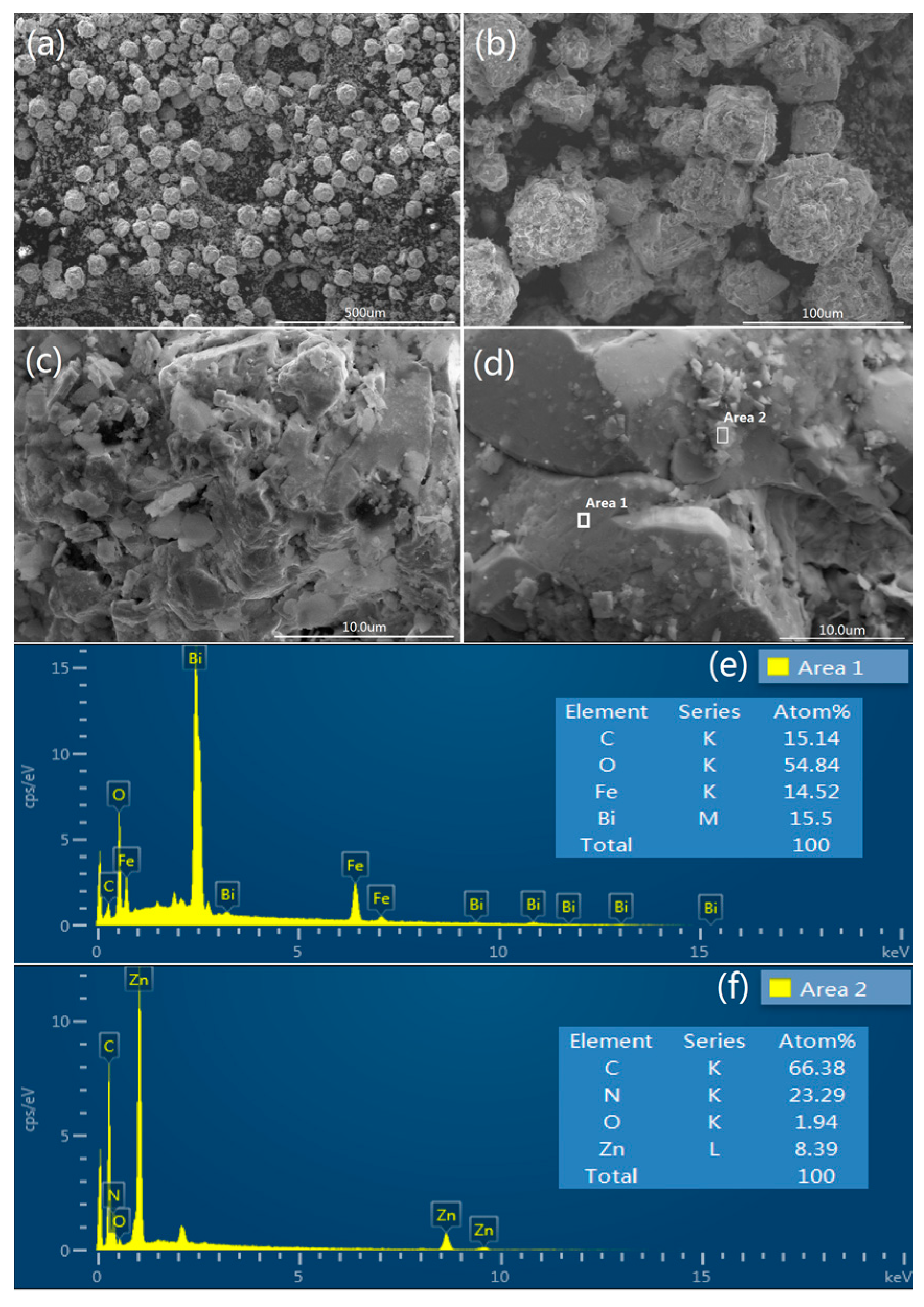
As seen in Figure 3a, spherical BiFeO3 particles are prepared by a hydrothermal process. Furthermore, no other morphologies can be detected, indicating a high uniformity of the product with the spherical morphology. The magnification of pure BiFeO3 shown in Figure 3b,c, spherical BiFeO3 are formed by the aggregation of many microcubes, which is consistent with a previous report [17]. It is clear that the surface morphology of BiFeO3 particle is very smooth in Figure 3c, which may be one of the reasons for the poor adsorption ability of the pure BiFeO3. Figure 3d shows the SEM images of prepared MOF (ZIF-8), pure MOF particles exhibit regular spherical morphology with an average diameter of 60~80 nm. SEM images of MOF/BiFeO3 composite in Figure 4a–d show lamellar MOF well disperse on the surface of BiFeO3 particle, the surface morphology of MOF/BiFeO3 composite changes obviously compared with the pure BiFeO3. The particle surface is coarse and porous, which allows the organic molecules to adsorb on the catalyst surface more easily and provide more reactive sites. These will contribute to the increase of the reactive sites, as well as the enhanced separation efficiency of the photogenerated electron–hole pairs.
Energy dispersive spectroscopy (EDS) analysis was carried out to identify the components of MOF/BiFeO3 composite. Figure 4e shows the diffraction peaks of Bi, Fe, O, and C elements corresponding to MOF/BiFeO3 composite observed in area 1 marked in Figure 4d. From the insert table in Figure 4e, it can be seen that the experimental atomic ratio of Bi, Fe, and O elements is close to the theoretical one of pure BiFeO3. A small amount of C element was detected and should be the result of a small amount of MOF adhering to the surface of the BiFeO3. Figure 4f shows the element contents of Area 2 marked in Figure 4d. The ratio of the elements in the insert table is close to the theoretical ZIF-8 and confirms that the lamellar MOF was successfully dispersed on the surface of BiFeO3 particle.
3.3. Enhanced Photocatalytic Performance
The photocatalytic activity of pure BiFeO3, pure MOF, and MOF/BiFeO3 composite photocatalysts under visible light irradiation were defined by measuring the photodegradation of MB aqueous solution. Where C0 and C are the initial and final concentration of MB, respectively. After 100 min of visible light irradiation, the maximum percentage of dye decomposition increased from 78% for pure BiFeO3 to 93% for MOF/BiFeO3 composite photocatalyst, as shown in Figure 5d. As a contrast, when pure MOF powders of the same amount were added to the photocatalytic experiments in Figure 5b, they absorbed about 48% of the dye in the dark environment for 30 min, and hardly underwent photocatalytic reactions under light irradiation. Degradation experiments showed that MOF has no photocatalytic capacity, despite excellent adsorption properties. After 40 min, dye adsorption reached the maximum, 50% of the MB dye was adsorbed. As shown in Figure 5a,c, compared with pure BiFeO3, the enhancement of MB photodegradation efficiency for MOF/BiFeO3 composite photocatalyst was mainly thanks to the promotion of adsorption capability. Approximately 32% MB was absorbed by composite photocatalyst in the first 30 min. However, for pure BiFeO3, only 1.5% was absorbed. The results of degradation experiments confirmed that the change of the surface morphology of the MOF/BiFeO3 composite photocatalyst can effectively enhance the photocatalytic degradation ability of MB molecules.
4. Photocatalytic Reaction Mechanism of MB over MOF/BiFeO3 Composite
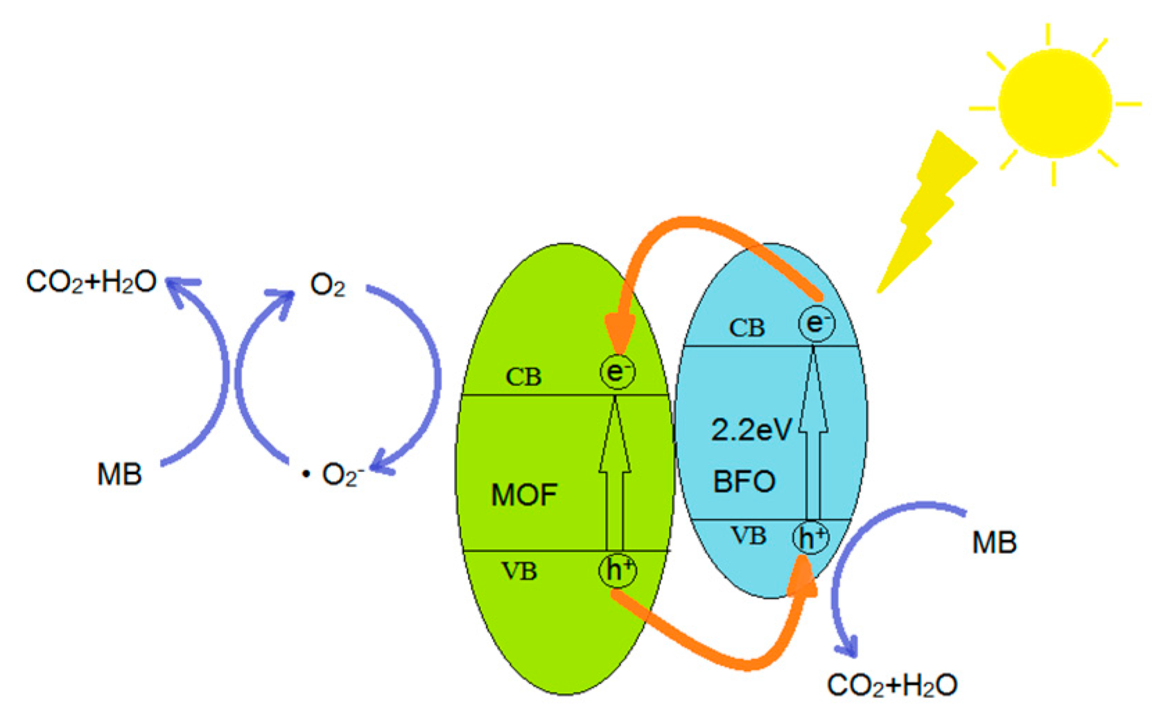
Under the irradiation of visible light, MOF/BiFeO3 composite photocatalyst can effectively absorb photons thanks to its narrow band gap. The photocatalytic process included a series of photochemical reactions through the first step of electrons (e−) and holes (h+) generation (Figure 6). The photogenerated holes in the valence band (VB) combine with absorbed H2O molecules to form strong oxidized hydroxyl radical (OH). In the presence of dissolved O2, the electrons in the conduction band (CB) can react with O2 to form superoxide radical (O2−) and hydrogen peroxide (H2O2). The OH radical has been deliberated to be the key active species accountable for the BiFeO3 photocatalytic process. The holes can also directly react with organic pollutants adsorbed on MOF/BiFeO3 composite and oxidize it to CO2 and H2O. The proposed mechanism (1–8) is as follows:
- Absorption of efficient photons by MOF/BiFeO3 composite photocatalyst
- Holes (h+) in valence band combine with absorbed H2O molecules which produces hydroxyl radical (·OH)
- Oxygen ionosorption
- Neutralization of by protons
- Transient hydrogen peroxide formation and dismutation of oxygen
- Decomposition of hydrogen peroxide
- Hydroxyl radical is further generated
- Oxidation of the MB molecules via successive attacks by ·OH and direct oxidation by reaction with holes
5. Conclusions
In this work, a novel MFO/BiFeO3 composite photocatalyst with high photocatalytic efficiency has been successfully synthesized through a hydrothermal method. Characterization of the composite photocatalyst confirmed that the MOF with strong adsorptive property was successfully immobilized on the BiFeO3 structure. The surface morphology of MOF/BiFeO3 composite changed obviously compared with the pure BiFeO3, a large surface area increased the number of active sites to promote the separation of e− and h+ pairs and improved the light absorption ability owing to multiple scattering effect. According to the degradation of MB, the introduction of MOF can enhance the adsorption capacity of BiFeO3 effectively, thereby enhancing the degradation of MB under visible light irradiation. This work proposed a new idea for efficient photocatalysts that can be used for purification of industrial waste.
Acknowledgments
This work was financially supported by National Science Foundation of China (Grant No. 51702218), National Natural Science Foundation of Guangdong, China (Grant Nos. 2016A030310054 and 2017A030310009), Youth Foundation of Shenzhen University (Grant No. 2016004), and Opening Funding of State Key Laboratory of New Ceramics and Fine Processing, School of Materials Science and Engineering of Tsinghua University (Grant No. KF201601).
Author Contributions
Yayun Li conceived and designed the experiments; Yunhui Si performed the experiments and wrote the manuscript; Jizhao Zou, Xinbo Xiong, Xierong Zeng and Ji Zhou helped perform the analysis with constructive discussions.
Conflicts of Interest
The authors declare no conflict of interest.
References
- Abe, R. Recent progress visible light irradiation on photocatalytic and photoelectrochemical water splitting under visible light irradiation. J. Photochem. Photobiol. C 2010, 11, 179–209. [Google Scholar] [CrossRef]
- Xu, J.H.; Wang, W.Z.; Sun, S.M.; Wang, L. Enhancing visible-light-induced photocatalytic activity by coupling with wide-band-gap semiconductor: A case study on Bi2WO6/TiO2. Appl. Catal. B Environ. 2012, 111, 126–132. [Google Scholar] [CrossRef]
- Devi, L.G.; Kavitha, R. A review on nonmetal ion doped titania for the photocatalytic degradation of organic pollutants under UV/solar light: Role of photogenerated charge carrier dynamics in enhancing the activity. Appl. Catal. B Environ. 2013, 140, 559–587. [Google Scholar] [CrossRef]
- Gao, F.; Chen, X.Y.; Yin, K.B.; Dong, S.; Ren, Z.F.; Yuan, F.; Yu, T.; Zou, Z.G.; Li, J.M. Visible-light photocatalytic properties of weak magnetic BiFeO3 nanoparticles. Adv. Mater. 2007, 19, 2889–2892. [Google Scholar] [CrossRef]
- Xue, Z.H.; Wang, T.; Chen, B.D.; Malkoske, T.; Yu, S.L.; Tang, Y.L. Degradation of tetracycline with BiFeO3 prepared by a simple hydrothermal method. Materials 2015, 8, 6360–6378. [Google Scholar] [CrossRef] [PubMed]
- Guo, R.Q.; Fang, L.; Dong, W.; Zheng, F.G.; Shen, M.G. Enhanced photocatalytic activity and ferromagnetism in Gd doped BiFeO3 nanoparticles. J. Phys. Chem. C 2010, 114, 21390–21396. [Google Scholar] [CrossRef]
- Zhang, X.; Wang, B.; Wang, X.; Xiao, X.; Wu, Z.W.; Zheng, J.; Ren, F.; Jiang, C.Z. Preparation of M@BiFeO3 nanocomposites (M = Ag, Au) bowl arrays with enhanced visible light photocatalytic activity. J. Am. Ceram. Soc. 2015, 98, 2255–2263. [Google Scholar] [CrossRef]
- Wang, B.; Wang, S.M.; Gong, L.X.; Zhou, Z.F. Structural, magnetic and photocatalytic properties of Sr2+-doped BiFeO3 nanoparticles based on an ultrasonic irradiation assisted self-combustion method. Ceram. Int. 2012, 38, 6643–6649. [Google Scholar] [CrossRef]
- Chauhan, S.; Kumar, M.; Chhoker, S.; Katyal, S.C.; Singh, H.; Jewariya, M.; Yadav, K.L. Multiferroic, magnetoelectric and optical properties of Mn doped BiFeO3 nanoparticles. Solid State Commun. 2012, 152, 525–529. [Google Scholar] [CrossRef]
- Hu, C.; Wang, Y.; Tang, H. Influence of adsorption on the photodegradation of various dyes using surface bond-conjugated TiO2 /SiO2 photocatalyst. Appl. Catal. B Environ. 2001, 35, 95–105. [Google Scholar]
- Oshikiri, M.; Boero, M.; Matsushita, A.; Ye, J. Water adsorption onto Y and V sites at the surface of the YVO4 photocatalyst and related electronic properties. J. Chem. Phys. 2009, 131, 034701. [Google Scholar] [CrossRef] [PubMed]
- Liu, X.; Guo, W.; Lu, Z.; Huo, P.; Yao, G.X.; Yan, Y.S. Surface molecular imprinting modified TiO2 photocatalyst for adsorption and photocatalytic degradation of salicylic acid. Fresen. Environ. Bull. 2014, 23, 1626–1634. [Google Scholar]
- Eddaoudi, M.; Kim, J.; Rosi, N.; Vodak, D.; Wachter, J.; O’Keeffe, M.; Yaghi, O.M. Systematic design of pore size and functionality in isoreticular MOFs and their application in methane storage. Science 2002, 295, 469–472. [Google Scholar] [CrossRef] [PubMed]
- Duren, T.; Sarkisov, L.; Yaghi, O.M.; Snurr, R.Q. Design of new materials for methane storage. Langmuir 2004, 20, 2683–2689. [Google Scholar] [CrossRef] [PubMed]
- Kuesgens, P.; Rose, M.; Senkovska, I.; Fröde, H.; Henschel, A.; Siegle, S.; Kaskel, S. Characterization of metal organic frameworks by water adsorption. Microporous Mesoporous Mater. 2009, 120, 325–330. [Google Scholar] [CrossRef]
- Wang, Y.G.; Xu, G.; Ren, Z.H.; Wei, X.; Weng, W.J.; Du, P.Y.; Shen, G.; Han, G.R. Mineralizer-assisted hydrothermal synthesis and characterization of BiFeO3 nanoparticles. J. Am. Ceram. Soc. 2007, 90, 2615–2617. [Google Scholar] [CrossRef]
- Li, S.; Lin, Y.H.; Zhang, B.P.; Nan, C.W.; Wang, Y. Photocatalytic and magnetic behaviors observed in nanostructured BiFeO3 particles. J. Appl. Phys. 2009, 105. [Google Scholar] [CrossRef]
Figure 1.
XRD patterns of (a) simulated MOF (ZIF-8) and (b) synthesized pure MOF (ZIF-8).
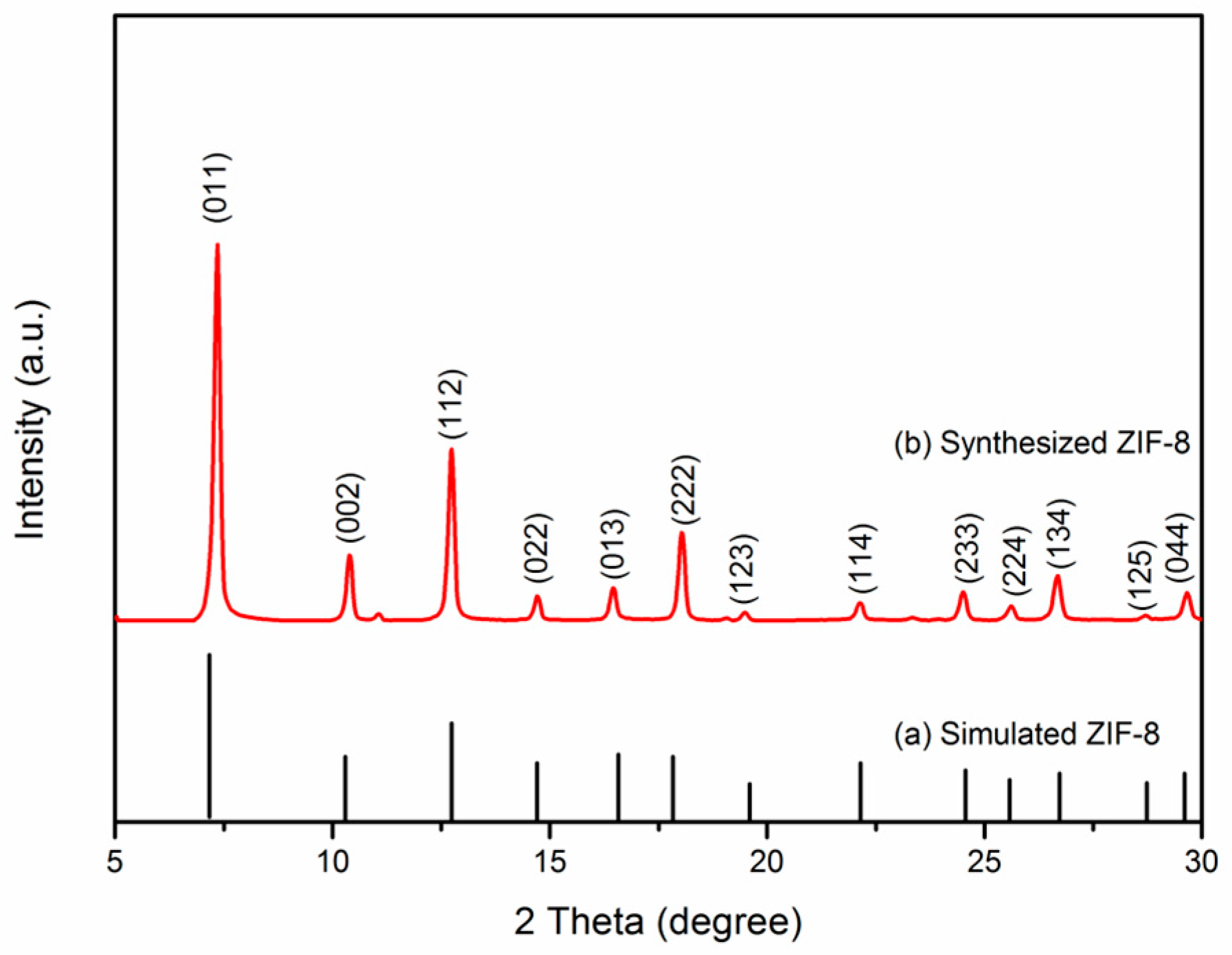
Figure 2.
XRD patterns of (a) pure BiFeO3 and (b) MOF/BiFeO3.
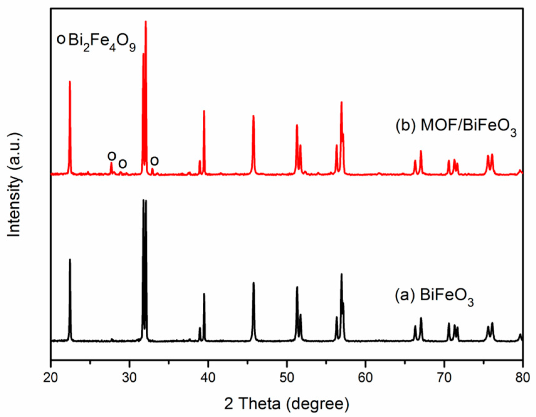
Figure 3.
SEM images of (a) pure BiFeO3; (b,c) the magnification of pure BiFeO3; and (d) pure MOF.
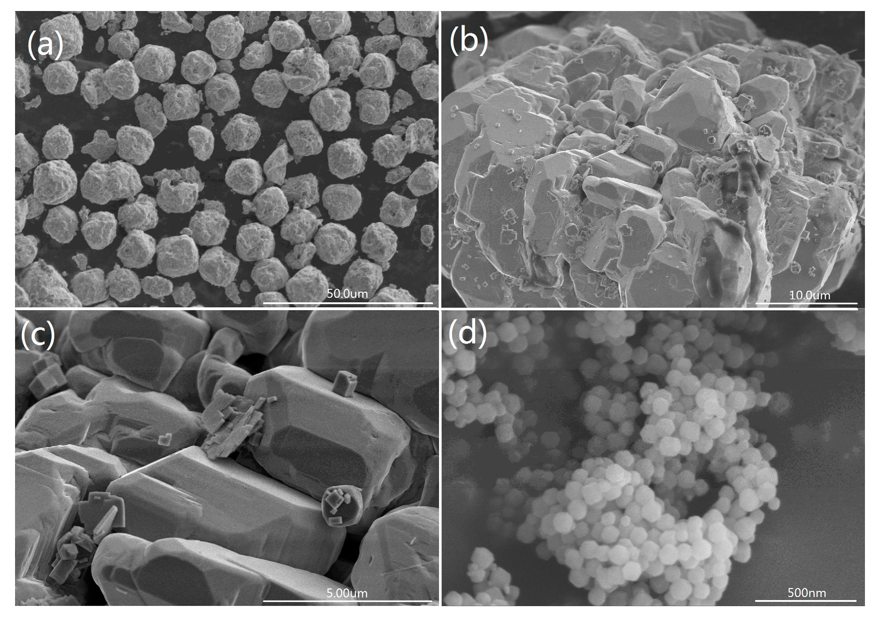
Figure 4.
SEM images of (a) MOF/BiFeO3 composite; (b–d) the magnification of MOF/BiFeO3 composite. EDS spectrum of MOF/BiFeO3 composite, (e) Area 1, (f) Area 2.
Figure 4.
SEM images of (a) MOF/BiFeO3 composite; (b–d) the magnification of MOF/BiFeO3 composite. EDS spectrum of MOF/BiFeO3 composite, (e) Area 1, (f) Area 2.

Figure 5.
Time dependent UV–vis spectrum changes of MB catalyzed by (a) pure BiFeO3; (b) pure MOF; and (c) MOF/BiFeO3 composite; (d) The photocatalytic degradation efficiencies of all samples to degrade MB.
Figure 5.
Time dependent UV–vis spectrum changes of MB catalyzed by (a) pure BiFeO3; (b) pure MOF; and (c) MOF/BiFeO3 composite; (d) The photocatalytic degradation efficiencies of all samples to degrade MB.
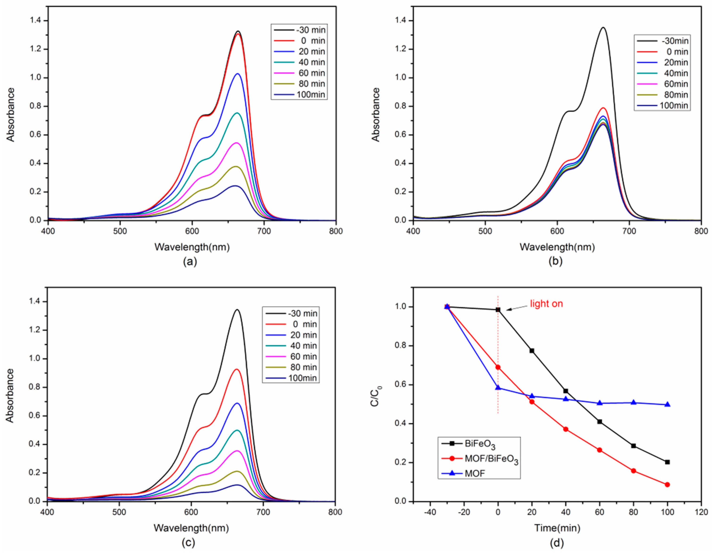
Figure 6.
Schematic diagram for photocatalytic degradation of MB.
© 2017 by the authors. Licensee MDPI, Basel, Switzerland. This article is an open access article distributed under the terms and conditions of the Creative Commons Attribution (CC BY) license (http://creativecommons.org/licenses/by/4.0/).
Share and Cite
MDPI and ACS Style
Si, Y.; Li, Y.; Zou, J.; Xiong, X.; Zeng, X.; Zhou, J. Photocatalytic Performance of a Novel MOF/BiFeO3 Composite. Materials 2017, 10, 1161. https://doi.org/10.3390/ma10101161
AMA Style
Si Y, Li Y, Zou J, Xiong X, Zeng X, Zhou J. Photocatalytic Performance of a Novel MOF/BiFeO3 Composite. Materials. 2017; 10(10):1161. https://doi.org/10.3390/ma10101161
Chicago/Turabian StyleSi, Yunhui, Yayun Li, Jizhao Zou, Xinbo Xiong, Xierong Zeng, and Ji Zhou. 2017. "Photocatalytic Performance of a Novel MOF/BiFeO3 Composite" Materials 10, no. 10: 1161. https://doi.org/10.3390/ma10101161
Note that from the first issue of 2016, this journal uses article numbers instead of page numbers. See further details here.