Production of High-Value Nanoparticles via Biogenic Processes Using Aquacultural and Horticultural Food Waste

1
Murdoch Applied Nanotechnology Research Group, Department of Physics, Energy Studies and Nanotechnology, School of Engineering and Energy, Murdoch University, Murdoch, Western Australia 6150, Australia
2
Department of Primary Industries and Regional Development, 3 Baron Hay Court, South Perth, Western Australia 6151, Australia
*
Author to whom correspondence should be addressed.
Materials 2017, 10(8), 852; https://doi.org/10.3390/ma10080852
Submission received: 19 May 2017
/
Revised: 13 July 2017
/
Accepted: 18 July 2017
/
Published: 25 July 2017
Abstract
:The quantities of organic waste produced globally by aquacultural and horticulture are extremely large and offer an attractive renewable source of biomolecules and bioactive compounds. The availability of such large and diverse sources of waste materials creates a unique opportunity to develop new recycling and food waste utilisation strategies. The aim of this review is to report the current status of research in the emerging field of producing high-value nanoparticles from food waste. Eco-friendly biogenic processes are quite rapid, and are usually carried out at normal room temperature and pressure. These alternative clean technologies do not rely on the use of the toxic chemicals and solvents commonly associated with traditional nanoparticle manufacturing processes. The relatively small number of research articles in the field have been surveyed and evaluated. Among the diversity of waste types, promising candidates and their ability to produce various high-value nanoparticles are discussed. Experimental parameters, nanoparticle characteristics and potential applications for nanoparticles in pharmaceuticals and biomedical applications are discussed. In spite of the advantages, there are a number of challenges, including nanoparticle reproducibility and understanding the formation mechanisms between different food waste products. Thus, there is considerable scope and opportunity for further research in this emerging field.
1. Introduction
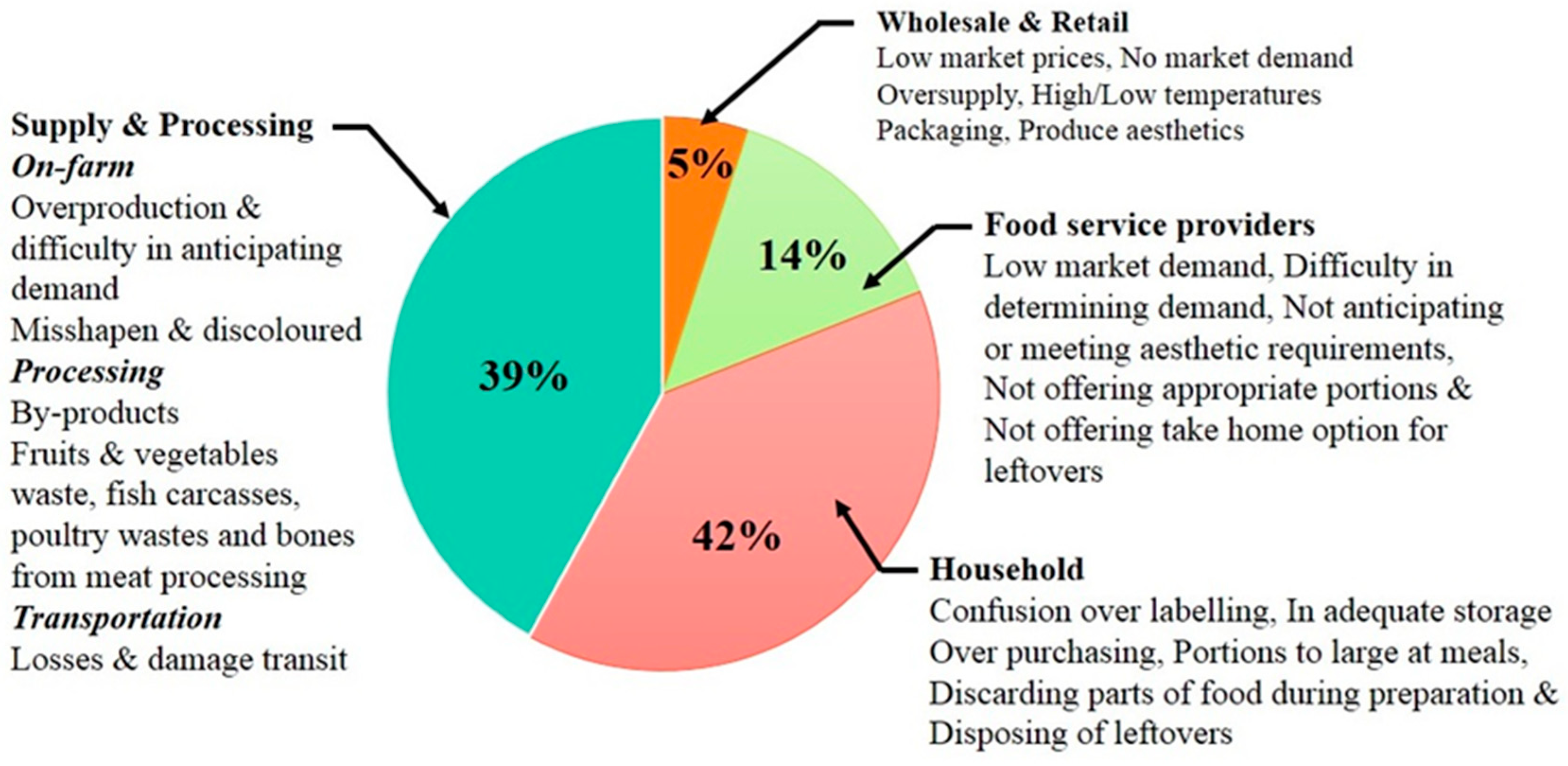
Two decades ago aquacultural and horticultural organic waste was not considered a major economic cost or resource loss to food processing industries [1]. However, recent public concerns about hunger, conservation, environmental degradation and the socioeconomic impact of food waste have accelerated research into developing strategies that can reduce food waste and promote effective waste utilisation methodologies [2]. In addition, global concerns regarding the limited natural resources currently available and the ability to effectively use these resources to feed the predicted population of 12.3 billion in 2100 has accelerated research into finding more effective resource utilisation and management strategies [3,4,5]. Therefore, efficient and cost = effective strategies are needed to reduce organic waste and develop better food waste utilisation practices that can assist in the overall management of the food supply chain [6,7]. In the context of this review, aquaculture relates to the industrial sector involved in farming marine and marine capture, while the horticultural sector principally focuses on the production, processing and retail sales of fruits and vegetables to consumers. From a practical point of view, food processing will always produce a certain amount of waste. However, the waste currently being produced during food processing is disproportionate compared to other processing industries. For example, Buzby et al. estimated in 2014 the total value of food losses for three large and expensive agricultural waste streams at the retail and consumer levels in the United States. The streams and their respective costs included grains (US$36.1 billion), vegetables (US$108.7 billion) and fruits (US$62.2 billion) [8]. Similarly, many other developed countries have waste trends in their respective agricultural sectors. For instance, in 2014 Segre and Falasconi estimated the Italian agricultural sector left around 17.7 million tonnes or 3.25% of its total produce in the ground [9]. Factors contributing to waste generation include produce sizing, aesthetic standards, produce quality, production surpluses and marketing as seen in Figure 1. Annually, in the USA these factors have resulted in around 2.7 million tonnes of fruits and vegetables not being harvested or sold [10]. In developing countries like India, between 18% and 40% of all fresh fruits and vegetables grown end up as waste. This large amount of waste equates to an annual financial loss of around US$71,481 million to the Indian agro-food industry [11]. From a European perspective, studies carried out in the Netherlands indicate the annual cost of food waste to be around €4.4 billion (US$4.9 billion) [12]. Surprisingly, many studies have shown that the agro-food sector can produce waste levels that are typically around 39% of total production, as seen in Figure 1 [13,14,15].
From the marine perspective, macroalgae or seaweed are plant-like organisms that are routinely washed up on beaches and shorelines in large quantities around the world. Edible seaweeds are consumed in many parts of the world since they are highly nutritional foods that are rich in proteins and a source material for many medicinal remedies [16,17,18]. Seaweed is a staple food in daily use throughout South-East Asia, and the health benefits derived from its consumption have resulted in numerous studies investigating the medicinal and pharmaceutical uses of seaweed [1,19,20]. Several studies have revealed that seaweed is rich in antioxidants, carbohydrates, carotenoids, polysaccharides, polyunsaturated fatty acids, proteins, vitamins and also contains numerous secondary metabolites [21,22,23]. These naturally occurring biological compounds have been used in traditional Chinese medicine for centuries [19], and recently several seaweed based extracts have been used to complement conventional treatments and supplement alternative therapies [24,25,26]. Furthermore, studies have reported anti-inflammatory and inhibitory properties being exhibited by several seaweed extracts [27,28]. These medicinal properties have also been found to reduce blood pressure levels [29], reduce the incidence of cardiovascular diseases [30], and suppress some forms of cancer [31,32]. In 2013, Tacon and Metian estimated that around 95.5% (12 million tonnes) of the total global production of marine plants is supplied by aquaculture (farming of marine plants), while the remaining 4.5% (0.44 million tonnes) is made up of marine capture [33]. The majority of aquaculture production (around 9 million tonnes) is destined for human consumption. The remaining tonnage undergoes processing to extract phycocolloids, a highly nutritious ingredient that is added to farm animal and aquaculture feeds [33,34]. While aquatic plants are an important commodity in the global food supply chain, there is very little data in the literature reporting levels of waste generation, waste management strategies and disposal protocols [15].
A number of utilisation strategies for processing waste fruits, vegetables and grains have been investigated [35,36,37,38]. The aim of these strategies is to maximise the value and practical benefits from food waste, which will ultimately reduce the amount of waste going to landfill [39]. Unfortunately, reviewing the literature reveals few studies that assess the economic benefits of the various waste management strategies operating at a commercial scale. However, the literature does clearly identify agricultural waste as an important source of chemicals, bioactive compounds and pharmaceuticals [40]. Importantly, the current high demand for pharmaceutical ingredients, enzymes, solvents and surfactants has resulted in many countries developing strategies for converting agricultural waste into products for chemical feedstock. This alternative, biologically based approach provides a large variety of chemical compounds from a wide range of renewable agricultural waste for recycling and processing by both chemical and pharmaceutical industries. For example, succinic acid can be obtained from crop waste like sugarcane, maize, rice, barley and potatoes [41]. Similarly, starch-based plastic can be produced from cassava, maize and wheat [42], fatty acids can be generated from coconut and oil palm [43], and polymers, lubricants, adhesives, solvents, and surfactants can be derived from rapeseed and sunflower [44]. These studies clearly demonstrate that materials commonly thought of as waste are rich in biologically active compounds that can be used to produce high-value chemical and pharmaceutical products. Furthermore, recent studies have also shown that both aquacultural horticultural waste can be used as renewable feedstock for the manufacture of high-value nanoparticles [45,46,47]. Food waste and nanoparticle synthesis sounds like an unlikely combination, but recent investigations in the literature have shown that naturally occurring biomolecules present in waste have the potential to produce nanoparticles (particles less than 100 nm) with unique medicinal and pharmaceutical properties [48,49,50].
Both aquacultural and horticultural food waste contain beneficial biomolecules and compounds that can play an active role in reducing precursor metal ions in aqueous solutions to form nanoparticles. The biomolecules also act as modelling agents that direct particle growth in particular orientations, while other biomolecules act as capping agents to prevent nanoparticle agglomeration [45,51]. Another interesting feature of nanoparticles synthesised from food waste is the potential to deliver reliable, sustainable and green chemistry-based technologies that are eco-friendly, thus reducing the human health and environmental degradation risks normally associated with the use of toxic solvents and chemicals during conventional physical and chemical manufacturing protocols [52]. The considerable interest in nanoparticles shown by the scientific community is due to their unique physiochemical properties and their application in a number of fields such as pharmaceuticals and biomedicine. The extremely small size and large surface area to volume ratio are two material features that give nanoparticles their new or enhanced properties compared to conventional bulk forms of the same substance [53]. For example, gold (Au) nanoparticles are already widely used in medicine for diagnostics [54,55,56], targeted delivery of pharmaceuticals [57,58,59,60] and tumour destruction via hyperthermia [61]. While silver (Ag) nanoparticles display a broad spectrum antimicrobial activity against many human and animal pathogens [62,63,64,65] and as a result are used as antimicrobial agents in a wide range of commercially available medical and consumer products [66,67,68], both platinum (Pt) and palladium (Pd) nanoparticles have been used as catalysts [69,70,71]. Furthermore, metal oxide nanoparticles such as copper oxide (Cu2O, CuO) and zinc oxide (ZnO) have displayed antimicrobial activity [72,73] and because of this ZnO has been used in a variety of food packaging applications [74]. Moreover, super-paramagnetic ferric oxide (Fe3O4) nanoparticles have the potential to be used in a wide variety of biomedical applications such as magnetic resonance imaging, hyperthermia treatments and as a carrier for anti-cancer drugs [75,76,77]. The utilisation of aquacultural and horticultural wastes for the biogenic synthesis of high-value products such as metal and metal oxide nanoparticles is a fairly new field of research. Accordingly, only a relative few articles have appeared in the literature reporting the use of various aquacultural and horticultural waste to produce nanoparticles using eco-friendly green chemistry-based technologies [78,79,80,81]. The present work summarises current research in this relatively new research field and discusses the various experimental parameters that govern nanoparticle formation and growth during biogenic synthesis. The remainder of the review discusses the potential applications of aquacultural and horticultural food-waste produced nanoparticles in fields such as pharmaceuticals and biomedicine.
2. Biogenic Synthesis of Nanoparticles Using Aquacultural and Horticultural Food Waste
The biogenic synthesis of a variety of metal and metal oxide nanoparticles using aquacultural and horticultural food waste can be considered an alternative, eco-friendly and viable green chemistry-based route. In recent years there have been numerous studies reporting the synthesis of nanoparticles using a diverse array of plants sources. However, relatively few studies have reported using aquacultural and horticultural food waste to produce value-added products such as nanoparticles. Like the plant sources reported in the literature, aquacultural and horticultural waste also contains a vast array of biomolecules such as alkaloids, amino acids, enzymes, phenolics, proteins, polysaccharides, saponins, tannins, terpinoids and vitamins that can all be used to assist in the creation of nanoparticles [82]. The biogenic synthesis of nanoparticles is a bottom-up approach, during which atoms and molecules combine to form precursor building blocks that subsequently self-assemble [83]. Reviewing the various studies reported in the literature, it is evident that plant-based synthesis procedures have successfully produced a variety of noble metal nanoparticles such as gold, silver, platinum and palladium [45]. A few studies have also reported the formation of noble metal nanoparticles from extracts taken from aquacultural and horticultural food waste. For instance, Dubey et al. have reported the formation of 16-nm Ag spheres and 11-nm Au nano-triangles when Tanacetum vulgare (tansy fruit) extract was allowed to react with aqueous solutions of AuCl4− ions and Ag+ ions respectively [84]. In addition, several other food waste extracts such as Pyrus sp. (pear fruit) and Mangifera indica (mango peel) have demonstrated their ability to reduce Au (III) ions to form Au nanoparticles [49,85]. Citrus sinensis (orange peel) and Ananas comosus (pineapple) extracts have shown the ability to reduce Ag+ ions in aqueous solutions to form Ag nanoparticles [78,86]. Likewise, Pt and Pd nanoparticles have been synthesised using Musa paradisiac (banana peel), tea and coffee extracts, and lignin [71,87,88].
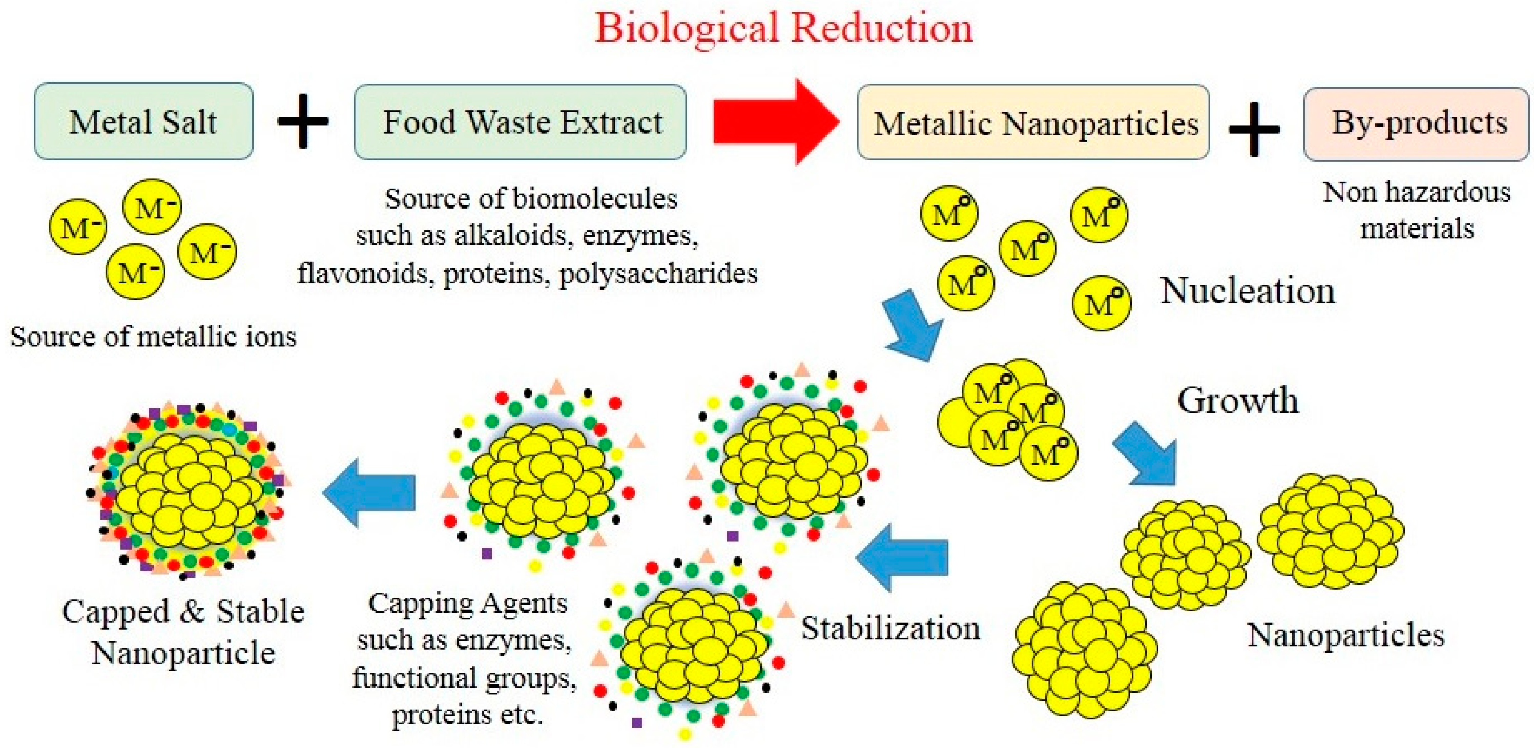
The advantage of using aquacultural and horticultural food waste is that it is readily available. This makes them a renewable feedstock that creates an alternative waste utilisation strategy for manufacturing high-value metal and metal oxide nanoparticles. Importantly, food waste offers a green, chemistry-based route that is rapid, cost-effective and eco-friendly. Metal nanoparticle production is a straightforward room temperature process that begins by mixing an aqueous metal salt solution with an aqueous solution containing a food waste extract as seen in Figure 2. Biogenic reduction starts immediately, and as the reduction process continues there is a distinctive colour change in the reaction mixture, indicating nanoparticle formation. For instance, a recent study by Kaviya et al. reported the formation of Ag nanoparticles when Citrus sinensis (orange) peel extract was used as the reducing agent. Reduction of Ag nanoparticles occurred within 20 min and their formation was clearly indicated by the reaction mixture changing colour from colourless to a yellowish-brown. Subsequent characterisation of the samples revealed nanoparticle morphology was spherical and their size was heavily dependent on reaction mixture temperature. At 25 °C the mean particle size was 35 ± 2 nm, while at 60 °C size was 10 ± 1 nm [89]. Studies indicate the fundamental nanoparticle formation mechanism created during biogenic synthesis begins with metal ions in solution transforming from their mono- or divalent oxidation states to from zero-valent states as seen in Figure 3. Biomolecules present in the food waste extract initiates metal ion reduction and then promotes nucleation [90,91,92]. Progressively, smaller neighbouring particles start assembling on their low energy faces that ultimately results in thermodynamically stable nanoparticle formation. During this stage food waste biomolecules also act as natural surfactants (capping agents), which influence the orientation and assembly of the smaller particles during subsequent growth as schematically presented in Figure 3. The modelling action produced by surfactants during biological synthesis explains why growth occurs along preferential planes [93]. These preferential growth planes result in morphologies such as spheres, cubes, triangles, hexagons, pentagons and wires being formed [82,94]. The number of experimental parameters known to govern the nanoparticle formation mechanism include: (1) the nature of the food waste extract; (2) concentration of food waste in the reaction mixture; (3) metal ion concentration in the source solution; (4) reaction mixture pH; (5) reaction mixture temperature; and (6) contact time. All these parameters are important and can directly influence nanoparticle formation and subsequently their physiochemical properties [95].
An important issue that has arisen in recent years is the possible adverse health effects produced by nanometre-scale materials. Studies have shown the physiochemical properties of nanometre scale materials of the same material can vary due to differences in parameters such as particle size, aggregation, chemical reactivity, concentration, dispersion and morphology [96,97]. Even small changes in these parameters can significantly influence nanoparticle behaviour and their subsequent interactions in particular environments. Generally, toxicity issues arise from the deposition of hazardous chemicals and solvents on the surface of the nanoparticles during many conventional physical and chemical manufacturing processes. The removal of these hazardous materials is extremely difficult and their presence can induce significant toxicological and inflammatory responses if used in biomedical applications [98]. Moreover, naked nanoparticles do not exist very long in the physiological environment of the human body and biomolecules such as proteins rapidly attach to their surface, forming a corona [99]. Therefore, synthesising biocompatible nanoparticles via food waste extracts offers a greener, less toxic and eco-friendly approach that avoids the use of toxic chemicals and solvents commonly used in conventional manufacturing. Furthermore, aquacultural and horticultural food waste is a renewable and relatively inexpensive feedstock. However, before aquacultural and horticultural food waste can be used commercially to manufacture high-value nanoparticles there needs to be more research into resolving a number of shortcomings. These shortcomings include: (1) developing an all-inclusive nanoparticle formation mechanism; (2) investigate the influence of experimental parameters on nanoparticle size, shape and dispersion, and (3) refine the biosynthesis process to improve reproducibility [100,101,102]. In terms of commercialisation: (1) develop technologies that overcome the limitations of scaling up the biosynthesis process; (2) developing a continuous supply route for suitable aquacultural and horticultural food waste; and (3) optimise the waste management chain to fully utilise the biomolecules and bioactive chemicals present in aquacultural and horticultural food waste [1,13,103].
3. Types of Nanoparticles Produced by Aquacultural and Horticultural Food Waste
The biogenic synthesis of metal and metal oxide nanoparticles via aquacultural and horticultural food waste is a new and emerging field of study. Food waste has the potential to produce a wide range of particle sizes and shapes using green, chemistry-based techniques [82,103,104]. At present, only a relatively small number of articles have appeared in the literature reporting the use of aquacultural and horticultural food waste being used to synthesise nanoparticles. The following sections summarise and discuss the current state of research as reported in the literature. A selection of recent studies reporting the biogenic synthesis of metal and metal oxide nanoparticles using various horticultural food waste products are summarised and presented in Table 1. Table 2 presents a selection of metal and metal oxide nanoparticles synthesised using a variety of marine alga (seaweeds) commonly produced by aquaculture.
3.1. Silver (Ag) Nanoparticles
The diversity of aquacultural and horticultural food waste has led a number of researchers to investigate their potential use in synthesising a variety of metal nanoparticles. In particular, Ag nanoparticles have paved a way into exploring this new field due to the exceptional antimicrobial properties displayed by Ag compounds. For centuries, Ag has been used as an antimicrobial agent in numerous medicinal preparations. In recent years Ag nanoparticles, with their unique physiochemical and enhanced antimicrobial properties, have been incorporated into a variety of biomedical protocols and pharmaceuticals [105,106]. The reason for these superior antimicrobial properties comes from the Ag nanoparticles ability to cause cell membrane damage and toxicological damage to cellular DNA [107,108]. Recently, several studies have reported the reduction of Ag+ ions in aqueous solutions containing aquacultural and horticultural food waste. For example, the formation of spherical Ag nanoparticles ranging in size from 5 to 35 nm was reported by Ahmad and Sharma using an extract taken from Ananas comosus (Pineapple) [79]. Similarly, spherical-shaped crystalline Ag nanoparticles ranging in size from 3 to 12 nm were synthesised by Konwarha et al. using an extract taken from Citrus sinensis (Orange) peel [86] and spherical Ag nanoparticles ranging in size from 5 to 20 nm have been produced using an extract taken from Citrus unshiu (Mandarin) peel by Basavegowda et al. [109]. In addition, Njagi et al. were able to use an aqueous solution containing Sorghum spp. (bran powder) extract to biologically synthesis spherical iron (Fe) and Ag nanoparticles that were typically around 10 nm in size [110]. Dubey et al. were able to synthesis both Ag and Au nanoparticles using Tanacetum vulgare (tansy fruit) [84]. Ag nanoparticles were spherical with a mean size of 16 nm and the Au nanoparticles were triangular plates that were typically around 11 nm in size. Likewise, Ankamwar et al. were able to use an Emblica officinalis (Indian Gooseberry) extract to synthesis both Ag and Au nanoparticles ranging in size from 10 nm to 25 nm [111]. Other researchers have investigated the use of marine algae, which are rich in polysaccharides, and other bioactive materials that can be used for synthesising nanoparticles. For example, Kannan et al. have reported the synthesis of Ag nanoparticles using Codium capitatum (seaweed). Their study revealed that two types of morphologies could be produced, namely spherical and cubic. Both morphologies ranged in size from 3 to 44 nm, with a mean particle size of 30 nm [112]. In a similar study by Castro et al., a green alga Spyrogira insignis was found to produce spherical Ag nanoparticles with a mean particle size of 30 nm [113]. Raeshkumar et al. have reported producing spherical Ag nanoparticles with a mean particle size of 14 nm using a brown seaweed, Padina tetrastromatica [114].
3.2. Gold (Au) Nanoparticles
Au nanoparticles are a very attractive material due to their wide application in fields such as catalytics, biomedicine, biosensors, pharmaceuticals, imaging and diagnostics [54,55,115,116,117]. Several recent studies have reported the reduction of aqueous chloroaurate solutions using a variety aquacultural and horticultural food waste products. For example, Krishnaswamy et al. have reported the formation of spherical Au nanoparticles ranging in size from 20 nm to 25 nm after using waste grape skins, stalks and seeds as the reducing agents [80]. Similarly, Ghodake et al. were able to synthesise triangular and hexagonal crystalline gold nanoparticles ranging in size from 200 nm to 500 nm using an extract taken from Pyrus sp. (pear) [85]. Likewise, Yang et al. have reported the formation of Au nanoparticles, ranging in size from 6.03 ± 2.77 nm to 18.01 ± 3.67 nm, using an extract taken from Mangifera indica (mango) peel [49]. Recently, Sharma et al. have produced Au nanoparticles using freshwater green alga (Prasiola crispa) and red alga (Lemanea fluviatilis) [118,119]. Studies have also shown that the type and concentration of biomolecules present in food waste can influence nanoparticle formation and their subsequent stability. A study by Huang et al. found that varying the concentration of sundried Cinnamomum camphora leaf extract or increasing the precursor chloroauric acid concentration in the reaction mixture resulted in nanoparticle shape changes (i.e., triangular to spherical) [120]. Similarly, Chandran et al. have reported varying the concentration of Aloe vera leaf extract in reaction mixtures containing chloroaurate ions to regulate nanoparticle size. The varying concentration not only regulated the size range between 50 and 350 nm, but also influenced the ratio of spherical to triangular nanoparticles produced [121]. Narayanan et al. were able to synthesis varying ratios of decahedral, hexagonal, triangular and spherical Au nanoparticles by changing the concentration of Coleus amboinicu leaf extract in the reaction mixture [122]. Furthermore, Ahmada et al. have reported that a low precursor Au concentration (1.53 mM) in a reaction mixture containing aqueous Elaise guineensis (oil palm) leaf extract produced spherical nanoparticles with a mean diameter of 27.89 ± 14.59 nm. However, a larger precursor Au concentration (4.055 mM) produced spherical, triangular, pentagonal and hexagonal nanoparticles with a mean particle diameter of 22.88 ± 8.21 nm [123]. The study also revealed a multilayer coating composed of carboxylic and phenolic compounds, which prevented the nanoparticles from agglomerating, thus indicating the importance of sufficient extract concentration to provide the necessary biomolecules needed for nanoparticle stabilization.
3.3. Other Types of Nanoparticles
In recent years a few studies have reported the formation of several other types of nanoparticles produced from aquacultural and horticultural food waste. For example, Bankar et al. have produced palladium (Pd) nanoparticles using an aqueous solution containing an extract taken from Musa paradisiac (banana) peel. The resulting nanoparticles were crystalline and irregular in shape, and had a mean size of 50 nm [88]. Similarly, Lakshmipathy et al. used an extract taken from watermelon rind to form Pd nanoparticles with a mean particle size of 96 nm [124]. Coccia et al. were able to produce both Pd and platinum (Pt) nanoparticles using lignin [71]. In addition, Nadagouda and Varma were able to use commercially available tea and coffee waste extracts to produce both Ag and Pd nanoparticles. Their study found that both types of nanoparticles were spherical and ranged in size from 5 nm to 100 nm, with the majority of the particles falling within the 20 to 60 nm range [87]. Furthermore, a recent study by Lunge et al. found that waste tea extracts could produce magnetic ferric oxide (Fe3O4) nanoparticles. The nanoparticles formed ranged in size from 5 to 25 nm and consisted of both cubes and pyramids [125]. Moreover, a study by Mahdavi et al. that used extracts from brown seaweed (Sargassum muticum) could be used to form ferric oxide (Fe3O4) nanoparticles. Their study also identified the water-soluble polysaccharide cell walls that contained amino, carboxyl and hydroxyl functional groups as the biomolecules that acted as both reducing and capping agents. The resulting crystalline nanoparticles were cubic in nature and had a mean particle size of 18 ± 4 nm [126]. Also using a brown seaweed extract (Bifurcaria bifurcata), Abboud et al. were able to produce both cuprous oxide (Cu2O) and cupric oxide nanoparticles (CuO). Nanoparticle morphology was predominantly spherical, with particles ranging in size from 5 to 45 nm [72]. Similarly, Khanehzaei et al. were able to produce spherical Cu-cored Cu2O nanoparticles with a mean particle size of 53 nm using extracts taken from red seaweed (Kappaphycus alvarezii) [127]. Other types of metal oxide nanoparticles produced from extracts taken from Citrus sinensis (orange) peels and Musa paradisiac (banana) peels include magnesium oxide (MgO) and manganese (II, III) oxide (Mn3O4) respectively. For example, Rao et al. were able to produce spherical MgO nanoparticles with a mean particle size of 29 nm using extracts taken from Citrus sinensis (orange) peel [128]. In a similar study, Yan et al. were able to use Musa paradisiac (banana) peel extract to form spherical Mn3O4 nanoparticles ranging in size from 20 nm to 50 nm. Their study also identified the super-capacitive properties of the nanoparticles and their potential use in high-stability Mn3O4-based electrodes [129].
4. Applications and Future Perspectives
Metal and metal oxide nanoparticles produced using conventional physical and chemical manufacturing processes, as mentioned earlier, have been used in variety of antimicrobials products, biosensors, photo-catalysts, pharmaceuticals, cancer therapy and food packaging applications. Aquacultural and horticultural food waste extracts have the potential to become another avenue for the green synthesis of various types of metal and metal oxide nanoparticles as seen in Table 1 and Table 2. With the diversity of food waste available and the ability to influence the reaction parameters during synthesis, there are many opportunities to produce novel metal-based nanoparticles with unique properties. However, despite the great interest shown in the biogenic synthesis of nanoparticles using aquacultural and horticultural food waste, there have only been a few practical applications reported. In spite of this, studying the properties of conventionally manufactured nanoparticles and comparing their properties with those produced using aquacultural and horticultural food waste should provide an indication of property differences and potential applications. The property differences arise from the use of hazardous chemicals and solvents (capping agents and surfactants) commonly used in many conventional manufacturing processes and their non-use in green biogenic synthesis procedures. Thus, biosynthesised nanoparticles should have a wider range of biomedical applications since they are free from relatively hazardous chemicals and solvents [97,98].
Noble metal nanoparticles, in particular Ag and Au, produced using a variety of techniques, are currently used in a wide range of detection, imaging, diagnostics and therapeutic applications [146,147,148]. In particular, using Au nanoparticles to promote DNA damage in cancerous cells through a variety of targeted therapeutic treatments is currently being applied in several therapeutic procedures [115,149,150]. Furthermore, both Ag and Au nanoparticles have a broad spectrum of antimicrobial properties that have been tested against a several human and animal pathogens [151,152,153]. With regards to horticultural food waste extracts, Jain et al. have used Carica papaya (pawpaw) to biosynthesise Ag nanoparticles and then evaluate their antimicrobial properties against a number of pathogens [131]. Similarly, Shanmugam et al. have biosynthesised Ag nanoparticles using the marine seaweed Sargassum wightii and then verified the nanoparticles’ antimicrobial activity against a number of human pathogens [154]. Ag nanoparticles produced in both food waste studies were found to have similar antimicrobial properties to those synthesised by conventional manufacturing processes. This is of particular importance since Ag nanoparticles produced by traditional methods are currently being used as antimicrobial agents in a wide range of medical and consumer products [66,67,68,155]. Thus, biosynthesising Ag nanoparticles from food waste is an alternative technique that can produce nanoparticles with properties similar to those produced by more conventional methods. Similarly, Ag and Pd nanoparticles were produced using waste tea and coffee extracts by Nadagouda and Varma [87]. During synthesis the individual metal salts were first reduced and then capped by polyphenols present in the respective extracts. Interestingly, recent studies have also revealed that Pd nanoparticles produced using food waste can be used as catalysts. The catalytic behaviour of produced Pd nanoparticles has been studied using oxidation and reduction reactions [71,124]. Furthermore, a recent study by Petla et al. identified and quantified the catalytic properties of Pd nanoparticles produced using soya leaf extract by successfully degrading several azo dyes [156].
The biogenic synthesis of less noble metals and metal oxide nanoparticles have also attracted the interest of several researchers. At present there are serious concerns about antibiotic resistance, complex manufacturing protocols, environmental pollution, and the high manufacturing costs associated with conventional pharmaceutical preparations [157]. Therefore, in recent years there has been a search for natural antimicrobial agents that can overcome the shortcomings of currently available antimicrobial pharmaceuticals. For example, copper (Cu) and Cu oxide nanoparticles are natural antimicrobial agents that have been biosynthesised using a variety of plant-derived extracts. A study by Lee et al. found Cu nanoparticles produced by a leaf extract from Magnolia Kobus formed spherical particles ranging in size from 40 to 100 nm and displayed antimicrobial properties towards Escherichia coli [158]. From a marine perspective, Abboud et al. have biosynthesised copper oxide nanoparticles using a brown algal extract (Bifurcaria bifurcata). The resulting nanoparticles were spherical, ranged in size from 5 to 45 nm and displayed good antibacterial properties towards both Enterobacter aerogenes and Staphylococcus aureus [72]. Furthermore, Nagarajan and Kuppusamy have biosynthesised zinc oxide (ZnO) nanoparticles from a brown seaweed extract (Sargassum myriocystum) that displayed antimicrobial properties against a number of bacteria and fungi [159]. However, at present no studies have appeared in the literature reporting the used of food waste to biosynthesise ZnO nanoparticles. The abovementioned antimicrobial studies demonstrate that biogenic synthesis can be used as an alternative method for producing nanoparticles with the potential to assist in the management of infectious diseases caused by bacteria.
However, the biosynthesis of metal and metal oxide nanoparticles using aquacultural and horticultural food waste extracts is still in its infancy. Consequently, only a few oxides have been studied and reported. Those recently reported include ferric oxide (Fe3O4), magnesium oxide (MgO) and manganese (II, III) oxide (Mn3O4). As mentioned earlier, Lunge et al. have successfully used waste tea extracts to produce (Fe3O4) nanoparticles. Their studies found that these cubic and pyramidal nanoparticles could be effectively used to remove arsenic metal ions from aqueous solutions [125]. Other studies reporting the biosynthesis of (Fe3O4) nanoparticles have involved using brown seaweed (Sargassum muticum) extracts [126,160]. Namvar et al. have used brown seaweed to produce (Fe3O4) nanoparticles that were subsequently assessed via in vitro cytotoxicity and anticancer testing for their activity against human cell lines for leukaemia, breast cancer, cervical cancer and liver cancer [160]. The results of their studies revealed that the accumulation of (Fe3O4) nanoparticles in treated cells tended to promote cell apoptosis, which suggested that an alternative cancer treatment protocol is possible using these nanoparticles. In terms of other oxides, Ganapathi Rao et al. have reported the biogenic synthesis of MgO nanoparticles from orange fruit waste [128] and Yan et al. have reported producing Mn3O4 nanoparticles with super-capacitive properties using Musa paradisiac (banana) peel extract [129].
The present review has demonstrated that the use of aquacultural and horticultural food waste extracts can be used to manufacture metal and metal oxide nanoparticles. Currently, this new and emerging field has produced relatively few study articles. The review has also shown that this new field needs to be fully explored and the development of eco-friendly and efficient green chemistry-based methods for recycling and utilising food waste needs to take place. Both aquacultural and horticultural food waste are produced in extremely large quantities around the world and offer an attractive and renewable source of biomolecules and bioactive compounds. The availability of such a large and diverse source of food waste creates a unique opportunity to develop new recycling and food waste utilisation strategies. One such value-adding strategy reported in the present work is the use of food waste to produce high-value metal and metal oxide nanoparticles. These high-value nanoparticles have the potential to be used in a wide range of current and future medical and pharmaceutical products.
5. Conclusions
The recycling and utilisation of food waste produced from aquacultural and horticultural industries has several advantages. The first is that the extremely large quantities of waste generated globally offer an attractive and renewable source of biomolecules and bioactive compounds. Processing food waste using chemistry-based strategies is eco-friendly, can significantly reduce the amount going to landfill, and creates a wide range of value-added products. One such strategy, presented in this review, is the biogenic synthesis of metal and metal oxide nanoparticles using aquacultural and horticultural food waste. The green biogenic synthesis route has several advantages over more traditional nanoparticle manufacturing processes. The procedure is straightforward, can be scaled up, and is eco-friendly. The most attractive feature of the biogenic synthesis route is that it can produce nanoparticles free from the toxic non-biodegradable commercial chemicals and surfactants that are commonly used in many conventional physical and chemical manufacturing processes. Studies reported and discussed in the present work have demonstrated that aquacultural and horticultural food waste extracts can be used to manufacture a wide variety of nanoparticles free from toxic chemicals and surfactants, thus, making these nanoparticles ideal candidates for pharmaceutical products and biomedical applications. This is unlike traditional nanoparticle manufacturing processes, which often leave detrimental surface coatings (solvents and surfactants) that severely limit their use in many therapeutic applications. For example, Ag and Au nanoparticles produced by food waste were reported to have a broad spectrum of antimicrobial properties against several human and animal pathogens. Furthermore, food waste has been reported by several researchers to produce Cu, Cu oxides, and ZnO nanoparticles. These nanoparticles were all found to display antimicrobial properties against several bacteria and fungi, while Fe3O4 nanoparticles synthesised using seaweed have displayed anticancer activity against a number of human cell lines. However, despite the many advantages of using food waste extracts to generate nanoparticles, there are a number of unresolved issues that need to be elucidated—for example, the variation of nanoparticle size and shape reproducibility when using different food waste extracts. Furthermore, there are also issues involving variations in formation mechanisms between different food waste products that need to be resolved. At present, only a few studies have reported using food waste to generate nanoparticles. The limited number of studies creates considerable scope and opportunity for further research in this emerging field.
Acknowledgments
Purabi Ghosh would like to acknowledge Murdoch University for providing her with a scholarship to undertake the present study. This work was partly supported by the Horticulture Innovation Australia Project Al14003 and Derek Fawcett would like to thank Horticulture Innovation Australia for the research fellowship.
Author Contributions
All authors have substantially contributed to the preparation of the manuscript.
Conflicts of Interest
The authors declare no conflict of interest.
References
- Baiano, A. Recovery of Biomolecules from Food Wastes—A Review. Molecules 2014, 19, 14821–14842. [Google Scholar] [CrossRef] [PubMed]
- Kantor, L.S.; Lipton, K.; Manchester, A.; Oliveira, V. Estimating and addressing America’s food losses. Food Rev. 1997, 20, 2–12. [Google Scholar]
- Beretta, C.; Stoessel, F.; Baier, U.; Hellweg, S. Quantifying food losses and the potential for reduction in Switzerland. Waste Manag. 2013, 33, 764–773. [Google Scholar] [CrossRef] [PubMed]
- Gerland, P.; Raftery, A.E.; Sevcikova, H.; Li, N.; Gu, D.; Spoorenberg, T.; Alkema, L.; Dosdick, B.K.; Chunn, J.; Lalic, N.; et al. World population stabilization unlikely this century. Science 2014, 346, 234–237. [Google Scholar] [CrossRef] [PubMed]
- Alesci, A.; Cicero, N.; Salvo, A.; Palombieri, D.; Zaccone, D.; Dugo, G.; Bruno, M.; Vadala, R.; Lauriano, E.R.; Perggolizzi, S. Extracts deriving from olive mill wastewater and their effects on the liver of goldfish Carassius auratus with hypercholesterolemic diet. Nat. Prod. Res. 2014, 28, 1343–1349. [Google Scholar] [CrossRef] [PubMed]
- Gustavsson, J.; Cederberg, C.; Sonesson, U.; van Otterdijk, R.; Meybeck, A. Global Food Losses and Food Waste: Extent, Causes and Prevention; Food and Agriculture Organisation of the United Nations: Rome, Italy, 2011. [Google Scholar]
- Ghosh, P.R.; Sharma, S.B.; Haigh, Y.T.; Barbara Evers, A.L.; Ho, G. An overview of food loss and waste: Why does it matter? COSMOS 2015, 11, 1–15. [Google Scholar] [CrossRef]
- Buzby, J.C.; Wells, H.F.; Hyman, J. The Estimated Amount, Value, and Calories of Postharvest Food Losses at the Retail and Consumer Levels in the United States; EIB-121; U.S. Department of Agriculture, Economic Research Service: Washington, DC, USA, 2014. [Google Scholar]
- Segre, A.; Falasconi, L. Save Food: Global Initiative on Food Loss and Waste Reduction, Background Paper on the Economics of Food Loss and Waste; Food and Agriculture Organization of the United Nations: Rome, Italy, 2014. [Google Scholar]
- Hirsch, J.; Harmanci, R. Food Waste: The Next Food Revolution. 2013. Available online: http://modernfarmer.com/2013/09/next-food-revolution-youre-eating/ (accessed on 19 July 2017).
- Hindustan Times. India Wastes Rs. 44,000 cr of Fruits, Vegetables and Grains Annually; Hindustan Times: Delhi, India, 2013. [Google Scholar]
- Food and Agriculture Organization of the United Nations. The State of Food Insecurity in the World High Food Prices and Food Security—Threats and Opportunities; Food and Agriculture Organization of the United Nations: Rome, Italy, 2008. [Google Scholar]
- Parfitt, J.; Barthel, M.; Macnaughton, S. Food waste within food supply chains: Quantification and potential for change to 2050. Philos. Trans. R. Soc. B Biol. Sci. 2010, 365, 3065–3081. [Google Scholar] [CrossRef] [PubMed]
- Waarts, Y.; Eppink, M.M.; Oosterkamp, E.B.; Hiller, S.; van der Sluis, A.; Timmermans, T. Reducing Food Waste: Obstacles Experienced in Legislation and Regulations; LEI Report 2011-059; Wageningen University & Research: Wageningen, The Netherlands, 2011. [Google Scholar]
- Ghosh, P.R.; Fawcett, D.; Sharma, S.B.; Poinern, G.E.J. Progress towards sustainable utilisation and management of food wastes in the global economy. Int. J. Food Sci. 2016. [Google Scholar] [CrossRef] [PubMed]
- Becker, E.W. Micro-algae as a source of protein. Biotechnol. Adv. 2007, 25, 207–210. [Google Scholar] [CrossRef] [PubMed]
- Evans, F.D.; Critchley, A.T. Seaweeds for animal production use. J. Appl. Phycol. 2014, 26, 891–899. [Google Scholar] [CrossRef]
- Salvo, A.; Giuffrida, D.; Rotondo, A.; De Pasquuale, P.; LaTorre, G.L.; Dugo, G. Determine and quantification of carotenoids in marine sponges Raspaciona aculeate and Dictyonelia marsilii present in the Ganzirri Lake (Messina) Italy. Nat. Prod. Res. 2017. [Google Scholar] [CrossRef] [PubMed]
- Liu, L.; Heinrich, M.; Myers, S.; Dworjanyn, S. Towards a better understanding of medicinal uses of the brown seaweed Sargassum in Traditional Chinese Medicine: A phytochemical and pharmacological review. J. Ethnopharmacol. 2012, 142, 591–619. [Google Scholar] [CrossRef] [PubMed]
- Smit, A.J. Medicinal and pharmaceutical uses of seaweed natural products: A review. J. Appl. Phycol. 2004, 16, 245–262. [Google Scholar] [CrossRef]
- Herrero, M.; Ibanez, E.; Senorans, J.; Cifuentes, A. Accelerated solvent extracts from Spirulina platensis microalga: Determination of their antioxidant activity and analysis by Micellar Electrokinetic Chromatography. J. Chromatogr. A 2003, 1047, 195–203. [Google Scholar]
- Mohamed, S.; Hashim, S.N.; Rahman, H.A. Seaweeds: A sustainable functional food for complementary and alternative therapy. Trends. Food Sci. Technol. 2012, 23, 83–96. [Google Scholar] [CrossRef]
- Renn, D. Biotechnology and the red seaweed polysaccharide industry: Status, needs and prospects. Trends Biotechnol. 1997, 15, 9–14. [Google Scholar] [CrossRef]
- Chanda, S.; Dave, R.; Kaneria, M.; Nagani, K. Seaweeds: A novel, untapped source of drugs from sea to combat infectious diseases. In Current Research, Technology and Education Topics in Applied Microbiology and Microbial Biotechnology; Mendez-Vilas, A., Ed.; Formatex Research Centre: Badajoz, Spain, 2010; pp. 473–480. [Google Scholar]
- Miyashita, K. The carotenoid fucoxanthin from brown seaweed affects obesity. Lipid Technol. 2009, 21, 186–190. [Google Scholar] [CrossRef]
- Zuercher, A.W.; Fritsche, R.; Corthesy, B.; Mercenier, A. Food products and allergy development, prevention and treatment. Curr. Opin. Biotechnol. 2006, 17, 198–203. [Google Scholar] [CrossRef] [PubMed]
- Ananthi, S.; Raghavendran, H.R.B.; Sunil, A.G.; Gayathri, V.; Ramakrishnan, G.; Vasanthi, H.R. In vitro antioxidant and in vivo anti-inflammatory potential of crude polysaccharide from Turbinaria ornata (Marine Brown Alga). Food Chem. Toxicol. 2010, 48, 187–192. [Google Scholar] [CrossRef] [PubMed]
- Khan, M.N.; Choi, J.S.; Lee, M.C.; Kim, E.; Nam, T.J.; Fuji, H.; Hong, Y.K. Anti-inflammatory activities of methanol extracts from various seaweed species. J. Environ. Biol. 2008, 29, 465–469. [Google Scholar] [PubMed]
- Wada, K.; Nakamura, K.; Tamai, Y. Seaweed intake and blood pressure levels in healthy pre-school Japanese children. Nutrition 2011, 10, 83–88. [Google Scholar] [CrossRef] [PubMed]
- Cohen, Z.; Vonshak, A. Fatty acid composition of Spirulina and Spirulina-like cyanobacteria in relation to their chemotaxonomy. Phytochemistry 1991, 30, 205–206. [Google Scholar] [CrossRef]
- Nishino, T.; Fukuda, A.; Nagumo, T.; Fujihara, T.; Kaji, E. Inhibition of the generation of thrombin and factor Xa by a fucoidan from the brown seaweed Ecklonia kurome. Thromb. Res. 1999, 96, 37–49. [Google Scholar] [CrossRef]
- Namvar, F.; Suhaila, M.; Gasemi Fard, S.; Behravan, J. Polyphenol-rich seaweed (Eucheuma cottonii) extract suppresses breast tumour via hormone modulation and apoptosis induction. Food Chem. 2012, 130, 376–382. [Google Scholar] [CrossRef]
- Tacon, A.G.J.; Metian, M. Fish matters: Importance of aquatic foods in human nutrition and global food supply. Rev. Fish. Sci. 2013, 21, 22–38. [Google Scholar] [CrossRef]
- Metian, A.; Taconand, M. Fishing for feed or fishing for food: Increasing global competition for small pelagic forage fish. AMBIO J. Hum. Environ. 2009, 38, 294–302. [Google Scholar] [CrossRef]
- Ghosh, P.R.; Fawcett, D.; Sharma, S.B.; Pierera, D.; Poinern, G.E.J. Survey of food waste generated by Western Australian fruit and vegetable producers: Options for minimization and utilization. Food Pub. Health 2016, 6, 115–122. [Google Scholar]
- Ravindran, R.; Jaiswal, A.K. Exploitation of food industry waste for high value products. Trends Biotechnol. 2016, 34, 58–69. [Google Scholar] [CrossRef] [PubMed]
- Banerjee, S.; Mudliar, S.; Sen, R.G.; Giri, B.; Satpute, D.; Chakrabatarti, T.; Pandey, R.A. Commercializing lignocellulosic bioethanol: Technology bottlenecks and possible remedies. Biofuels Bioprod. Biorefin. 2010, 4, 77–93. [Google Scholar] [CrossRef]
- Galbe, M.; Sassner, P.; Wingren, A.; Zacchi, G. Process engineering economics of bioethanol production. Adv. Biochem. Eng. Biotechnol. 2007, 108, 303–327. [Google Scholar] [PubMed]
- Loehr, R. Agricultural Waste Management: Problems, Processes, and Approaches; Elsevier: Amsterdam, The Netherlands, 2012. [Google Scholar]
- Galanakis, C.M. Recovery of high added-value components from food wastes: Conventional, emerging technologies and commercialized applications. Trends Food Sci. Technol. 2012, 26, 68–87. [Google Scholar] [CrossRef]
- Langeveld, J.W.A.; Dixon, J.; Jaworski, J.F. Development perspectives of the bio-based economy: A review. Crop Sci. 2010, 50, S142–S151. [Google Scholar] [CrossRef]
- Balagopalan, C. Cassava utilization in food, feed and industry. In Cassava: Biology, Production and Utilization; Hillocks, R.J., Thresh, J.M., Eds.; CABI: Oxfordshire, UK, 2001; pp. 301–318. [Google Scholar]
- Gunstone, F. Vegetable Oils in Food Technology: Composition, Properties and Uses; John Wiley & Sons: Chichester, UK, 2011. [Google Scholar]
- Johansson, D. Renewable Raw Materials—A Way to Reduced Greenhouse Gas Emissions for the EU Industry. DG Enterprise; European Commission: Brussels, Belgium, 2000. [Google Scholar]
- Shah, M.; Fawcett, D.; Sharma, S.; Tripathy, S.; Poinern, G.E.J. Green synthesis of metallic nanoparticles via biological entities. Materials 2015, 8, 7278–7308. [Google Scholar] [CrossRef]
- Dahl, J.A.; Maddux, B.L.; Hutchison, J.E. Toward greener nanosynthesis. Chem. Rev. 2007, 107, 2228–2269. [Google Scholar] [CrossRef] [PubMed]
- Kulkarni, N.; Muddapur, U. Biosynthesis of metal nanoparticles: A review. J. Nanotechnol. 2014, 1–8. [Google Scholar] [CrossRef]
- Madhumitha, G.; Rajakumar, G.; Roopan, S.M.; Rahuman, A.A.; Priya, K.M.; Khan, F.R.; Khanna, V.G.; Saral, A.M.; Velayutham, K.; Jayaseelan, C.; et al. Acaricidal, insecticidal, and larvicidal efficacy of fruit peel aqueous extract of Annona squamosa and its compounds against blood-feeding parasites. Parasitol. Res. 2012, 111, 2189–2199. [Google Scholar] [CrossRef] [PubMed]
- Yang, N.; Wei-Hong, L.; Hao, L. Biosynthesis of Au nanoparticles using agricultural waste mango peel extract and it’s in vitro cytotoxic effect on two normal cells. Mater. Lett. 2014, 134, 67–70. [Google Scholar] [CrossRef]
- Kim, H.; Kim, H.; Mosaddik, A.; Gyawall, R.; Ahn, K.S.; Cho, S.K. Induction of apoptosis by ethanolic extract of mango peel and comparative analysis of the chemical constitutes of mango peel and flesh. Food Chem. 2012, 133, 416–422. [Google Scholar] [CrossRef] [PubMed]
- Fawcett, D.; Verduin, J.J.; Shah, M.; Sharma, S.B.; Poinern, G.E.J. A Review of Current Research into the Biogenic Synthesis of Metal and Metal Oxide Nanoparticles via Marine Algae and Seagrasses. J. Nanosci. 2017, 2017, 1–15. [Google Scholar] [CrossRef]
- Anastas, P.T.; Warner, J.C. Green Chemistry: Theory and Practice; Oxford University Press: Oxford, UK, 2000. [Google Scholar]
- Perez, J.; Bax, L.; Escolano, C. Roadmap Report on Nanoparticles; Willems & van den Wildenberg: Barcelona, Spain, 2005. (In Espana) [Google Scholar]
- Torres-Chavolla, E.; Ranasinghe, R.J.; Alocilja, E.C. Characterization and functionalization of biogenic gold nanoparticles for biosensing enhancement. IEEE Trans. Nanobiotechnol. 2010, 9, 533–538. [Google Scholar] [CrossRef]
- Liu, X.Q.; Dai, Q.; Austin, L.; Coutts, J.; Knowles, G.; Zou, J.H.; Chen, H.; Huo, Q. A one-step homogeneous immunoassay for cancer biomarker detection using gold nanoparticle probes coupled with dynamic light scattering. J. Am. Chem. Soc. 2008, 130, 2780–2782. [Google Scholar] [CrossRef] [PubMed]
- Puvanakrishnan, P.; Park, J.; Chatterjee, D.; Krishnan, S.; Tunnell, J.W. In vivo tumor targeting of gold nanoparticles: Effect of particle type and dosing strategy. Int. J. Nanomed. 2012, 7, 1251–1258. [Google Scholar] [CrossRef] [PubMed]
- Bhumkar, D.R.; Joshi, H.M.; Sastry, M.; Pokharkar, V.B. Chitosan reduced gold nanoparticles as novel carriers for transmucosal delivery of insulin. Pharm. Res. 2007, 24, 1415–1426. [Google Scholar] [CrossRef] [PubMed]
- Paciotti, G.F.; Mayer, L.; Weinreich, D.; Gola, D.; Pavel, N.; McLaughlin, R.E.; Tamakin, L. Colloidal gold: A novel nanoparticle vector for tumour directed drug delivery. Drug Deliv. 2004, 11, 169–183. [Google Scholar] [CrossRef] [PubMed]
- Bhattacharya, R.; Murkherjee, P. Biological properties of ‘naked’ metal nanoparticles. Adv. Drug Deliv. Rev. 2008, 60, 128–136. [Google Scholar] [CrossRef] [PubMed]
- Virkutyte, J.; Varma, R.S. Green synthesis of metal nanoparticles: Biodegradable polymers and enzymes in stabilization and surface functionalization. Chem. Sci. 2011, 2, 837–846. [Google Scholar] [CrossRef]
- Huang, X.; Jian, P.K.; El-Sayed, I.H.; El-Sayed, M.A. Determination of the minimum temperature required for selective photothermal destruction of cancer cells with the use of immune-targeted gold nanoparticles. Photochem. Photobiol. 2006, 82, 412–417. [Google Scholar] [CrossRef] [PubMed]
- Asha Rani, P.V.; Mun, G.L.K.; Hande, M.P.; Valiyaveettil, S. Cytotoxicity and genotoxicity of silver nanoparticles in human cells. ACS Nano 2009, 3, 279–290. [Google Scholar] [CrossRef] [PubMed]
- Li, W.R.; Xie, X.B.; Shi, Q.S.; Duan, S.S.; Ouyang, Y.S.; Chen, Y.B. Antibacterial effect of silver nanoparticles on Staphylococcus aureus. Biometals 2011, 24, 135–141. [Google Scholar] [CrossRef] [PubMed]
- Shah, M.; Poinern, G.E.J.; Fawcett, D. Biogenic synthesis of silver nanoparticles via indigenous Anigozanthos manglesii (red and green kangaroo paw) leaf extract and its potential antibacterial activity. Int. J. Res. Med. Sci. 2016, 4, 3427–3432. [Google Scholar] [CrossRef]
- Shah, M.; Poinern, G.E.J.; Fawcett, D. Biosynthesis of silver nanoparticles using indigenous Xanthorrhoea glauca leaf extract and their antibacterial activity against Escherichia coli and Staphylococcus epidermis. Int. J. Res. Med. Sci. 2016, 4, 2886–2892. [Google Scholar] [CrossRef]
- Tran, Q.H.; Nguyen, V.Q.; Le, A.T. Silver nanoparticles: Synthesis, properties, toxicology, applications and perspectives. Adv. Nat. Sci. Nanosci. Nanotechnol. 2013, 4, 1–21. [Google Scholar] [CrossRef]
- Rai, M.; Yadav, A.; Gade, A. Silver nanoparticles as a new generation of antimicrobials. Biotechnol. Adv. 2009, 27, 76–83. [Google Scholar] [CrossRef] [PubMed]
- Pollini, M.; Paladini, F.; Catalno, M.; Taurino, A.; Licciull, A.; Maffezzoli, A.; Sannino, A. Antibacterial coatings on haemodialysis catheters by photochemical deposition of silver nanoparticles. J. Mater. Sci. Mater. Med. 2011, 22, 2005–2012. [Google Scholar] [CrossRef] [PubMed]
- Cheong, S.; Watt, J.D.; Tilley, R.D. Shape control of platinum and palladium nanoparticles for catalysis. Nanoscale 2010, 2, 2045–2053. [Google Scholar] [CrossRef] [PubMed]
- Lin, X.; Wu, M.; Wu, B.; Kuga, S.; Endo, T.; Huang, Y. Platinum nanoparticles using wood nanomaterials: Eco-friendly synthesis, shape control and catalytic activity for p-nitrophenol reduction. Green Chem. 2011, 13, 283–287. [Google Scholar] [CrossRef]
- Coccia, F.; Tonucci, L.; Bosco, D.; Bressan, M.; d’Alessandro, N. One pot synthesis of lignin-stabilized platinum and palladium nanoparticles and their catalytic behaviours in oxidation and reduction reactions. Green Chem. 2012, 14, 1073–1078. [Google Scholar] [CrossRef]
- Abboud, Y.; Saffaj, T.; Chagraoui, A.; Ei-Bouari, A.; Brouzi, K.; Tanane, O.; Ihssane, B. Biosynthesis, characterization and antimicrobial activity of copper oxide nanoparticles (CONPs) produced using brown alga extract (Bifurcaria bifurcata). Appl. Nanosci. 2014, 4, 571–576. [Google Scholar] [CrossRef]
- Namvar, F.; Rahman, H.S.; Mohamad, R.; Azizi, S.; Tahir, P.M.; Chartrand, M.S.; Yeap, S.K. Cytotoxic Effects of Biosynthesized Zinc Oxide Nanoparticles on Murine Cell Lines. Evid. Based Complement. Altern. Med. 2015, 1–11. [Google Scholar] [CrossRef] [PubMed]
- Espitia, P.J.P.; Soares, N.F.F.; dos Reis Coimbra, J.S.; de Andrade, N.J.; Cruz, R.S.; Medeiros, E.A.A. Zinc oxide nanoparticles: Synthesis, antimicrobial activity and food packaging applications. Food Bioprocess Technol. 2012, 5, 1447–1464. [Google Scholar] [CrossRef]
- Gupta, A.K.; Gupta, M. Synthesis and surface engineering of iron oxide nanoparticles for biomedical applications. Biomaterials 2005, 26, 3995–4021. [Google Scholar] [CrossRef] [PubMed]
- Wang, X.; Wei, F.; Liu, A.; Wang, L.; Wang, J.C.; Ren, L.; Liu, W.; Tu, Q.; Li, L.; Wang, J. Cancer stem cell labeling using poly (l-lysine)-modified iron oxide nanoparticles. Biomaterials 2012, 33, 3719–3732. [Google Scholar] [CrossRef] [PubMed]
- Santhosh, P.B.; Ulrih, N.P. Multifunctional superparamagnetic iron oxide nanoparticles: Promising tools in cancer theranostics. Cancer Lett. 2012, 336, 8–17. [Google Scholar] [CrossRef] [PubMed]
- Edison, T.J.I.; Sethuraman, M. Instant green synthesis of silver nanoparticles using Terminalia chebula fruit extract and evaluation of their catalytic activity on reduction of methylene blue. Process Biochem. 2012, 47, 1351–1357. [Google Scholar] [CrossRef]
- Ahmad, N.; Sharma, S. Green Synthesis of Silver Nanoparticles Using Extracts of Ananas comosus. Green Sustain. Chem. 2012, 2, 141–147. [Google Scholar] [CrossRef]
- Krishnaswamy, K.; Vali, H.; Orsat, V. Value-adding to grape waste: Green synthesis of gold nanoparticles. J. Food Eng. 2014, 142, 210–220. [Google Scholar] [CrossRef]
- Malhotra, A.; Sharma, N.; Kumar, N.N.; Dolma, K.; Sharma, D.; Nandanwar, H.S.; Choudhury, A.R. Multi-analytical approach to understand bio-mineralization of gold using rice bran: A novel and economical route. RSC Adv. 2014, 4, 39484–39490. [Google Scholar]
- Akhtar, M.S.; Panwar, J.; Yun, Y.S. Biogenic Synthesis of Metallic Nanoparticles by Plant Extracts. ACS Sustain. Chem. Eng. 2013, 1, 591–602. [Google Scholar] [CrossRef]
- Thakkar, K.N.; Mhatre, S.S.; Parikh, R.Y. Biological synthesis of metallic nanoparticles. Nanomed. Nanotechnol. Biol. Med. 2010, 6, 257–262. [Google Scholar] [CrossRef] [PubMed]
- Dubey, S.P.; Lahtinen, M.; Sillanpaa, M. Tansy fruit mediated greener synthesis of silver and gold nanoparticles. Process Biochem. 2010, 45, 1065–1071. [Google Scholar] [CrossRef]
- Ghodake, G.; Deshpande, N.; Lee, Y.; Jin, E. Pear fruit extract-assisted room-temperature biosynthesis of gold nanoplates. Colloids Surf. B Biointerfaces 2010, 75, 584–589. [Google Scholar] [CrossRef] [PubMed]
- Konwarha, R.; Gogoia, B.; Philip, R.; Laskarb, M.A.; Karaka, N. Biomimetic preparation of polymer- supported free radical scavenging, cytocompatible and antimicrobial ‘green’ silver nanoparticles using aqueous extract of Citrus sinensis peel. Colloids Surf. B Biointerfaces 2011, 84, 338–345. [Google Scholar] [CrossRef] [PubMed]
- Nadagouda, M.N.; Varma, R.S. Green synthesis of silver and palladium nanoparticles at room temperature using coffee and tea extract. Green Chem. 2008, 10, 859–862. [Google Scholar] [CrossRef]
- Bankar, A.; Joshi, B.; Kumar, A.R.; Zinjarde, S. Banana peeled extract mediated novel route for the synthesis of palladium nanoparticles. Mater. Lett. 2010, 64, 1951–1953. [Google Scholar] [CrossRef]
- Kaviya, S.; Santhanalakshmi, J.; Viswanathan, B.; Multhumary, J.; Srinivvasan, K. Biosynthesis of silver nanoparticles using Citrus sinensis peel extract and its antibacterial activity. Spectrochim. Acta A Mol. Biomol. Spectrosc. 2011, 79, 594–598. [Google Scholar] [CrossRef] [PubMed]
- Poinern, G.E.J.; Chapman, P.; Le, X.; Fawcett, D. Green biosynthesis of gold nanometre scale plates using the leaf extracts from an indigenous Australian plant Eucalyptus macrocarpa. Gold Bull. 2013, 46, 165–173. [Google Scholar] [CrossRef]
- Malik, P.; Shankar, R.; Malik, V.; Sharma, N.; Mukherjee, T.K. Green Chemistry Based Benign Routes for Nanoparticle Synthesis. J. Nanopart. 2014, 2014, 302429. [Google Scholar] [CrossRef]
- Poinern, G.E.J.; Shah, M.; Thomsen, G.; Fawcett, D. Microscopy study of Xanthorrhoea glauca leaves and preliminary investigation into biogenic synthesis of silver nanoparticles. Int. J. Sci. 2016, 2, 58–62. [Google Scholar]
- Chiu, C.Y.; Ruan, L.; Huang, Y. Biomolecular specificity controlled nanomaterial synthesis. Chem. Soc. Rev. 2013, 42, 2512–2527. [Google Scholar] [CrossRef] [PubMed]
- Poinern, G.E.J.; Shah, M.; Sharma, S.B.; Fawcett, D. Biogenic synthesis of gold and silver nanoparticles using the leaf extract from Eucalyptus macrocarpa. Int. J. Sci. 2015, 4, 27–33. [Google Scholar]
- Dwivedi, A.D.; Gopal, K. Plant-mediated biosynthesis of silver and gold nanoparticles. J. Biomed. Nanotechnol. 2011, 7, 163–164. [Google Scholar] [CrossRef] [PubMed]
- Jiang, J.; Oberdörster, G.; Biswas, P. Characterization of size, surface charge, and agglomeration state of nanoparticle dispersions for toxicological studies. J. Nanopart. Res. 2009, 11, 77–89. [Google Scholar] [CrossRef]
- Elsaesser, A.; Vyvyan Howard, C. Toxicology of nanoparticles. Adv. Drug Deliv. Rev. 2012, 64, 129–137. [Google Scholar] [CrossRef] [PubMed]
- Gautama, A.; van Veggel, F.C.J.M. Synthesis of nanoparticles, their biocompatibility, and toxicity behaviour for biomedical applications. J. Mater. Chem. B 2013, 1, 5186–5200. [Google Scholar] [CrossRef]
- Cedervall, T.; Lynch, I.; Lindman, S.; Berggard, T.; Thulin, E.; Nilsson, H.; Dawson, K.A.; Linse, S. Understanding the nanoparticle-protein corona using methods to quantify exchange rates and affinities of proteins for nanoparticles. Proc. Natl. Acad. Sci. USA 2007, 104, 2050–2055. [Google Scholar] [CrossRef] [PubMed]
- Jha, A.K.; Prasad, K.; Kulkarni, A.R. Plant system: Nature’s nanofactory. Colloids Surf. B Biointerfaces 2009, 73, 219–223. [Google Scholar] [CrossRef] [PubMed]
- Makarov, V.V.; Love, A.J.; Sinitsyna, O.V.; Makarova, S.S.; Yaminsky, I.V.; Tallansky, M.E.; Kalinina, N.O. Green Nanotechnologies: Synthesis of Metal Nanoparticles Using Plants. Acta Nat. 2014, 6, 35–44. [Google Scholar]
- Seabra, A.B.; Dura, N. Nanotoxicology of metal oxide nanoparticles. Metals 2015, 5, 934–975. [Google Scholar] [CrossRef]
- Mittal, A.K.; Chisti, Y.; Banerjee, U.C. Synthesis of metallic nanoparticles using plants. Biotechnol. Adv. 2013, 31, 346–356. [Google Scholar] [CrossRef] [PubMed]
- Asmathunisha, N.; Kathiresan, K. A review on biosynthesis of nanoparticles by marine organisms. Colloids Surf. B Biointerfaces 2013, 103, 283–287. [Google Scholar] [CrossRef] [PubMed]
- Chen, X.; Schluesener, H.J. Nanosilver: A nanoproduct in medical applications. Toxicol. Lett. 2008, 176, 1–12. [Google Scholar] [CrossRef] [PubMed]
- Sotiriou, G.A.; Pratsinis, S.E. Engineering nanosilver as an antibacterial, biosensor and bioimaging material. Curr. Opin. Chem. Eng. 2011, 1, 3–10. [Google Scholar] [CrossRef] [PubMed]
- Hussain, S.M.; Hess, K.L.; Gearhart, J.M.; Geiss, K.T.; Schlager, J.J. In vitro toxicity of nanoparticles in BRL 3A rat liver cells. Toxicol. In Vitro 2005, 19, 975–983. [Google Scholar] [CrossRef] [PubMed]
- Vasir, J.K.; Labhasetwar, V. Quantification of the force of nanoparticle–cell membrane interactions and its influence on intracellular trafficking of nanoparticles. Biomaterials 2008, 29, 4244–4252. [Google Scholar] [CrossRef] [PubMed]
- Basavegowda, N.; Rok Lee, Y. Synthesis of silver nanoparticles using Satsuma mandarin (Citrus unshiu) peel extract: A novel approach towards waste utilization. Mater. Lett. 2013, 109, 31–33. [Google Scholar] [CrossRef]
- Njagi, E.C.; Huang, H.; Stafford, L.; Genuino, H.; Galindo, H.M.; Collins, J.B.; Hoag, G.E.; Sulb, S.L. Biosynthesis of iron and silver nanoparticles at room temperature using aqueous Sorghum bran extracts. Langmuir 2010, 27, 264–271. [Google Scholar] [CrossRef] [PubMed]
- Ankamwar, B.; Damle, C.; Ahmad, A.; Sastry, M. Bio-synthesis of gold and silver using Emblica officinalis fruit extract, their phase transfer and transmetallation in an organic solution. J. Nanosci. Nanotechnol. 2005, 5, 1665–1671. [Google Scholar] [CrossRef] [PubMed]
- Kannan, R.R.R.; Stirk, W.A.; van Staden, J. Synthesis of silver nanoparticles using the seaweed Codium capitatum P.C. Silva (Chlorophyceae). S. Afr. J. Bot. 2013, 86, 1–4. [Google Scholar] [CrossRef]
- Castro, L.; Blázquez, M.L.; Munoz, J.A.; Gonzaliez, F.; Ballester, A. Biological synthesis of metallic nanoparticles using algae. IET Nanobiotechnol. 2013, 7, 109–116. [Google Scholar] [CrossRef] [PubMed]
- Raeshkumar, S.; Kannan, C.; Amnadurai, G. Synthesis and characterisation of antimicrobial silver nanoparticles using marine brown seaweed Padina tetrastromatica. Drug Invent. Today 2012, 4, 511–513. [Google Scholar]
- Cai, W.; Gao, T.; Hong, H.; Sun, J. Applications of gold nanoparticles in cancer nanotechnology. Nanotechnol. Sci. Appl. 2008, 1, 17–32. [Google Scholar] [CrossRef] [PubMed]
- Ghosh, P.; Han, G.; De, M.; Kim, C.K.; Rotello, V.M. Gold nanoparticles in delivery applications. Adv. Drug Deliv. Rev. 2008, 60, 1307–1315. [Google Scholar] [CrossRef] [PubMed]
- Mikami, Y.; Dhakshinamoorthy, A.; Alvaro, M.; Garcia, H. Catalytic activity of unsupported gold nanoparticles. Catal. Sci. Technol. 2013, 3, 58–69. [Google Scholar] [CrossRef]
- Sharma, B.; Purkayastha, D.D.; Hazra, S.; Thajamanbi, M.; Bhattacharjee, C.R.; Ghosh, N.N.; Rout, J. Biosynthesis of gold nanoparticles using a freshwater green alga, Prasiola crispa. Mater. Lett. 2014, 116, 94–97. [Google Scholar] [CrossRef]
- Sharma, B.; Purkayastha, D.D.; Hazra, S.; Thajamanbi, M.; Bhattacharjee, C.R.; Ghosh, N.N.; Rout, J. Biosynthesis of fluorescent gold nanoparticles using an edible freshwater red alga, Lemanea fluviatilis (L.) C. Ag and antioxidant activity of biomatrix loaded nanoparticles. Bioprocess Biosyst. Eng. 2014, 37, 2559–2565. [Google Scholar] [CrossRef] [PubMed]
- Huang, J.; Li, Q.; Sun, D.; Lu, Y.; Su, Y.; Yang, X.; Wang, H.; Wang, Y.; Shao, W.; He, N.; et al. Biosynthesis of silver and gold nanoparticles by novel sundried Cinnamomum camphora leaf. Nanotechnology 2007, 18, 1–11. [Google Scholar] [CrossRef]
- Chandran, S.P.; Chaudhary, M.; Pasricha, R.; Ahmad, A.; Sastry, M. Synthesis of gold nanotriangles and silver nanoparticles using Aloe vera plant extract. Biotechnol. Prog. 2006, 22, 577–583. [Google Scholar] [CrossRef] [PubMed]
- Narayanan, K.B.; Sakthivel, N. Phytosynthesis of gold nanoparticles using leaf extract of Coleus amboinicus Lour. Mater. Charact. 2010, 61, 1232–1238. [Google Scholar] [CrossRef]
- Ahmada, T.; Irfana, M.; Bhattacharjeea, S. Parametric Study on Gold Nanoparticle Synthesis Using Aqueous Elaise Guineensis (Oil palm) Leaf Extract: Effect of Precursor Concentration. Procedia Eng. 2016, 148, 1396–1401. [Google Scholar] [CrossRef]
- Lakshmipathy, R.; Palakshi Reddy, B.; Sarada, N.C.; Chidambaram, K.; Khadeer Pasha, S. Watermelon rind-mediated green synthesis of noble palladium nanoparticles: Catalytic application. Appl. Nanosci. 2015, 5, 223–228. [Google Scholar] [CrossRef]
- Lunge, S.; Singh, S.; Sinha, A. Magnetic iron oxide (Fe3O4) nanoparticles from tea waste for arsenic removal. J. Magnet. Mag. Mater. 2014, 356, 21–31. [Google Scholar] [CrossRef]
- Mahdavi, M.; Namvar, F.; Ahmad, M.; Mohamad, R. Green biosynthesis and characterization of magnetic iron oxide (Fe3O4) nanoparticles using seaweed (Sargassum muticum) aqueous extract. Molecules 2013, 18, 5954–5964. [Google Scholar] [CrossRef] [PubMed]
- Khanehzaei, H.; Ahmad, M.B.; Shameli, K.; Ajdari, Z. Synthesis and Characterization of Cu@Cu2O Core Shell Nanoparticles Prepared in Seaweed Kappaphycus alvarezii Media. Int. J. Electrochem. Sci. 2014, 9, 8189–8198. [Google Scholar]
- Ganapathi Rao, K.; Ashok, C.H.; Venkateswara Rao, K.; Shilpa Chakra, C.H.; Akshaykranth, A. Eco-Friendly Synthesis of MgO Nanoparticles from Orange Fruit Waste. Int. J. Adv. Res. Phys. Sci. 2015, 2, 1–6. [Google Scholar]
- Yan, D.; Zhang, H.; Chen, L.; Zhu, G.; Wang, Z.; Xu, H.; Yu, A. Super-capacitive properties of Mn3O4 nanoparticles biosynthesized from banana peel extract. RSC Adv. 2014, 4, 23649–23652. [Google Scholar] [CrossRef]
- Mude, N.; Ingle, A.; Gade, A.; Rai, M. Synthesis of silver nanoparticles using Callus extract of Carica papaya—A First Report. J. Plant Biochem. Biotechnol. 2008, 18, 83–86. [Google Scholar] [CrossRef]
- Jain, D.; Daima, H.K.; Kachhwaha, S.; Kothari, S. Synthesis of plant-mediated silver nanoparticles using papaya fruit extract and evaluation of their antimicrobial activities. Dig. J. Nanomater. Biostruct. 2009, 4, 557–563. [Google Scholar]
- Sriram, T.; Pandidurai, V. Synthesis of silver nanopar- ticles from leaf extract of Psidium guajava and its antibacterial activity against pathogens. Int. J. Curr. Microbiol. App. Sci. 2014, 3, 146–152. [Google Scholar]
- Abubakar, A.S.; Salisu, I.B.; Chahal, S.; Pudake, R.N. Biosynthesis and characterization of silver nano particles using black carrot root extract. Int. J. Curr. Res. Rev. 2014, 6, 5–8. [Google Scholar]
- Ahamed, M.; Khan, M.; Siddiqui, M.K.J.; Al-Salhiu, M.A.; Alrokayan, S.A. Green synthesis, characterization and evaluation of biocompatibility of silver nanoparticles. Phys. E Low Dimens. Syst. Nanostruct. 2011, 43, 1266–1271. [Google Scholar] [CrossRef]
- Ndikau, M.; Noah, N.M.; Andala, D.M.; Masika, E. Green Synthesis and Characterization of Silver Nanoparticles Using Citrullus lanatus Fruit Rind Extract. Int. J. Anal. Chem. 2017, 2017, 8108504. [Google Scholar] [CrossRef] [PubMed]
- Patra, J.K.; Baek, K.H. Novel green synthesis of gold nanoparticles using Citrullus lanatus rind and investigation of proteasome inhibitory activity, antibacterial, and antioxidant potential. Int. J. Nanomed. 2015, 10, 7253–7264. [Google Scholar]
- Pattanayak, M.; Muralikrishnan, T.; Nayak, P.L. Green Synthesis of Gold Nanoparticles Using Daucus Carota (carrot) Aqueous Extract. World J. Nano Sci. Technol. 2014, 3, 52–58. [Google Scholar]
- Ghule, K.; Ghule, A.V.; Liu, J.Y.; Ling, Y.C. Microscale size triangular gold prisms synthesized using Bengal gram beans (Cicer arietinum L.) extract and HAuCl4.3H2O: A green biogenic approach. J. Nanosci. Nanotechnol. 2006, 6, 3746–3751. [Google Scholar] [CrossRef] [PubMed]
- Castro, L.; Blazquez, M.L.; Munoz, J.A.; Gonzaliez, F.; Garcia-Balboa, C.; Ballester, A. Biosynthesis of gold nanowires using sugar beet pulp. Process Biochem. 2011, 46, 1076–1082. [Google Scholar] [CrossRef]
- Ramkumara, V.S.; Pugazhendhib, A.; Gopalakrishnanb, K.; Sivagurunathanc, P.; Sarataled, G.D.; Dunge, T.N.B.; Kannapiranf, E. Biofabrication and characterization of silver nanoparticles using aqueous extract of seaweed Enteromorpha compressa and its biomedical properties. Biotechnol. Rep. 2017, 14, 1–7. [Google Scholar] [CrossRef] [PubMed]
- Mmola, M.; Roes-Hill, M.L.; Durrell, K.; Bolton, J.J.; Sibuyi, N.; Meyer, M.E.; Beukes, D.R.; Antunes, E. Enhanced Antimicrobial and Anticancer Activity of Silver and Gold Nanoparticles Synthesised Using Sargassum incisifolium Aqueous Extracts. Molecules 2016, 21, 1633. [Google Scholar] [CrossRef] [PubMed]
- Rajeshkumar, S.; Malarkodi, C.; Gananajobitha, G.; Paulkumar, K.; Vanaja, M.; Kannan, C.; Annadurai, G. Seaweed-mediated synthesis of gold nanoparticles using Turbinaria conoides and its characterization. J. Nanostruct. Chem. 2013, 3, 1–7. [Google Scholar] [CrossRef]
- Arockiya Aarthi Rajathi, F.; Parthiban, C.; Ganesh Kumar, V.; Anantharaman, P. Biosynthesis of antibacterial gold nanoparticles using brown alga, Stoechospermum marginatum (kützing). Spectrochim. Acta Part A Mol. Biomol. Spectrosc. 2012, 99, 166–173. [Google Scholar] [CrossRef] [PubMed]
- Parker, H.L.; Dodson, J.R.; Budarin, V.L.; Clark, J.H.; Hunt, A.J. Direct synthesis of Pd nanoparticles on alginic acid and seaweed supports. Green Chem. 2015, 17, 2200–2207. [Google Scholar] [CrossRef]
- Francavilla, M.; Pineda, A.; Romero, A.A.; Colmenares, J.C.; Vargas, C.; Monteleone, M.; Lugue, R. Efficient and simple reactive milling preparation of photocatalytically active porous ZnO nanostructures using biomass derived polysaccharides. Green Chem. 2014, 16, 2876–2885. [Google Scholar] [CrossRef]
- Youns, M.; Hoheisel, J.D.; Efferth, T. Therapeutic and diagnostic applications of nanoparticles. Curr. Drug Targets 2011, 12, 357–365. [Google Scholar] [CrossRef] [PubMed]
- Chen, X.J.; Sanchez-Gaytan, B.L.; Qian, Z.X.; Park, S.J. Noble metal nanoparticles in DNA detection and delivery. Wiley Interdiscip. Rev. Nanomed. Nanobiotechnol. 2012, 4, 273–290. [Google Scholar] [CrossRef] [PubMed]
- Doria, G.; Conde, J.; Veigas, B.; Glestas, L.; Almelda, C.; Assuncao, M.; Rosa, J.; Baptista, P.V. Noble metal nanoparticles for bio-sensing applications. Sensors 2012, 12, 1657–1687. [Google Scholar] [CrossRef] [PubMed]
- Fortina, P.; Kricka, L.J.; Graves, D.J.; Park, J.; Hyslop, T.; Tam, F.; Halas, N.; Surrey, S.; Waldman, S.A. Applications of nanoparticles to diagnostics and therapeutics in colorectal cancer. Trends Biotechnol. 2007, 25, 145–152. [Google Scholar] [CrossRef] [PubMed]
- Zheng, Y.; Sache, L. Gold nanoparticles enhance DNA damage induced by anti-cancer drugs and radiation. Radiat. Res. 2009, 172, 114–119. [Google Scholar] [CrossRef] [PubMed]
- Zhao, Y.; Tian, Y.; Cui, Y.; Liu, W.; Ma, W.; Jiang, X. Small molecule-capped gold nanoparticles as potent antibacterial agents that target gram-negative bacteria. J. Am. Chem. Soc. 2010, 132, 12349–12356. [Google Scholar] [CrossRef] [PubMed]
- Ali, D.M.; Thajuddin, N.; Jeganathan, K.; Gunasekaran, M. Plant extract mediated synthesis of silver and gold nanoparticles and its antibacterial activity against clinically isolated pathogens. Colloids Surf. B Biointerfaces 2011, 85, 360–365. [Google Scholar]
- Jacob, S.; Finub, J.; Narayanan, A. Synthesis of silver nanoparticles using Piper longum leaf extracts and its cytotoxic activity against Hep-2 cell line. Colloids Surf. B Biointerfaces 2011, 91, 212–214. [Google Scholar] [CrossRef] [PubMed]
- Shanmugam, N.; Rajkamal, P.; Cholan, S.; Kannadasan, N.; Sathishkumar, K.; Viruthagiri, G.; Sundaramanickam, A. Biosynthesis of silver nanoparticles from marine seaweed Sargassum wightii and their antimicrobial activity against some human pathogens. Appl. Nanosci. 2014, 4, 881–888. [Google Scholar] [CrossRef]
- Haider, A.; Kang, I.K. Preparation of silver nanoparticles and their industrial and biomedical applications: A comprehensive Review. Adv. Mater. Sci. Eng. 2015, 2015, 165257. [Google Scholar] [CrossRef]
- Petla, R.K.; Vivekanandhan, S.; Misra, M.; Mohanty, A.K.; Satyanarayana, N. Soybean (Glycine Max) leaf extract based green synthesis of palladium nanoparticles. J. Bioma. Nanobiotechnol. 2012, 3, 14–19. [Google Scholar] [CrossRef]
- Buffet-Bataillon, S.; Tattevin, P.; Bonnaure-Mallet, M.; Jolivet-Gougeon, A. Emergence of resistance to antibacterial agents: The role of quaternary ammonium compounds-a critical review. Int. J. Antimicrob. Ag 2012, 5, 381–389. [Google Scholar] [CrossRef] [PubMed]
- Lee, H.J.; Lee, G.; Jang, N.R.; Yun, J.H.; Song, J.Y.; Kim, B.S. Biological synthesis of copper nanoparticles using plant extract. Nanotechnology 2011, 1, 371–374. [Google Scholar]
- Nagarajan, S.; Kuppusamy, K.A. Extracellular synthesis of zinc oxide nanoparticle using seaweeds of Gulf of Mannar, India. J. Nanobiotechnol. 2013, 11, 1–11. [Google Scholar] [CrossRef] [PubMed]
- Namvar, F.; Rahman, H.S.; Mohamad, R.; Baharara, J.; Mahdavi, M.; Amini, E.; Chartrand, M.S.; Yeap, S.K. Cytotoxic effect of magnetic iron oxide nanoparticles synthesized via seaweed aqueous extract. Int. J. Nanomed. 2014, 9, 2479–2488. [Google Scholar] [CrossRef] [PubMed]

Figure 2.
Schematic representation of the biogenic synthesis of nanoparticles using aquacultural and horticultural food waste extracts.
Figure 2.
Schematic representation of the biogenic synthesis of nanoparticles using aquacultural and horticultural food waste extracts.
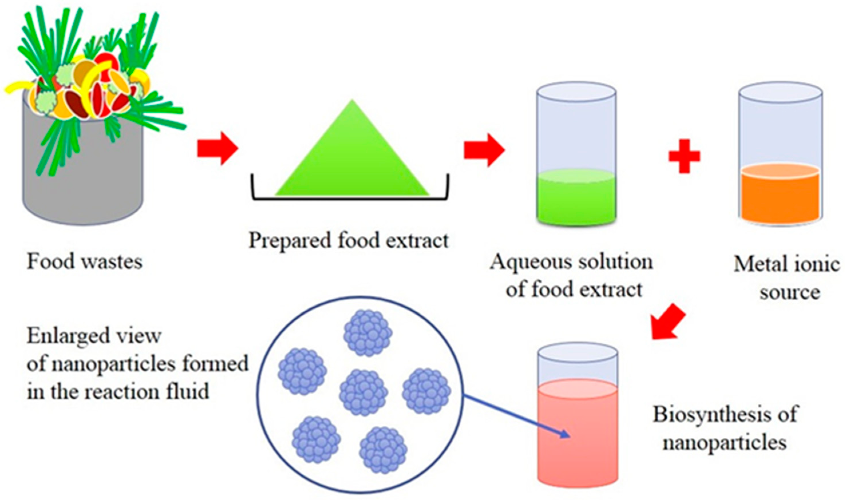
Figure 3.
Schematic diagram of the mechanism behind the biogenic synthesis of metallic nanoparticles.
Figure 3.
Schematic diagram of the mechanism behind the biogenic synthesis of metallic nanoparticles.

Table 1.
A selection of nanoparticles biosynthesised by horticultural waste sources.
| Nanoparticle | Size & Morphology | Food Source | Year | Reference |
|---|---|---|---|---|
| Ag | 5 to 35 nm, Spherical | Ananas comosus (Pineapple) | 2012 | [79] |
| Ag & Au | Ag: 16 nm, Spherical, Au: 11 nm, triangular | Tanacetum vulgare (tansy fruit) | 2010 | [84] |
| Ag | 3 to 12 nm, Spherical | Citrus sinensis (orange) peel | 2011 | [86] |
| Ag | 35 ± 2 nm @ 25 °C, Spherical 10 ± 1 nm @ 60 °C, Spherical | Citrus sinensis (orange) peel | 2011 | [89] |
| Ag | 5 to 20 nm, Spherical | Citrus unshiu (mandarin) peel | 2013 | [109] |
| Ag | 10 nm, Quasi-spherical | Sorghum spp. (bran) (aw) | 2010 | [110] |
| Ag & Au | Ag: 10 nm to 20 nm, Spherical Au: 15 nm to 25 nm, Spherical | Emblica officinalis (Indian Gooseberry) | 2005 | [111] |
| Ag | Large nanoclusters | Musa paradisiac (banana) peel | 2010 | [88] |
| Ag | 60 to 80 nm, Spherical | Carica papaya (pawpaw) | 2008 | [130] |
| Ag | 15 nm, Cubic | Carica papaya (pawpaw) | 2009 | [131] |
| Ag | 0.1 µm to 0.5 µm, Granular | Psidium guajava (guava) | 2014 | [132] |
| Ag | 4.32 nm to 17.65 nm, Spherical | Daucus carrota L. (Black Carrot) | 2014 | [133] |
| Ag | 4 nm to 22 nm, Spherical | Allium sativum (garlic clove) | 2011 | [134] |
| Ag | 17.96 ± 0.16 nm, Spherical | Citrullus lanatus rind | 2017 | [135] |
| Au | 20 to 140 nm, Spherical | Citrullus lanatus rind | 2015 | [136] |
| Au | 20 to 25 nm, Quasi-spherical | Grape skin, stalk and seed waste | 2014 | [80] |
| Au | 50 to 100 nm, Spherical | Rice bran (aw) | 2014 | [81] |
| Au | 200 to 500 nm, Triangular, hexagonal | Pyrus sp. (pear) | 2010 | [85] |
| Au | 6.03 ± 2.77 to 18.01 ± 3.67 nm, Spherical | Mangifera indica (mango) peel | 2014 | [49] |
| Au | 432.3 nm, Shape not specified | Daucus carota, subsp. Sativus (Carrot) | 2014 | [137] |
| Au | Micro-scale, Triangular | Cicer arietinum L. (Bean extract) | 2006 | [138] |
| Au | pH 9: 10 nm, Spherical, pH 10: 25 nm, Spherical, rods, pH 11: 15 nm diameter nanowires of varying length | Beta vulgaris (sugar beet pulp) | 2011 | [139] |
| Pd | 50 nm, Crystalline, irregular shape | Musa paradisiac (banana) peel | 2010 | [88] |
| Pd & Ag | 20 nm to 60 nm, Spherical | Various commercially available tea/coffee extracts | 2008 | [87] |
| Pd & Pt | 16 to 20 nm, Spherical | Lignin (aw) | 2012 | [71] |
| Pd | 96 nm, Spherical | Citrullus lanatus (watermelon) rind | 2015 | [124] |
| Fe3O4 | 5 to 25 nm, Cubes & Pyramids | Tea Waste | 2014 | [125] |
| MgO | 29 nm, Spherical | Citrus sinensis (orange) peel | 2015 | [128] |
| Mn3O4 | 20 nm to 50 nm, Spherical | Musa paradisiac (banana) peel | 2014 | [49] |
Note: (aw) indicates agricultural waste.
Table 2.
A selection of nanoparticles biosynthesised using marine plant sources.
| Nanoparticle | Size & Shape | Marine Alga | Year | Reference |
|---|---|---|---|---|
| Ag | 3 to 44 nm, Spherical and Cubic | Codium capitatum | 2013 | [112] |
| Ag | 30 nm, Spherical | Spyrogira insignis | 2013 | [113] |
| Ag | 4 to 24 nm, Spherical | Enteromorpha compressa | 2017 | [140] |
| Ag Au | 20 nm, Spherical 5 to 260 nm, Triangles, Spheres and Hexagons | Sargassum incisifolium | 2016 | [141] |
| Au | 6 to 10 nm, Spherical & Triangular | Turbinaria conoides | 2013 | [142] |
| Au | 18.7 to 93.7 nm, Spherical | Stoechospermum marginatum | 2012 | [143] |
| Pd | 4 to 6 nm, Spherical | Laminaria digitata | 2015 | [144] |
| Cu2O, CuO | 5 to 45 nm, Spherical | Bifurcaria bifurcata | 2014 | [72] |
| Cu/Cu2O | 53 nm, Spherical | Kappaphycus alvarezii | 2014 | [127] |
| Fe3O4 | 18 ± 4 nm, Cubic | Sargassum muticum | 2013 | [126] |
| ZnO | 18 to 50 nm, Hexagonal | Gracilaria gracilis | 2014 | [145] |
© 2017 by the authors. Licensee MDPI, Basel, Switzerland. This article is an open access article distributed under the terms and conditions of the Creative Commons Attribution (CC BY) license (http://creativecommons.org/licenses/by/4.0/).
Share and Cite
MDPI and ACS Style
Ghosh, P.R.; Fawcett, D.; Sharma, S.B.; Poinern, G.E.J. Production of High-Value Nanoparticles via Biogenic Processes Using Aquacultural and Horticultural Food Waste. Materials 2017, 10, 852. https://doi.org/10.3390/ma10080852
AMA Style
Ghosh PR, Fawcett D, Sharma SB, Poinern GEJ. Production of High-Value Nanoparticles via Biogenic Processes Using Aquacultural and Horticultural Food Waste. Materials. 2017; 10(8):852. https://doi.org/10.3390/ma10080852
Chicago/Turabian StyleGhosh, Purabi R., Derek Fawcett, Shashi B. Sharma, and Gerrard E. J. Poinern. 2017. "Production of High-Value Nanoparticles via Biogenic Processes Using Aquacultural and Horticultural Food Waste" Materials 10, no. 8: 852. https://doi.org/10.3390/ma10080852
Note that from the first issue of 2016, this journal uses article numbers instead of page numbers. See further details here.