Development of Biomedical Polymer-Silicate Nanocomposites: A Materials Science Perspective

Abstract
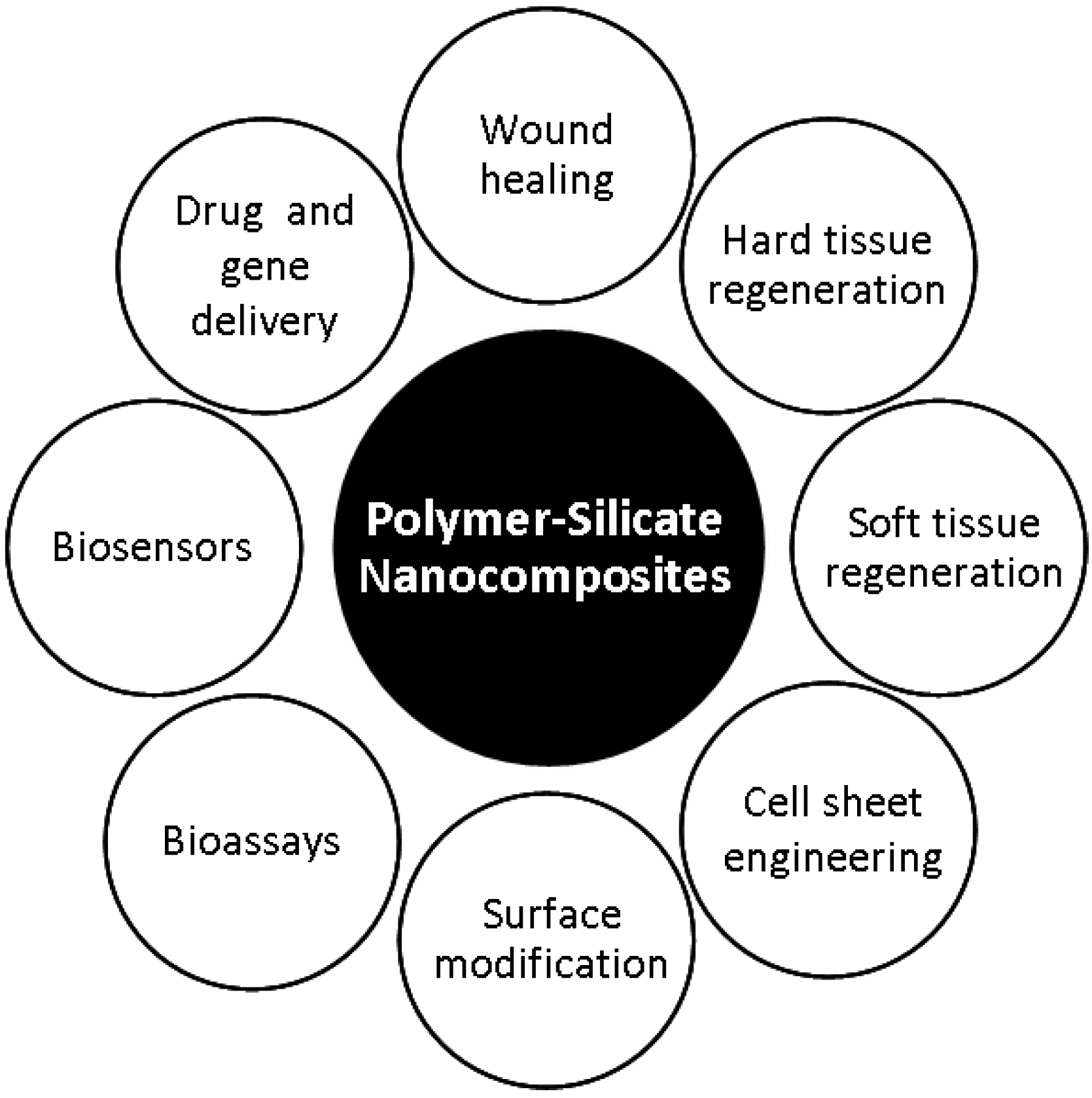
:1. Introduction

2. Biomedical Polymers Reinforced with Clay Based Silicate Nanoparticles

| Nanoparticles | Polymer | Experimental observations | Ref |
|---|---|---|---|
| MMT | PLG | Toughness and elongation of the nanocomposites enhanced due to addition of nanoparticles. Physical cross-linking between polymer and nanoparticles triggered a toughening mechanism via multiple crazing and shear yielding | [30] |
| MMT | PLLA | Increase in tensile modulus observed with addition of MMT. Enhanced surface interaction between nanoparticles and polymer decreased polymer crystallinity and promoted degradation of the nanocomposite | [31] |
| MMT | PLLA | MMT improved structural integrity of the nanocomposites | [33,34] |
| MMT | PLA | MMT improved compression properties and hydrophilicity of the polymeric matrix | [35] |
| MMT | PLLA | Higher amounts of MMT and fully exfoliated structures gave rise to stiffer materials. Addition of MMT suppressed polymer crystallization due to enhanced surface interactions | [36] |
| MMT | Gelatin-chitosan | Lower degradation rate and enhanced cell adhesion observed after addition of MMT to the polymer blend | [37] |
| Cloisite | Ethylene vinyl acetate | 10% clay concentration produced materials with the higher moduli and enhanced cell proliferation | [38] |
| Cloisite | Polyurethanes | Nanocomposites had a 5 fold lower permeability towards water vapor and enhanced mechanical properties | [40,41] |
3. Polymer Silicate Nanocomposite Hydrogels with Biomedical Potential
| Nanoparticles | Polymer | Experimental observations | Ref |
|---|---|---|---|
| Laponite | PNIPAM | Ultrahigh elongation with near-complete recovery, rapid de-swelling responses to temperature changes and large equilibrium swellings were observed due to addition of Laponite to the polymeric matrix. | [42,46,47,48] |
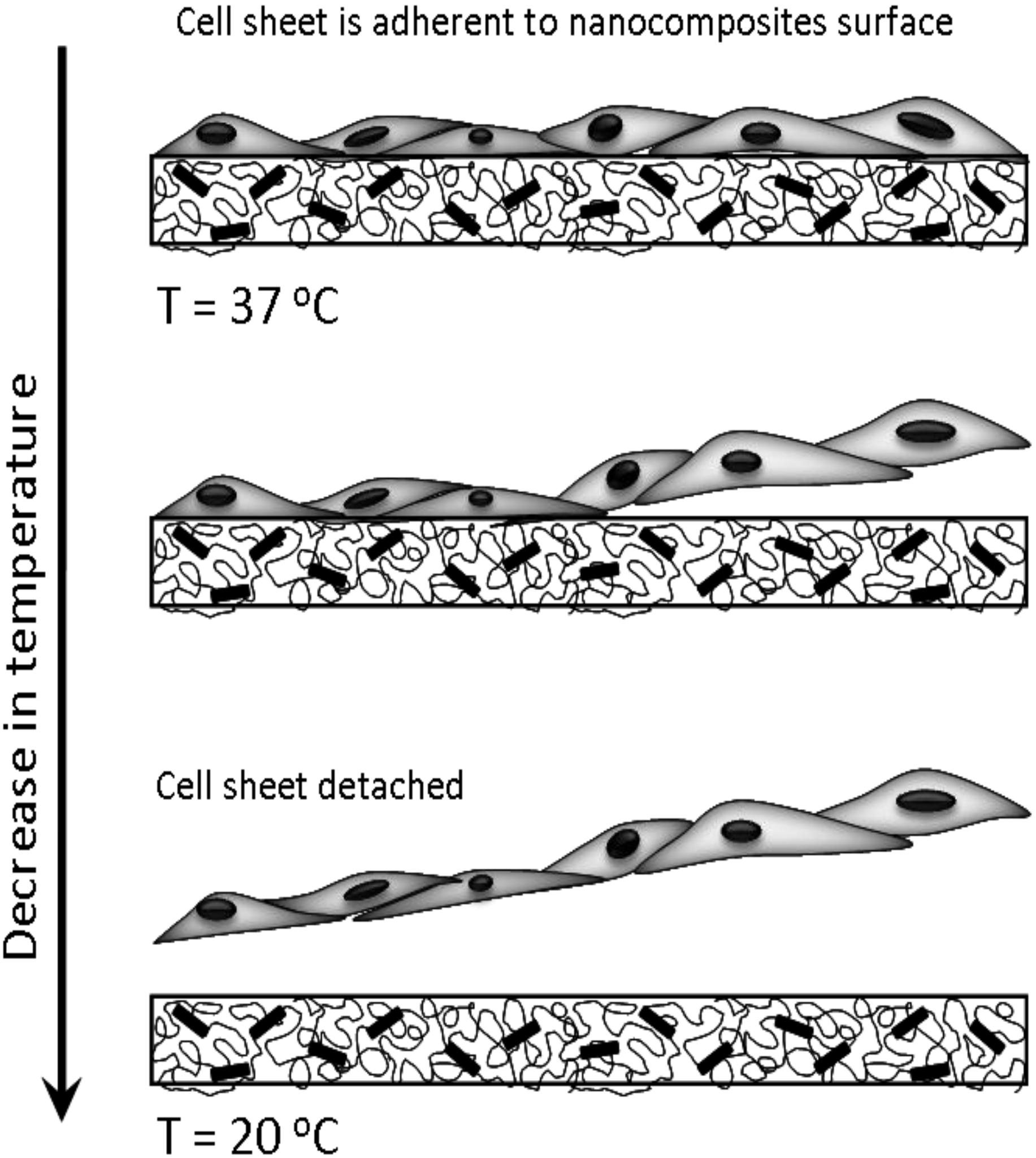
| Laponite | PNIPAM | Cell sheet easily detached by changing temperature. | [43] |
| Laponite | PEO | Cells cultured on the surfaces of PEO-Laponite gels attached and proliferated easily. | [53,54] |

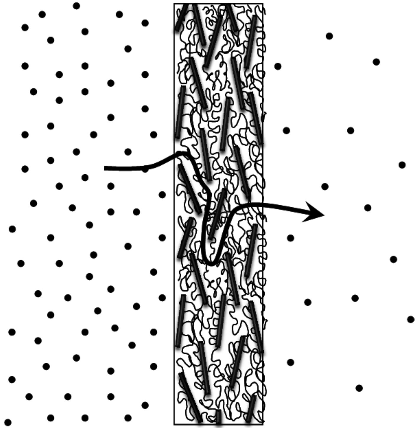
4. Polymer Layered Silicate Nanocomposite Developments for Drug Delivery Applications

| Nanoparticles | Polymer | Experimental observations | Ref |
|---|---|---|---|
| Cloisite | Poly(ethylene-co-vinyl acetate) | Addition of nanoparticle resulted in slower release of dexamethasone. Moreover, release kinetics were dependent on the aspect ratio and degree of dispersion of the nanoparticle | [56] |
| Laponite | Pluronic | A temperature dependent sol-gel transition was observed in the nanocomposites. Laponite enhanced the dissolution resistant properties of the hydrogels and release of entrapped macromolecular drug was slowed down | [57] |
| Bentonite | Acrylic acid-PEG methyl ether acrylate | Elution kinetics strongly depended on the interactions between the surface charges of the clay and the drug | [58] |
| Laponite | PEO-polyamide | Molecular interactions between Laponite and drug resulted in sustained release profiles | [59] |
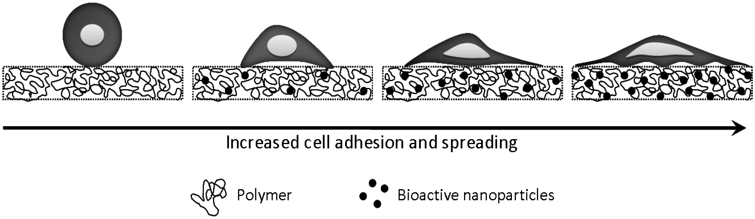
5. Polymer Bioactive Glass Nanocomposites for Tissue Engineering and Repair

| Nanoparticles | Polymer | Experimental observations | Ref |
|---|---|---|---|
| Bioglass | P3HB | Nanocomposite supported osteoblast cell attachment, proliferation and differentiation. | [76] |
| Bioglass | P3HB | Addition of nanoparticles enhanced modulus and strength of the nanocomposite compared to microcomposite. Addition of bioglass resulted in deposition of hydroxyapatite when submersed in simulated body fluid. | [77] |
| Wollastonite | PCL | Addition of wollastonite improved the nanocomposite Young’s modulus, tensile strength and fracture toughness. Nanocomposites supported in vitro formation of apatite. | [78,79] |
| Bioglass | PLA | Addition of bioglass fiber enhanced in vitro bioactivity of the nanocomposite. Significant increase in alkaline phosphatase activity observed in nanocomposite compared to pure PLA. | [80] |
| Bioglass | Poly L-lactide | Increase in bioglass concentration reduced water absorption capacity but enhanced degradation rate. | [81] |
| Bioglass | Chitosan & Chitosan-Gelatin | Bioactive nanocomposite scaffolds promoted osteoblast cell adhesion and spreading. | [82,83,84] |
| Silica | Chitosan | Improved mechanical properties observed due to addition of bioglass. Bioglass aided in significant increase in cell adhesion, proliferation and alkaline phosphatase activity. Enhanced bone regeneration observed when the nanocomposite was implanted in vivo. | [85] |
| Silica | Collagen | Improved bioactivity of the material; accelerated the formation of bone-like apatite and led to the differentiation of human monocytes into osteoclast-like cells. | [86,87] |
| Silica | Chitin | Chitinous organic matrix provided a template for bio-directed deposition of the silicate mineral phase. | [88] |
| Silica | Silk | High toughness and strength due to deposition of silica. | [89] |
| Wollastonite | Silk | Wollastonite enhanced both the mechanical strength and bioactivity of the nanocomposites. In vitro cell attachment and proliferation were also observed on the nanocomposites. | [90] |
6. Future Trends and Challenges
Abbreviations
| MMT | Montmorillonite clay, layered silicate |
| P3HB | Poly(3-hydroxybutyrate) |
| PCL | Polycaprolactone |
| PEG | Poly(ethylene glycol) |
| PEO | Poly(ethylene oxide) |
| PNIPAM | Poly(N-isopropylacrylamide) |
| PLA | Poly lactic acid |
| PLG | Poly(lactic–Cco-glycolide) |
| PLLA | Poly L-lactic acid |
Acknowledgements
References and Notes
- Kohane, D.S.; Langer, R. Polymeric biomaterials in tissue engineering. Pediat. Res. 2008, 63, 487–491. [Google Scholar] [CrossRef] [PubMed]
- Ratner, B.D.; Hoffman, A.S.; Schoen, F.J.; Lemons, J.E. Biomaterials Science: An Introduction to Materials In Medicine; Elsevier: Amsterdam, The Netherlands, 2004; pp. 67–80. [Google Scholar]
- Temenoff, J.S.; Mikos, A.G. Biomaterials. In The Intersection of Biology and Materials Science; Pearson Prentice Hall: New Jersey, NJ, USA, 2008; pp. 10–12. [Google Scholar]
- Mulhaupt, R. Hermann Staudinger and the origin of macromolecular chemistry. Angew.Chem.Int. Ed. 2004, 43, 1054–1063. [Google Scholar] [CrossRef]
- Flory, P.J. Principles of Polymer Chemistry; Cornell University Press: New York, NY, USA, 1967; pp. 3–25. [Google Scholar]
- Huebsch, N.; Mooney, D.J. Inspiration and application in the evolution of biomaterials. Nature 2009, 462, 426–432. [Google Scholar] [CrossRef] [PubMed]
- Langer, R.; Tirrell, D.A. Designing materials for biology and medicine. Nature 2004, 428, 487–492. [Google Scholar] [CrossRef] [PubMed]
- Peppas, N.A. Intelligent Biomaterials as Pharmaceutical Carriers in Mirofabricated and Nanoscale Devices. MRS Bull. 2006, 31, 888–893. [Google Scholar] [CrossRef]
- Peppas, N.A.; Hilt, J.Z.; Khademhosseini, A.; Langer, R. Hydrogels in biology and medicine: From molecular principles to bionanotechnology. Advan. Mater. 2006, 18, 1345–1360. [Google Scholar] [CrossRef]
- Ratner, B.D.; Bryant, S.J. Biomaterials: Where we have been and where we are going. Annu. Rev. Biomed. Eng. 2004, 6, 41–75. [Google Scholar] [CrossRef] [PubMed]
- Okada, A.; Usuki, A. Twenty years of polymer-clay nanocomposites. Macromol. Mater. Eng. 2006, 291, 1449–1476. [Google Scholar] [CrossRef]
- Vaia, R.A.; Gianellis, E.P. Polymer Nanocomposites: Status and Applications. MRS Bull. 2001, 62, 394–401. [Google Scholar] [CrossRef]
- Wagner, H.D. Nanocomposites—Paving the way to stronger materials. Nat. Nanotech. 2007, 2, 742–744. [Google Scholar] [CrossRef]
- Hule, R.A.; Pochan, D.J. Polymer nanocomposites for biomedical applications. MRS Bull. 2007, 32, 354–358. [Google Scholar] [CrossRef]
- Schexnailder, P.J.; Schmidt, G. Nanocomposite Polymer Hydrogels. Colloid Polym. Sci. 2009, 287, 1–11. [Google Scholar] [CrossRef]
- Williams, D.F. On the nature of biomaterials. Biomaterials 2009, 30, 5897–5909. [Google Scholar] [CrossRef] [PubMed]
- Hench, L.L.; Polak, J.M. Third-generation biomedical materials. Science 2002, 295, 1014–1017. [Google Scholar] [CrossRef] [PubMed]
- Ma, X.M.; Elisseeff, J. Scaffolding in Tissue Engineering; CRC Press, Taylor and Francis: Boca Raton, FL, USA, 2006; pp. 3–625. [Google Scholar]
- Hirst, A.R.; Escuder, B.; Miravet, J.F.; Smith, D.K. High-Tech Applications of Self-Assembling Supramolecular Nanostructured Gel-Phase Materials: From Regenerative Medicine to Electronic Devices. Angew. Chem. Int. Ed. 2008, 47, 8002–8018. [Google Scholar] [CrossRef]
- Stuart, M.A.C.; Huck, W.T.S.; Genzer, J.; Muller, M.; Ober, C.; Stamm, M.; Sukhorukov, G.B.; Szleifer, I.; Tsukruk, V.V.; Urban, M.; Winnik, F.; Zauscher, S.; Luzinov, I.; Minko, S. Emerging applications of stimuli-responsive polymer materials. Nat. Mater. 2010, 9, 101–113. [Google Scholar] [CrossRef] [PubMed]
- Fratzl, P.; Weinkamer, R. Nature’s hierarchical materials. Prog. Mater. Sci. 2007, 52, 1263–1334. [Google Scholar] [CrossRef]
- Gao, H.; Ji, B.; Jäger, I.L.; Arzt, E.; Fratzl, P. Materials become insensitive to flaws at nanoscale: Lessons from nature. Proc. Nat. Acad. Sci.USA 2003, 100, 5597–5600. [Google Scholar] [CrossRef] [PubMed]
- Tang, Z.Y.; Kotov, N.A.; Magonov, S.; Ozturk, B. Nanostructured artificial nacre. Nat. Mater. 2003, 2, 413–418. [Google Scholar] [CrossRef] [PubMed]
- Weiner, S.; Wagner, H.D. The material bone: Structure-mechanical function relations. Annu. Rev. Mater. Sci. 1998, 28, 271–298. [Google Scholar] [CrossRef]
- Mitragotri, S.; Lahann, J. Physical approaches to biomaterial design. Nat. Mater. 2009, 8, 15–23. [Google Scholar] [CrossRef] [PubMed]
- Sheldon, B.W.; Curtin, W.A. Nanoceramic composites: Tough to test. Nat. Mater. 2004, 3, 505–506. [Google Scholar] [CrossRef] [PubMed]
- Vaia, R.; Baur, J. Adaptive Composites. Science 2008, 319, 420–421. [Google Scholar] [CrossRef] [PubMed]
- Vaia, R.A.; Wagner, H.D. Framework for nanocomposites. Mater. Today 2004, 7, 32–37. [Google Scholar] [CrossRef]
- Faucheu, J.; Gauthier, C.; Chazeau, L.; Cavaille, J.Y.; Mellon, V.; Lami, E.B. Miniemulsion polymerization for synthesis of structured clay/polymer nanocomposites: Short review and recent advances. Polymer 2010, 51, 6–17. [Google Scholar] [CrossRef]
- Xu, W.; Raychowdhury, S.; Jiang, D.D.; Retsos, H.; Giannelis, E.P. Dramatic improvements in toughness in poly(lactide-co-glycolide) nanocomposties. Small 2008, 4, 662–669. [Google Scholar] [CrossRef] [PubMed]
- Lee, J.H.; Park, T.G.; Park, H.S.; Lee, D.S.; Lee, Y.K.; Yoon, S.C.; Nam, J.D. Thermal and mechanical characteristics of poly (L-lactic acid) nanocomposite scaffold. Biomaterials 2003, 24, 2773–2778. [Google Scholar] [CrossRef] [PubMed]
- Ray, S.S.; Yamada, K.; Okamoto, M.; Ueda, K. Polylactide-layered silicate nanocomposite: A novel biodegradable material. Nano Lett. 2002, 2, 1093–1096. [Google Scholar] [CrossRef]
- Lee, Y.H.; Lee, J.H.; An, I.G.; Kim, C.; Lee, D.S.; Lee, Y.K.; Nam, J.D. Electrospun dual-porosity structure and biodegradation morphology of Montmorillonite reinforced PLLA nanocomposite scaffolds. Biomaterials 2005, 26, 3165–3172. [Google Scholar] [CrossRef] [PubMed]
- Tsivintzelis, I.; Marras, S.I.; Zuburtikudis, I.; Panayiotou, C. Porous poly (L-lactic acid) nanocomposite scaffolds prepared by phase inversion using supercritical CO2 as antisolvent. Polymer 2007, 48, 6311–6318. [Google Scholar] [CrossRef]
- Ozkoc, G.; Kemaloglu, S.; Quaedflieg, M. Production of Poly(lactic acid)/Organoclay Nanocomposite Scaffolds by Microcompounding and Polymer/Particle Leaching. Polym. Compos. 2010, 31, 674–683. [Google Scholar]
- Krikorian, V.; Pochan, D.J. Poly (L-lactic acid)/layered silicate nanocomposite: Fabrication, characterization and properties. Chem. Mater. 2003, 15, 4317–4324. [Google Scholar] [CrossRef]
- Zhuang, H.; Zheng, J.P.; Gao, H.; De Yao, K. In vitro biodegradation and biocompatibility of gelatin/montmorillonite-chitosan intercalated nanocomposite. J. Mater.Sci. Mater. Med. 2007, 18, 951–957. [Google Scholar] [CrossRef] [PubMed]
- Lewkowitz-Shpuntoff, H.M.; Wen, M.C.; Singh, A.; Brenner, N.; Gambino, R.; Pernodet, N.; Isseroff, R.; Rafailovich, M.; Sokolov, J. The effect of organo clay and adsorbed FeO3 nanoparticles on cells cultured on Ethylene Vinyl Acetate substrates and fibers. Biomaterials 2009, 30, 8–18. [Google Scholar] [CrossRef] [PubMed]
- Yang, M.J.; Zhang, Z.; Hahn, C.; Laroche, G.; King, M.W.; Guidoin, R. Totally implantable artificial hearts and left ventricular assist devices: Selecting impermeable polycarbonate urethane to manufacture ventricles. J. Biomed. Mater. Res. 1999, 48, 13–23. [Google Scholar] [CrossRef] [PubMed]
- Xu, R.J.; Manias, E.; Snyder, A.J.; Runt, J. New biomedical poly(urethane urea)—layered silicate nanocomposites. Macromolecules 2001, 34, 337–339. [Google Scholar] [CrossRef]
- Xu, R.J.; Manias, E.; Snyder, A.J.; Runt, J. Low penneability biomedical polyurethane nanocomposites. J.Biomed. Mater. Res. Part A 2003, 64A, 114–119. [Google Scholar] [CrossRef]
- Haraguchi, K.; Takehisa, T. Nanocomposite hydrogels: A unique organic-inorganic network structure with extraordinary mechanical, optical, and swelling/de-swelling properties. Advan. Mater. 2002, 14, 1120–1124. [Google Scholar] [CrossRef]
- Haraguchi, K.; Takehisa, T.; Ebato, M. Control of cell cultivation and cell sheet detachment on the surface of polymer/clay nanocomposite hydrogels. Biomacromolecules 2006, 7, 3267–3275. [Google Scholar] [CrossRef] [PubMed]
- Schmidt, G.; Nakatani, A.I.; Butler, P.D.; Karim, A.; Han, C.C. Shear orientation of viscoelastic polymer-clay solutions probed by flow birefringence and SANS. Macromolecules 2000, 33, 7219–7222. [Google Scholar] [CrossRef]
- Haraguchi, K.; Li, H.J.; Matsuda, K.; Takehisa, T.; Elliott, E. Mechanism of forming organic/inorganic network structures during in-situ free-radical polymerization in PNIPA-clay nanocomposite hydrogels. Macromolecules 2005, 38, 3482–3490. [Google Scholar] [CrossRef]
- Haraguchi, K.; Li, H.J. Mechanical properties and structure of polymer-clay nanocomposite gels with high clay content. Macromolecules 2006, 39, 1898–1905. [Google Scholar] [CrossRef]
- Haraguchi, K.; Farnworth, R.; Ohbayashi, A.; Takehisa, T. Compositional effects on mechanical properties of nanocomposite hydrogels composed of poly(N,N-dimethylacrylamide) and clay. Macromolecules 2003, 36, 5732–5741. [Google Scholar] [CrossRef]
- Haraguchi, K.; Takehisa, T.; Fan, S. Effects of clay content on the properties of nanocomposite hydrogels composed of poly(N-isopropylacrylamide) and clay. Macromolecules 2002, 35, 10162–10171. [Google Scholar] [CrossRef]
- Haraguchi, K.; Song, L.Y. Microstructures formed in co-cross-linked networks and their relationships to the optical and mechanical properties of PNIPA/clay nanocomposite gels. Macromolecules 2007, 40, 5526–5536. [Google Scholar] [CrossRef]
- Loizou, E.; Butler, P.D.; Porcar, L.; Talmon, Y.; Kesselman, E.; Schmidt, G. Large scale structures in polymer-clay hydrogels. Macromolecules 2005, 38, 2047–2049. [Google Scholar] [CrossRef]
- Harris, J.M. Poly(ethylene glycol) Chemistry: Biotechnical and Biomedical Applications; Plenum Press: New York, NY, USA, 1992; pp. 1–12. [Google Scholar]
- Schexnailder, P.; Loizou, E.; Porcar, L.; Butler, P.; Schmidt, G. Heterogeneity in nanocomposite hydrogels from poly(ethylene oxide) cross-linked with silicate nanoparticles. Phys. Chem. Chem. Phys. 2009, 11, 2760–2766. [Google Scholar] [CrossRef] [PubMed]
- Gaharwar, A.K.; Schexnailder, P.; Kaul, V.; Akkus, O.; Zakharov, D.; Seifert, S.; Schmidt, G. Highly extensible bio-nanocomposite films with direction-dependent properties. Advan. Funct. Mater. 2010, 20, 429–436. [Google Scholar] [CrossRef]
- Jin, Q.; Schexnailder, P.; Gaharwar, A.K.; Schmidt, G. Silicate cross-linked bio-nanocomposite hydrogels from peo and chitosan. Macromol. Biosci. 2009, 9, 1028–1035. [Google Scholar] [CrossRef] [PubMed]
- Yano, K.; Usuki, A.; Okada, A. Synthesis and properties of polyimide-clay hybrid films. J. Polym. Sci. A-Polym. Chem. 2000, 35, 2289–2294. [Google Scholar] [CrossRef]
- Cypes, S.H.; Saltzman, W.M.; Giannelis, E.P. Organosilicate-polymer drug delivery systems: controlled release and enhanced mechanical properties. J. Control. Release 2003, 90, 163–169. [Google Scholar] [CrossRef] [PubMed]
- Wu, C.J.; Schmidt, G. Thermosensitive and dissolution properties in nanocomposite polymer hydrogels. Macromol. Rapid Commun. 2009, 30, 1492–1497. [Google Scholar] [CrossRef] [PubMed]
- Lee, W.F.; Chen, Y.C. Effect of bentonite on the physical properties and drug-release behavior of poly (AA-co-PEGMEA)/bentonite nanocomposite hydrogels for mucoadhesive. J. Appl. Polym. Sci. 2004, 91, 2934–2941. [Google Scholar] [CrossRef]
- Takahashi, T.; Yamada, Y.; Kataoka, K.; Nagasaki, Y. Preparation of a novel PEG-clay hybrid as a DDS material: Dispersion stability and sustained release profiles. J. Control. Release 2005, 107, 408–416. [Google Scholar] [CrossRef] [PubMed]
- Misra, S.K.; Valappil, S.P.; Roy, I.; Boccaccini, A.R. Polyhydroxyalkanoate (PHA)/inorganic phase composites for tissue engineering applications. Biomacromolecules 2006, 7, 2249–2258. [Google Scholar] [CrossRef] [PubMed]
- Carlisle, E.M.; Alpenfels, W.F. Silicon Requirement for Normal Growth of Cartilage in Culture. Fed. Proc. 1980, 39, 787. [Google Scholar]
- Hench, L.L.; Paschall, H.A. Direct chemical bond of bioactive glass-ceramic materials to bone and muscle. J. Biomed. Mater. Res. 1973, 7, 25–42. [Google Scholar] [CrossRef] [PubMed]
- Schwarz, K. Bound form of silicon in glycosaminoglycans and polyuronides - (polysaccharide matrix connective tissue). Proc. Nat. Acad. Sci. 1973, 70, 1608–1612. [Google Scholar] [CrossRef] [PubMed]
- Schwarz, K.; Milne, D.B. Growth-promoting effects of silicon in rats. Nature 1972, 239, 333–334. [Google Scholar] [CrossRef] [PubMed]
- Hench, L.L. Genetic design of bioactive glass. J. Euro. Ceram. Soc. 2009, 29, 1257–1265. [Google Scholar] [CrossRef]
- Vogel, M.; Voigt, C.; Gross, U.M.; Müller-Mai, C.M. In vivo comparison of bioactive glass particles in rabbits. Biomaterials 2001, 22, 357–362. [Google Scholar] [CrossRef] [PubMed]
- Kokubo, T. Bioactive glass ceramics: properties and applications. Biomaterials 1991, 12, 155–163. [Google Scholar] [CrossRef] [PubMed]
- Valerio, P.; Pereira, M.M.; Goes, A.M.; Leite, M.F. The effect of ionic products from bioactive glass dissolution on osteoblast proliferation and collagen production. Biomaterials 2004, 25, 2941–2948. [Google Scholar] [CrossRef] [PubMed]
- Xynos, I.D.; Edgar, A.J.; Buttery, L.D.K.; Hench, L.L.; Polak, J.M. Gene-expression profiling of human osteoblasts following treatment with the ionic products of Bioglass® 45S5 dissolution. J. Biomed. Mater. Res. A 2001, 55, 151–157. [Google Scholar] [CrossRef]
- Jones, J.R.; Ehrenfried, L.M.; Hench, L.L. Optimising bioactive glass scaffolds for bone tissue engineering. Biomaterials 2006, 27, 964–973. [Google Scholar] [CrossRef] [PubMed]
- Mansur, H.S.; Costa, H.S. Nanostructured poly (vinyl alcohol)/bioactive glass and poly (vinyl alcohol)/chitosan/bioactive glass hybrid scaffolds for biomedical applications. Chem. Eng. J. 2008, 137, 72–83. [Google Scholar] [CrossRef]
- Blaker, J.J.; Gough, J.E.; Maquet, V.; Notingher, I.; Boccaccini, A.R. In vitro evaluation of novel bioactive composites based on Bioglass®-filled polylactide foams for bone tissue engineering scaffolds. J. Biomed. Mater. Res. A 2003, 67A, 1401–1411. [Google Scholar] [CrossRef] [PubMed]
- Verrier, S.; Blaker, J.J.; Maquet, V.; Hench, L.L.; Boccaccini, A.R. PDLLA/Bioglass® composites for soft-tissue and hard-tissue engineering: an in vitro cell biology assessment. Biomaterials 2004, 25, 3013–3021. [Google Scholar] [CrossRef] [PubMed]
- Day, R.M.; Boccaccini, A.R.; Shurey, S.; Roether, J.A.; Forbes, A.; Hench, L.L.; Gabe, S.M. Assessment of polyglycolic acid mesh and bioactive glass for soft-tissue engineering scaffolds. Biomaterials 2004, 25, 5857–5866. [Google Scholar] [CrossRef] [PubMed]
- Webster, T.J.; Ergun, C.; Doremus, R.H.; Siegel, R.W.; Bizios, R. Enhanced functions of osteoblasts on nanophase ceramics. Biomaterials 2000, 21, 1803–1810. [Google Scholar] [CrossRef] [PubMed]
- Misra, S.K.; Ansari, T.; Mohn, D.; Valappil, S.P.; Brunner, T.J.; Stark, W.J.; Roy, I.; Knowles, J.C.; Sibbons, P.D.; Jones, E.V.; Boccaccini, A.R.; Salih, V. Effect of nanoparticulate bioactive glass particles on bioactivity and cytocompatibility of poly(3-hydroxybutyrate) composites. J. Roy. Soc. Interface 2010, 7, 453–465. [Google Scholar] [CrossRef]
- Misra, S.K.; Mohn, D.; Brunner, T.J.; Stark, W.J.; Philip, S.E.; Roy, I.; Salih, V.; Knowles, J.C.; Boccaccini, A.R. Comparison of nanoscale and microscale bioactive glass on the properties of P(3HB)/Bioglass® composites. Biomaterials 2008, 29, 1750–1761. [Google Scholar] [CrossRef] [PubMed]
- Kotela, I.; Podporska, J.; Soltysiak, E.; Konsztowicz, K.J.; Blazewicz, M. Polymer nanocomposites for bone tissue substitutes. Ceram. Int. 2009, 35, 2475–2480. [Google Scholar] [CrossRef]
- Wei, J.; Heo, S.J.; Liu, C.; Kim, D.H.; Kim, S.E.; Hyun, Y.T.; Shin, J.W.; Shin, J.W. Preparation and characterization of bioactive calcium silicate and poly (ε-caprolactone) nanocomposite for bone tissue regeneration. J. Biomed. Mater. Res. Part A 2008, 90A, 702–712. [Google Scholar]
- Kim, H.W.; Lee, H.H.; Chun, G.S. Bioactivity and osteoblast responses of novel biomedical nanocomposites of bioactive glass nanofiber filled poly(lactic acid). J. Biomed. Mater. Res. Part A 2008, 85A, 651–663. [Google Scholar] [CrossRef]
- El-Kady, A.M.; Ali, A.F.; Farag, M.M. Development, characterization, and in vitro bioactivity studies of sol-gel bioactive glass/poly(L-lactide) nanocomposite scaffolds. Mater. Sci. Eng.C 2010, 30, 120–131. [Google Scholar] [CrossRef]
- Peter, M.; Binulal, N.S.; Soumya, S.; Nair, S.V.; Furuike, T.; Tamura, H.; Jayakumar, R. Nanocomposite scaffolds of bioactive glass ceramic nanoparticles disseminated chitosan matrix for tissue engineering applications. Carbohyd. Polym. 2010, 79, 284–289. [Google Scholar] [CrossRef]
- Peter, M.; Binulal, N.S.; Nair, S.V.; Selvamurugan, N.; Tamura, H.; Jayakumar, R. Novel biodegradable chitosan–gelatin/nano-bioactive glass ceramic composite scaffolds for alveolar bone tissue engineering. Chem. Eng. J. 2010, 158, 353–361. [Google Scholar] [CrossRef]
- Peter, M.; Kumar, P.T.S.; Binulal, N.S.; Nair, S.V.; Tamura, H.; Jayakumar, R. Development of novel [alpha]-chitin/nanobioactive glass ceramic composite scaffolds for tissue engineering applications. Carbohyd. Polym. 2009, 78, 926–931. [Google Scholar] [CrossRef]
- Lee, E.J.; Shin, D.S.; Kim, H.E.; Kim, H.W.; Koh, Y.H.; Jang, J.H. Membrane of hybrid chitosan-silica xerogel for guided bone regeneration. Biomaterials 2009, 30, 743–750. [Google Scholar] [CrossRef] [PubMed]
- Heinemann, S.; Heinemann, C.; Bernhardt, R.; Reinstorf, A.; Nies, B.; Meyer, M.; Worch, H.; Hanke, T. Bioactive silica-collagen composite xerogels modified by calcium phosphate phases with adjustable mechanical properties for bone replacement. Acta Biomater. 2009, 5, 1979–1990. [Google Scholar] [CrossRef] [PubMed]
- Heinemann, S.; Heinemann, C.; Ehrlich, H.; Meyer, M.; Baltzer, H.; Worch, H.; Hanke, T.A. novel biomimetic hybrid material made of silicified collagen: perspectives for bone replacement. Advan. Eng. Mater. 2007, 9, 1061–1068. [Google Scholar] [CrossRef]
- Ehrlich, H.; Janussen, D.; Simon, P.; Bazhenov, V.V.; Shapkin, N.P.; Erler, C.; Mertig, M.; Born, R.; Heinemann, S.; Hanke, T.; Worch, H.; Vournakis, J.N. Nanostructural organization of naturally occurring composites-Part II: Silica-chitin-based biocomposites. J. Nanomater. 2008. [Google Scholar] [CrossRef]
- Foo, W.P.; Patwardhan, S.V.; Belton, D.J.; Kitchel, B.; Anastasiades, D.; Huang, J.; Naik, R.R.; Perry, C.C.; Kaplan, D.L. Novel nanocomposites from spider silk-silica fusion (chimeric) proteins. Proc. Nat. Acad. Sci. 2006, 103, 9428–9433. [Google Scholar] [CrossRef] [PubMed]
- Zhu, H.L.; Shen, J.Y.; Feng, X.X.; Zhang, H.P.; Guo, Y.H.; Chen, J.Y. Fabrication and characterization of bioactive silk fibroin/wollastonite composite scaffolds. Mater. Sci. Eng.C 2010, 30, 132–140. [Google Scholar] [CrossRef]
- Balazs, A.C.; Emrick, T.; Russell, T.P. Nanoparticle polymer composites: Where two small worlds meet. Science 2006, 314, 1107–1110. [Google Scholar] [CrossRef] [PubMed]
- Winey, K.I.; Vaia, R.A. Polymer nanocomposites. MRS Bull. 2007, 32, 314–319. [Google Scholar] [CrossRef]
© 2010 by the authors; licensee MDPI, Basel, Switzerland. This article is an open-access article distributed under the terms and conditions of the Creative Commons Attribution license (http://creativecommons.org/licenses/by/3.0/).
Share and Cite
Wu, C.-J.; Gaharwar, A.K.; Schexnailder, P.J.; Schmidt, G. Development of Biomedical Polymer-Silicate Nanocomposites: A Materials Science Perspective. Materials 2010, 3, 2986-3005. https://doi.org/10.3390/ma3052986
Wu C-J, Gaharwar AK, Schexnailder PJ, Schmidt G. Development of Biomedical Polymer-Silicate Nanocomposites: A Materials Science Perspective. Materials. 2010; 3(5):2986-3005. https://doi.org/10.3390/ma3052986
Chicago/Turabian StyleWu, Chia-Jung, Akhilesh K. Gaharwar, Patrick J. Schexnailder, and Gudrun Schmidt. 2010. "Development of Biomedical Polymer-Silicate Nanocomposites: A Materials Science Perspective" Materials 3, no. 5: 2986-3005. https://doi.org/10.3390/ma3052986