Biosynthesis and Characterization of Nanocellulose-Gelatin Films

Abstract
:1. Introduction
2. Results and Discussion
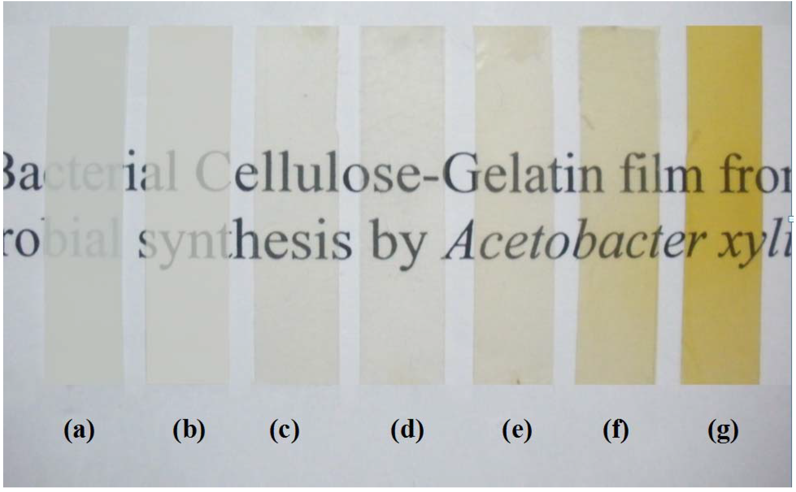
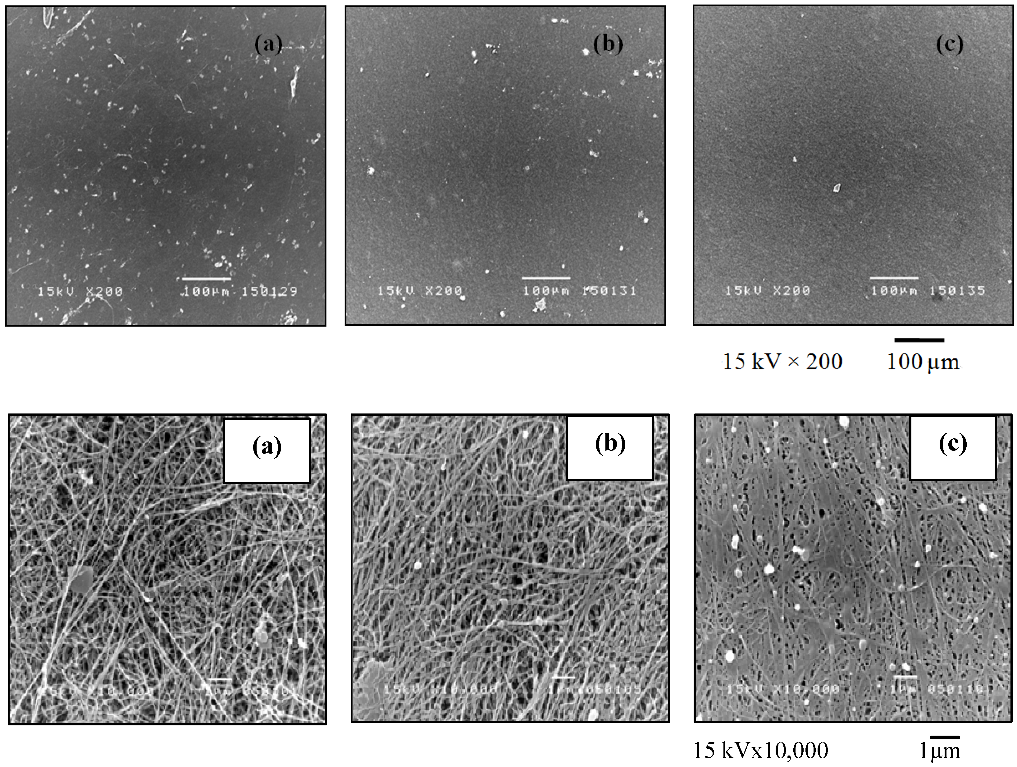
2.1. Morphology

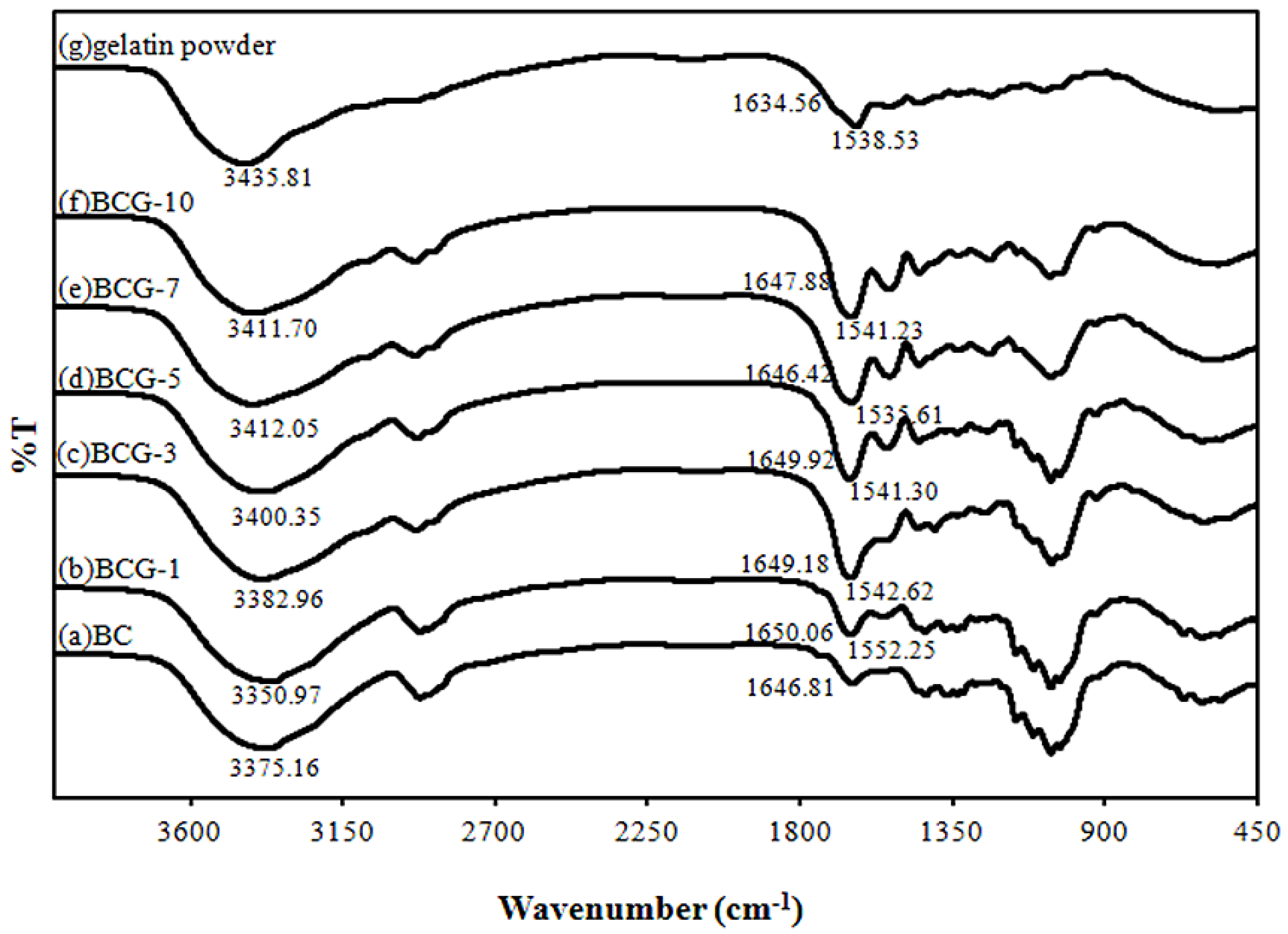
2.2. FTIR Analysis
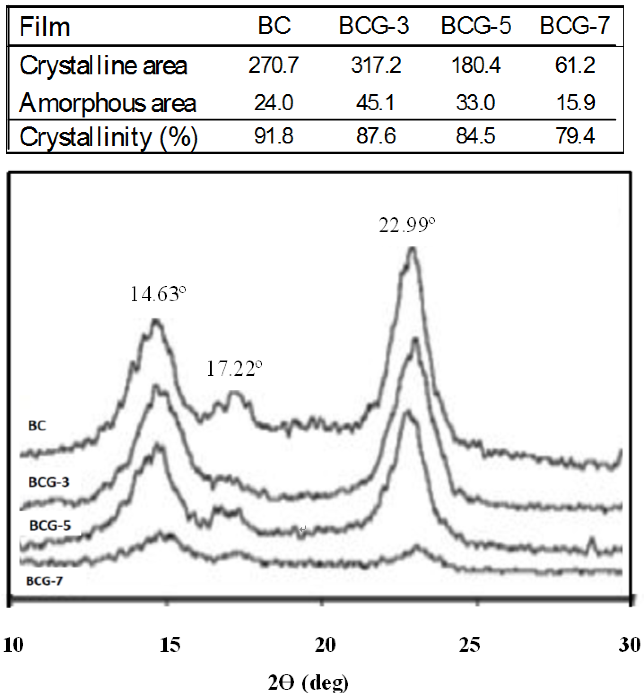
2.3. Crystallinity

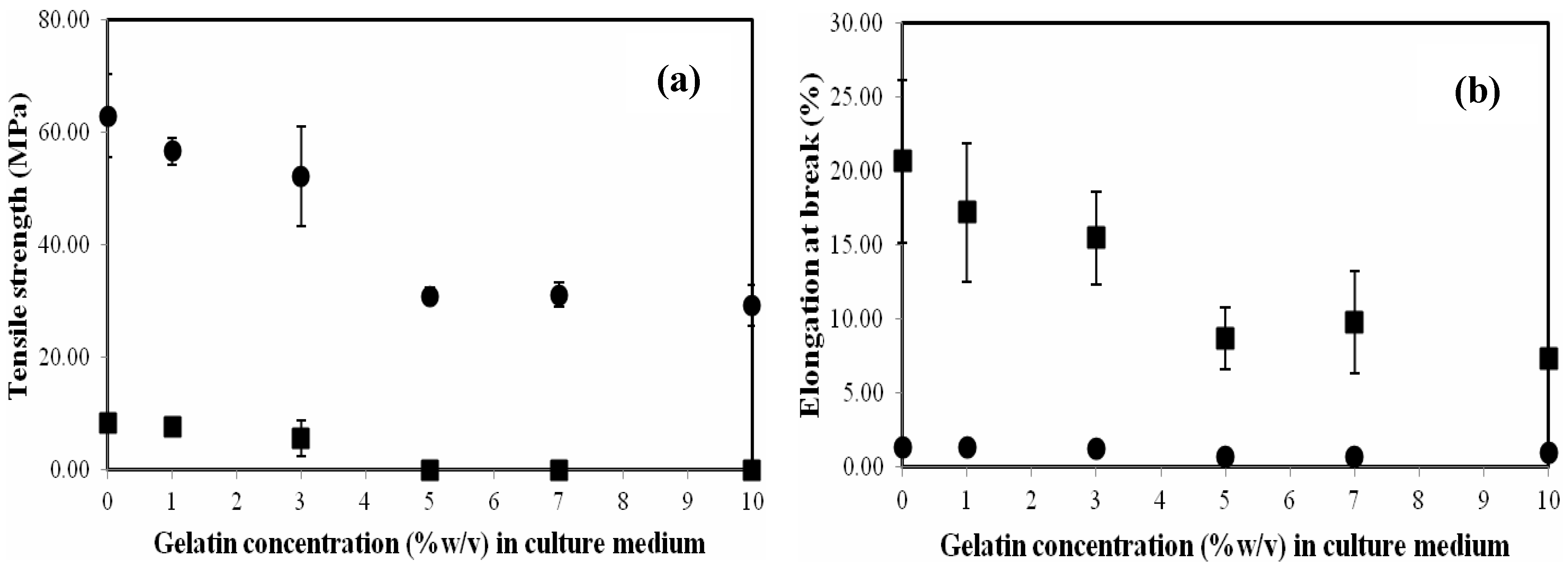
2.4. Mechanical Property
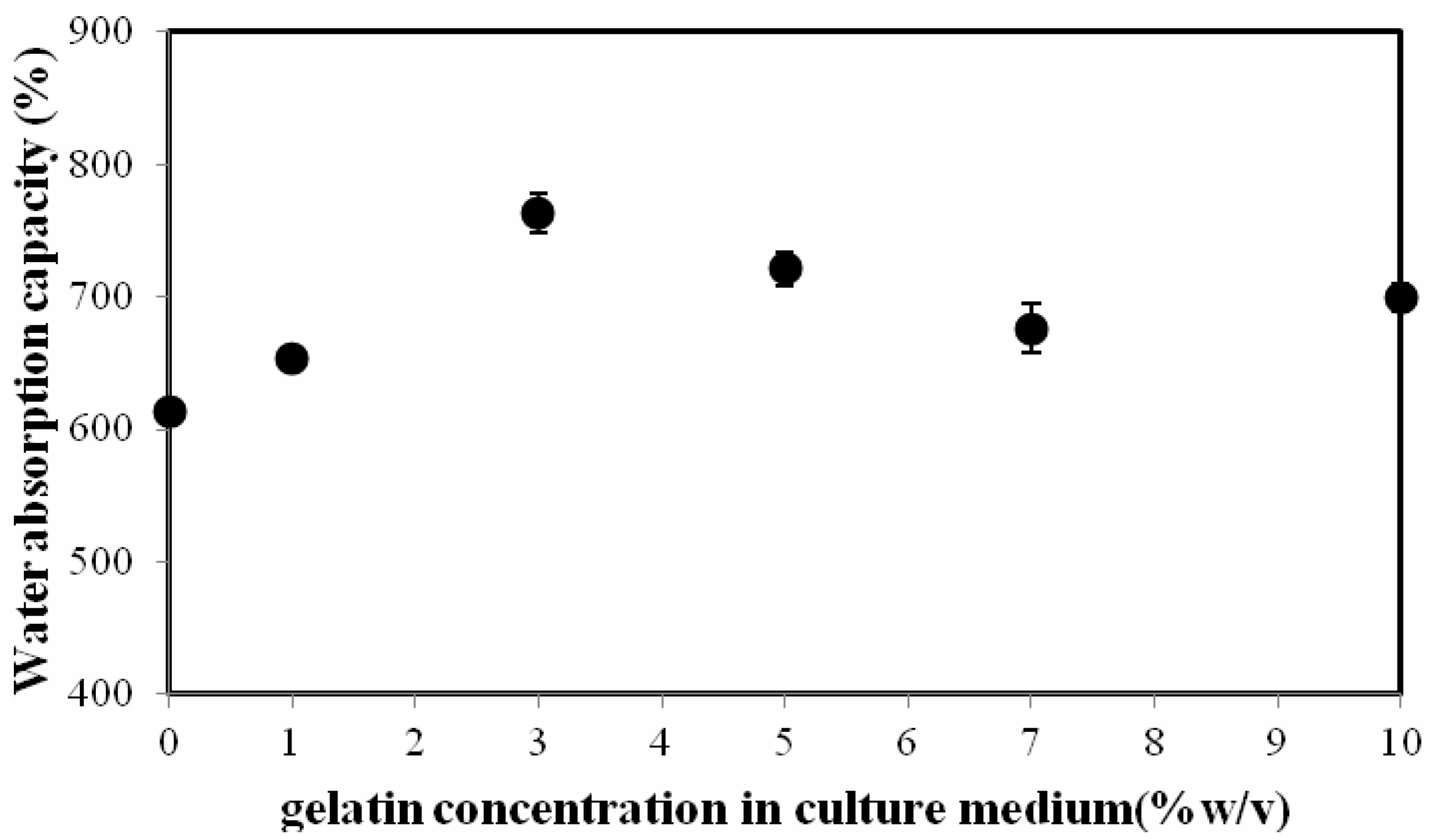
2.5. Water Absorption Capacity (WAC)

2.6. Gas (Vapor) Transmission Rate
2.7. Cytoxicity

| Type of films | Absorbance | ||
|---|---|---|---|
| 0 h | 24 h | 48 h | |
| BC | 0.298 ± 0.037 | 0.376 ± 0.029 | 0.481 ± 0.036 |
| BCG-10 | 0.256 ± 0.020 | 0.342 ± 0.044 | 0.589 ± 0.052 |
3. Experimental Section
3.1. Microbial Strains
3.2. Culture Media and Method
3.3. Characterizations
4. Conclusions
Acknowledgements
References
- Hoenich, N. Cellulose for medical applications. BioResources 2006, 1, 270–280. [Google Scholar]
- Brown, E.E.; Laborie, M.P.G. Bioengineering bacterial cellulose/poly(ethylene oxide) nanocomposites. Biomacromolecules 2007, 8, 3074–3081. [Google Scholar] [CrossRef] [PubMed]
- Jonas, R.; Farah, L.F. Production and application of microbial cellulose. Polym. Degrad. Stab. 1998, 59, 101–106. [Google Scholar] [CrossRef]
- Saied, H.E.; Basta, A.H.; Gobran, R.H. Research progress in friendly environmental technology for the production of cellulose products (bacterial cellulose and its application). Polym. Plast. Technol. Eng. 2004, 43, 797–820. [Google Scholar] [CrossRef]
- Iguchi, M.; Yamanaka, S.; Budhiono, A. Bacterial cellulose-masterpiece of nature’s arts. J. Mater. Sci. 2000, 35, 261–270. [Google Scholar] [CrossRef]
- Chen, S.; Zou, Y.; Yan, Z.; Shen, W.; Shi, S.; Zhang, X.; Wang, H. Carboxymethylated-bacterial cellulose for copper and lead ion removal. J. Hazard. Mater. 2009, 161, 1355–1359. [Google Scholar] [CrossRef] [PubMed]
- Fontana, J.D.; de Souza, A.M.; Fontana, C.K.; Torriani, I.L.; Moreschi, J.C.; Gallotti, B.J.; de Souza, S.J.; Narcisco, G.P.; Bichara, J.A.; Farah, L.F.X. Acetobacter cellulose pellicle as a temporary skin substitute. Appl. Biochem. Biotechnol. 1990, 24–25, 253–263. [Google Scholar]
- Klemm, D.; Schumann, D.; Udhardt, U.; Marsch, S. Bacterial synthesized cellulose artificial blood vessels for microsurgery. Prog. Polym. Sci. 2001, 26, 1561–1603. [Google Scholar] [CrossRef]
- Svensson, A.; Nicklasson, E.; Harrah, T.; Panilaitis, B.; Kaplan, D.L.; Brittberg, M.; Gatenholm, P. Bacterial cellulose as a potential scaffold for tissue engineering of cartilage. Biomaterials 2005, 26, 419–431. [Google Scholar] [CrossRef] [PubMed]
- Czaja, W.; Krystynowicz, A.; Bielecki, S. Microbial cellulose—The natural power to heal wounds. Biomaterials 2006, 27, 145–151. [Google Scholar] [CrossRef] [PubMed]
- Peña, C.; de la Caba, K.; Eceiza, A.; Ruseckaite, R.; Mondragon, I. Enhancing water repellence and mechanical properties of gelatin films by tannin addition. Biores. Technol. 2010, 101, 6836–6842. [Google Scholar] [CrossRef]
- Dong, Z.; Wang, Q.; Du, Y. Alginate/gelatin blend films and their properties for drug controlled release. J. Membr. Sci. 2006, 280, 37–44. [Google Scholar] [CrossRef]
- Lien, S.M.; Ko, L.Y.; Huang, T.J. Effect of pore size on ECM secretion and cell growth in gelatin scaffold for articular cartilage tissue engineering. J. Acta Biomater. 2009, 5, 670–679. [Google Scholar] [CrossRef]
- Tabata, Y.; Ikada, Y. Vascularization effect of basic fibroblast growth factor released from gelatin hydrogels with different biodegradabilities. Biomaterials 1999, 20, 2169–2175. [Google Scholar] [CrossRef] [PubMed]
- Yakimets, I.; Wellner, N.; Smith, A.C.; Wilson, R.H.; Farhat, I.; Mitchell, J. Mechanical properties with respect to water content of gelatin films in glassy state. Polymer 2005, 46, 12577–12585. [Google Scholar] [CrossRef]
- Phisalaphong, M.; Jatupaiboon, N. Biosynthesis and characterization of bacterial cellulose-chitosan film. Carbohydr. Polym. 2008, 74, 482–488. [Google Scholar] [CrossRef]
- Kingkaew, J.; Jatupaiboon, N.; Sanchavanakit, N.; Pavasant, P.; Phisalaphong, M. Biocompatibility and growth of human keratinocytes and fibroblasts on biosynthesized cellulose-chitosan film. J. Biomater. Sci. Polym. Ed. 2010, 21, 1009–1021. [Google Scholar] [CrossRef] [PubMed]
- Kanjanamosit, N.; Muangnapoh, C.; Phisalaphong, M. Biosynthesis and characterization of bacteria cellulose-alginate film. J. Appl. Polym. Sci. 2009, 115, 1581–1588. [Google Scholar] [CrossRef]
- Wiegand, C.; Elsner, P.; Hipler, U.C.; Klemm, D. Protease and ROS activities influenced by a composite of bacterial cellulose and collagen type I in vitro. Cellulose 2006, 13, 689–696. [Google Scholar] [CrossRef]
- Watanabe, K.; Eto, Y.; Takano, S.; Nakamori, S.; Shibai, H.; Yamanaka, S. A new bacterial cellulose substrate for mammalian cell culture. Cytotechnology 1993, 13, 107–114. [Google Scholar] [CrossRef] [PubMed]
- Andradea, F.K.; Pertilea, R.N.; Douradoa, F.; Gama, F.M. Bacterial cellulose: Properties, production and applications. In Cellulose: Structure and Properties; Lejeune, A., Deprez, T., Eds.; Nova Science Publishers Inc.: New York, NY, USA, 2010; Chapter 18; pp. 427–458. [Google Scholar]
- Wang, J.; Wan, Y.; Han, J.; Lei, X.; Yan, T.; Gao, C. Nanocomposite prepared by immobilising gelatin and hydroxyapatite on bacterial cellulose nanofibres. Micro Nano Lett. 2011, 6, 133–136. [Google Scholar] [CrossRef]
- Chang, S.T.; Chen, L.C.; Lin, S.B.; Chen, H.H. Nano-biomaterials application: Morphology and physical properties of bacterial cellulose/gelatin composites via crosslinking. Food Hydrocoll. 2012, 27, 137–144. [Google Scholar] [CrossRef]
- Nakayama, A.; Kakugo, A.; Gong, J.P.; Osada, Y.; Takai, M.; Erata, T.; Kawano, S. High mechanical strength double-network hydrogel with bacterial cellulose. Adv. Funct. Mater. 2004, 14, 1124–1128. [Google Scholar] [CrossRef]
- Yano, H.; Sugiyama, J.; Nakagaito, A.N.; Nogi, M.; Matsuura, T.; Hikita, M.; Handa, K. Optically transparent composites reinforced with networks of bacterial nanofibers. Adv. Mater. 2005, 17, 153–155. [Google Scholar] [CrossRef]
- Gea, S.; Bilotti, E.; Reynolds, C.T.; Soykeabkeaw, N.; Peijs, T. Bacterial cellulose-poly(vinyl alcohol) composites prepared using an in situ process. Mater. Lett. 2010, 64, 901–904. [Google Scholar] [CrossRef]
- Wang, S.; Chow, Y.; Tan, D.W.; Zhang, D.; Zhang, Y. Nanostructured and transparent polymer membranes with thermosensitivity for wound dressing and cell grafting. Adv. Mater. 2004, 16, 1790–1794. [Google Scholar] [CrossRef]
- Cai, Z.; Kim, J. Bacterial cellulose/poly (ethylene glycol) composite. Cellulose 2010, 17, 83–91. [Google Scholar] [CrossRef]
- Kim, J.; Cai, Z.; Chen, Y. Biocompatible bacterial cellulose composites for biomedical application. J. Nanotechnol. Eng. Med. 2010, 1, 1–7. [Google Scholar]
- Liang, X.H.; Guo, Y.Q.; Gu, L.Z.; Ding, E.Y. Crystalline amorphous phase transition of poly(ethylene glycol)/cellulose blend. Macromolecules 1995, 28, 6551–6555. [Google Scholar] [CrossRef]
- Liang, S.M.; Wu, J.J.; Tian, H.F.; Zhang, L.N.; Xu, J. High-strength cellulose/poly (ethylene glycol) gels. ChemSusChem 2008, 1, 558–563. [Google Scholar] [CrossRef] [PubMed]
- Shibazaki, H.; Saito, M.; Kuga, S.; Okano, T. Native cellulose II production by Acetobacter xylinum under physical constraints. Cellulose 1998, 5, 165–173. [Google Scholar] [CrossRef]
- Hirai, A.; Tsuji, M.; Horii, F. TEM study of band-like cellulose assemblies produced by Acetobacter xylinum at 4 °C. Cellulose 2002, 9, 105–113. [Google Scholar] [CrossRef]
- Hesse, S.; Kondo, T. Behavior of cellulose production of Acetobacter xylinum in 13C-Enriched cultivation media including movements on nematic ordered cellulose templates. Carbohydr. Polym. 2005, 60, 457–465. [Google Scholar] [CrossRef]
- Hong, F.; Qiu, K. An alternative carbon source from konjac powder for enhancing production of bacterial cellulose in static cultures by a model strain Acetobacter xylinus ATCC 23770. Carbohydr. Polym. 2008, 72, 545–549. [Google Scholar] [CrossRef]
- Haigler, C.H.; White, A.R.; Brown, R.M., Jr.; Cooper, K.M. Alteration of in vivo cellulose ribbon assembly by carboxymethyl-cellulose and other cellulose derivatives. J. Cell Biol. 1982, 94, 64–69. [Google Scholar] [CrossRef] [PubMed]
- Keenan, T.R. Gelatin. Encycl. Polym. Sci. Technol. 2003, 6, 311–324. [Google Scholar]
- Saibuatong, O.; Phisalaphong, M. Novo Aloe vera-bacterial cellulose composite film from biosynthesis. Carbohydr. Polym. 2010, 79, 455–460. [Google Scholar] [CrossRef]
© 2013 by the authors; licensee MDPI, Basel, Switzerland. This article is an open access article distributed under the terms and conditions of the Creative Commons Attribution license (http://creativecommons.org/licenses/by/3.0/).
Share and Cite
Taokaew, S.; Seetabhawang, S.; Siripong, P.; Phisalaphong, M. Biosynthesis and Characterization of Nanocellulose-Gelatin Films. Materials 2013, 6, 782-794. https://doi.org/10.3390/ma6030782
Taokaew S, Seetabhawang S, Siripong P, Phisalaphong M. Biosynthesis and Characterization of Nanocellulose-Gelatin Films. Materials. 2013; 6(3):782-794. https://doi.org/10.3390/ma6030782
Chicago/Turabian StyleTaokaew, Siriporn, Sutasinee Seetabhawang, Pongpun Siripong, and Muenduen Phisalaphong. 2013. "Biosynthesis and Characterization of Nanocellulose-Gelatin Films" Materials 6, no. 3: 782-794. https://doi.org/10.3390/ma6030782