The Development of Novel Near-Infrared (NIR) Tetraarylazadipyrromethene Fluorescent Dyes

Abstract
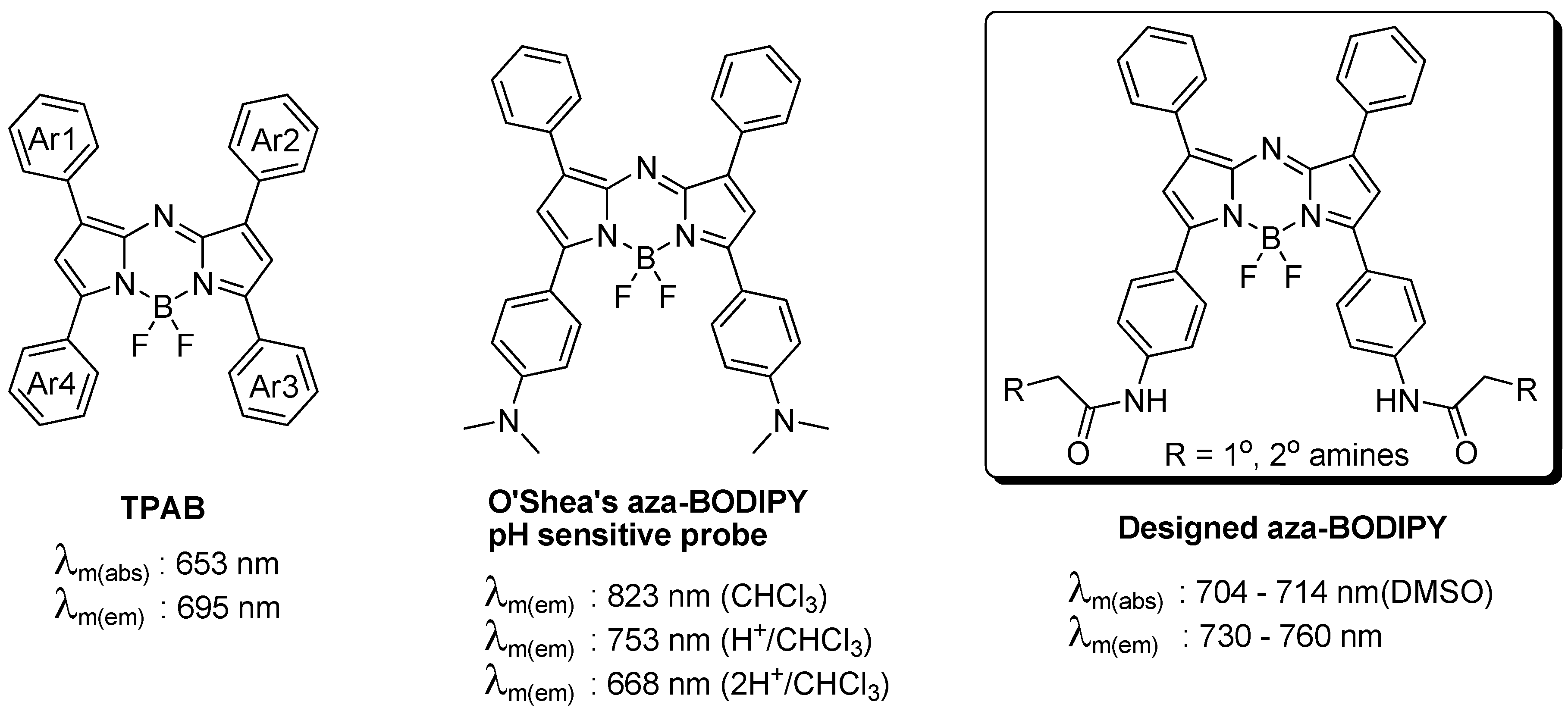
:1. Introduction
2. Experimental Section
2.1. Materials & Method
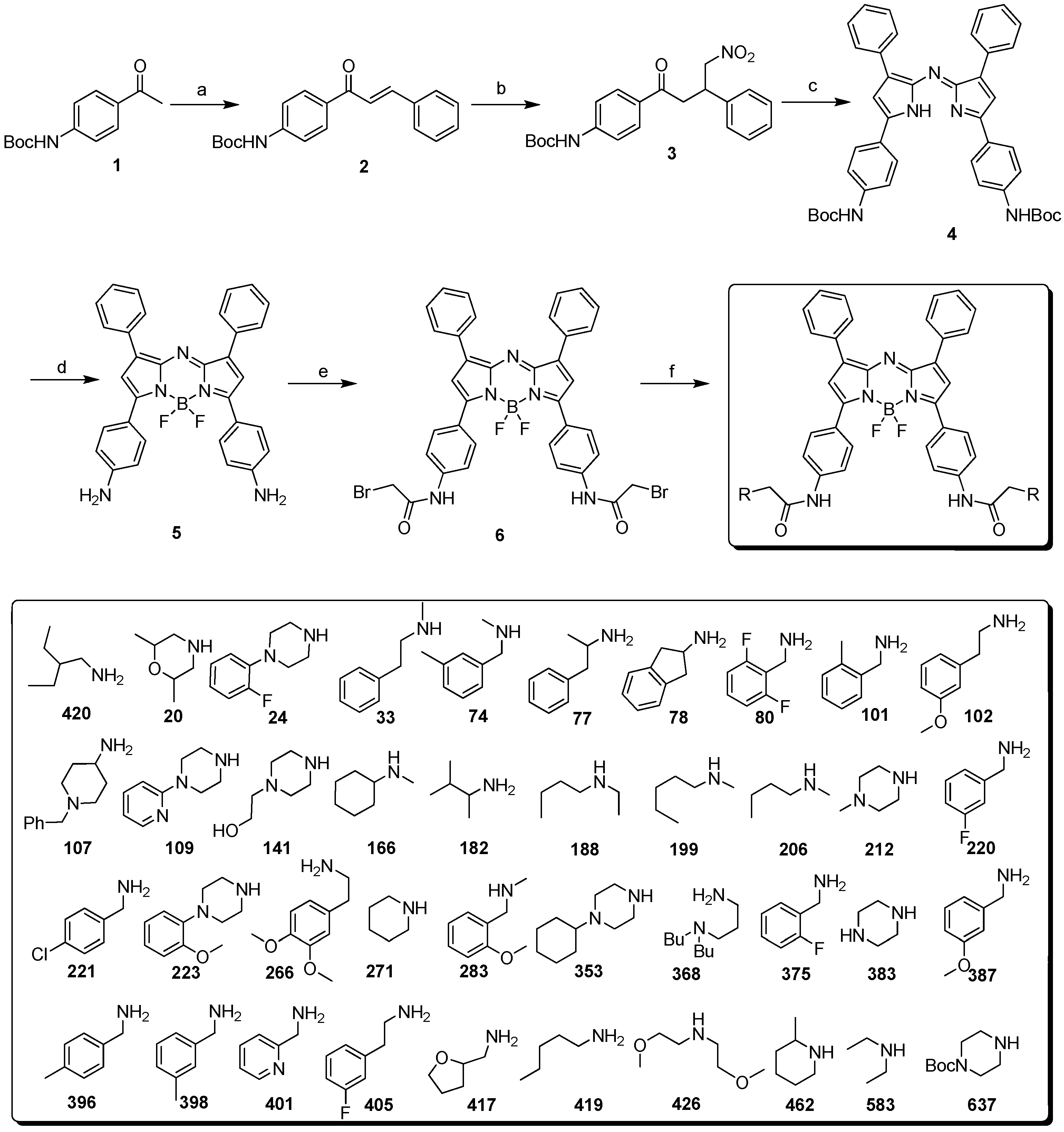
2.2. Synthesis
3. Results and Discussion
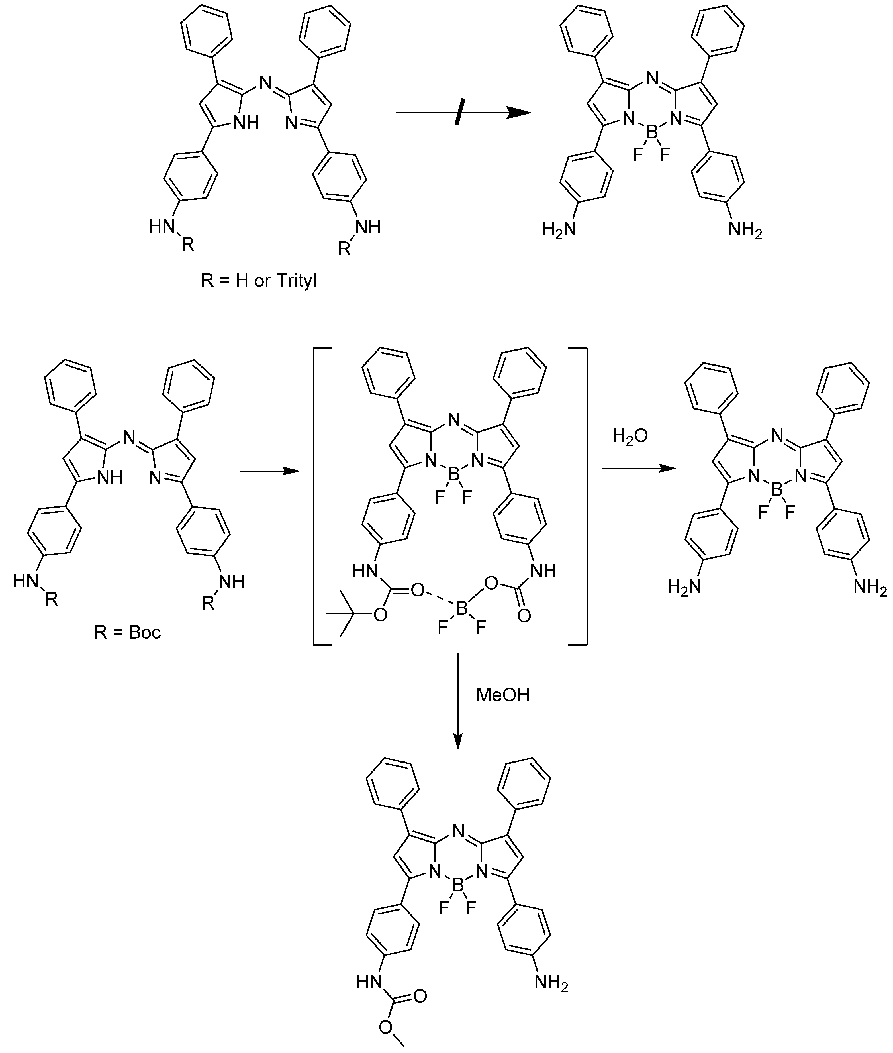
3.1. Synthesis


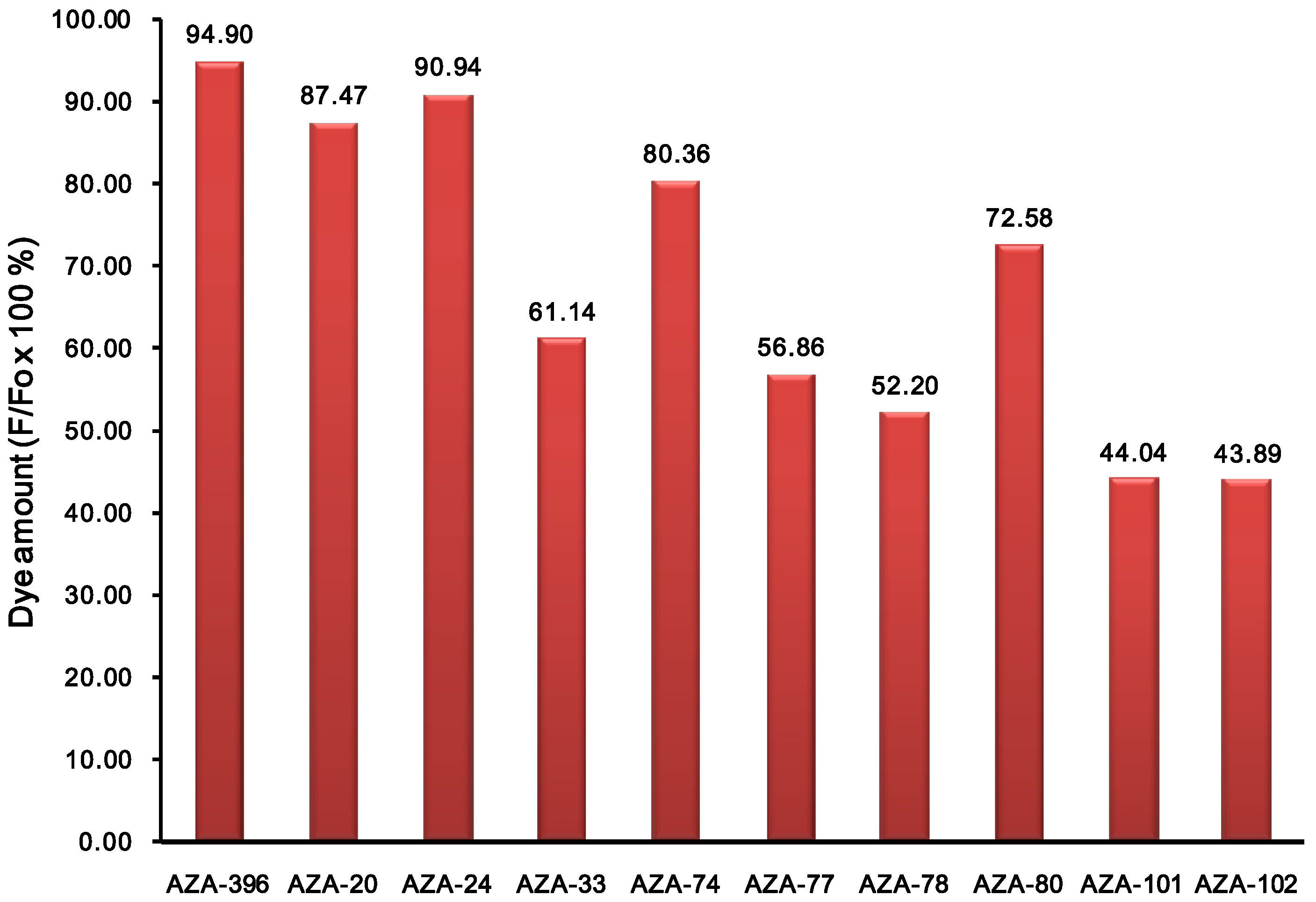
3.2. Spectroscopic Properties
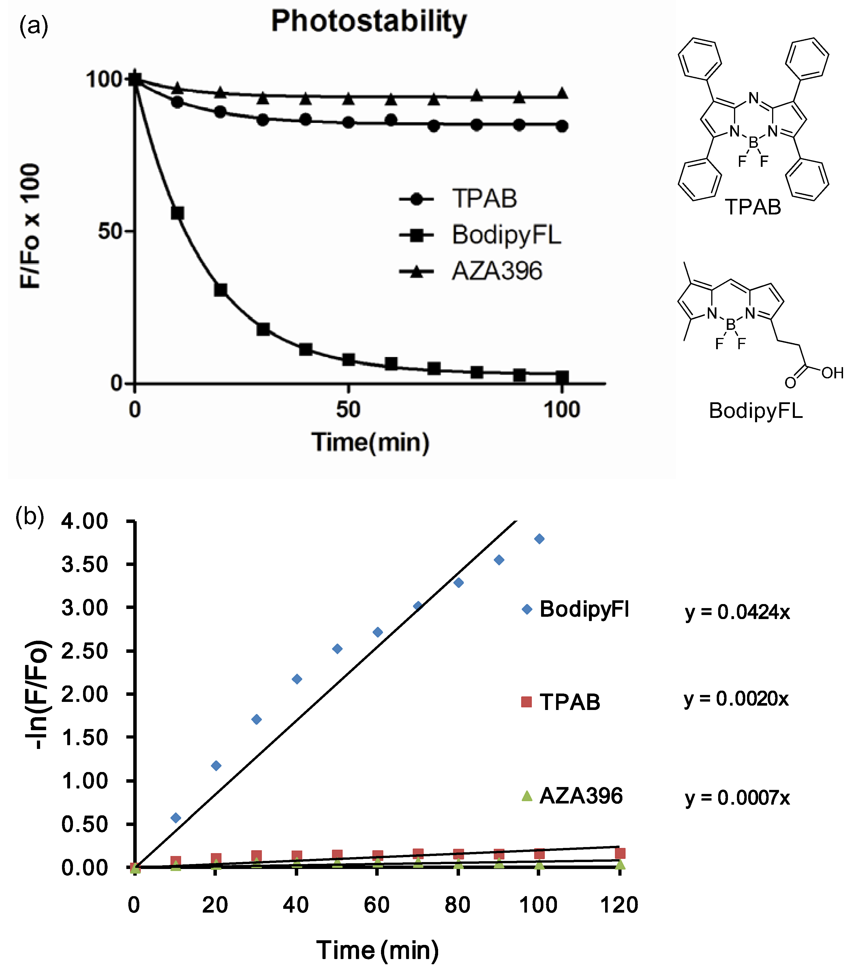
3.3. Photostability

4. Conclusions
Supplementary Materials
Supplementary File 1Acknowledgments
References
- Stephens, D.J.; Allan, V.J. Light microscopy techniques for live cell imaging. Science 2003, 300, 82–86. [Google Scholar] [CrossRef] [PubMed]
- Lippincott-Schwartz, J.; Patterson, G.H. Development and use of fluorescent protein markers in living cells. Science 2003, 300, 87–91. [Google Scholar] [CrossRef] [PubMed]
- Rieder, C.L.; Khodjakov, A. Mitosis through the microscope: Advances in seeing inside live dividing cells. Science 2003, 300, 91–96. [Google Scholar] [CrossRef] [PubMed]
- Frangioni, J.V. In vivo near-infrared fluorescence imaging. Curr. Opin. Chem. Biol. 2003, 7, 626–634. [Google Scholar] [CrossRef] [PubMed]
- Das, R.K.; Samanta, A.; Ha, H.H.; Chang, Y.T. Solid phase synthesis of ultra-photostable cyanine NIR dye. RSC Adv. 2011, 1, 573–575. [Google Scholar] [CrossRef]
- Maiti, K.K.; Samanta, A.; Vendrell, M.; Soh, K.S.; Olivo, M.; Chang, Y.T. Multiplex cancer cell detection by SERS nanotags with cyanine and triphenylmethine Raman reporters. Chem. Commun. 2011, 47, 3514–3516. [Google Scholar] [CrossRef]
- Samanta, A.; Vendrell, M.; Das, R.; Chang, Y.T. Development of photostable near-IR cyanine dyes. Chem. Commun. 2010, 46, 7406–7408. [Google Scholar] [CrossRef]
- Samanta, A.; Vendrell, M.; Yun, S.W.; Guan, Z.; Xu, Q.H.; Chang, Y.T. A photostable near-infrared protein labeling dye for in vivo imaging. Chem. Asian J. 2011, 6, 1353–1357. [Google Scholar] [CrossRef] [PubMed]
- Samanta, A.; Maiti, K.K.; Soh, K.S.; Liao, X.; Vendrell, M.; Dinish, U.S.; Yun, S.W.; Bhuvaneswari, R.; Kim, H.; Rautela, S.; et al. Ultrasensitive near-infrared Raman reporters for SERS-based in vivo cancer detection. Angew. Chem. Int. Ed. 2011, 50, 6089–6092. [Google Scholar] [CrossRef]
- McDonnell, S.O.; O’Shea, D.F. Near-infrared sensing properties of dimethlyamino-substituted BF2-azadipyrromethenes. Org. Lett. 2006, 8, 3493–3496. [Google Scholar] [CrossRef] [PubMed]
- Jokic, T.; Borisov, S.M.; Saf, R.; Nielsen, D.A.; Kuhl, M.; Klimant, I. Highly photostable near-infrared fluorescent pH indicators and sensors based on BF2-chelated tetraarylazadipyrromethene dyes. Anal. Chem. 2012, 84, 6723–6730. [Google Scholar] [CrossRef] [PubMed]
- Lee, J.S.; Vendrell, M.; Chang, Y.T. Diversity-oriented optical imaging probe development. Curr. Opin. Chem. Biol. 2011, 15, 760–767. [Google Scholar] [CrossRef] [PubMed]
- Loudet, A.; Bandichhor, R.; Wu, L.; Burgess, K. Functionalized BF2 Chelated Azadipyrromethene Dyes. Tetrahedron 2008, 64, 3642–3654. [Google Scholar] [CrossRef] [PubMed]
© 2013 by the authors; licensee MDPI, Basel, Switzerland. This article is an open access article distributed under the terms and conditions of the Creative Commons Attribution license (http://creativecommons.org/licenses/by/3.0/).
Share and Cite
Lee, S.-C.; Zhai, D.; Mukherjee, P.; Chang, Y.-T. The Development of Novel Near-Infrared (NIR) Tetraarylazadipyrromethene Fluorescent Dyes. Materials 2013, 6, 1779-1788. https://doi.org/10.3390/ma6051779
Lee S-C, Zhai D, Mukherjee P, Chang Y-T. The Development of Novel Near-Infrared (NIR) Tetraarylazadipyrromethene Fluorescent Dyes. Materials. 2013; 6(5):1779-1788. https://doi.org/10.3390/ma6051779
Chicago/Turabian StyleLee, Sung-Chan, Duanting Zhai, Parag Mukherjee, and Young-Tae Chang. 2013. "The Development of Novel Near-Infrared (NIR) Tetraarylazadipyrromethene Fluorescent Dyes" Materials 6, no. 5: 1779-1788. https://doi.org/10.3390/ma6051779