Communication

Redox Response of Reduced Graphene Oxide-Modified Glassy Carbon Electrodes to Hydrogen Peroxide and Hydrazine

Graduate School of Pharmaceutical Sciences, Tohoku University, Aramaki, Aoba-ku, Sendai 980-8578, Japan
*
Author to whom correspondence should be addressed.
Materials 2013, 6(5), 1840-1850; https://doi.org/10.3390/ma6051840
Submission received: 12 April 2013 / Revised: 26 April 2013 / Accepted: 28 April 2013 / Published: 7 May 2013

Abstract

:
The surface of a glassy carbon (GC) electrode was modified with reduced graphene oxide (rGO) to evaluate the electrochemical response of the modified GC electrodes to hydrogen peroxide (H2O2) and hydrazine. The electrode potential of the GC electrode was repeatedly scanned from −1.5 to 0.6 V in an aqueous dispersion of graphene oxide (GO) to deposit rGO on the surface of the GC electrode. The surface morphology of the modified GC electrode was characterized by scanning electron microscopy (SEM) and atomic force microscopy (AFM). SEM and AFM observations revealed that aggregated rGO was deposited on the GC electrode, forming a rather rough surface. The rGO-modified electrodes exhibited significantly higher responses in redox reactions of H2O2 as compared with the response of an unmodified GC electrode. In addition, the electrocatalytic activity of the rGO-modified electrode to hydrazine oxidation was also higher than that of the unmodified GC electrode. The response of the rGO-modified electrode was rationalized based on the higher catalytic activity of rGO to the redox reactions of H2O2 and hydrazine. The results suggest that rGO-modified electrodes are useful for constructing electrochemical sensors.

1. Introduction

A variety of functional materials have been utilized to modify the surface of electrodes for constructing electrochemical devices, which include catalysts [1,2], redox mediators [3,4], polymers [5,6,7,8], proteins [9,10,11], and DNA [12,13]. Recently, carbon nanomaterials have been widely employed for this purpose because of their high catalytic activity in redox reactions [14,15,16,17,18,19,20,21,22]. In this context, we have studied the catalytic activity of carbon nanotubes (CNTs) deposited on the surface of metal and carbon electrodes, and demonstrated high catalytic activity of CNTs in the oxidation of hydrogen peroxide (H2O2) [20]. CNT-modified electrodes have successfully been used to construct choline and lactate biosensors by combining the electrodes with enzymes [21,22]. In some cases, carbon CNTs have been combined with other functional materials to enhance catalytic activity [23,24].
Recently, graphene has attracted considerable attention from diverse fields in science and technology because of its excellent electrical and mechanical properties [25,26,27,28,29,30]. Graphene is a two-dimensional nanosheet consisting of sp2-hybridized carbon atoms arranged in a honeycomb structure. Graphene materials exhibit many advantages over CNTs, including higher surface area, structural simplicity, higher conductivity, and lower production cost. Thus, graphene materials have been applied to the development of optical and electrochemical sensors [31,32,33,34], energy storage [35,36], fuel cells [37], controlled release [38,39,40], and so forth. In the present study, we have prepared reduced grapheme oxide (rGO)-modified glassy carbon (GC) electrodes and have evaluated their catalytic activity with respect to redox reactions of H2O2 and hydrazine. rGO-modified electrodes were prepared by electrodeposition of graphene oxide (GO), in which GO was electrochemically reduced to form a thin layer of insoluble rGO on the surface of the GC electrode. The rGO-modified GC electrodes exhibited high catalytic activity to oxidation of H2O2 and hydrazine. We discuss the possible use of rGO-modified electrodes for constructing electrochemical sensors.

2. Experimental Section

2.1. Reagents

Graphite powder was obtained from Nakarai Co. (Kyoto, Japan). Hydrogen peroxide (30% aqueous solution) and hydrazine monohydrate were purchased from Santoku Chemical Co. (Tokyo, Japan) and Tokyo Kasei Co. (Tokyo, Japan), respectively. All other reagents were of the highest grade available and were used without further purification. We synthesized GO in accordance with the Hammers method [41].

2.2. Apparatus

We used atomic force microscopy (AFM, SPM-9600, Shimadzu Co., Kyoto, Japan) and scanning electron microscopy (SEM, S-3200N, Hitachi Co., Tokyo, Japan) for imaging GO and the surface of rGO-modified electrodes. All electrochemical measurements were performed using an electrochemical analyzer (Model 660B, ALS Co., Tokyo, Japan).

2.3. Deposition of rGO on the Surface of a GC Electrode

We deposited GO on the surface of a GC disk electrode (3-mm diameter) in accordance with a published procedure with slight modifications [42]. Briefly, the GC electrode was immersed in a dispersion of GO (0.3 mg/mL) in 0.7 mM pH 9 phosphate buffer. We then repeatedly scanned the electrode potential from −1.5 to 0.6 V (vs. Ag/AgCl electrode) at 10 mV s−1 with gentle stirring under a nitrogen atmosphere (~20 °C). The modified GC electrode thus prepared was rinsed thoroughly in working buffer before use.

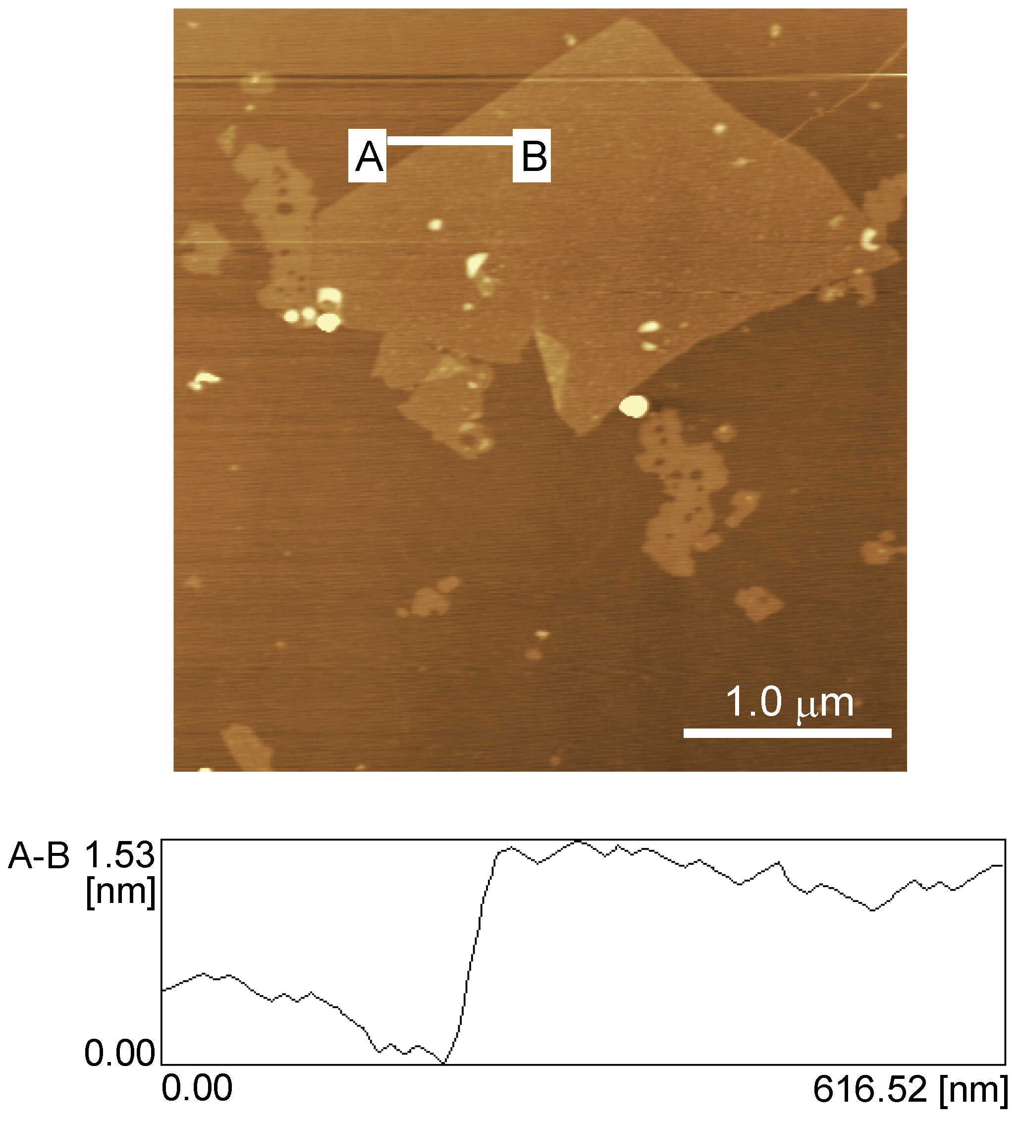
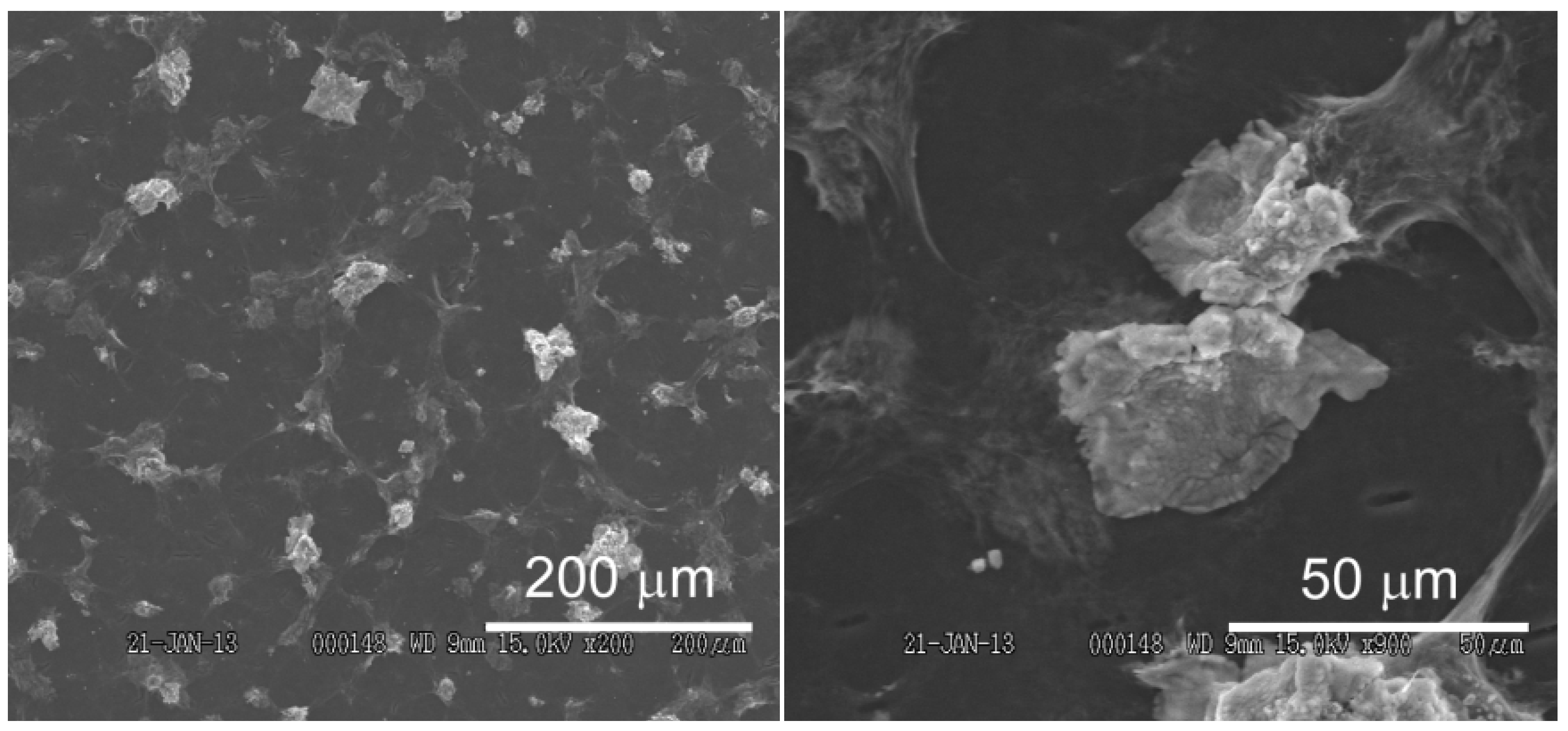
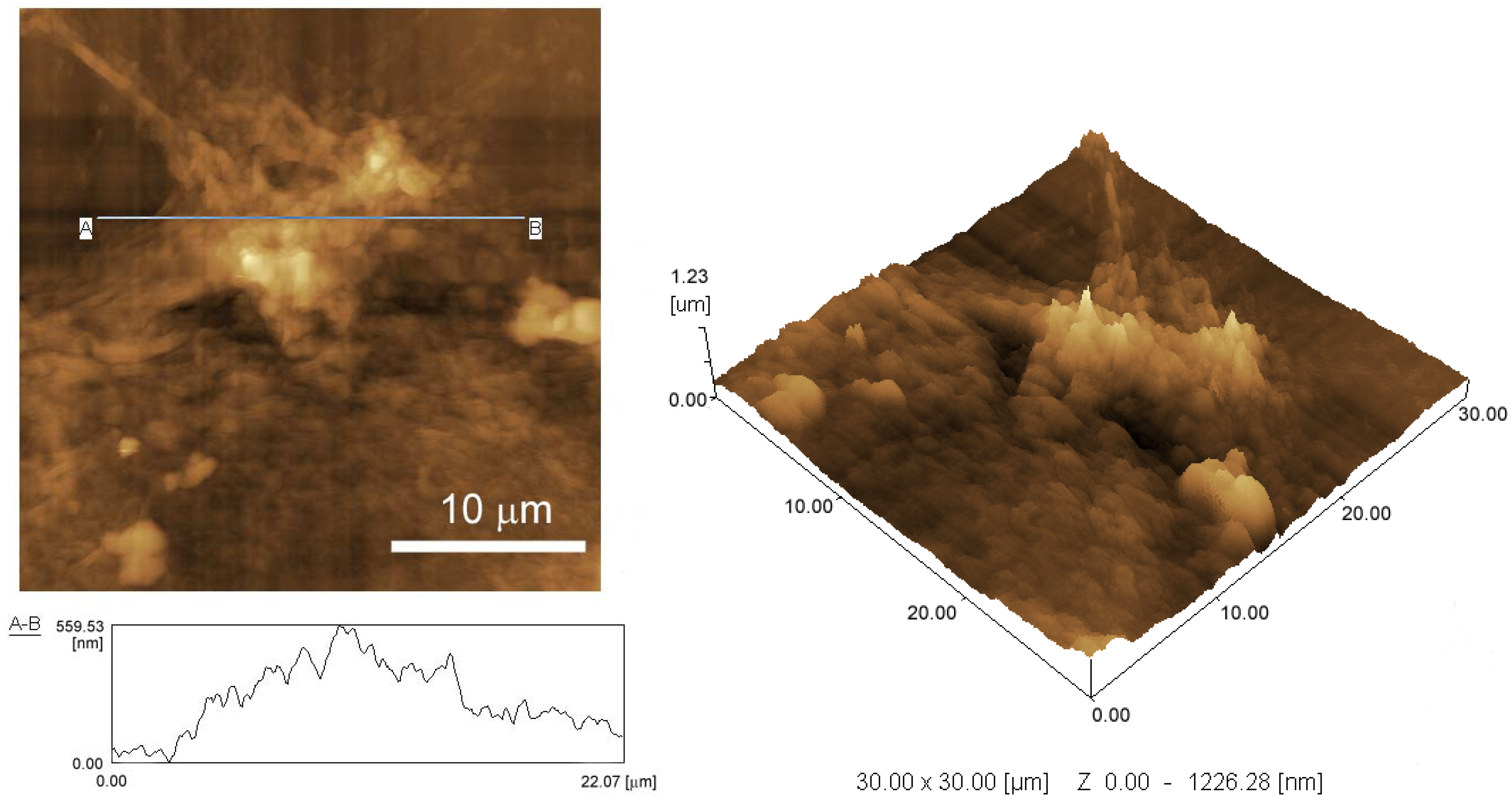
2.4. AFM and SEM imaging

AFM images of GO were recorded in air at room temperature (~20 °C) using a SPM-9600 instrument operating in dynamic (tapping) mode. The sample for AFM observation was prepared on a mica surface. We obtained SEM images of the deposited rGO for platinum-sputtered samples (prepared on a GC plate) with an S-3200N instrument operating at 15 kV. AFM was also employed to study the surface morphology of the rGO-deposited GC plate.

2.5. Electrochemical Measurements

We measured the electrochemical response of rGO-modified and unmodified GC electrodes in a glass cell using the GC electrode as the working electrode, a platinum wire as the counter electrode, and a Ag/AgCl electrode (3.3 M KCl) as the reference electrode. All measurements were performed in air at room temperature (~20 °C).

3. Results and Discussion

3.1. Preparation of rGO-Modified GC Electrode

Prior to depositing rGO on the surface of a GC electrode, we characterized the synthesized GO using AFM. Figure 1 shows a typical AFM image and height profile of a GO sheet deposited onto a mica substrate from an aqueous dispersion. The AFM image demonstrates that the GO sheet has a thickness of 1.0–1.5 nm and a length in the range of several micrometers. The observed thickness of the GO sheet agrees well with the reported average thickness of fully exfoliated GO sheets [43,44].
We electrochemically deposited the GO sheets on the surface of a GC electrode. GO is converted to rGO during electrodeposition [42]. The surface morphology of the modified GC electrode was studied by SEM and AFM. Figure 2 shows SEM images of the surface of an rGO-modified GC electrode. The surface of the GC electrode was covered with an rGO layer containing large aggregates 10–30 μm in length (Figure 2B). Figure 3 shows AFM images and a height profile of the rGO aggregate, demonstrating that the rGO aggregate has a height of >500 nm. The SEM and AFM images clearly show that the surface of an rGO-modified GC electrode is rather rough, which is attributable to rGO aggregate formation. It is likely that, during electrodeposition, GO was electrochemically reduced to form highly hydrophobic rGO when the electrode potential was negatively scanned, which resulted in deposition of aggregated rGO on the electrode surface.
Figure 1. Atomic force microscopy (AFM) image of graphene oxide (GO) and its height profile.
Materials 06 01840 g001
Figure 2. Scanning electron microscopy (SEM) images of the surface of an rGO-modified glassy carbon (GC) electrode.
Materials 06 01840 g002

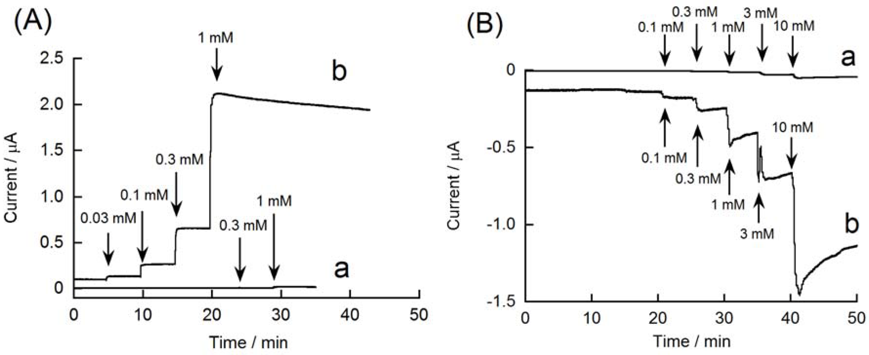
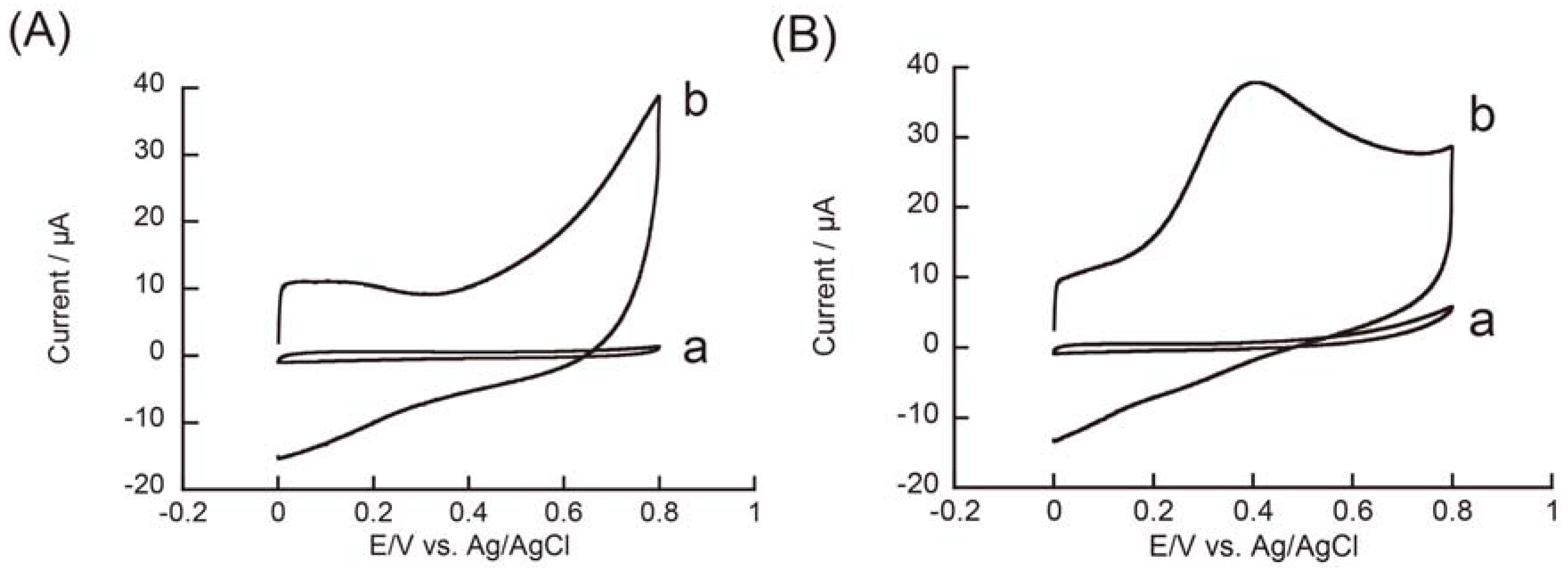
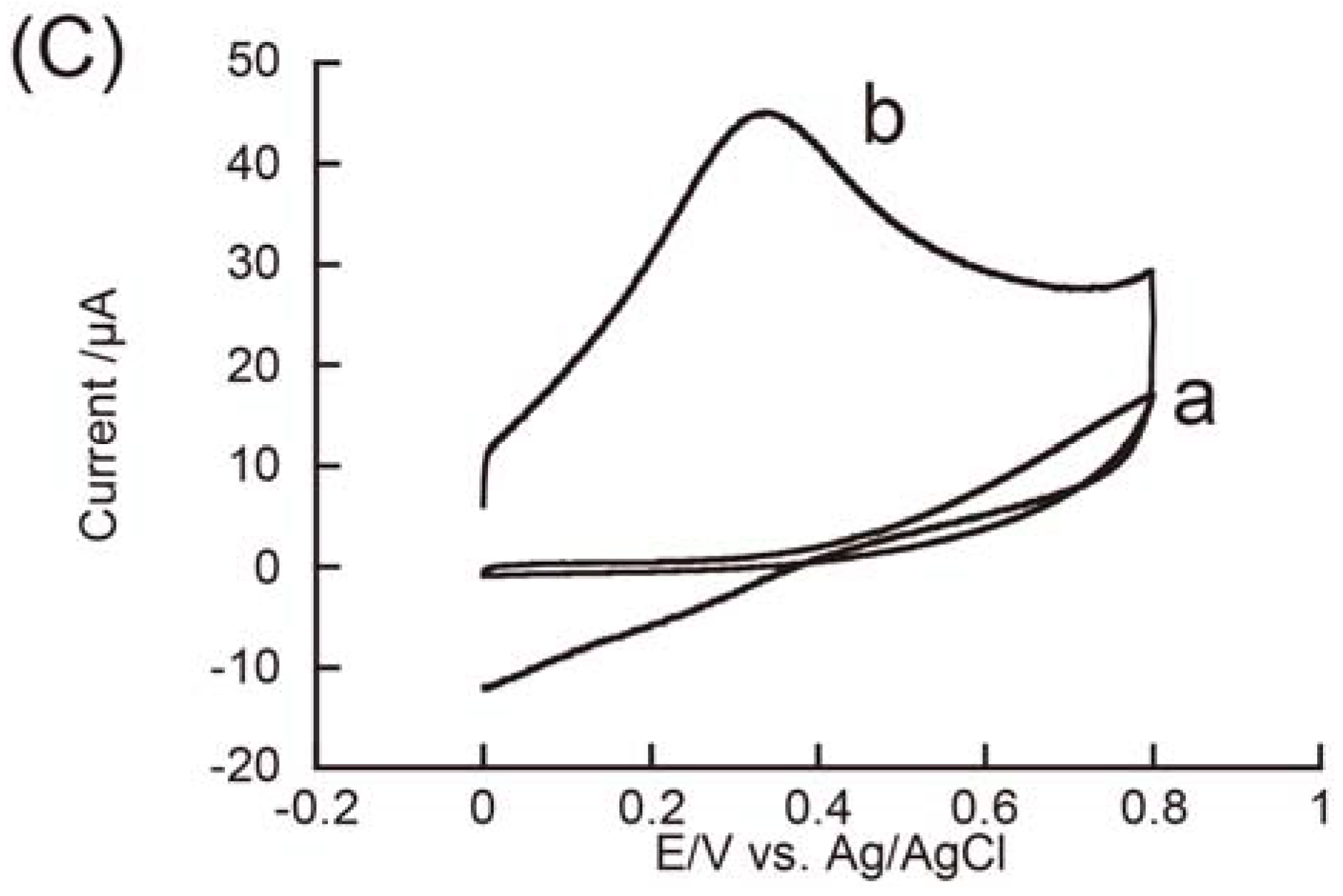
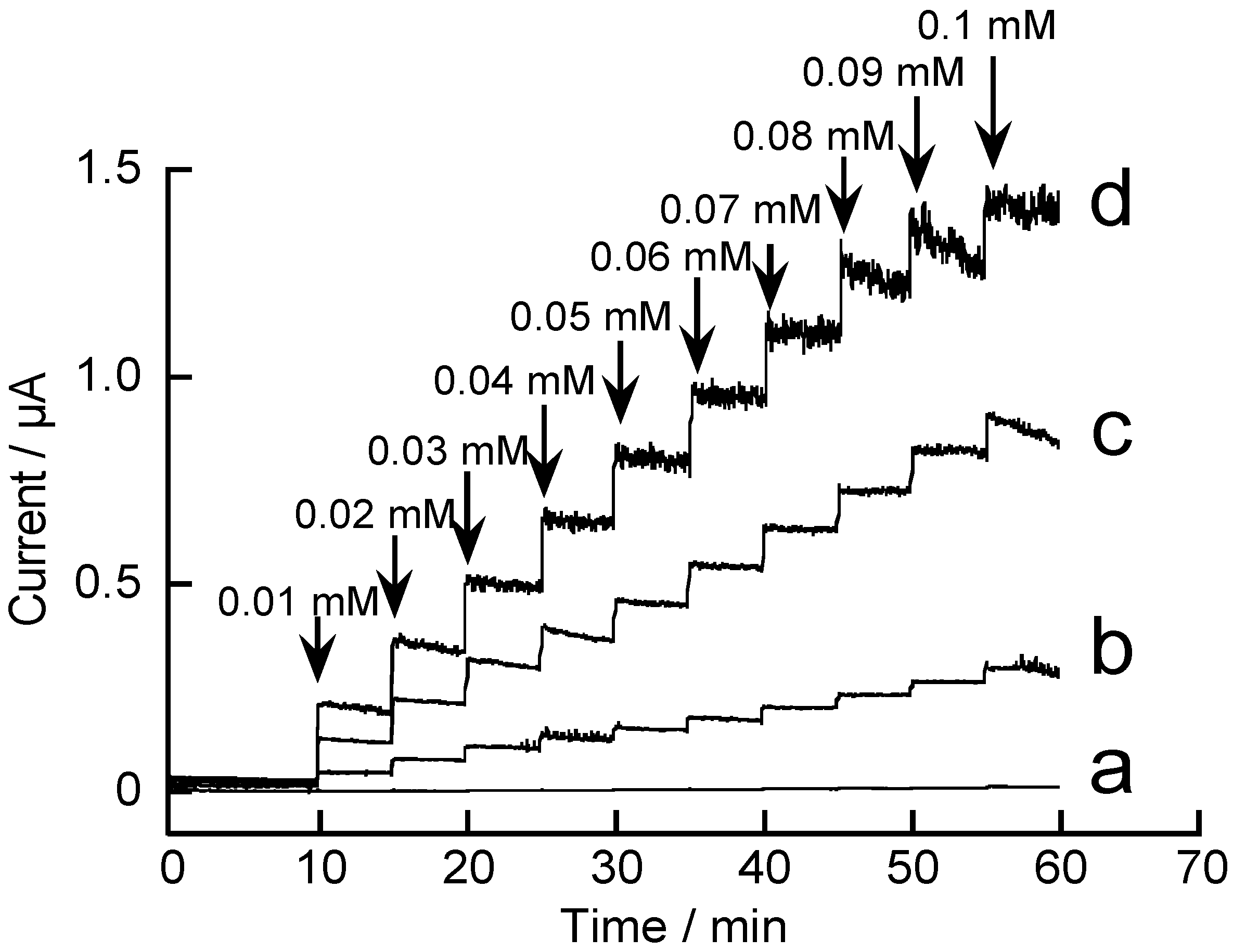
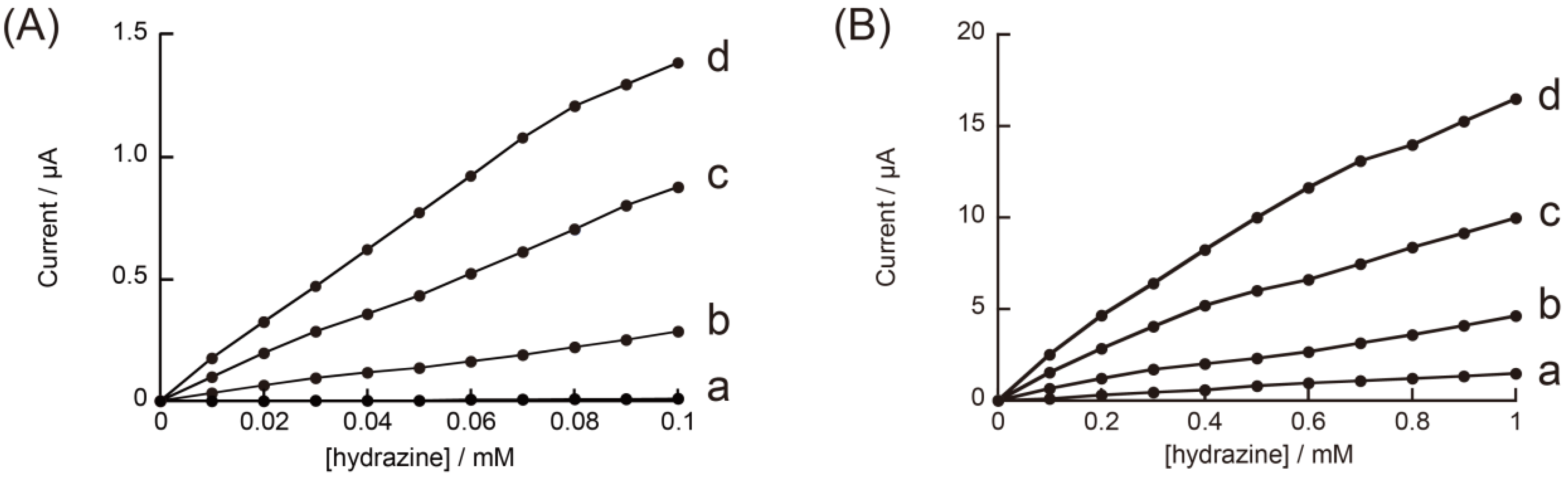
3.2. Redox Reactions of H2O2 and Hydrazine on an rGO-Modified Electrode

The rGO-modified electrode may exhibit high catalytic activity in redox reactions because a large quantity of rGO was deposited on the GC electrode. Edge-plane-like defective sites on rGO facilitate electron transfer to molecules in solution [45]. To evaluate the catalytic activity of rGO-modified GC electrodes, we recorded cyclic voltammograms (CVs) of H2O2 (Figure 4). The CV of H2O2 on an rGO-modified GC electrode exhibited oxidation and reduction currents, the onset potentials of which were 0.4 and 0.1 V, respectively, whereas the unmodified GC electrode exhibited virtually no redox response to H2O2 in the potential range tested. Thus, the rGO-modified GC electrode exhibited a significantly higher response to H2O2 than the unmodified electrode. These results suggest that an rGO-modified electrode can be used for amperometric quantitation of H2O2.
Figure 3. AFM images of rGO aggregates deposited on a GC electrode and its height profile.
Materials 06 01840 g003
Figure 4. Cyclic voltammograms (CVs) of 3 mM H2O2 on (a) unmodified GC and (b) rGO-modified electrodes in 0.1 M pH 7.4 phosphate buffer. Scan rate: 0.1 V·s−1.
Materials 06 01840 g004
Figure 5A depicts the amperometric response of rGO-modified and unmodified GC electrodes at 0.6 V. The oxidation current of H2O2 increased with increasing H2O2 concentration, whereas the response of the unmodified GC electrode was negligible. The rGO-modified electrode can also be operated at −0.1 V to detect a reduction current of H2O2 (Figure 5B). The reduction current increased in accordance with the concentration of H2O2. Yang and coworkers have recently reported that GC electrodes modified with GO-Ag composite exhibit amperometric response to 0.0016–9.0 mM H2O2 [46]. These results suggest that an rGO-modified electrode can be used for constructing biosensors by combining oxidase enzymes, such as glucose oxidase, lactate oxidase, and cholesterol oxidase, because these enzymes generate H2O2 as reaction products.
Figure 5. Amperometric responses of (a) unmodified and (b) rGO-modified GC electrodes to H2O2, recorded at (A) 0.6 V and (B) −0.1 V.
Materials 06 01840 g005
We also studied the redox reaction of hydrazine on an rGO-modified electrode. Figure 6 shows CVs of hydrazine recorded on rGO-modified and unmodified GC electrodes at pH 5.0, 7.0, and 9.0. The unmodified GC electrode exhibited no redox peak over the potential range of 0–0.8 V under the present experimental conditions, although the oxidation current slightly increased at 0.6–0.8 V in pH 9.0 medium. In contrast, we observed well-defined oxidation peaks in CVs at 0.35–0.4 V in pH 7.0 and 9.0 solutions on an rGO-modified electrode. No reduction peak was observed in the CVs during the reverse scan, demonstrating that the redox process was irreversible. We did not observe an oxidation peak at pH 5.0 in the potential range of 0–0.8 V, although the oxidation current gradually increased at 0.4–0.8 V. The oxidation current in CVs recorded at pH 7.0 and 9.0 was remarkably high. In this context, Wang and coworkers also reported an enhanced redox response of hydrazine on GC electrodes modified with polymer-coated graphene [47]. Electrocatalytic oxidation of hydrazine was pH-dependent, that is, higher in neutral or basic solutions [48], in accordance with the pH-dependent response of hydrazine on the rGO-modified electrode.
Figure 6. CVs of 3 mM hydrazine recorded on (a) unmodified GC and (b) rGO-modified electrodes at (A) pH 5.0; (B) 7.0; and (C) 9.0.
Materials 06 01840 g006aMaterials 06 01840 g006b
It is interesting to evaluate the amperometric response of rGO-modified GC electrodes to hydrazine. Figure 7 shows the amperometric response of the rGO-modified GC electrodes to 0.01–0.1 mM hydrazine in solution at pH 7.0.
Figure 7. Amperometric responses of (a) unmodified and (bd) rGO-modified electrodes to hydrazine, recorded at 0.4 V in 0.1 M pH 7.0 phosphate buffer. The rGO-modified electrodes were prepared by scanning electrode potential (b) six; (c) seven; and (d) eight times.
Materials 06 01840 g007
In this experiment, we used three types of rGO-modified electrodes, in which the quantity of rGO loaded on the electrode surface was regulated by changing the number of scans of electrode potential upon electrode position of rGO. The quantity of the deposited rGO on an electrode depends on the number of scans during electrodeposition [42]. Higher quantities of rGO can be deposited on the electrode surface by increasing the number of potential scans. Thus, we fabricated three different types of rGO-modified GC electrodes by changing the number of potential scans (i.e., six, seven, and eight) during electrodeposition. The rGO-modified electrode prepared by scanning the electrode potential eight times exhibited the highest response among the modified electrodes tested. The response of the modified electrode prepared by scanning six times was significantly lower than that of the other electrodes, suggesting that scanning the electrode potential seven or eight times is required for depositing an adequate quantity of rGO on the electrode surface. The results show that rGO-modified electrodes are sensitive to hydrazine in the concentration range of 0.01–0.1 mM. In a separate measurement, we have confirmed that the modified electrodes also exhibit an amperometric response to hydrazine in a higher concentration range, 0.1–1.0 mM (data not shown). Figure 8 shows calibration graphs of the electrodes to hydrazine in the concentration range of 0.01–0.1 and 0.1–1.0 mM. The results suggest a possible use of rGO-modified GC electrodes for quantitating hydrazine at submillimolar concentrations. Recently, electrochemical oxidation of hydrazine on GC electrodes modified with metal nanoparticle-rGO composite has been reported [49].
Figure 8. Calibration graphs of (a) unmodified and (bd) rGO-modified electrodes to hydrazine, recorded at 0.4 V in 0.1 M pH 7.0 phosphate buffer. Hydrazine concentration: (A) 0.01–0.1 mM; (B) 0.1–1 mM.
Materials 06 01840 g008

4. Conclusions

We have demonstrated that rGO-modified GC electrodes can be prepared by electrodeposition of GO by scanning the electrode potential. The rGO-modified GC electrodes exhibited high sensitivity in voltammetric and amperometric measurements of H2O2 and hydrazine. The sensitivity of the modified electrodes can be tuned by regulating the loading of rGO on the electrode surface. Therefore, the rGO-modified GC electrodes are promising with respect to electrochemical sensor development. Further research for improving the performance characteristics of rGO-modified GC electrodes is now in progress in our laboratory.

Acknowledgments

This work was supported in part by JSPS KAKENHI Grant Number 24659013.

References

  1. Zayats, M.; Katz, E.; Willner, I. Electrical contacting of flavoenzymes and NAD(P)+-dependent enzymes by reconstitution and affinity interactions on phenylboronic acid monolayers associated with Au-electrodes. J. Am. Chem. Soc. 2002, 124, 14724–14735. [Google Scholar] [CrossRef] [PubMed]
  2. Shi, H.; Yang, Y.; Huang, J.; Zhao, Z.; Xu, X.; Anzai, J.; Osa, T.; Chen, Q. Amperometric choline biosensors prepared by layer-by-layer deposition of choline oxidase on the Prussian blue-modified platinum electrode. Talanta 2006, 70, 852–858. [Google Scholar] [CrossRef] [PubMed]
  3. Wang, B.; Anzai, J. Redox reactions of ferricyanide ions in layer-by-layer deposited polysaccharide films: A significant effect of the type of polycation in the films. Langmuir 2007, 23, 7378–7384. [Google Scholar] [CrossRef] [PubMed]
  4. Liu, A.; Anzai, J. Ferrocene-containing polyelectrolyte multilayer films: Effects of electrochemically inactive surface layers on the redox properties. Langmuir 2003, 19, 4043–4046. [Google Scholar] [CrossRef]
  5. Egawa, Y.; Seki, T.; Takahashi, S.; Anzai, J. Electrochemical and optical sugar sensors based on phenylboronic acid and its derivatives. Mater. Sci. Eng. C 2011, 31, 1257–1264. [Google Scholar] [CrossRef]
  6. Ge, C.; Doherty, W.J., III; Mendes, S.B.; Armstrong, N.R.; Saavedra, S.S. Voltammetric and waveguide spectroelectrochemical characterization of ultrathin poly(aniline)/poly(acrylic acid) films self-assembled on indium-tin oxide. Talanta 2005, 65, 1126–1131. [Google Scholar] [CrossRef] [PubMed]
  7. Sato, K.; Kodama, D.; Naka, Y.; Anzai, J. Electrochemically induced disintegration of layer-by-layer-assembled thin films composed of 2-iminobiotin-labeled poly(ethyleneimine) and avidin. Biomacromolecules 2006, 7, 3302–3305. [Google Scholar] [CrossRef] [PubMed]
  8. Recksiedler, C.L.; Deore, B.A.; Freund, M.S. A novel layer-by-layer approach for the fabrication of conducting polymer/RNA multilayer films for controlled release. Langmuir 2006, 22, 2811–2815. [Google Scholar] [CrossRef] [PubMed]
  9. Takahashi, S.; Sato, K.; Anzai, J. Layer-by-layer construction of protein architectures through avidin-biotin and lectin-sugar interactions for biosensor applications. Anal. Bioanal. Chem. 2012, 402, 1749–1758. [Google Scholar] [CrossRef] [PubMed]
  10. Leech, D.; Kavanagh, P.; Shuhmann, W. Enzymatic fuel cells: Recent progress. Electrochim. Acta 2012, 84, 223–234. [Google Scholar] [CrossRef]
  11. Sato, K.; Takahashi, S.; Anzai, J. Layer-by-layer thin films and microcapsules for biosensors and controlled release. Anal. Sci. 2012, 28, 929–938. [Google Scholar] [CrossRef] [PubMed]
  12. Budnikov, H.C.; Evtugyn, G.A.; Gennady, A.; Porfireva, A.V. Electrochemical DNA sensors based on electropolymerized materials. Talanta 2012, 102, 137–155. [Google Scholar] [CrossRef] [PubMed]
  13. Liu, A.; Anzai, J. Use of polymeric indicator for electrochemical DNA sensors: Poly(4-vinylpyridine) derivative bearing [Os(5,6-dimethyl-1,10-phenanthroline)2Cl]2+. Anal. Chem. 2004, 76, 2975–2980. [Google Scholar] [CrossRef] [PubMed]
  14. Wang, J.; Musameh, M. Electrochemical detection of trace insulin at carbon-nanotube-modified electrodes. Anal. Chim. Acta 2004, 511, 33–36. [Google Scholar] [CrossRef]
  15. Trojanowicz, M. Analytical applications of carbon nanotubes: A review. Trend Anal. Chem. 2006, 25, 480–489. [Google Scholar] [CrossRef]
  16. Rivas, G.A.; Rubianes, M.D.; Rodriguez, M.C.; Ferreyra, N.F.; Luque, G.L.; Pedano, M.L.; Miscoria, S.A.; Parrado, C. Carbon nanotubes for electrochemical biosensing. Talanta 2007, 74, 291–307. [Google Scholar] [CrossRef] [PubMed]
  17. Mita, D.G.; Attanasio, A.; Arduini, F.; Diano, N.; Grano, V.; Bencivenga, U.; Rossi, S.; Amine, A.; Moscone, D. Enzymatic determination of BPA by means of tyrosinase immobilized on different carbon carriers. Biosens. Bioelectron. 2007, 23, 60–65. [Google Scholar] [CrossRef] [PubMed]
  18. Ensafi, A.A.; Allafchian, A.R.; Razaei, B. A sensitive and selective voltammetric sensor based on multiwall carbon nanotubes decorated with MgCr2O4 for the determination of azithromycin. Colloid Surf. B 2013, 103, 468–474. [Google Scholar] [CrossRef]
  19. Arvand, M.; Gholizadeh, T.M. Simultaneous voltammetric determination of tyrosine and paracetamol using a carbon nanotube-graphene nanosheet nanocomposite modified electrode in human blood serum and pharmaceuticals. Colloid Surf. B 2013, 103, 84–93. [Google Scholar] [CrossRef]
  20. Huang, J.; Yang, Y.; Shi, H.; Song, Z.; Zhao, Z.; Anzai, J.; Osa, T.; Chen, Q. Multi-walled carbon nanotube-based glucose biosensors prepared by a layer-by-layer technique. Mater. Sci. Eng. C 2006, 26, 113–117. [Google Scholar] [CrossRef]
  21. Song, Z.; Huang, J.; Wu, B.; Shi, H.; Anzai, J.; Chen, Q. Amperometric aqueous sol-gel biosensor for low-potential stable choline detection at multi-walled carbon nanotube modified platinum electrode. Sens. Actuators B 2006, 115, 626–633. [Google Scholar] [CrossRef]
  22. Huang, J.; Song, Z.; Li, J.; Yang, Y.; Shi, H.; Wu, B.; Anzai, J.; Osa, T.; Chen, Q. A highly-sensitive L-lactate biosensor based on sol-gel film combined with multi-walled carbon nanotubes (MWCNTs) modified electrode. Mater. Sci. Eng. C 2007, 27, 29–34. [Google Scholar] [CrossRef]
  23. Munge, B.; Liu, G.; Collins, G.; Wang, J. Multiple enzyme layers on carbon nanotubes for electrochemical detection down to 80 DNA copies. Anal. Chem. 2005, 77, 4662–4666. [Google Scholar] [CrossRef] [PubMed]
  24. Yan, X.B.; Chen, X.J.; Tay, B.K.; Khor, K.A. Transparent and flexible glucose biosensor via layer-by-layer assembly of multi-wall carbon nanotubes and glucose oxidase. Electrochem. Commun. 2007, 9, 1269–1275. [Google Scholar] [CrossRef]
  25. Chavez-Valdez, A.; Shaffer, M.S.P.; Boccaccini, A.R. Applications of graphene electrophoretic deposition: A review. J. Phys. Chem. B 2013, 117, 1502–1515. [Google Scholar] [CrossRef] [PubMed]
  26. Yang, W.; Ratinac, K.R.; Ringer, S.P.; Thordarson, P.; Gooding, J.J.; Braet, F. Carbon nanomaterials in biosensors: Should you use nanotubes or graphene? Angew. Chem. Int. Ed. 2010, 49, 2114–2138. [Google Scholar] [CrossRef]
  27. Inagaki, M.; Kim, Y.A.; Endo, M. Graphene: Preparation and structural perfection. J. Mater. Chem. 2011, 21, 3280–3294. [Google Scholar] [CrossRef]
  28. Guo, S.; Dong, S. Graphene nanosheet: Synthesis, molecular engineering, thin film, hybrids, and energy and analytical applications. Chem. Soc. Rev. 2011, 40, 2644–2672. [Google Scholar] [CrossRef] [PubMed]
  29. Brownson, D.A.C.; Munro, L.J.; Kampouris, D.K.; Banks, C.E. Electrochemistry of grapheme: Not such a beneficial electrode materials? RSC Adv. 2011, 1, 978–988. [Google Scholar] [CrossRef]
  30. Brownson, D.A.C.; Kampouris, D.K.; Banks, C.E. Graphene electrochemistry: Fundamental concepts through to prominent applications. Chem. Soc. Rev. 2012, 41, 6944–6976. [Google Scholar] [CrossRef] [PubMed]
  31. Shao, Y.; Wang, J.; Wu, H.; Liu, J.; Aksay, I.A.; Lin, Y. Graphene based electrochemical sensors and biosensors: A review. Electroanalysis 2010, 22, 1027–1036. [Google Scholar] [CrossRef]
  32. Ratinac, K.R.; Yang, W.; Gooding, J.J.; Thordarson, P.; Braet, F. Graphene and related materials in electrochemical sensing. Electroanalysis 2011, 23, 803–826. [Google Scholar] [CrossRef]
  33. Yeo, M.Y.; Park, S.Y.; In, I. Temperature-dependent optical transmittance of chemically reduced graphene oxide/hydroxypropyl cellulose assembly. Chem. Lett. 2012, 41, 197–199. [Google Scholar] [CrossRef]
  34. Zhu, X.; Liu, Q.; Zhu, X.; Li, C.; Xu, M.; Liang, Y. Reduction of graphene oxide via ascorbic acid and its application for simultaneous detection of dopamine and ascorbic acid. Int. J. Electrochem. Sci. 2012, 7, 5172–5184. [Google Scholar]
  35. Shao, Y.; Wang, J.; Engelhard, M.; Wang, C.; Lin, Y. Facile and controllable electrochemical reduction of graphene oxide and its applications. J. Mater. Chem. 2012, 20, 743–748. [Google Scholar] [CrossRef]
  36. Yin, S.; Zhang, Y.; Kong, J.; Zou, C.; Li, C.M.; Lu, X.; Ma, J.; Boey, F.Y.C.; Chen, X. Assembly of graphene sheets into hierarchical structures for high-performance energy storage. ACS Nano 2011, 5, 3831–3838. [Google Scholar] [CrossRef] [PubMed]
  37. Muthuswamy, N.; de la Fuente, J.L.G.; Ochal, P.; Giri, R.; Raaen, S.; Sunde, S.; Ronning, M.; Chen, D. Towards a highly-efficient fuel-cell catalyst: Optimization of Pt particle size, supports and surface-oxygen group concentration. Phys. Chem. Chem. Phys. 2013, 15, 3803–3813. [Google Scholar] [CrossRef] [PubMed]
  38. Pandey, H.; Parashar, V.; Parashar, R.; Prakash, R.; Ramteke, P.W.; Pandey, A.C. Controlled drug release characteristics and enhanced antibacterial effect of graphene nanosheets containing gentamicin sulfate. Nanoscale 2011, 3, 4104–4108. [Google Scholar] [CrossRef] [PubMed]
  39. Kurapati, R.; Raichur, A.M. Near-infrared light-responsive graphene oxide composite multilayer capsules: a novel route for remote controlled drug delivery. Chem. Commun. 2013, 49, 734–736. [Google Scholar] [CrossRef]
  40. Yuan, B.; Zhu, T.; Zhang, Z.; Jiang, Z.; Ma, Y. Self-assembly of multilayered functional films based on graphene oxide sheets for controlled release. J. Mater. Chem. 2011, 21, 3471–3476. [Google Scholar] [CrossRef]
  41. Hammers, W.S., Jr.; Offeman, R.E. Preparation of graphitic oxide. J. Am. Chem. Soc. 1958, 80, 1339–1339. [Google Scholar] [CrossRef]
  42. Chen, L.; Tang, Y.; Wang, K.; Liu, C.; Luo, S. Direct electrodeposition of reduced graphene oxide on glassy carbon electrode and its electrochemical application. Electrochem. Commun. 2011, 13, 133–137. [Google Scholar] [CrossRef]
  43. Guo, H.; Wang, X.; Qian, Q.; Wang, F.; Xie, X. A green approach to the synthesis of graphene nanosheets. ACS Nano 2009, 3, 2653–2659. [Google Scholar] [CrossRef] [PubMed]
  44. Mahmoud, W.E. Morphology and physical properties of poly(ethylene oxide) loaded graphene nanocomposites prepared by two different techniques. Eur. Polym. J. 2011, 47, 1534–1540. [Google Scholar] [CrossRef]
  45. Banks, C.E.; Compton, R.G. Exploring the electrocatalytic sites of carbon nanotubes for NADH detection: an edge plane pyrolytic graphite electrode study. Analyst 2005, 130, 1232–1239. [Google Scholar] [CrossRef] [PubMed]
  46. Zhao, B.; Liu, Z.; Fu, W.; Yang, H. Construction of 3D electrochemically reduced grapheme oxide-silver nanocomposite film and application as nonenzymatic hydrogen peroxide sensor. Electrochem. Commun. 2013, 27, 1–4. [Google Scholar] [CrossRef]
  47. Wang, C.; Zhang, L.; Guo, Z.; Xu, J.; Wang, H.; Zhai, K.; Zhuo, X. A novel hydrazine electrochemical sensor based on the high specific surface area graphene. Microchim. Acta 2010, 169, 1–6. [Google Scholar] [CrossRef]
  48. Perez, E.F.; Neto, G.O.; Tanaka, A.A.; Kubota, L.T. Electrochemical sensor for hydrazine based on silica modified with nickel tetrasulfonated phthalocyanine. Electroanalysis 1998, 10, 111–115. [Google Scholar] [CrossRef]
  49. Wan, Q.; Liu, Y.; Wang, Z.; Wei, W.; Li, B.; Zou, J.; Yang, N. Graphene nanoplatelets supported metal nanoparticles for electrochemical oxidation of hydrazine. Electrochem. Commun. 2013, 29, 29–32. [Google Scholar] [CrossRef]

