Control blocks treated with only DCM, only water, or only the liquid culture media showed an average of less than 1% change in their L*a*b* values between untreated and treated states on both external and internal faces.
3.1. External
In terms of visual color change, all treatments covered all surfaces of the test blocks, with the green colorant from
Chlorociboria aeruginosa (
Figure 1) being particularly striking. However, color change was much more apparent on the aniline dye blocks. The liquid cultures did not perform as well, with only the pink colorant of
S. cuboideum and the yellow colorant of
S. ganodermophthorum coloring all blocks, with lodgepole pine showing a strong color contrast (
Figure 2). No visual external coloration occurred from liquid cultures of
C. aeruginosa, although significant delta
E* values did occur on red alder and Oregon maple, indicating that color change took place, but it was not sufficient to effect a visual color change.
Figure 1.
Coloring of external surface of test blocks. (A) Noble fir with no treatment; (B) aniline dye carried in ethanol on noble fir; (C) wood-agar colorant carried in dichloromethane (DCM) from C. aeruginosa on noble fir.
Figure 1.
Coloring of external surface of test blocks. (A) Noble fir with no treatment; (B) aniline dye carried in ethanol on noble fir; (C) wood-agar colorant carried in dichloromethane (DCM) from C. aeruginosa on noble fir.
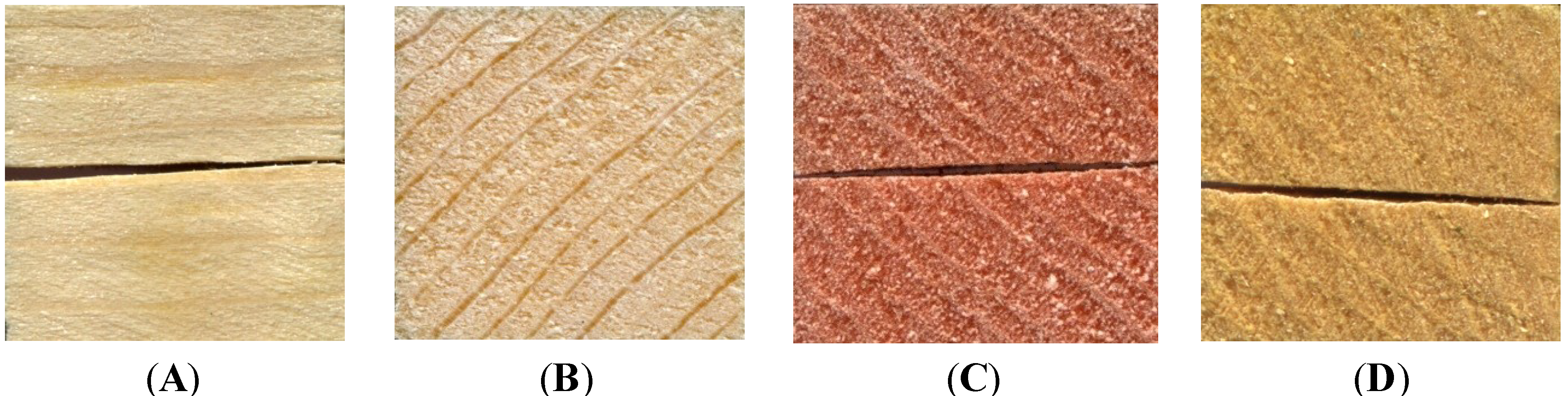
Figure 2.
Coloring of external surface of test blocks from liquid cultures. (A) lodgepole pine with no treatment; (B) lodgepole pine with C. aeruginosa; (C) lodgepole pine with S. cuboideum; (D) lodgepole pine with S. ganodermophthorum.
Figure 2.
Coloring of external surface of test blocks from liquid cultures. (A) lodgepole pine with no treatment; (B) lodgepole pine with C. aeruginosa; (C) lodgepole pine with S. cuboideum; (D) lodgepole pine with S. ganodermophthorum.
The three-way ANOVA comparing the external color change for all four colorants (red, green, yellow, aniline dye) using L*a*b* data found all the independent variables significant at p < 0.0001 and all interactions significant at p < 0.0001 with the exception of the interaction of wood and color, which was significant at p = 0.0009. Despite the high significance, the Tukey HSD identified few significant differences. The noble fir blocks treated with aniline dye showed the greatest increase in delta E* values (558.15), but the value was not significantly different from sugar maple with aniline dye (525.19), sugar maple with yellow colorant in DCM (475.68), lodgepole pine with aniline dye (474.63), port orford cedar with aniline dye (415.07), lodgepole pine with red colorant in liquid culture (378.21), cottonwood with yellow colorant in DCM (368.13), red alder with green colorant in DCM, port orford cedar with yellow colorant in DCM (306.11) and Oregon maple with green colorant in DCM (302.87).
Although the aniline dye did produce significantly more external color change on the wood in most instances, it is important to note that all of the colorants were capable of changing the color of the test block surfaces, with the red and yellow colorants being the most versatile. The way the colorant was carried made a substantial difference, as no visual color change appeared on the surface of blocks pressure treated with the liquid culture of
C. aeruginosa. The visual failure of
C. aeruginosa in liquid culture was most likely due to the aggregation effect common with the green colorant xylindein [
4]. At lower concentrations xylindein can be carried by many solvents, including water. However, there is a threshold at which the xylindein preferentially binds to either itself or the container and can only be mobilized with certain organic solvents. It is likely, due to the age of the liquid cultures, that the xylindein had already moved past the point of aggregation. Experiments with younger cultures may produce different results; however, this would also reduce the concentration of xylindein in solution.
In terms of external dyeing potential, the colorants produced by the fungi
S. cuboideum and
S. ganodermophthorum are just as efficient at surface coverage as a standard aniline dye of similar
L* values (
Table 1). Woodworkers and other craftspeople wishing to utilize natural, permanent dyes for their works should encounter few issues with using the extracted fungal colorants for surface dyeing of wood, especially if they pair the most effective coloration method with wood species. For example, very high delta
E* values occurred in sugar maple and port orford cedar with yellow colorant in DCM, lodgepole pine with red colorant in liquid culture, cottonwood with yellow colorant in liquid culture, and red alder and Oregon maple with green colorant in DCM.
Table 1.
ANOVA results for L*a*b* and percent coverage data by wood species. Different letters indicate statistically significant differences at alpha = 0.05 within each wood species and test column.
Table 1.
ANOVA results for L*a*b* and percent coverage data by wood species. Different letters indicate statistically significant differences at alpha = 0.05 within each wood species and test column.
| Wood | Method | Color | External Delta E* | Internal Delta E* | Internal % Coverage |
|---|
| Ash | aniline dye | blue-green | 144.84 (A) | 53.17 (A) | 16.00 (A) |
| liquid | red | 168.89 (A) | 25.59 (B) | 0 (B) |
| blue-green | 22.68 (A) | 18.16 (B) | 0 (B) |
| yellow | 16.63 (A) | 18.89 (B) | 0 (B) |
| wood-agar | red | 100.40 (A) | 1.57 (B) | 0 (B) |
| blue-green | 100.40 (A) | 11.22 (B) | 0 (B) |
| yellow | 23.52 (A) | 18.89 (B) | 0 (B) |
| Chinkapin | aniline dye | blue-green | 178.24 (A) | 35.68 (A) | 58.67 (A) |
| liquid | red | 128.94 (ABC) | 10.18 (CD) | 0 (B) |
| blue-green | 38.95 (E) | 11.84 (CD) | 0 (B) |
| yellow | 64.09 (CDE) | 8.81 (D) | 0 (B) |
| wood-agar | red | 134.84 (AB) | 27.06 (BC) | 0 (B) |
| blue-green | 110.52 (BCD) | 27.06 (AB) | 0 (B) |
| yellow | 53.38 (DE) | 5.38 (D) | 0 (B) |
| Cottonwood | aniline dye | blue-green | 282.86 (A) | 57.47 (B) | 58.67 (A) |
| liquid | red | 264.71 (A) | 0.76 (D) | 0 (B) |
| blue-green | 64.71 (A) | 2.39 (D) | 0 (B) |
| yellow | 368.13 (A) | 97.24 (A) | 0 (B) |
| wood-agar | red | 131.78 (A) | 29.62 (C) | 2.9 (B) |
| blue-green | 99.47 (A) | 36.61 (BC) | 0 (B) |
| yellow | 45.49 (A) | 1.43 (D) | 0 (B) |
| Lodgepole pine | aniline dye | blue-green | 474.46 (A) | 65.26 (A) | 55.33 (A) |
| liquid | red | 378.21 (B) | 4.69 (C) | 8.67 (B) |
| blue-green | 87.95 (D) | 2.39 (C) | 0 (B) |
| yellow | 185.69 (C) | 3.79 (C) | 0 (B) |
| wood-agar | red | 142.78 (CD) | 16.94 (B) | 0 (B) |
| blue-green | 113.85 (D) | 24.94 (B) | 0 (B) |
| yellow | 182.58 (C) | 2.44 (C) | 0 (B) |
| Noble fir | aniline dye | blue-green | 558.15 (A) | 46.96 (A) | 52.33 (A) |
| liquid | red | 182.16 (B) | 4.28 (B) | 6.67 (B) |
| blue-green | 61.95 (B) | 18.02 (B) | 0 (B) |
| yellow | 110.23 (B) | 5.84 (B) | 0 (B) |
| wood-agar | red | 202.50 (B) | 5.22 (B) | 0.83 (B) |
| blue-green | 227.11 (B) | 6.34 (B) | 0 (B) |
| yellow | 160.88 (B) | 2.79 (B) | 8.57 (B) |
| Oregon maple | aniline dye | blue-green | 162.74 (BC) | 39.91 (A) | 39.33 (A) |
| liquid | red | 136.04 (BC) | 6.70 (B) | 0.67 (B) |
| blue-green | 58.97 (C) | 6.44 (B) | 0 (B) |
| yellow | 70.05 (C) | 5.61 (B) | 0 (B) |
| wood-agar | red | 205.97 (AB) | 31.85 (A) | 0.57 (B) |
| blue-green | 302.87 (A) | 30.76 (A) | 0 (B) |
| yellow | 57.28 (C) | 5.89 (B) | 3.67 (B) |
| Port orford cedar | aniline dye | blue-green | 415.10 (A) | 61.75 (A) | 60.00 (A) |
| liquid | red | 246.50 (A) | 0.95 (D) | 12.33 (B) |
| blue-green | 55.0 (A) | 1.24 (D) | 0 (B) |
| yellow | 112.10 (A) | 1.73 (D) | 0 (B) |
| wood-agar | red | 187.10 (A) | 10.95 (C) | 4.3 (B) |
| blue-green | 165.50 (A) | 21.62 (B) | 4.03 (B) |
| yellow | 306.10 (A) | 3.50 (D) | 9.33 (B) |
| Red alder | aniline dye | blue-green | 142.76 (BC) | 40.38 (B) | 0 (A) |
| liquid | red | 78.27 (C) | 8.59 (C) | 0 (A) |
| blue-green | 78.27 (C) | 8.37 (C) | 0 (A) |
| yellow | 43.49 (C) | 11.92 (C) | 0 (A) |
| wood-agar | red | 259.61 (AB) | 57.68 (A) | 0 (A) |
| blue-green | 318.11 (A) | 41.27 (B) | 0 (A) |
| yellow | 150.56 (ABC) | 5.61 (C) | 1.33 (A) |
| Sweet cherry | aniline dye | blue-green | 153.23 (B) | 90.35 (A) | 8.33 (A) |
| liquid | red | 63.87 (C) | 11.36 (BC) | 0 (B) |
| blue-green | 45.34 (C) | 10.73 (BC) | 0 (B) |
| yellow | 32.11 (C) | 9.30 (C) | 0 (B) |
| wood-agar | red | 241.47 (A) | 94.77 (A) | 0 (B) |
| blue-green | 193.19 (AB) | 53.88 (AB) | 0 (B) |
| yellow | 50.48 (C) | 20.73 (BC) | 0 (B) |
| Sugar maple | aniline dye | blue-green | 525.19 (A) | 52.94 (A) | 12.00 (B) |
| liquid | red | 158.52 (B) | 17.61 (BC) | 10.67 (B) |
| blue-green | 49.53 (B) | 12.60 (CD) | 0 (C) |
| yellow | 29.53 (B) | 15.42 (CD) | 0 (C) |
| wood-agar | red | 218.02 (B) | 9.94 (D) | 0 (C) |
| blue-green | 201.77 (B) | 12.43 (CD) | 0 (C) |
| yellow | 475.68 (A) | 23.10 (B) | 18.33 (A) |
3.2. Internal
Internal results were much more variable than external results. Pressure treatment of any chemical into wood has challenges that can increase depending upon the unique anatomy of each wood species. Generally, species with low heartwood, few extractives, and large earlywood vessels can be pressure treated much more easily than those with more cellular blockage. Additionally, compounds that aggregate are far less likely to be pressure treated into wood, as more aggregation leads to larger compounds that cannot move as readily through wood. Fungi selected for this experiment produce colorants specific to their role in nature—to thrive inside wood through the accumulation of their anti-fungal colorants within the wood structure [
7,
8,
9,
10]. It was assumed that the aniline dyes would show much greater penetration, as the fungal colorants are meant to be delivered by the fungal hyphae or in very small concentrations through the wood via water conduction. In the case of visual internal color change this assumption was confirmed. All wood species showing more internal visual color from the aniline dye blocks than with the other treatments. The notable exception is sugar maple where there was significantly more visual internal color from the yellow colorant in DCM than from the aniline dye (
Table 1). Why sugar maple was the only species to have significant visual internal color not from aniline dye is unknown. However, every laboratory spalting test to date has found sugar maple to be the superior wood species for spalting visual color changes [
3]. When growing fungi directly on the wood this is attributed to the higher sucrose content of sugar maple [
11]. However, as no live cultures were utilized in this experiment, it is unknown why the yellow colorant carried in DCM performed so well.
Interestingly, the results presented in
Table 1 show that in some instances, the fungal colorants are able to create a significant color change; however, that color change may not be visible to the naked eye. The three-way ANOVA for internal color change was highly significant, with all independent variables and interactions significant at
p < 0.0001 except color, which was significant at
p = 0.0011. The highest delta
E* values came from cottonwood treated with the liquid culture yellow (97.24), which had an internal visual surface yellow coverage of 0%, and sweet cherry pressure treated with red (94.77) that also showed no visual color change. The delta
E* values of these two were not significantly higher than sweet cherry treated with aniline dye (90.35), which showed a visual percent coverage of 8.33%.
In the case of the sweet cherry, it is likely that the red color change was masked visually by the natural red color of the wood. The same may also be true of the cottonwood, which is naturally a pale yellow to white. Enlarged scanned images of the blocks with high delta E* readings but no visual color change (increased from 14 mm to 15 cm) revealed small flecks of the target color within the wood, all sufficiently separated so that a gross color change could not be visualized on the small sample. While this confirms the presence of the colorant and validates the larger delta E* readings it does not address the needed results—that a color change take place on the wood that is visual even on small areas. Hence, there is a need for a threshold of delta E* at which visual color changes can be discerned.
Different thresholds have been used to determine how much change in delta
E* constitutes a visual color change, with companies responsible for matching paint colors to consumer samples using as low as delta
E* = 1.0 as the maximum score before color change is visually detectible [
12]. It is obvious from this research that a much higher threshold needs to be set for visual color changes to be seen in wood, taking into account the high color variability even within the same wood species, and the potential desire to increase the color saturation of a given species (turning a red wood “more red”, for example).
3.3. Comparing Percent Coverage to CIE Lab Values
To determine the delta
E* threshold at which visual color change can be discerned, the internal block faces that received a “0” for percent internal coverage were also analyzed using the chroma meter. As the delta
E* values and corresponding visual color change varied so widely with species, thresholds were set for each species individually. For each wood species, the lowest internal visual color coverage numbers was noted, as was its associated delta
E* value. If there were no higher delta
E* values within that wood species showed 0% internal color coverage, the value was recorded as the threshold value in
Table 2. If a higher delta
E* value was found that did not have an associated internal color coverage, the next highest internal visual color coverage number was used until the point was found at which there were no higher delta
E* values without corresponding internal color coverage numbers. Thresholds were identified as the lowest delta
E* value corresponding to a >1% visual internal coverage for the given species. Hypothetically, delta
E* values at or above these species-specific thresholds will be associated with a visual color change.
Table 2.
Minimum delta E* values required for a visual color change, by wood species.
Table 2.
Minimum delta E* values required for a visual color change, by wood species.
| Wood | Threshold (Delta E*) |
|---|
| ash | 54 |
| chinkapin | 36 |
| cottonwood | 58 |
| lodgepole pine | 66 |
| noble fir | 47 |
| Oregon maple | 40 |
| port orford cedar | 22 |
| red alder | NA |
| sweet cherry | 91 |
| sugar maple | 18 |
Table 2 provides a useful baseline guide of the lowest delta
E* value required for a visual color change by wood species, regardless of colorant utilized. However, due to the highly variable nature of wood, these values may not routinely represent minimum delta
E* values. Additionally, given the potential for differences in anatomy for each wood block, delta
E* may not be an ideal measurement of internal color change for pressure treated woods.


