Article

Root Infection of Canker Pathogens, Fusarium circinatum and Diplodia sapinea, in Asymptomatic Trees in Pinus radiata and Pinus pinaster Plantations

by
Laura Hernandez-Escribano
1,2,
Eugenia Iturritxa
3,
Ana Aragonés
3,
Nebai Mesanza
3,
Mónica Berbegal
4,
Rosa Raposo
1,5 and
Margarita Elvira-Recuenco
1,*
1
Instituto Nacional de Investigación y Tecnología Agraria y Alimentaria, Centro de Investigación Forestal (INIA-CIFOR), Ctra. La Coruña, Km. 7.5, 28040 Madrid, Spain
2
Doctorado en Biotecnología y Recursos Genéticos de Plantas y Microorganismos Asociados, Departamento de Biotecnología-Biología Vegetal, Escuela Técnica Superior de Ingeniería Agronómica, Alimentaria y de Biosistemas, Universidad Politécnica de Madrid, 28040 Madrid, Spain
3
NEIKER, Granja Modelo de Arkaute, Apdo 46, 01080 Vitoria-Gasteiz, Spain
4
Instituto Agroforestal Mediterráneo, Universitat Politècnica de València, Camino de Vera s/n, 46022 Valencia, Spain
5
Instituto de Gestión Forestal Sostenible (iuFOR), Universidad de Valladolid-INIA, Avenida de Madrid 44, 34071 Palencia, Spain
*
Author to whom correspondence should be addressed.
Forests 2018, 9(3), 128; https://doi.org/10.3390/f9030128
Submission received: 31 January 2018 / Revised: 28 February 2018 / Accepted: 6 March 2018 / Published: 8 March 2018

Abstract

:
The existence of a latent stage within host tissue of the pine pathogens Fusarium circinatum and Diplodia sapinea, the causal agents of pitch canker and shoot blight disease respectively, has previously been cited. However, studies on this cryptic phase in each disease lifecycle has only been focused on the host aerial parts but not on the roots. Therefore, our objective was to analyze the presence of both pathogens in roots of non-symptomatic mature trees in plantations where the pathogens are known to be causing canker symptoms. For that, we sampled roots from ten non-symptomatic and ten symptomatic trees in three Pinus radiata and one Pinus pinaster plantations in Basque Country, Spain. Both pathogens were isolated from roots of non-symptomatic trees in a higher frequency than from roots of symptomatic trees, 23.3% and 6.6% respectively for D. sapinea and 16.6% and 3.3% respectively for F. circinatum. Neither pathogens was detected in the P. pinaster plantation. The two pathogens were never isolated from the same tree. A high molecular variability was observed for D. sapinea isolates with six different haplotypes and two mating types for the eleven characterized isolates, but only one haplotype and mating type was found for F. circinatum, with all isolates of both fungi being proved pathogenic. These results evidence the importance root infection may have in the disease lifecycle and, therefore, disease management.

1. Introduction

Fusarium circinatum Nirenberg and O’Donnell 1998 [1] and Diplodia sapinea (Fr.) Fuckel [2] are two of the most important pathogens that affect Pinus species causing canker diseases, and can result in significant economic losses in nurseries, plantations and natural forests worldwide [3,4,5,6,7,8]. Disease establishment and severity of pine pitch canker seem to be determined by climatic conditions. A temperature range of 20–25 °C and high humidity favor spore germination, mycelial growth and infection [3,9]. In Europe, F. circinatum at present is restricted to Northern Spain, where it is a non-native pathogen and the first report was in 2005 by Landeras et al. [10], and a few areas in Portugal [11], though there were previous reports in Italy [12] and France [13], while D. sapinea is widely distributed where Pinus spp. are present [8,14,15,16]. Diplodia sapinea is among the most common causes of damage to plantation-grown pines in Spain [17,18] and disease outbreaks usually depend on climatic condition such as hail, drought or heat wave [19,20]. Diplodia sapinea is a mesophilic species with an optimum temperature for growth near 30 °C [21,22] and its growth rate should be favoured by climate warming [14]. In Spain both pathogens have a mayor impact in Pinus radiata D. Don, highly susceptible to both fungi [23,24], whereas Pinus pinaster Ait. shows moderate resistance [5]. Pinus radiata is a non-native species grown for timber production and occupies large extensions in Northern Spain, especially in Basque Country. Pinus pinaster represents the dominant species in the Mediterranean area, especially in the Atlantic coast of France, Portugal and Spain. Thus, the economic and environmental losses that both pathogens cause are of outmost importance.
Fusarium circinatum, the causal agent of pitch canker disease, can infect pines at any stage of plant development, from seedlings to mature trees. In mature trees, the fungus produces branch dieback, and as disease progresses sunken cankers appear in stem or branches with abundant production of resin. Multiple infections of branches may happen and then, it causes severe canopy defoliation [3]. The pathogen can naturally infect reproductive structures that causes mortality of female flowers, misshaped cones, and seed deterioration [25]. Infected seeds are one of the ways of long-distance transmission of the pathogen with various transmission rates cited [23,26,27]. At seedling stage the fungus produces pre- and post-emergence damping-off, which often causes seedling mortality in nurseries [23,26,28]. Diplodia sapinea, the causal agent of shoot (tip) blight disease, is a common and widely distributed pathogen. The most obvious symptoms are needle and shoot blight, seedling damping off, stem cankers and blue stain [7,29,30,31,32]. It is commonly isolated from seedlings in nurseries [33,34] and several organs of pines such as needles, branches, mature wood, cones and seeds [35,36,37,38]. Conidia from pycnidia are easily released in the presence of moisture and disseminated by wind and rain splash [39]. Pine cones infection may become an important source of inoculum and being of primary importance for disease development in plantations [40]. If the infection is severe, both pathogens can produce tree mortality.
Although the occurrence of symptoms makes both pathogens visually detectable, the fact is that several studies have described the existence of a latent stage of both fungi within host tissue, without causing visible damage [26,37,39,41,42]. Both fungi, F. circinatum and D. sapinea, are commonly isolated from seedlings in nurseries. Fusarium circinatum has been reported to remain in this symptomless stage in P. radiata seedlings for up to 2 years [43]. Elvira-Recuenco et al. [26] detected the fungus in asymptomatic P. radiata seedlings 475 days post-inoculation. However, this cryptic infection can become active over time and produce disease, therefore seedlings can serve as vehicle of dissemination of the fungus [41]. Diplodia sapinea is frequently isolated from apparently healthy tissue in both seedlings and adult trees and frequently produces disease when the host is exposed to stress situations such as pruning cuts and drought [44]. It is known that D. sapinea can be released from its quiescent stage in the host by water stress [44], and severe drought in 2003 may have allowed D. sapinea to become epidemic in Central Europe [45]. Persistence of D. sapinea and F. circinatum in the healthy tissues of host trees makes particularly difficult to formulate or impose measures to control movement of the fungus to new environments.
Presence of F. circinatum and D. sapinea in roots has been underestimated in the lifecycle of both pathogens and existing research on the role that infected roots play in symptom expression and disease development is limited. Fusarium circinatum has been detected in roots of pine seedlings [23,28] causing disease symptoms. Likewise, the fungus has the ability to colonize roots of pine seedlings as an endophyte, that is, without causing apparent symptoms [26,41,42], but it has never been reported in roots from asymptomatic mature trees. Seedling roots can be colonized from infected soil [41,42] or by transmission from infected emerged seeds [26], and aerial symptoms are not visible until the fungus reaches the collar region [41]. In addition, results indicate that this occurs when the fungus population in the root is above a certain level [26]. Diplodia sapinea exists as a latent pathogen without visible symptoms on pines in many parts of the world. It can be isolated from healthy tissue of pine trees at all stages from seedling to maturity, mostly in branches, twigs and reproductive organs. The fungus can also be isolated from asymptomatic wood of mature trees [46]. Extensive losses caused by a root disease of Pinus elliottii Engelm. and Pinus taeda Lin. in South Africa and Swaziland, related to stress were observed in the 70 s. Isolations from root and trunk lesions yielded D. sapinea which suggested that it was an important factor in the disease [29].
Our main objective is to determine the presence of F. circinatum and D. sapinea in roots of non-symptomatic mature trees (no cankers) in P. radiata and P. pinaster plantations where the pathogens are known to be causing canker symptoms. In addition, we will characterize morphologically and molecularly by microsatellite analysis all isolates obtained from roots of both symptomatic (cankers were present) and non-symptomatic trees. Furthermore, field-collected soil in one of the plantations will be tested for the presence of both fungi. These results will provide information to elucidate the role of root colonization in the lifecycle of these pathogens.

2. Materials and Methods

2.1. Plant Material Sampling

In four Pinus plantations in Basque Country (NE Spain) showing symptoms of pitch canker disease, roots from ten non-symptomatic and ten symptomatic trees were sampled. Trees were considered as “non-symptomatic” when resinous cankers were not present, in the main stem or in the principal branches. On the contrary, trees with at least one resinous canker were considered as “symptomatic”. In addition, trees were evaluated for the presence/absence of lower and/or upper dead branches as an additional index of a possible infection by D. sapinea. Three of the plantations were P. radiata, located at Bizkaia (100 × 55 m of surface and 7.27 km to the coast), Gipuzkoa (130 × 80 m and 6.08 km) and Araba (160 × 70 m and 32.24 km). The fourth one was a P. pinaster plantation located at Gipuzkoa (90 × 65 m and 3.99 km) (Figure S1). Data on average, minimum and maximum temperatures and precipitation per month in the period 2008–2017 are given per plantation in Figure S2. Diplodia sapinea is known to be present in the area of the study since 1999 [47] and F. circinatum since 2015 [48]. Disease incidence for each plantation estimated by percentage of trees with cankers is shown in Figure 1.
Roots were sampled at a maximum distance of 1 m from the collar region of the tree and within the first 20 cm of depth. Approximately 100 g of roots were collected for each sample with a root diameter of 0.2 to 5 mm. All roots sampled were apparently healthy and no differences were observed between roots collected from non-symptomatic or symptomatic pine trees. Plant material was stored at 4 °C until processing in the following days.

2.2. Isolation in Roots and Morphological Identification

Roots were carefully washed under tap water to remove any adhered soil particles. For surface disinfestation, roots were dipped into 70% ETOH for 1 min, submerged in a 30% commercial bleach with Tween 20 (1 drop/100 mL) solution during 15 min and soaked twice in sterile distilled water. The thinnest roots (less than 1 mm diameter) were immersed in the same commercial bleach solution but for 10 instead of 15 min. Surface disinfested roots were aseptically transferred to sterilized filter paper and when dried, transversally cut into 5 mm segments and placed in petri dishes with Fusarium Selective Medium (FSM) [49], in which D. sapinea could also grow normally. The thickest roots were first longitudinally divided in two and then cut into 5 mm pieces. Three to 6 petri dishes per sampled root were cultured depending on the root thickness. Generally, one isolate was obtained per sampled tree, and 2 isolates in trees where fungi were isolated with more frequency. Plates were incubated in darkness at 25 °C and evaluated every three days.
For morphological fungal identification, putative colonies of F. circinatum growing on FSM were transferred to a Spezieller Nahrstoffarmer Agar (SNA) medium [50] to confirm the species. Plates were incubated for 7–10 days at 25 °C and then microscopically inspected for the formation of coiled sterile hyphae characteristic of F. circinatum [1]. Putative colonies of D. sapinea were transferred to Potato Dextrose agar (PDA) plates, which were incubated for 7 days at 25 ℃ and mycelial growth characteristics observed. Isolates were then grown on 2% water agar with sterilized pine needles at 25 ℃ under near-ultraviolet light (near-UV light) to induce sporulation [51]. Conidia characteristics defined by shape, color, presence of septa, width and length were also observed. According to mycelial growth and conidia characteristics, morphological identification of D. sapinea was verified [2] and other D. species reported to be closely related to D. sapinea by phyhlogenetic analysis [52,53,54,55] discarded, such as: D. intermedia, D. crataegicola [56], D. galiicola [56], D. guayanensis [57] and D. seriata. Diplodia scrobiculata presents similar morphological characteristics to D. sapinea, and specific primers to differentiate both species [58] were used as described below. Monosporic isolates were obtained and conserved on paper (Whatman n° 1) at −20 °C until use.

2.3. Molecular Identification

Molecular confirmation of F. circinatum and D. sapinea was done by PCR using the specific primers CIRC1A (CTTGGCTCGAGAAGGG) and CIRC4A (ACCTACCCTACACCTCTCACT) for F. circinatum as described by Schweigkofler et al. [59] and the specific primers CTTATATATCAAACTATGCTTTG-TA and GCTTACACTTTCATTTATAGACC for D. sapinea as described by Smith and Stanosz [58]. PCR was done in a final volume per reaction of 25 µL with 1 µL DNA template (PCR reactives Bioline). Fungal DNA from both pathogens was extracted from mycelia growing on PDA using E.Z.N.A. Plant DNA Kit (Omega Biotek, Norcross, GA, USA), following the manufacturer’s instructions and stored at −20 °C. PCR amplification was performed in a Veriti 96 well Thermal Cycler (Applied Biosystems, Waltham, MA, USA), and its product was visualized with UV light in a 1% agarose gel (Agarose MS-12, Pronadisa, Shah Alam, Malaysia), stained with RedSafe (RedSafe Nucleic Staining Acid Solution, Intron Biotechnology, Seongnam, Korea). A 100 bp ladder was used as molecular weight marker (Biotools, Jupiter, FL, USA). Fusarium circinatum isolates amplified a 360 bp fragment and D. sapinea isolates a 650 bp fragment (Figure S3).

2.4. Mating Type Determination

For F. circinatum, idiomorphs MAT-1 or MAT-2 were determined for each isolate. A multiplex PCR assay was performed using primers MAT1p1 (AGAAACTGACTGATACATCAAGGGG)-MAT1p3 (TCATAAGAAGTGTTGAAGGAATCACAG) and GcHMG1 (CTTTACCGTAAGGAGCGTCACCAT)-GcHMG2 (TGATCCGCCATCTGCTTGTAGAGT) for alleles MAT-1 y MAT-2, respectively, as described by Wallace and Cover [60] and Schweigkofler et al. [59]. A final volume of 25 µL with 1 µL of DNA template per reaction was used following amplification conditions described by Berbegal et al. [61]. PCR products were visualized in a 1.5% agarose gel under UV light. A 100 bp ladder was used as molecular weight marker. MAT-1 isolates amplified a 380 bp fragment, while MAT-2 produced a 190 bp amplicon.
For D. sapinea, idiomorphs MAT1-1 or MAT1-2 were determined for each isolate. A conventional PCR was used in two separated reactions, with the primers described by Bihon et al. [6], for the MAT1-1-1 gene, primers DipM1f: 50-CAA GCC ATC GAC CGA AAC and DipM1r: 50-GAA GAA GCG CAC CCT CAA T; and the MAT1-2-1 gene, primers DipHMGf: 50-ACA AAG TTC AGC GGA GCG and DipHMGr: 50-CCT CCG CAG GTC ACT CAT [6]. The reaction mixtures had a total volume of 20 µL and contained 15 ng of genomic DNA, 1X PCR buffer (IBIANLab, Zaragoza, Spain), 200 nM each of the dNTPs, 4 pmol of each primer and 0.5 units of Taq DNA polymerase (IBIANLab). The amplification reactions were carried out in a 2720 thermal cycler (Applied Biosystems) programmed for an initial denaturation of 5 min at 95 °C followed by 35 cycles of 30 s at 95 °C, 30 s at 58 °C and 45 s at 72 °C, and a final extension of 7 min at 72 °C. The resulting PCR products were separated by electrophoresis at 100 V for 30 min on a 2% (wt/vol) agarose gel containing 4 µL gelRedTMnucleic acid (10000Xin water, Biotium, Fremont, CA, USA) in 1× TBE buffer and visualized under UV light. A 100 bp ladder was used as molecular weight marker. MAT1-1-1 isolates produced a 416 bp amplicon, while MAT1-2-1 isolates produced a 390 bp amplicon.

2.5. Haplotypes

For F. circinatum haplotype determination, primers for eight SSR loci (FCM-2, FCM-4, FCM-6, FCM-7, FCM-19, FCM-24, FCM-25 and FCM-26) previously characterized and selected for reproducible polymorphism were used [61]. Fluorescent labelling for each SSR locus was performed in one reaction using three primers [62]: an SSR forward primer with a M13 tail (TGTAAAACGACGGCCAGT) at the 5′ end, an SSR reverse primer, and the universal fluorochrome 6-carboxy-fluorescein (FAM) labelled M13 primer. A reference isolate representing the single haplotype identified in Basque Country (isolate CECT20759) [61,63] and a negative control with no DNA were also included. The PCR reaction was performed as described by Berbegal et al. [61] in a final volume of 10 µL. Amplifications were conducted in 96 well plates and PCR products were purified by adding ExoSAP-IT PCR Product Cleanup (Affimetrix, Santa Clara, CA, USA) following the manufacturer’s instructions. PCR products were sized by capillary electrophoresis (Macrogen Europe Lab, kyoto, Japan). Allele calls were performed using the Geneious R10 10.0.9 Software (Biomatters ApS, Europe, Silkeborg, Denmark).
For D. sapinea, 10 microsatellite loci SS1-SS5-SS9-SS10-SS11-SS12-SS13-SS14-SS15-SS16 [14] were amplified for the 11 D. sapinea isolates. Positive controls with known DNA and negative controls without DNA were included. All SSR-PCR products were multiplexed and run in a single lane. SSR-PCR was conducted with a PCR mixture containing 2X QIAGEN® Multiplex PCR kit, 200 nM (each) primer, 15 ng of DNA template and water to a final volume of 13 µL. The reactions were carried out in a 2720 thermal cycler (Applied Biosystems, Foster City, CA, USA) programmed for an initial denaturation of 5 min at 95 °C followed by 15 cycles of 30 s at 95 °C, 30 s at 58 °C and 45 s at 72 °C, 20 cycles of 30 s at 95 °C, 30 s at 55 °C and 45 s at 72 °C and a final extension of 7 min at 72 °C. An amount of 1 µL of a dilution 1:50 of these multiplexed PCR products was separated on an ABI Prism 3130 Genetic analyser. The mobility of SSR products were compared to that of internal size standards (LIZ-500) and alleles estimated by GeneMapper v. 4.0 computer software (Applied Biosystems, Foster City, CA, USA).

2.6. Pathogenicity Tests

Pathogenicity tests for F. circinatum and D. sapinea were conducted with all isolates obtained from roots and one virulent strain of F. circinatum (Isolate CECT20759) [63] as positive control in F. circinatum tests and one virulent strain of D. sapinea (Isolate INIA 111) in D. sapinea tests. Two-year-old P. radiata commercial seedlings were acclimatized before inoculations in a greenhouse during 2 weeks at 22–25 °C and a photoperiod of 12 h light/12 h darkness.
Each isolate was cultured on PDA for 7–10 days in darkness. For F. circinatum, mycelium of each isolate was scrapped off the agar plate with a sterile needle and inoculated by wound in the pine stem parallel to the stem axis [64]. The wound was made approximately at 10 cm above soil level. A sterile needle without inoculum was used to wound plants as negative control. Plants were covered with a plastic bag during 24 h in order to maintain high humidity and favor fungal infection. Six seedlings were randomly inoculated per each isolate. Lesion length was measured at the 5th post-inoculation week. Re-isolation of the pathogen from 2 plants of each isolate on FSM and SNA were conducted to confirm that inoculated isolates were responsible for the lesions observed. For D. sapinea, a piece of agar with mycelium was removed with a sterile needle and placed on the tip of the main stem previously cut off approximately 2–3 cm. A piece of agar without mycelium was used as negative control. Three seedlings were randomly inoculated per each isolate. Lesion length was measured at the 5th post-inoculation week. Re-isolation of the pathogen from some plants on PDA was conducted to confirm that inoculated isolates were responsible for the lesions observed.

2.7. Isolation from Field-Collected Soil

Soil from Bizkaia plantation (P. radiata) was tested for the presence or absence of F. circinatum and D. sapinea. Ten soil samples were collected in 10 points of the plantation by drawing two diagonals across the plantation surface and sampling at five points per diagonal approximately at the same distance from each other. Approximately 500 g within 15 cm of the soil surface were collected per soil sample and any debris in the soil was removed. Each sample was air dried, pass through a 2 mm sieve, divided in two subsamples and a part of each subsample was kept at 100 °C for 48 h to calculate the dry weight later. A suspension per subsample was prepared by adding 10 g of soil sample to 100 mL sterile distilled water and shaken at 150 rpm for 30 min. Three serial 10 fold dilutions were prepared and 400 µL of each suspension was added to 4 petri dishes containing FSM with some modification (3 g/L streptomycin sulphate, 50 mg/L Rose Bengal, 1 g/L PCNB, 1 g/L neomycin). Plates were incubated in darkness at 25 °C and checked during the next 3 weeks. Putative colonies of F. circinatum and D. sapinea were transferred to SNA or PDA medium respectively, in order to identify them. The experiment was repeated twice.

3. Results

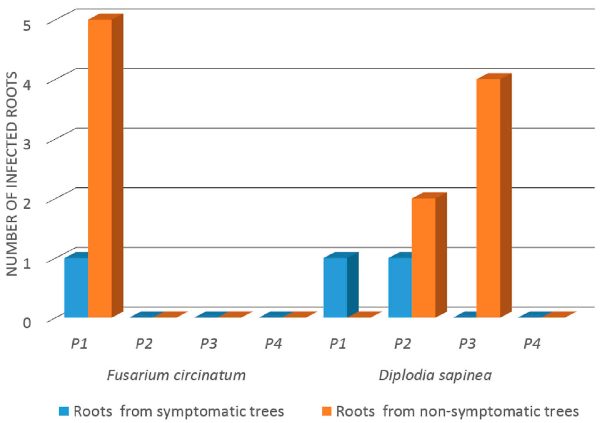
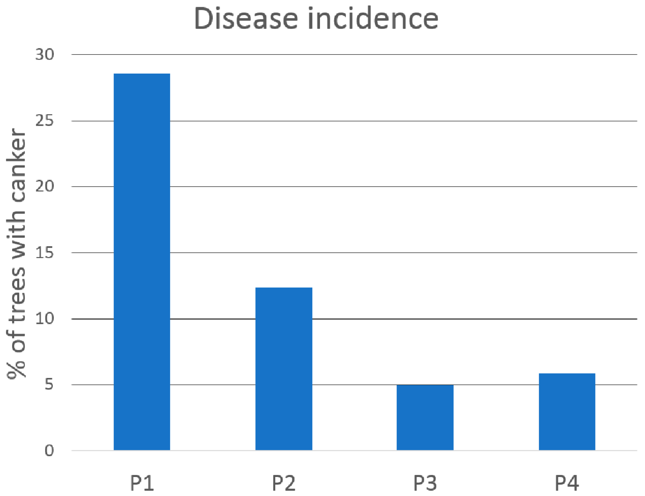
3.1. Incidence of F. circinatum and D. sapinea in Roots

Fusarium circinatum was only isolated from roots at the Bizkaia plantation (6 isolates from 6 trees) where half of sampled roots of non-symptomatic tress (5 roots out of 10) were colonized by the pathogen. However, in roots of symptomatic trees only one sampled root was colonized by the fungus (Figure 2). This plantation had the highest percentage of trees with cankers. Fusarium circinatum was not detected in any of the other three locations for either symptomatic or non-symptomatic trees.
Diplodia sapinea was isolated (11 isolates from 8 trees) from non-symptomatic trees at Gipuzkoa-R and Araba plantations and not at Bizkaia one (Table 1). Twenty percent (2 roots out of 10) and 40% (4 roots out of 10) of non-symptomatic sampled trees were colonized by D. sapinea at Gipuzkoa-R and Araba plantation, respectively (Figure 2). Diplodia sapinea was also isolated from roots of symptomatic trees, one root at Bizkaia and one at Gipuzkoa-R plantation. Two of the 11 isolates came from a tree with upper dead branches and 3 isolates from 3 trees with lower dead branches, with 2 of these trees presenting also cankers. The two plantations with the highest canker rate were the same plantations where D. sapinea was isolated from roots of symptomatic trees.
The pathogens were not isolated from roots of either non-symptomatic or symptomatic trees at P. pinaster plantation (Gipuzkoa-P).
The Bizkaia plantation was the only plantation where both pathogens were detected, but they were not isolated from the same tree.

3.2. Isolate Characterization: Genetic Variability and Pathogenicity

3.2.1. Mating Types

Regarding determination of F. circinatum mating types, results showed that all isolates were exclusively MAT-2. Regarding D. sapinea, two idiomorphs were identified, MAT1-1 and MAT1-2, both present in Gipuzkoa-R and Araba plantations, with the genes MAT1-1-1 for 8 isolates and 1-2-1 for 3 isolates (Table 1). Two different idiomorphs were found in the same tree, ID tree number 237 at Araba and ID tree 231 at Gipuzkoa-R.

3.2.2. Haplotypes

Genotype diversity analysis of F. circinatum isolates by SSR markers revealed that all isolates were identical, and they did not show any polymorphism for any SSR loci. Haplotypes were not differentiated on the basis of their origin, roots of non-symptomatic or symptomatic trees. All isolates presented only peak sizes of 178, 153, 251, 190, 173, 124, 204 and 244 bp resulted for the SSR loci FCM-2, FCM-4, FCM-6, FCM-7, FCM-19, FCM-24, FCM-25 and FCM-26, respectively. No peak was detected in the negative controls.
Genotype diversity analysis of D. sapinea by 10 SSR markers revealed a high polymorphism with 6 different haplotypes for the 11 isolates tested. One of the haplotypes was common for 6 isolates, present in the three plantations where D. sapinea was detected, and each of the other 5 haplotypes was represented by one isolate. There was high diversity in Araba plantation (4 haplotypes for 6 isolates) and Gipuzkoa-R (3 haplotypes for 5 isolates) (Table 1).

3.2.3. Pathogenicity Test

All F. circinatum and D. sapinea isolates from non-symptomatic and symptomatic trees were pathogenic to P. radiata. The lesion size measured at the 5th week after inoculation with F. circinatum ranged from 4.05 ± 0.73 (mean ± standard error) to 4.76 ± 1.11 cm, and the mean lesion caused by the pathogenic reference isolate was 4.98 ± 1.65 cm. The lesion size measured at the 5th week after inoculation with D. sapinea ranged from 1.27 ± 0.886 to 6.92 ± 1.062 cm and the mean lesion caused by the pathogenic reference isolate was 5.88 ± 3.52 cm. No lesions were developed in the non-inoculated plants. Both fungi were successfully recovered from the sampled inoculated pines and its identity was morphologically confirmed. Though the D. sapinea isolate causing the shortest lesion came from a non-symptomatic tree and the isolate causing the longest lesion came from a symptomatic tree, there were also isolates from non-symptomatic trees causing large lesions (more than 4 cm).

3.3. Detection in Field-Collected Soil

Neither F. circinatum nor D. sapinea were isolated from any of the ten soil samples at Bizkaia plantation where symptomatic P. radiata trees were present.

4. Discussion

In the present study, we report for the first time the presence of F. circinatum in roots of asymptomatic P. radiata mature trees. In previous studies the pathogen has been detected within roots of seedlings that appear healthy under controlled conditions and in nurseries [23,26,41,42]. The results presented here are obtained from pine roots colonized by F. circinatum under field conditions. A previous work [26] determined that pine seedlings can remain symptomless 475 days after inoculation under controlled conditions. In another study, [41], colonized pine seedlings remained symptomless for up to 52 weeks from initial exposure to the pathogen. Seedlings are moved from nurseries to forest plantations approximately when seedlings are 1 year old, but seedlings can remain symptomless for a longer period of time. Diplodia sapinea has been isolated from healthy tissue of pine trees from seedling to maturity [36,39], though to our knowledge there is only one report by Wingfield and Knox-Davies [29] on the presence of D. sapinea in tree roots. In their study, the fungus was first isolated from trees where a root disease was observed causing extensive losses and also aboveground symptoms. Later, root systems of healthy trees in contact with diseased roots were also infected. In the present study, D. sapinea was detected in a higher proportion from roots of trees with no cankers, also with no any dead of lower or upper leaves. The same pattern was observed for F. circinatum. Although the number of sampled roots are rather low for statistically significant analysis of pathogen presence, these observations raise the question how these roots became infected and the implications of root colonization in the disease cycle for both fungi.
Since F. circinatum was only isolated at the Bizkaia plantation, with almost 30% of trees with cankers, the highest rate of the four plantations tested, it seems that the presence of the fungus in roots of both symptomatic and asymptomatic trees is related to disease incidence, with no detections for plantations lower than 13%. However, for D. sapinea, canker rate only seems to be related to the presence of the fungus in roots of symptomatic trees, and its presence in roots of asymptomatic trees was the highest at the P. radiata plantation with the lowest canker rate. According to Garbelotto et al. [65], different inoculum loads were associated with trees displaying different levels of disease symptoms. This could be one of the reasons why F. circinatum was only isolated at the Bizkaia plantation. Proximity to the sea and mild temperatures are known to be optimal for disease development caused by F. circinatum [66]. Distance to the coast is similar for all plantations, except the Araba plantation. However, all plantations had similar climatic conditions (Figure S2), and therefore, no assumptions can be made about influence of these factors in the presence of F.circinatum in the plantations tested. D. sapinea is known to happen in a wide range of conditions.
We did not isolate any of the two pathogens studied from the soils sampled in a P. radiata plantation with symptoms of pitch canker disease and shoot blight. Although results have to be interpreted with caution due to the relatively low number of samples, they suggest that the ability of both fungi to survive in soil after a disease outbreak under field conditions is limited, thus, we propose that roots are not infected from soilborne inoculum. The lack of chlamydospores or other survival structures could explain why these fungi do not persist in soil. In a previous study [48], soils of 8 P. radiata plantations with symptoms of pitch canker disease in Basque Country, were tested for the presence of F. circinatum, but the fungus was never detected in any of the soils sampled. In the same study, survival of fungal conidia in infected soil was measured under controlled temperature, but no conidia were recovered after 8 months at temperatures of 20 and 30 °C. One possible explanation for our isolation of F. circinatum and D. sapinea from roots of symptomless mature trees and not from the soil could be that some symptomless but infected seedlings may be moved from nurseries to plantations and once there, the pathogen can persist in a latent stage for a long period in the root system. This latent stage within root tissue for F. circinatum could be related to fungal population in roots. Elvira-Recuenco et al. [26] found that F. circinatum population present in roots of non-symptomatic pine seedlings was lower than the population obtained in the symptomatic ones. Furthermore, following stress induced when moving asymptomatic but infected pine seedlings from nurseries to forest plantations, colonization by both fungi could progress and disease may develop, symptoms become visible and the pathogen can spread horizontally by airborne spores or via insects from pine to pine.
Other possible way of root infection is that infested debris or herbaceous plants on soil act as a source of inoculum. F. circinatum was recovered after 507 days, up to 27% of the wood pieces from naturally colonized branches and needles placed on soil at two locations [48]. Fusarium circinatum is also able to colonize herbaceous hosts as an endophyte and senescent segments of this hosts fallen in the soil but infected by the fungus can cause disease in pine seedlings growing next to them [67]. Asexual fruiting bodies of D. sapinea are commonly encountered on dead tissue and the conidia are easily transmitted by wind, rain splash and humans to new areas [7,39]. Also it is commonly encountered as a saprophyte on cone bracts and coarse woody debris [35,46,68,69]. Oblinger et al. [70] demonstrated that planting red pine seedlings next to debris infested with D. sapinea could provide a persistent source of inoculum. Therefore, one of the explanations for the root colonization observed by both pathogens in the present study is that colonized debris on/in soil may infect roots by spore washing or by walking, cattle, or machinery moving.
Also further research on the role that roots of felled trees may play in the dispersal of both diseases is needed. In case of F. circinatum, several measures were undertaken when the pathogen was first detected in Europe [10], like an eradication and control program in Spain (Real Decree 637/2006, Ministry of Agriculture, Fisheries and Food, Spain), which included cutting down and elimination of the aerial part of symptomatic trees and demarcation of buffering areas. However, root system of these trees remained in the plantation and therefore it should be investigated if root colonization of felled trees might be an important factor in disease dispersal for both pathogens.
There was a high variability of D. sapinea isolates with 6 different haplotypes within 11 isolates and both idiomorphs present. Also, within the same tree, different haplotypes and idiomorphs were found. High variability of D. sapinea has been found in previous studies [46,47]. No correlation of a certain idiomorph or haplotype was found with isolation from non-symptomatic or symptomatic tree. So it remains unclear the role this variability may play in root colonization and symptom development. No variability found for F. circinatum, confirms the presence of one haplotype and mating type described for the Basque Country in previous studies for Pinus spp. [61,63] and herbaceous hosts [67]. The genetic homogeneity of F. circinatum isolates sampled in this study can be explained by the relatively short history of the presence of the fungus in the area [63].
Fusarium circinatum and D. sapinea were never isolated from the same tree root in this study. However, it is known that both pathogens causing symptoms can coexist in the same area, even in the same pine tree [5,47]. In addition, Whitehill et al. [30] described the role of Ips pini insect as vector of D. sapinea. In the same way, one species of the same genus, Ips sexdentatus, was found to be associated with F. circinatum in Spain [71,72]. It should be considered that both fungi are canker pathogens and they produce branch dieback, wilting and canopy defoliation. The similarity of symptoms often leads to a confusing identification when they coexist in the same tree under field conditions. Furthermore, pine trees are known to be more susceptible to pathogens under stress conditions, so stress produced by infection of F. circinatum could make the tree more vulnerable to D. sapinea infection, or vice versa. Besides F. circinatum was only detected at Bizkaia plantation, the most heavily infected plantation with F. circinatum and it was in the same plantation where D. sapinea was less frequently detected and only from one symptomatic tree. This might implies a strong competition of both pathogens when they are in a latent stage in the root system.
Interestingly, none of the pathogens were isolated from P. pinaster roots of non-symptomatic and symptomatic trees, and this could be due to differences in host susceptibility and not to the highest proximity to the coast of this plantation. Based on lesion length measurement after stem inoculation with a known virulent isolate, several studies have reported P. radiata as highly susceptible to both pathogens [5] while P. pinaster has shown moderate resistance [5]. Studies on incidence and disease severity on six Pinus spp. in northern Spain forests, including P. radiata and P. pinaster [5], revealed 100% incidence of D. sapinea for all Pinus species but only a medium-high severity for P. radiata, while F. circinatum was only detected in P. radiata and P. pinaster with an incidence of 16.8% and 1.5% respectively in a field survey, but again only a medium-high severity was found in P. radiata. Besides, no colonization in root systems of P. pinaster for both symptomatic and non-symptomatic trees would indicate that maybe there is also a defense strategy for P. pinaster not to be colonized by both pathogens as an endophyte without causing any symptoms in the tree.
The physiological and ecological role these fungi play as endophytes in pine roots are unknown. However, recent studies showed that when F. circinatum inhabits roots of P. radiata seedlings as an endophyte, that is, without causing visible symptoms, growth rate of the plant seedlings is enhanced and the mycorrhizal community associated become more extensive [41]. Furthermore, when roots were previously exposed to F. circinatum, seedlings expressed high resistance to stem infection [41]. Bonello et al. [73] pointed out that the lesion length of trees previously inoculated with the pathogen was shorter than lesions of trees with no prior exposure to the fungus. We isolated F. circinatum and D. sapinea in a higher proportion from non-symptomatic than symptomatic trees (one or more cankers were present) and also from trees with no dead upper and/or lower branches. Therefore, it is possible that the presence of F. circinatum or D. sapinea in roots confers to mature trees less vulnerability to future infections. This could explain why the fungus was isolated in high proportion from roots of mature non-symptomatic trees in the present study. These findings, in addition to the recent detection of F. circinatum colonizing as endophyte in grasses [74,75] and other family species [67] suggest that an endophytic stage of F. circinatum is an important phase of its lifecycle which precedes a pathogenic lifestyle and further exploration of the ecological role these pathogens may have within their hosts is needed. This situation is not uncommon and has been described for other pathogens. One example could be found in banana, were Deightoniella torulosa or Colletotrichum sp. can remain latent inside the host before the outbreak of disease symptoms [76]. Similarly, the same strain of Leptosphaeria maculans was found as endophyte in completely asymptomatic Arabidopsis thaliana plants under natural conditions, but the fungus become necrotrophic in a more stressful situation [77]. Diplodia sapinea has been reported by many authors as a latent and opportunistic pathogen in Pinus spp. that displays symptoms under stressed conditions [6]. It can persist on or within hosts without causing symptoms [8,68,78]. Asymptomatic persistence of D. sapinea on or in stems of red pine seedlings [79] and jack pine seedlings [34] has been also described. Isolation from naturally regenerated seedlings of P. patula in South Africa was done but not from roots since they were not tested [46].

5. Conclusions

We report the presence of the pine pathogens, Fusarium circinatum and Diplodia sapinea, in roots of non-symptomatic trees of Pinus radiata in the Basque Country, Spain. Latent phase for both pathogens in roots is of importance considering that isolation frequency was higher in non-symptomatic trees than in symptomatic ones, and all isolates where shown to be pathogenic. Host susceptibility seems to be involved in the presence of both pathogens in roots since they were not isolated from any roots in the Pinus pinaster plantation, pine species reported to have moderate resistance to both pathogens. The fact that there was no coincident isolation of both pathogens in the same tree, raises the question of potential interactions between both pathogens, such as competition. Negative detection in soil shows that this is not the inoculum source for roots but more likely seeds or seedlings from nursery, or debris in/on soil. We conclude root infection is a relevant factor in the disease cycle for both pathogens, and this finding needs to be considered hereafter in the epidemiology and management of both diseases.

Supplementary Materials

The following are available online at https://www.mdpi.com/1999-4907/9/3/128/s1, Figure S1: Map location of sampled Pinus plantations. P. radiata plantations: P1 (Bizkaia), P2 (Gipuzkoa-R), P3 (Araba). P. pinaster plantations: P4 (Gipuzkoa-P), Figure S2: Climatological conditions at each plantation: temperature average, minimum and maximum temperatures and precipitation per month in the period 2008-2017. P. radiata plantations: P1 (Bizkaia), P2 (Gipuzkoa-R), P3 (Araba). P. pinaster plantations: P4 (Gipuzkoa-P), Figure S3: Electrophoresis gels of Diplodia sapinea PCR amplicons (650 bp) using the specific primers CTTATATATCAAACTATGCTTTG-TA and GCTTACACTTTCATTTATAGACC for D. sapinea as described by Smith and Stanosz 2006.

Acknowledgments

We acknowledge María Teresa Morales Clemente for her excellent technical assistance. Laura Hernández-Escribano was supported by a fellowship from INIA (FPI-INIA). Financial support for this research was provided by project RTA2013-00048-C03-01, RTA2017-00063-C04-01 and C04-03 (National Progamme I + D + I, INIA, Spain) and the Project Healthy Forest LIFE14 ENV/ES/000179. This article is-based upon work from COST Action FP1406, Pine pitch canker—strategies for management of Gibberella circinata in greenhouses and forests (PINESTRENGTH), supported by COST (European Cooperation in Science and Technology).

Author Contributions

E.I. developed conceptual ideas, designed the study, conducted field sampling, experimental work and data analysis and revised the manuscript, A.A. and N.M. participated in molecular characterization of D. sapinea isolates, M.B. participated in molecular characterization of F. circinatum isolates, L.H.-E. and M.E.-R. developed conceptual ideas, designed the study, conducted experimental work and data analysis in lab experiments, and wrote the paper, and R.R. developed conceptual ideas, designed the study, and revised the manuscript.

Conflicts of Interest

The authors declare no conflict of interest.

References

  1. Nirenberg, H.I.; O’Donnell, K. New Fusarium species and combinations within the Gibberella fujikuroi species complex. Mycologia 1998, 90, 434. [Google Scholar] [CrossRef]
  2. Phillips, A.J.L.; Alves, A.; Abdollahzadeh, J.; Slippers, B.; Wingfield, M.J.; Groenewald, J.Z.; Crous, P.W. The Botryosphaeriacea: Genera and species known from culture. Stud. Mycol. 2013, 76, 51–167. [Google Scholar] [CrossRef] [PubMed]
  3. Wingfield, M.J.; Hammerbacher, A.; Ganley, R.J.; Steenkamp, E.T.; Gordon, T.R.; Wingfield, B.D.; Coutinho, T.A. Pitch canker caused by Fusarium circinatum—A growing threat to pine plantations and forests worldwide. Australas. Plant Pathol. 2008, 37, 319–334. [Google Scholar] [CrossRef]
  4. Burgess, T.I.; Wingfield, M.J.; Wingfield, B.D. Global distribution of Diplodia pinea genotypes revealed using simple sequence repeat (SSR) markers. Australas. Plant Pathol. 2004, 33, 513–519. [Google Scholar] [CrossRef]
  5. Iturritxa, E.; Ganley, R.J.; Raposo, R.; García-Serna, I.; Mesanza, N.; Kirkpatrick, S.C.; Gordon, T.R. Resistance levels of Spanish conifers against Fusarium circinatum and Diplodia pinea. For. Pathol. 2013, 43, 488–495. [Google Scholar] [CrossRef]
  6. Bihon, W.; Wingfield, M.J.; Slippers, B.; Duong, T.A.; Wingfield, B.D. MAT gene idiomorphs suggest a heterothallic sexual cycle in a predominantly asexual and important pine pathogen. Fungal Genet. Biol. 2014, 62, 55–61. [Google Scholar] [CrossRef] [PubMed]
  7. Swart, W.J.; Wingfield, M.J. Biology and control of Sphaeropsis sapinea on Pinus species in South Africa. Plant Dis. 1991, 75, 761–766. [Google Scholar] [CrossRef]
  8. Blodgett, J.T.; Kruger, E.L.; Stanosz, G.R. Sphaeropsis sapinea and water stress in a red pine plantation in Central Wisconsin. Phytopathology 1997, 87, 429–434. [Google Scholar] [CrossRef] [PubMed]
  9. Inman, A.R.; Kirkpatrick, S.C.; Gordon, T.R.; Pathology, P.; Shaw, D.V. Limiting effects of low temperature on growth and spore germination in Gibberella circinata, the cause of pitch canker in pine species. Plant Dis. 2008, 92. [Google Scholar] [CrossRef]
  10. Landeras, E.; García, P.; Fernández, Y.; Braña, M. Outbreak of pitch canker caused by Fusarium circinatum on Pinus spp. in Northern Spain. Plant Dis. 2005, 89, 1015. [Google Scholar] [CrossRef]
  11. Bragança, H.; Diogo, E.; Moniz, F.; Amaro, P. First report of pitch canker on pines caused by Fusarium circinatum in Portugal. Plant Dis. 2009, 93, 1079. [Google Scholar] [CrossRef]
  12. Carlucci, A.; Colatruglio, L.; Frisullo, S. First report of pitch canker caused by Fusarium circinatum on Pinus halepensis and P. pinea in Apulia (Southern Italy). Plant Dis. 2007, 91, 1683. [Google Scholar] [CrossRef]
  13. European and Mediterranean Plant Protection Organization. EPPO Reporting Service; No. 05; EPPO: Paris, France, 1 May 2006. [Google Scholar]
  14. Bihon, W.; Slippers, B.; Burgess, T.; Wingfield, M.J.; Wingfield, B.D. Diverse sources of infection and cryptic recombination revealed in South African Diplodia pinea populations. Fungal Biol. 2012, 116, 112–120. [Google Scholar] [CrossRef] [PubMed]
  15. Kay, S.J.; Ah Chee, A.; Sale, P.O.; Taylor, J.T.; Hadar, E.; Hadar, Y.; Farrell, R.L. Variation among New Zealand isolates of Sphaeropsis sapinea. For. Pathol. 2002, 32, 109–121. [Google Scholar] [CrossRef]
  16. Smith, H.; Wingfield, M.J.; de Wet, J.; Coutinho, T. A Genotypic diversity of Sphaeropsis sapinea from South Africa and Northern Sumatra. Plant Dis. 2000, 84, 139–142. [Google Scholar] [CrossRef]
  17. Nadal, M.; Moret, A.; Ferrer, R. Léxico de las Enfermedades de las Plantas Producidas por Hongos; Phytoma: Valencia, España, 2005. [Google Scholar]
  18. García-serna, I.; Iturritxa, E. Diversidas de aislados de Diplodia Pinea procedentes de Chile, Nueva Zelanda y las comunidades de País Vasco y Navarra. Cuad. Soc. Esp. Cienc. For. 2008, 26, 161–164. [Google Scholar]
  19. Zwolinski, J.B.; Swart, W.J.; Wingfield, M.J. Economic impact of a post-hail outbreak of dieback induced by Sphaeropsis sapinea. For. Pathol. 1990, 20, 405–411. [Google Scholar] [CrossRef]
  20. Stanosz, G.R.; Trobaugh, J.; Guthmiller, M.A.; Sanosz, J.C. Sphaeropsis shoot blight and altered nutrition in red pine plantations treated with paper mill waste sludge. For. Ecol. Manag. 2004, 34, 245–253. [Google Scholar] [CrossRef]
  21. Desprez-Loustau, M.L.; Robin, C.; Reynaud, G.; Déqué, M.; Badeau, V.; Piou, D.; Husson, C.; Marçais, B. Simulating the effects of a climate-change scenario on the geographical range and activity of forest-pathogenic fungi. Can. J. Plant Pathol. 2007, 29, 101–120. [Google Scholar] [CrossRef]
  22. Keen, A.; Smits, T.F.C. Application of a mathematical function for a temperature optimum curve to establish differences in growth between isolates of a fungus. Neth. J. Plant Pathol. 1989, 95, 37–49. [Google Scholar] [CrossRef]
  23. Storer, A.J.; Gordon, T.R.; Clark, S.L. Association of the pitch canker fungus, Fusarium subglutinans f.sp. pini, with Monterey pine seeds and seedlings in California. Plant Pathol. 1998, 47, 649–656. [Google Scholar] [CrossRef]
  24. Iturritxa, E.; Mesanza, N.; Elvira-Recuenco, M.; Serrano, Y.; Quintana, E.; Raposo, R. Evaluation of genetic resistance in Pinus to pitch canker in Spain. Australas. Plant Pathol. 2012, 41, 601–607. [Google Scholar] [CrossRef]
  25. Barrows-Broaddus, J. Colonization of clones and seed of loblolly pine following inoculation with Fusarium subglutinans. Plant Dis. 1990, 74, 1002–1005. [Google Scholar] [CrossRef]
  26. Evira-Recuenco, M.; Iturritxa, E.; Raposo, R. Impact of seed transmission on the infection and development of pitch canker disease in Pinus radiata. Forests 2015, 6, 3353–3368. [Google Scholar] [CrossRef]
  27. Anderson, R.L. New method for assessing contamination of slash and loblolly pine seeds by Fusarium moniliforme var. subglutinans. Plant Dis. 1986, 70, 452–453. [Google Scholar] [CrossRef]
  28. Viljoen, A.; Wingfield, M.J.; Marasas, W.F.O. First report of Fusarium subglutinans f.sp. pini on pine seedlings in South Africa. Plant Dis. 1994, 78, 309–312. [Google Scholar] [CrossRef]
  29. Wingfield, M.; Knox-Davies, P. Association of Diplodia pinea with a root disease of pines in South Africa. Plant Dis. 1980, 64, 221–223. [Google Scholar] [CrossRef]
  30. Whitehill, J.G.; Lehman, J.S.; Bonello, P. Ips pini (Curculionidae: Scolytinae) is a vector of the fungal pathogen, Sphaeropsis sapinea (Coelomycetes), to Austrian pines, Pinus nigra (Pinaceae). Environ. Entomol. 2007, 36, 114–120. [Google Scholar] [CrossRef]
  31. Sinclair, W.A.; Johnson, W.T.; Lyon, H.H. Diseases of Trees and Shrubs; Cornell University Press: Ithaca, NY, USA, 1987; ISBN 0801415179. [Google Scholar]
  32. Stanosz, G.R.; Swart, W.J.; Smith, D.R. RAPD marker and isozyme characterization of Sphaeropsis sapinea from diverse coniferous hosts and locations. Mycol. Res. 1999, 103, 1193–1202. [Google Scholar] [CrossRef]
  33. Palmer, M.A.; Nicholls, T.H. Shoot blight and collar rot of Pinus resinosa caused by Sphaeropsis sapinea in forest tree nurseries. Plant Dis. 1985, 69, 739–740. [Google Scholar] [CrossRef]
  34. Stanosz, G.R.; Smith, D.R.; Leisso, R. Diplodia shoot blight and asymptomatic persistence of Diplodia pinea on or in stems of jack pine nursery seedlings. For. Pathol. 2007, 37, 145–154. [Google Scholar] [CrossRef]
  35. Flowers, J.L.; Nuckles, E.; Hartman, J.R.; Vaillancourt, L.J. Latent infection of Austrian and Scots pine tissues by Sphaeropsis sapinea. Plant Dis. 2001, 85, 1107–1112. [Google Scholar] [CrossRef]
  36. Flowers, J.; Hartman, J.; Vaillancourt, L. Detection of latent Sphaeropsis sapinea infections in austrian pine tissues using nested-polymerase chain reaction. Phytopathology 2003, 93, 1471–1477. [Google Scholar] [CrossRef] [PubMed]
  37. Smith, H.; Wingfied, M.J.; Coutinho, T.A. The role of latent Sphaeropsis sapinea infections in post-hail associated die-back of Pinus patula. For. Ecol. Manag. 2002, 164, 177–184. [Google Scholar] [CrossRef]
  38. Vujanovic, V.; St-Arnaud, M.; Neumann, P.J. Susceptibility of cones and seeds to fungal infection in a pine (Pinus spp.) collection. For. Pathol. 2000, 30, 305–320. [Google Scholar] [CrossRef]
  39. Bihon, B.W.; Slippers, B.; Burgess, T.; Wingfield, M.J.; Wingfield, B.D. Sources of Diplodia pinea endophytic infections in Pinus patula and P. radiata seedlings in South Africa. For. Pathol. 2011, 41, 370–375. [Google Scholar] [CrossRef]
  40. Fabre, B.; Piou, D.; Desprez-Loustau, M.L.; Marçais, B. Can the emergence of pine Diplodia shoot blight in France be explained by changes in pathogen pressure linked to climate change? Glob. Chang. Biol. 2011, 17, 3218–3227. [Google Scholar] [CrossRef]
  41. Swett, C.L.; Kirkpatrick, S.C.; Gordon, T. Evidence for a hemibiotrophic association of the pitch canker pathogen, Fusarium circinatum, with Pinus radiata. Plant Dis. 2016, 1–27. [Google Scholar] [CrossRef]
  42. Martín-Rodrigues, N.; Sanchez-Zabala, J.; Salcedo, I.; Majada, J.; González-Murua, C.; Duñabeitia, M.K. New insights into radiata pine seedling root infection by Fusarium circinatum. Plant Pathol. 2015, 64, 1336–1348. [Google Scholar] [CrossRef]
  43. Swett, C.L.; Gordon, T.R. Exposure to a pine pathogen enhances growth and disease resistance in Pinus radiata seedlings. For. Pathol. 2017, 47, 1–10. [Google Scholar] [CrossRef]
  44. Stanosz, G.; Blodgett, J.; Smith, D.; Kruger, E. Water stress and Sphaeropsis sapinea as a latent pathogen of red pine seedlings. New Phytol. 2001, 149, 531–538. [Google Scholar] [CrossRef]
  45. Blaschke, M.; Cech, T.L. Absterbende Weißkiefern—eine langfristige Folge des Trockenjahres 2003? (Declining scots pines: A consequence of the drought in 2003?). Forstsch. Aktuell 2007, 40, 32–34. [Google Scholar]
  46. Bihon, W.; Burgess, T.; Slippers, B.; Wingfield, M.J.; Wingfield, B.D. Distribution of Diplodia pinea and its genotypic diversity within asymptomatic Pinus patula trees. Australas. Plant Pathol. 2011, 40, 540–548. [Google Scholar] [CrossRef]
  47. García Serna, I. Diplodia pinea (Desm.) Kickx y Fusarium circinatum Nirenberg & O’Donnell, Principales Hongos de Chancro de las Masas Forestales de Pinus radiata D. Don del País Vasco. Ph.D. Thesis, Universidad del País Vasco, Leioa, Spain, 2011. [Google Scholar]
  48. Serrano, Y.; Iturritxa, E.; Elvira-Recuenco, M.; Raposo, R. Survival of Fusarium circinatum in soil and Pinus radiata needle and branch segments. Plant Pathol. 2016, 66, 934–940. [Google Scholar] [CrossRef]
  49. Aegerter, B.J.; Gordon, T.R. Rates of pitch canker induced seedling mortality among Pinus radiata families varying in levels of genetic resistance to Gibberella circinata (anamorph Fusarium circinatum). For. Ecol. Manag. 2006, 235, 14–17. [Google Scholar] [CrossRef]
  50. Nirenberg, H.I. A simplified method for identifying Fusarium spp. occurring on wheat. Can. J. Bot. 1981, 59, 1599–1609. [Google Scholar] [CrossRef]
  51. Slippers, B.; Crous, P.W.; Denman, S.; Coutinho, T.A.; Wingfield, B.D.; Wingfield, M.J. Combined multiple gene genealogies and phenotypic characters differentiate several species previously identified as Botryosphaeria dothidea. Mycologia 2004, 96, 83–101. [Google Scholar] [CrossRef] [PubMed]
  52. Alves, A.; Linaldeddu, B.T.; Deidda, A.; Scanu, B.; Phillips, A.J.L. The complex of Diplodia species associated with Fraxinus and some other woody hosts in Italy and Portugal. Fungal Divers. 2014, 67, 143–156. [Google Scholar] [CrossRef]
  53. Hyde, K.D.; Nilsson, R.H.; Alias, S.A.; Ariyawansa, H.A.; Blair, J.E.; Cai, L.; de Cock, A.W.A.M.; Dissanayake, A.J.; Glockling, S.L.; Goonasekara, I.D.; et al. One stop shop: Backbones trees for important phytopathogenic genera: I (2014). Fungal Divers. 2014, 67, 21–125. [Google Scholar] [CrossRef]
  54. Dissanayake, A.; Phillips, A.; Li, X.; KD, H. Botryosphaeriaceae : Current status of genera and species. Mycosphere 2016, 7, 1001–1073. [Google Scholar] [CrossRef]
  55. Linaldeddu, B.T.; Maddau, L.; Franceschini, A.; Phillips, A.J.L. Botryosphaeriaceae species associated with lentisk dieback in Italy and description of Diplodia insularis sp. nov. Mycosphere 2016, 7, 962–977. [Google Scholar] [CrossRef]
  56. Ariyawansa, H.A.; Hyde, K.D.; Jayasiri, S.C.; Buyck, B.; Chethana, K.W.T.; Dai, D.Q.; Dai, Y.C.; Daranagama, D.A.; Jayawardena, R.S.; Lücking, R.; et al. Hernawati Fungal diversity notes 111–252—Taxonomic and phylogenetic contributions to fungal taxa. Fungal Divers. 2015, 75, 27–274. [Google Scholar] [CrossRef]
  57. Úrbez-Torres, J.R.; Castro-Medina, F.; Mohali, S.R.; Gubler, W.D. Botryosphaeriaceae species associated with cankers and dieback symptoms of Acacia mangium and Pinus caribaea var. hondurensis in Venezuela. Plant Dis. 2016, 100, 2455–2464. [Google Scholar] [CrossRef]
  58. Smith, D.R.; Stanosz, G.R. A species-specific PCR assay for detection of Diplodia pinea and D. scrobiculata in dead red and Jack pines with collar rot symptoms. Plant Dis. 2006, 90, 307–313. [Google Scholar] [CrossRef]
  59. Schweigkofler, W.; O’Donnell, K.; Garbelotto, M. Detection and quantification of airborne conidia of Fusarium circinatum, the causal agent of pine pitch canker, from two California sites by using a real-time PCR approach combined with a simple spore trapping method. Appl. Environ. Microbiol. 2004, 70, 3512–3520. [Google Scholar] [CrossRef] [PubMed]
  60. Wallace, M.M.; Covert, S.F. Molecular mating type assay for Fusarium circinatum. Appl. Environ. Microbiol. 2000, 66, 5506–5508. [Google Scholar] [CrossRef] [PubMed]
  61. Berbegal, M.; Armengol, J.; Grünwald, N.J. Evidence for multiple introductions and clonality in spanish populations of Fusarium circinatum. Phytopathology 2013, 103, 851–861. [Google Scholar] [CrossRef] [PubMed]
  62. Schuelke, M. An economic method for the fluorescent labeling of PCR fragments. Nat. Biotechnol. 2000, 18, 233–234. [Google Scholar] [CrossRef] [PubMed]
  63. Iturritxa, E.; Ganley, R.J.; Wright, J.; Heppe, E.; Steenkamp, E.T.; Gordon, T.R.; Wingfield, M.J. A genetically homogenous population of Fusarium circinatum causes pitch canker of Pinus radiata in the Basque Country, Spain. Fungal Biol. 2011, 115, 288–295. [Google Scholar] [CrossRef] [PubMed]
  64. Elvira-Recuenco, M.; Iturritxa, E.; Majada, J.; Alia, R.; Raposo, R. Adaptive potential of maritime pine (Pinus pinaster) populations to the emerging pitch canker pathogen, Fusarium circinatum. PLoS ONE 2014, 9, 1–21. [Google Scholar] [CrossRef] [PubMed]
  65. Garbelotto, M.; Smith, T.; Schweigkofler, W. Variation in rates of spore deposition of Fusarium circinatum, the causal agent of pine pitch canker, over a 12-month-period at two locations in Northern California. Phytopathology 2008, 98, 137–143. [Google Scholar] [CrossRef] [PubMed]
  66. Serra-Varela, M.J.; Alía, R.; Pórtoles, J.; Gonzalo, J.; Soliño, M.; Grivet, D.; Raposo, R. Incorporating exposure to pitch canker disease to support management decisions of Pinus pinaster Ait. in the face of Climate change. PLoS ONE 2017, 12, 1–18. [Google Scholar] [CrossRef] [PubMed]
  67. Hernandez-Escribano, L.; Iturritxa, E.; Elvira-Recuenco, M.; Berbegal, M.; Campos, J.A.; Renobales, G.; García, I.; Raposo, R. Herbaceous plants in the understory of a pitch canker-affected Pinus radiata plantation are endophytically infected with Fusarium circinatum. Fungal Ecol. 2018, 32, 65–71. [Google Scholar] [CrossRef]
  68. Smith, H.; Wingfield, M.J.; Crous, P.W.; Coutinho, T. Sphaeropsis sapinea and Botryosphaeria dothidea endophytic in Pinus spp. and Eucalyptus spp. in South Africa. S. Afr. J. Bot. 1996, 62, 86–88. [Google Scholar] [CrossRef]
  69. Santini, A.; Pepori, A.; Ghelardini, L.; Capretti, P. Persistence of some pine pathogens in coarse woody debris and cones in a Pinus pinea forest. For. Ecol. Manag. 2008, 256, 502–506. [Google Scholar] [CrossRef]
  70. Oblinger, B.W.; Smith, D.R.; Stanosz, G.R. Red pine harvest debris as a potential source of inoculum of Diplodia shoot blight pathogens. For. Ecol. Manag. 2011, 262, 663–670. [Google Scholar] [CrossRef]
  71. Romón, P.; Troya, M.; Fernández De Gamarra, M.E.; Eguzkitza, A.; Iturrondobeitia, J.C.; Goldarazena, A. Fungal communities associated with pitch canker disease of Pinus radiata caused by Fusarium circinatum in northern Spain: Association with insects and pathogen-saprophyte antagonistic interactions. Can. J. Plant Pathol. 2008, 30, 241–253. [Google Scholar] [CrossRef]
  72. Bezos, D.; Martínez-Álvarez, P.; Fernández, M.; Diez, J.J. Epidemiology and management of Pine Pitch Canker disease in Europe—A Review. Balt. For. 2017, 23, 279–293. [Google Scholar]
  73. Eyles, A.; Bonello, P.; Ganley, R.; Mohammed, C. Induced resistance to pests and pathogens in trees. New Phytol. 2010, 185, 893–908. [Google Scholar] [CrossRef] [PubMed]
  74. Swett, C.L.; Gordon, T.R. First report of grass species (Poaceae) as naturally occurring hosts of the pine pathogen Gibberella circinata. Plant Dis. 2012, 96, 908. [Google Scholar] [CrossRef]
  75. Swett, C.L.; Porter, B.; Fourie, G.; Steenkamp, E.T.; Gordon, T.R.; Wingfield, M.J. Association of the pitch canker pathogen Fusarium circinatum with grass hosts in commercial pine production areas of South Africa. South. For. 2014, 76, 161–166. [Google Scholar] [CrossRef]
  76. Photita, W.; Lumyong, S.; Lumyong, P.; Mckenzie, E.; Hyde, K. Are some endophytes of Musa acuminata latent pathogens? Fungal Divers. 2004, 16, 131–140. [Google Scholar]
  77. Junker, C.; Draeger, S.; Schulz, B. A fine line—Endophytes or pathogens in Arabidopsis thaliana. Fungal Ecol. 2012, 5, 657–662. [Google Scholar] [CrossRef]
  78. Flowers, J.L.; Hartman, J.R.; Vaillancourt, L.J. Histology of Diplodia pinea in diseased and latently infected Pinus nigra shoots. For. Pathol. 2006, 36, 447–459. [Google Scholar] [CrossRef]
  79. Stanosz, G.R.; Smith, D.R.; Albers, J.S. Surveys for asymptomatic persistence of Sphaeropsis sapinea on or in stems of red pine seedlings from seven Great Lakes region nurseries. For. Pathol. 2005, 35, 233–244. [Google Scholar] [CrossRef]
Figure 1. Disease incidence for each plantation measured as the percentage of trees with cankers. P1: Bizkaia plantation, 560 sampled tress; P2: Gipuzkoa-R plantation, 500 sampled trees; P3: Araba plantation, 302 sampled trees; P4: Gipuzkoa-P plantation, 170 sampled trees.
Forests 09 00128 g001
Figure 2. Number of roots of symptomatic (in blue) and non-symptomatic trees (in orange) from which F. circinatum and D. sapinea were isolated in each plantation. Pinus radiata plantations: P1 (Bizkaia), P2 (Gipuzkoa-R), P3 (Araba). Pinus pinaster plantations: P4 (Gipuzkoa-P).
Forests 09 00128 g002
Table 1. Fungal species, location of the plantation, number of cankers in the main stem of the tree, absence/presence of dead upper branches (0/1), absence/presence of dead lower branches (0/1), bp peak for each SSR loci and mating type determination for each isolate. IC: INIA isolate code; NC: number of cankers; DUB: dead upper branches; DLB: dead lower branches.
SpeciesICLocationTree CodeNCDUBDLBHaplotype SSR bp Peaks 1MAT 2
Diplodia sapinea176Araba224000409-500-267-312-173-112-157-162-71-1081-1-1
Diplodia sapinea178Araba224000409-500-267-312-173-112-157-162-71-1081-1-1
Diplodia sapinea175Araba231000409-500-263-312-173-112-157-162-71-1081-1-1
Diplodia sapinea181Araba237010409-500-267-312-173-112-157-162-71-1081-1-1
Diplodia sapinea183Araba237010409-500-263-312-173-112-157-171-69-1081-2-1
Diplodia sapinea177Araba248001409-500-263-312-173-112-157-162-69-1081-1-1
Diplodia sapinea182Bizkaia335201409-500-267-312-173-112-157-162-71-1081-2-1
Diplodia sapinea209Gipuzkoa-R63101409-500-263-312-173-112-157-171-71-1081-1-1
Diplodia sapinea212Gipuzkoa-R231000409-500-263-314-173-112-157-162-71-1081-2-1
Diploida sapinea213Gipuzkoa-R231000409-500-267-312-173-112-157-162-71-1081-1-1
Diplodia sapinea211Gipuzkoa-R336000409-500-267-312-173-112-157-162-71-1081-1-1
Fusarium circinatum104Bizkaia286000178-153-251-190-173-124-204-2442
Fusarium circinatum105Bizkaia484000178-153-251-190-173-124-204-2442
Fusarium circinatum106Bizkaia486000178-153-251-190-173-124-204-2442
Fusarium circinatum107Bizkaia570000178-153-251-190-173-124-204-2442
Fusarium circinatum108Bizkaia459101178-153-251-190-173-124-204-2442
Fusarium circinatum109Bizkaia4010178-153-251-190-173-124-204-2442
1 Diplodia sapinea SSR: SS1-SS5-SS9-SS10-SS11-SS12-SS13-SS14-SS15-SS16; Bihon et al. [14]. Fusarium circinatum SSR: FCM-2, FCM-4, FCM-6, FCM-7, FCM-19, FCM-24, FCM-25, FCM-26; Berbegal et al. [61]. 2 Diplodia sapinea MAT1-1-1 and MAT1-2-1; Bihon et al. [6]. Fusarium circinatum MAT-1 and MAT-2; Wallace and Cover [60], Schweigkofler et al. [59].

