A Survey on West Nile and Usutu Viruses in Horses and Birds in Poland

 ,
,
Abstract
:1. Introduction
2. Material and Methods
2.1. Ethics Statement
2.2. Specimens
2.3. Virus Isolation and Identification
2.4. Serology
2.5. Statistical Analysis
3. Results
WNV or USUV Could Not Be Isolated from Any of the Bird Tissue Samples Tested
4. Discussion
Acknowledgments
Author Contributions
Conflicts of Interest
References
- Di Sabatino, D.; Bruno, R.; Sauro, F.; Danzetta, M.L.; Cito, F.; Iannetti, S.; Narcisi, V.; De Massis, F.; Calistri, P. Epidemiology of West Nile Disease in Europe and in the Mediterranean Basin from 2009 to 2013. Biomed. Res. Int. 2014, 1–10. [Google Scholar] [CrossRef] [PubMed]
- Ashraf, U.; Ye, J.; Ruan, X.; Wan, S.; Zhu, B.; Cao, S. Usutu Virus: An Emerging Flavivirus in Europe. Viruses 2015, 7, 219–238. [Google Scholar] [CrossRef] [PubMed]
- Golnik, W.; Bażanów, B.; Florek, M.; Pawęska, J. Prevalence of equine arteritis and West Nile virus—Specific antibodies in thoroughbred horses in Poland. Ann. UMCS Sect. DDD 2008, 26, 1–4. [Google Scholar] [CrossRef]
- Greene, S.E.; Reid, A. West Nile Virus. (American Society for Microbiology 2013. FAQ). Available online: https://microbewiki.kenyon.edu/index.php/West_Nile_Virus_in_Birds (accessed on 16 April 2016).
- Rappole, J.H.; Hubálek, Z. Migratory birds and West Nile virus. J. Appl. Microbiol. 2003, 94, 47–58. [Google Scholar] [CrossRef]
- Ludolfs, D.; Niedrig, M.; Paweska, J.T.; Schmitz, H. Reverse ELISA for the detection of anti West Nile virus IgG antibodies in humans. Eur. J. Clin. Microbiol. Infect. Dis. 2007, 26, 467–473. [Google Scholar] [CrossRef] [PubMed]
- Venter, M.; Burt, F.J.; Blumberg, L.; Fickl, H.; Paweska, J.; Swanepoel, R. Cytokine induction after laboratory acquired West Nile Virus infection. N. Eng. J. Med. 2009, 360, 1260–1262. [Google Scholar] [CrossRef] [PubMed]
- Venter, M.; Human, S.; Zaayman, D.; Gerdes, G.H.; Williams, J.; Steyl, J.; Leman, P.A.; Paweska, J.T.; Setzkorn, H.; Rous, G.; et al. Lineage 2 West Nile virus as a cause of fatal neurological disease in horses, South Africa. Emerg. Infect. Dis. 2009, 15, 877–884. [Google Scholar] [CrossRef] [PubMed]
- Venter, M.; Steyl, J.; Human, S.; Weyer, J.; Zaayman, D.; Blumberg, L.; Leman, P.; Paweska, J.; Swanepoel, R. Transmission of West Nile virus during horse autopsy. Emerg. Infect. Dis. 2010, 16, 573–575. [Google Scholar] [CrossRef] [PubMed]
- Campbell, G.L.; Marfin, A.A.; Lanciotti, R.S.; Gubler, D.J. West Nile virus. Lancet Infect. Dis. 2002, 2, 519–529. [Google Scholar] [CrossRef]
- Komar, N.; Langevin, S.; Hinten, S.; Nemeth, N.; Edwards, E.; Hettler, D.; Davis, B.; Bowen, R.; Bunning, M. Experimental infection of North American birds with the New York 1999 strain of West Nile virus. Emerg. Infect. Dis. 2003, 9, 311–322. [Google Scholar] [CrossRef] [PubMed]
- Niczyporuk, J.S.; Samorek-Salomonowicz, E.; Kozdruń, W.; Mizak, Z. The survey of wild birds for West Nile virus in Poland. Pol. J. Vet. Sci. 2011, 14, 573–577. [Google Scholar] [CrossRef] [PubMed]
- Hubálek, Z.; Wegner, E.; Halouzka, J.; Tryjanowski, P.; Jerzak, L.; Sikutová, S.; Rudolf, I.; Kruszewicz, A.G.; Jaworski, Z.; Wlodarczyk, R. Serologic survey of potential vertebrate hosts for West Nile virus in Poland. Viral Immunol. 2008, 21, 247–253. [Google Scholar] [CrossRef] [PubMed]
- Niczyporuk, J.S.; Samorek-Salamonowicz, E.; Lecollinet, S.; Pancewicz, S.A.; Kozdruń, W.; Czekaj, H. Occurrence of West Nile virus antibodies in wild birds, horses, and humans in Poland. Biomed. Res. Int. 2015, 2015, 234181. [Google Scholar] [CrossRef] [PubMed]
- Vázquez, A.; Jiménez-Clavero, M.A.; Franco, L.; Donoso-Mantke, O.; Sambri, V.; Niedrig, M.; Zeller, H.; Tenorio, A. Usutu virus–potential risk of human disease in Europe. Eurosurveillance 2011, 16, e19935. [Google Scholar]
- Moniuszko-Malinowska, A.; Czupryna, P.; Dunaj, J.; Zajkowska, J.; Siemieniako, A.; Pancewicz, S. West Nile virus and Usutu—A threat to Poland. Przegl. Epidemiol. 2016, 70, 7–10. [Google Scholar] [PubMed]
- Epp, T.; Waldner, C.; West, K.; Townsend, H. Factors associated with West Nile virus disease fatalities in horses. Can. Vet. J. 2007, 48, 1137–1145. [Google Scholar] [PubMed]
- Pecorari, M.; Longo, G.; Gennari, W.; Grottola, A.; Sabbatini, A.; Tagliazucchi, S.; Savini, G.; Monaco, F.; Simone, M.; Lelli, R.; et al. First human case of Usutu virus neuroinvasive infection, Italy, August–September 2009. Eurosurveillance 2009, 14, 19446. [Google Scholar] [PubMed]
- Weissenböck, H.; Kolodziejek, J.; Url, A.; Lussy, H.; Rebel-Bauder, B.; Nowotny, N. Emergence of Usutu virus, an African mosquito-borne flavivirus of the Japanese encephalitis virus group, central Europe. Emerg. Infect. Dis. 2002, 8, 652–656. [Google Scholar] [CrossRef] [PubMed]
- Buckley, A.; Dawson, A.; Moss, S.R.; Hinsley, S.A.; Bellamy, P.E.; Gould, E.A. Serological evidence of West Nile virus, Usutu virus and Sindbis virus infection of birds in the UK. J. Gen. Virol. 2003, 84, 2807–2817. [Google Scholar] [CrossRef] [PubMed]
- Buckley, A.; Dawson, A.; Gould, E.A. Detection of seroconversion to West Nile virus, Usutu virus and Sindbis virus in UK sentinel chickens. Virol. J. 2006, 3, 71. [Google Scholar] [CrossRef] [PubMed] [Green Version]
- Rizzoli, A.; Rosa, R.; Rosso, F.; Buckley, A.; Gould, E. West Nile virus circulation detected in northern Italy in sentinel chickens. Vector Borne Zoonotic Dis. 2007, 7, 411–417. [Google Scholar] [CrossRef] [PubMed]
- Lelli, R.; Savini, G.; Teodori, L.; Filipponi, G.; Di Gennaro, A.; Leone, A.; Di Gialleonardo, L.; Venturi, L.; Caporale, V. Serological evidence of USUTU virus occurrence in north-eastern Italy. Zoonoses Public Health 2008, 55, 361–367. [Google Scholar] [CrossRef] [PubMed]
- Llopis, I.V.; Rossi, L.; Di Gennaro, A.; Mosca, A.; Teodori, L.; Tomassone, L.; Grego, E.; Monaco, F.; Lorusso, A.; Savini, G. Further circulation of West Nile and Usutu viruses in wild birds in Italy. Infect. Genet. Evol. 2015, 32, 292–297. [Google Scholar] [CrossRef] [PubMed]
- Vittecoq, M.; Lecollinet, S.; Jourdain, E.; Thomas, F.; Blanchon, T.; Arnal, A.; Lowenski, S.; Gauthier-Clerc, M. Recent circulation of West Nile virus and potentially other closely related flaviviruses in Southern France. Vector Borne Zoonotic Dis. 2013, 13, 610–613. [Google Scholar] [CrossRef] [PubMed]
- Petrović, T.; Blazquez, A.B.; Lupulović, D.; Lazić, G.; Escribano-Romero, E.; Fabijan, D.; Kapetanov, M.; Lazić, S.; Saiz, J. Monitoring West Nile virus (WNV) infection in wild birds in Serbia during 2012: First isolation and characterization of WNV strains from Serbia. Eurosurveillance 2013, 18, 20622. [Google Scholar] [CrossRef] [PubMed]
- Chaintoutis, S.C.; Dovas, C.I.; Papanastassopoulou, M.; Gewehr, S.; Danis, K.; Beck, C.; Lecollinet, S.; Antalis, V.; Kalaitzopoulou, S.; Panagiotopoulos, T.; et al. Evaluation of a West Nile virus suirvellance and early warning system in Greece, based on domestic pigeons. Comp. Immunol. Microbiol. Infect. Dis. 2014, 37, 131–141. [Google Scholar] [CrossRef] [PubMed]
- Engel, D.; Jost, H.; Wink, M.; Börstler, J.; Bosch, S.; Garigliany, M.M.; Jöst, A.; Czajka, C.; Lühken, R.; Ziegler, U.; et al. Reconstruction of the Evolutionary History and Dispersal of Usutu Virus, a Neglected Emerging Arbovirus in Europe and Africa. MBio 2016, 7, e01938-15. [Google Scholar] [CrossRef] [PubMed]
- Meister, T.; Lussy, H.; Bakonyi, T.; Šikutová, S.; Rudolf, I.; Vogl, W.; Winkler, H.; Frey, H.; Hubálek, Z.; Nowotny, N.; et al. Serological evidence of continuing high Usutu virus (Flaviviridae) activity and establishment of herd immunity in wild birds in Austria. Vet. Microbiol. 2008, 127, 237–248. [Google Scholar] [CrossRef] [PubMed]
- Weissenböck, H.; Bakonyi, T.; Rossi, G.; Mani, P.; Nowotny, N. Usutu virus, Italy, 1996. Emerg. Infect. Dis. 2013, 19, 274–277. [Google Scholar] [CrossRef] [PubMed]
- Alsaleh, K.; Khou, C.; Frenkiel, M.P.; Lecollinet, S.; Vàzquez, A.; de Arellano, E.R.; Després, P.; Pardigon, N. The E glycoprotein plays an essential role in the high pathogenicity of European-Mediterranean IS98 strain of West Nile virus. Virology 2016, 492, 53–65. [Google Scholar] [CrossRef] [PubMed]
- Ziegler, U.; Jöst, H.; Müller, K.; Fischer, D.; Rinder, M.; Tietze, D.T.; Danner, K.J.; Becker, N.; Skuballa, J.; Hamann, H.P.; et al. Epidemic Spread of Usutu Virus in Southwest Germany in 2011 to 2013 and Monitoring of Wild Birds for Usutu and West Nile Viruses. Vector Borne Zoonotic Dis. 2015, 15, 481–487. [Google Scholar] [CrossRef] [PubMed]
- Becker, N.; Jost, H.; Ziegler, U.; Eiden, M.; Höper, D.; Emmerich, P.; Fichet-Calvet, E.; Ehichioya, D.U.; Czajka, C.; Gabriel, M.; et al. Epizootic emergence of Usutu virus in wild and captive birds in Germany. PLoS ONE 2012, 7, e32604. [Google Scholar] [CrossRef]
- Hubálek, Z.; Halouzka, J.; Juricová, Z.; Sikutová, S.; Rudolf, I.; Honza, M.; Janková, J.; Chytil, J.; Marec, F.; Sitko, J. Serologic survey of birds for West Nile flavivirus in southern Moravia (Czech Republic). Vector Borne Zoonotic Dis. 2008, 8, 659–666. [Google Scholar] [CrossRef] [PubMed]
- Hubálek, Z.; Rudolf, I.; Čapek, M.; Bakonyi, T.; Betášová, L.; Nowotny, N. Usutu virus in blackbirds (Turdus merula), Czech Republic, 2011–2012. Transbound. Emerg. Dis. 2012, 61, 273–276. [Google Scholar] [CrossRef] [PubMed]
- Bażanów, B.; Jackulak, N.; Florek, M.; Staroniewicz, Z. Equid Herpesvirus-Associated Abortion in Poland between 1977–2010. J. Equine Vet. Sci. 2012, 32, 747–751. [Google Scholar] [CrossRef]
- Stasiak, K.; Rola, J.; Ploszay, G.; Socha, W.; Zmudzinski, J.F. Detection of the neuropathogenic variant of equine herpesvirus 1 associated with abortions in mares in Poland. BMC Vet. Res. 2015, 11, 102. [Google Scholar] [CrossRef] [PubMed]
- Timoney, J.; Gillespie, J.H.; Scott, F.W.; Barlough, J.E. Laboratory Diagnosis of Viral Infection in Hagan and Bruner’s Microbiology and Infectious Diseases of Domestic Animals, 8th ed.; Cornell University Press: Ithaca, NY, USA, 1988; pp. 467–468. [Google Scholar]
- Kuno, G.; Chang, G.-J.J.; Tsuchiya, K.R.; Karabatsos, N.; Cropp, C.B. Phylogeny of the genus flavivirus. J. Virol. 1998, 72, 73–83. [Google Scholar] [PubMed]
- Pearce, M.C.; Venter, M.; Schouwstra, T.; Van Eeden, C.; Jansen van Vuren, P.; Paweska, J.T.; Liu, B.; du Plessis, A. Serum neutralizing antibody response of seronegative horses against lineage 1 and lineage 2 West Nile virus following vaccination with an inactivated lineage 1 West Nile virus vaccine. J. S. Afr. Vet. Assoc. 2013, 84, 1–4. [Google Scholar] [CrossRef]
- Juricova, Z.; Pinowski, J.; Literak, I.; Hahm, K.H.; Romanowski, J. Antibodies to alphavirus, flavivirus, and bunyavirus arboviruses in house sparrows (Passer domesticus) and tree sparrows (Passermontanus) in Poland. Avian Dis. 1998, 42, 182–185. [Google Scholar] [CrossRef] [PubMed]
- Hubalek, Z.; Ludvikova, E.; Jahn, P.; Treml, F.; Rudolf, I.; Svobodova, P.; Sikutova, S.; Betasova, L.; Bires, J.; Mojzis, M.; et al. West Nile virus equine serosurvey in the Czech and Slovak Republics. Vector-Borne Zoonotic Dis. 2013, 13, 733–738. [Google Scholar] [CrossRef] [PubMed]
- Csank, T.; Bhide, K.; Bencurova, E.; Dolinska, S.; Drzewniokova, P.; Major, P.; Korytar, L.; Bockova, E.; Bhide, M.; Pistl, J. Detection of West Nile virus and tick-borne encephalitis virus in birds in Slovakia, using a universal primer set. Arch. Virol. 2016, 161, 1679–1683. [Google Scholar] [CrossRef] [PubMed]
- Ziegler, U.; Skrypnyk, A.; Keller, M.; Staubach, C.; Bezymennyi, M.; Damiani, A.M.; Osterrieder, N.; Groschup, M.H. West Nile Virus Antibody Prevalence in Horses of Ukraine. Viruses 2013, 5, 2469–2482. [Google Scholar] [CrossRef] [PubMed]
- Ziegler, U.; Seidowski, D.; Angenvoort, J.; Eiden, M.; Muller, K.; Nowotny, N.; Groschup, M.H. Monitoring of West Nile Virus Infections in Germany. Zoonoses Public Health 2012, 59, 95–101. [Google Scholar] [CrossRef] [PubMed]
- Samoilova, T.I.; Votiakov, V.I.; Titov, L.P. Virologic and serologic investigations of West Nile virus circulation in Belarus. Cent. Eur. J. Public Health 2003, 11, 55–62. [Google Scholar] [PubMed]
- Wodak, E.; Richter, S.; Bago, Z.; Revilla-Fernandez, S.; Weissenbock, H.; Nowotny, N.; Winter, P. Detection and molecular analysis of West Nile virus infections in birds of prey in the eastern part of Austria in 2008 and 2009. Vet. Microbiol. 2011, 149, 358–366. [Google Scholar] [CrossRef] [PubMed]
- Hermanowska-Szpakowicz, T.; Grygorczuk, S.; Kondrusik, M.; Zajkowska, J.; Pancewicz, S. Infections caused by West Nile virus. Przegl. Epidemiol. 2006, 60, 93–98. [Google Scholar] [PubMed]
- Saiz, J.C.; Blazquez, A.B. Usutu virus: Current knowledge and future perspectives. Virus Adapt. Treat. 2017, 9, 27–40. [Google Scholar] [CrossRef]
- Barbic, L.; Vilibic-Cavlek, T.; Listes, E.; Stevanovic, V.; Gjenero-Margan, I.; Ljubin-Sternak, S.; Pem-Novosel, I.; Listes, I.; Mlinaric-Galinovic, G.; di Gennaro, A.; et al. Demonstration of Usutu Virus Antibodies in Horses, Croatia. Vector Borne Zoonotic Dis. 2013, 13, 772–774. [Google Scholar] [CrossRef] [PubMed]
- Lupulovic, D.; Martín-Acebes, M.A.; Lazic, S.; Alonso-Padilla, J.; Blázquez, A.B.; Escribano-Romero, E.; Petrovic, T.; Saiz, J.C. First serological evidence of West Nile virus activity in horses in Serbia. Vector Borne Zoonotic Dis. 2011, 11, 1303–1305. [Google Scholar] [CrossRef] [PubMed]
- Simpson, D.I. The susceptibility of Arvicanthis abyssinicus (Ruppell) to infection with various arboviruses. Trans. R. Soc. Trop. Med. Hyg. 1966, 60, 248–254. [Google Scholar] [CrossRef]
- Savini, G.; Monaco, F.; Terregino, C.; Di Gennaro, A.; Bano, L.; Pinoni, C.; De Nardi, R.; Bonilauri, P.; Pecorari, M.; Di Gialleonardo, L.; et al. Usutu virus in Italy: An emergence or a silent infection? Vet. Microbiol. 2011, 15, 264–274. [Google Scholar] [CrossRef] [PubMed]
- Kubica-Biemat, B. Distribution of mosquitoes (Diptera: Culicidae) in Poland. Eur. Mosq. Bull. 1999, 5, 1–17. [Google Scholar]
- CBDK, Centralna Baza Danych Koniowatych—Central Equine Database. Available online: http://cbdk.pl/liczba-koniowatych-2013/ (accessed on 10 January 2014).
- Avibase—The World Bird Database. Available online: http://avibase.bsc-eoc.org/checklist.jsp?region=PL (accessed on 24 June 2003).
 Site and number of sera collected;
  USUV positive serum samples;
  WNV positive serum samples;
  USUV/WNV positive serum samples.
Site and number of sera collected; USUV positive serum samples; WNV positive serum samples; USUV/WNV positive serum samples.
USUV/WNV positive serum samples.
Site and number of sera collected; USUV positive serum samples; WNV positive serum samples; USUV/WNV positive serum samples.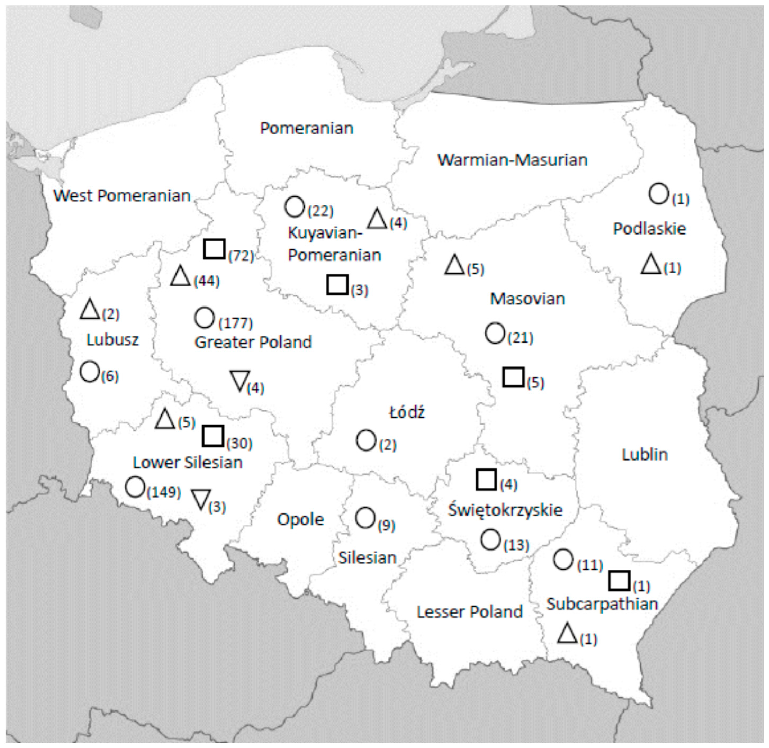


| Common Name | Scientific Name | Virus Isolation a | Serology No. Tested/No. Positive | ||
|---|---|---|---|---|---|
| No. Tested/No. Positive WNV/No. Positive USUV | WNV | USUV | WNV/USUV | ||
| White-tailed eagle | Haliaeetus albicilla | 10/0/0 | 3/2 | 3/0 | 3/0 |
| Common buzzard | Buteo buteo | 6/0/0 | 1/0 | 1/0 | 1/0 |
| Goshawk | Accipiter gentilis | 1/0/0 | 10/3 | 10/1 | 10/1 |
| Peregrine falcon | Falco peregrines | 1/0/0 | n.t. | n.t. | n.t. |
| Capercaillies | Tetrao urogallus | 4/0/0 | n.t. | n.t. | n.t. |
| Mute swan | Cygnus olor | 4/0/0 | n.t. | n.t. | n.t. |
| Saker falcon | Falco cherrug | 1/0/0 | n.t. | n.t. | n.t. |
| Crossbreed peregrine falcon/gyr falcon | Falco peregrinus/Falco rusticolus | 1/0/0 | n.t. | n.t. | n.t. |
| European herring gul | Larus argentatus | 1/0/0 | n.t. | n.t. | n.t. |
| Mallard | Anas platyrhynchos | 1/0/0 | n.t. | n.t. | n.t. |
| Voivodeship | No. Tested | No. Positive (% Positive) | ||
|---|---|---|---|---|
| Horses Which Do Not Move Outside Poland | Travelling Horses | Total | ||
| Masovian | 21 | WNV 5 (23.80) | 0 (0.00) | 5 (23.80) |
| USUV 2 (9.52) | 3 (14.28) | 5 (23.80) | ||
| Podlaskie | 1 | WNV0 (0.00) | 1 (100.00) | 1 (100.00) |
| USUV 0 (0.00) | 0 (0.00) | 0 (0.00) | ||
| Subcarpathian | 11 | WNV 0 (0.00) | 1 (9.09) | 1 (9.09) |
| USUV 1 (9.09) | 0 (0.00) | 1 (9.09) | ||
| Świętokrzyskie | 13 | WNV 0 (0.00) | 0 (0.00) | 0 (0.00) |
| USUV 2 (15.38) | 2 (15.38) | 4 (30.76) | ||
| Silesian | 9 | WNV 0 (0.00) | 0 (0.00) | 0 (0.00) |
| USUV 0 (0.00) | 0 (0.00) | 0 (0.00) | ||
| Łódź | 2 | WNV 0 (0.00) | 0 (0.00) | 0 (0.00) |
| USUV 0 (0.00) | 0 (0.00) | 0 (0.00) | ||
| Kuyavian-Pomeranian | 22 | WNV 4 (18.18) | 0 (0.00) | 4 (18.18) |
| USUV 3 (13.63) | 0 (0.00) | 3 (13.63) | ||
| Greater Poland | 177 | WNV 7 (3.95) | 37 (20.90) | 44 (24.85) |
| USUV 40 (22.59) | 32 (18.07) | 72 (40.67) | ||
| WNV/USUV1 (0.56) | 3 (1.69) | 4 (2.25) | ||
| Lower Silesian | 149 | WNV 3 (2.01) | 2 (1.34) | 5 (3.35) |
| USUV 19 (12.75) | 11 (7.38) | 30 (20.13) | ||
| WNV/USUV (0.00) | 3 (2.01) | 3 (2.01) | ||
| Lubusz | 6 | WNV 2 (33.33) | 0 (0.00) | 2 (33.33) |
| USUV 0 (0.00) | 0 (0.00) | 0 (0.00) | ||
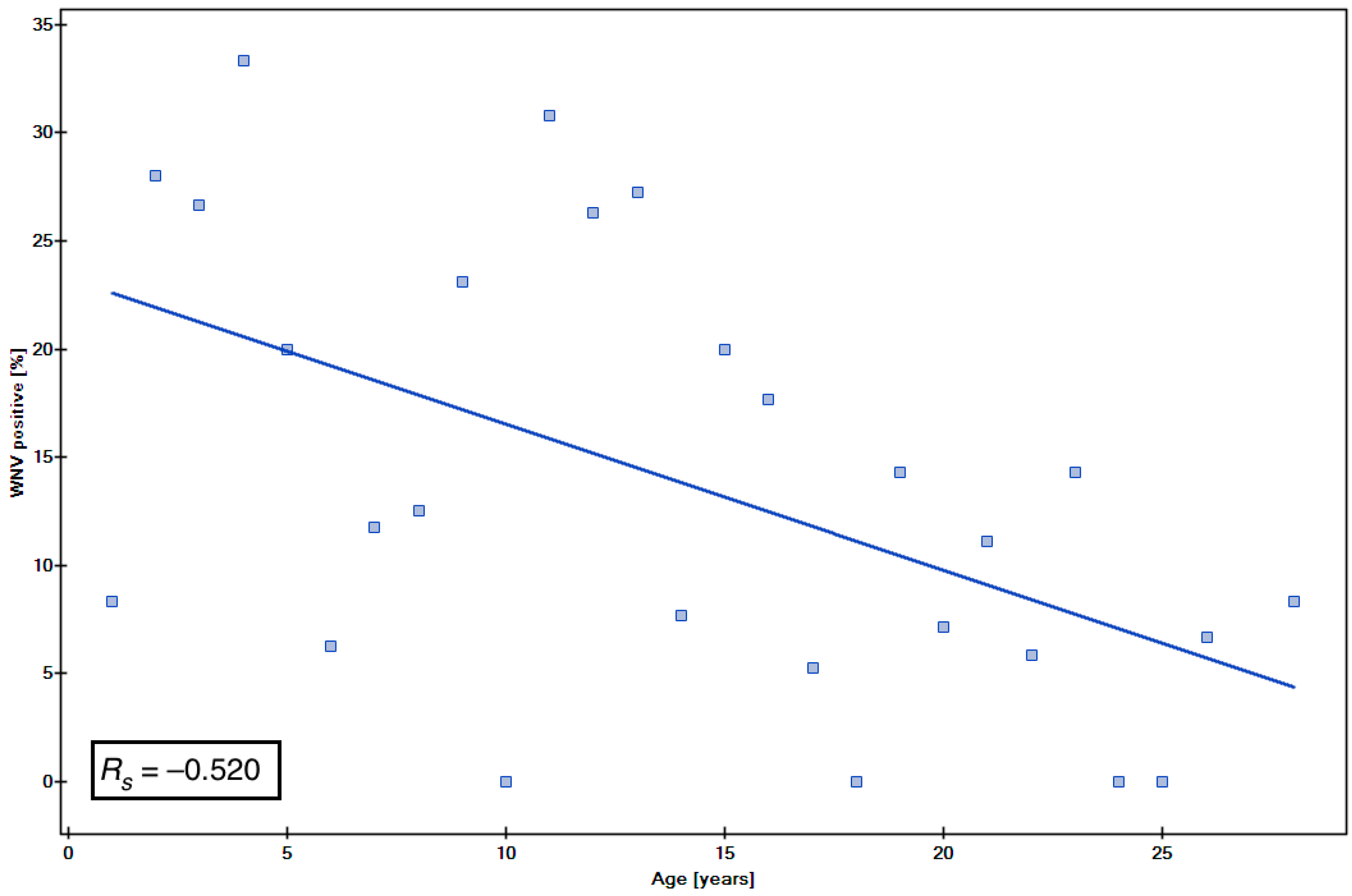
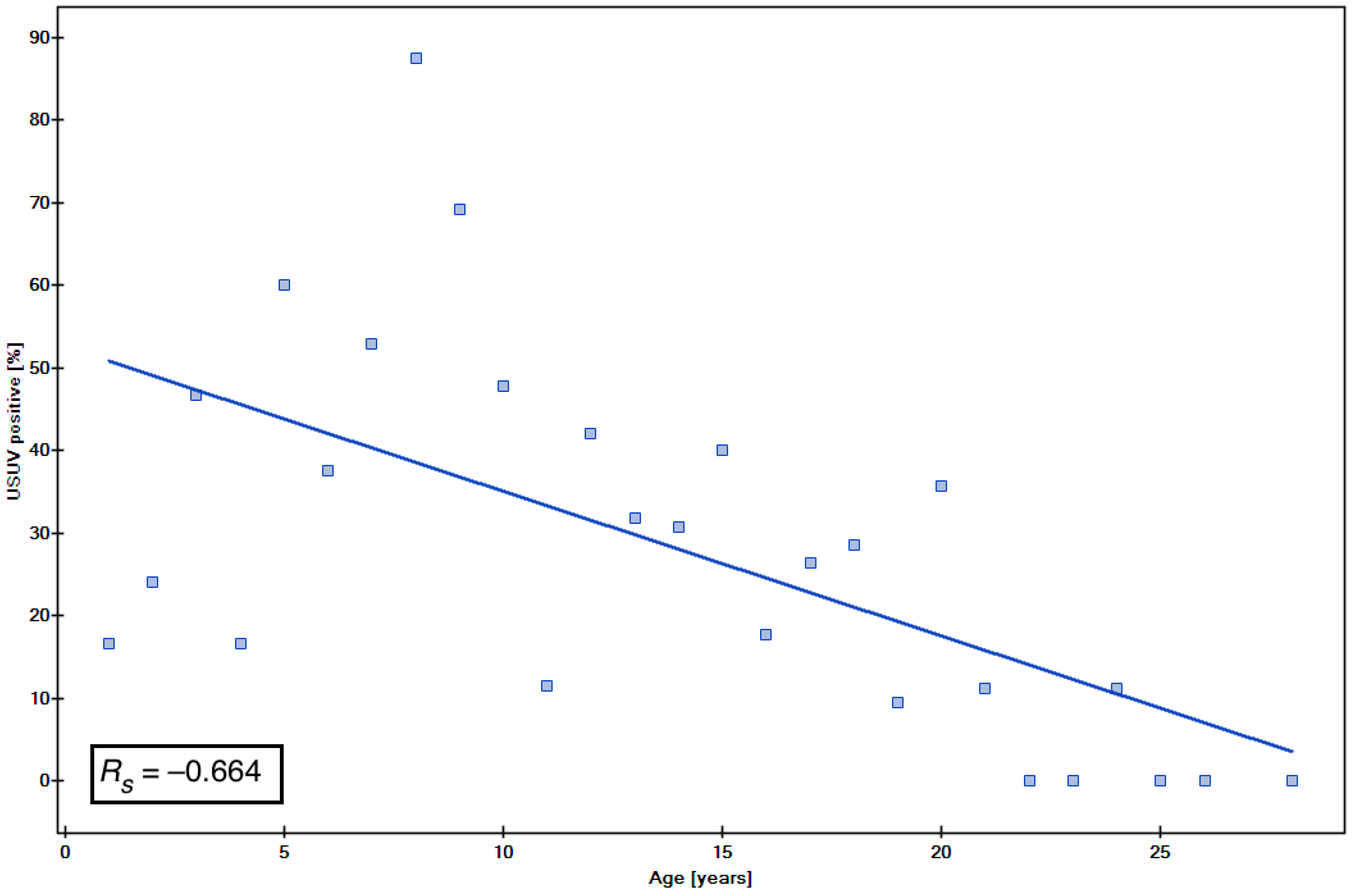
| Age (yrs) | No. Tested | No. Positive (%) | ||
|---|---|---|---|---|
| Horses Which Do Not Move Outside Poland | Travelling Horses | Total | ||
| 1–3 | 52 | WNV 6 (11.53) USUV 7 (13.46) | 6 (11.53) | 12 (23.07) |
| 8 (15.38) | 15 (28.84) | |||
| 4–6 | 43 | WNV 1 (2.32) USUV 9 (20.93) | 7 (16.27) | 8 (18.60) |
| 8 (18.60) | 17 (39.53) | |||
| 7–9 | 38 | WNV 1 (2.63) USUV 13 (34.21) | 5 (13.15) | 6 (15.78) |
| 12 (31.57) | 25 (65.78) | |||
| 10–12 | 68 | WNV 4 (5.88) USUV 16 (23.52) | 9 (13.23) | 13 (19.11) |
| 6 (8.82) | 22 (32.35) | |||
| 13–15 | 50 | WNV 4 (8.00) USUV 11 (22.00) | 6 (12.00) | 10 (20.00) |
| 6 (12.00) | 17 (34.00) | |||
| >15 | 160 | WNV 5 (3.12) USUV 11 (6.87) | 8 (5.00) | 13 (8.12) |
| 8 (5.00) | 19 (11.87) | |||
© 2018 by the authors. Licensee MDPI, Basel, Switzerland. This article is an open access article distributed under the terms and conditions of the Creative Commons Attribution (CC BY) license (http://creativecommons.org/licenses/by/4.0/).
Share and Cite
Bażanów, B.; Jansen van Vuren, P.; Szymański, P.; Stygar, D.; Frącka, A.; Twardoń, J.; Kozdrowski, R.; Pawęska, J.T. A Survey on West Nile and Usutu Viruses in Horses and Birds in Poland. Viruses 2018, 10, 87. https://doi.org/10.3390/v10020087
Bażanów B, Jansen van Vuren P, Szymański P, Stygar D, Frącka A, Twardoń J, Kozdrowski R, Pawęska JT. A Survey on West Nile and Usutu Viruses in Horses and Birds in Poland. Viruses. 2018; 10(2):87. https://doi.org/10.3390/v10020087
Chicago/Turabian StyleBażanów, Barbara, Petrus Jansen van Vuren, Piotr Szymański, Dominika Stygar, Agnieszka Frącka, Jan Twardoń, Roland Kozdrowski, and Janusz T. Pawęska. 2018. "A Survey on West Nile and Usutu Viruses in Horses and Birds in Poland" Viruses 10, no. 2: 87. https://doi.org/10.3390/v10020087