Article

The Incidence and Genetic Diversity of Apple Mosaic Virus (ApMV) and Prune Dwarf Virus (PDV) in Prunus Species in Australia

1
Agriculture Victoria, AgriBio, La Trobe University, Melbourne VIC 3083, Australia
2
School of Applied Systems Biology, AgriBio, La Trobe University, Melbourne VIC 3083, Australia
3
Animal, Plant and Soil Sciences Department, AgriBio, La Trobe University, Melbourne VIC 3083, Australia
*
Author to whom correspondence should be addressed.
Viruses 2018, 10(3), 136; https://doi.org/10.3390/v10030136
Submission received: 23 February 2018 / Revised: 14 March 2018 / Accepted: 17 March 2018 / Published: 19 March 2018
(This article belongs to the Special Issue Fruit Tree Viruses and Viroids)

Abstract

:
Apple mosaic virus (ApMV) and prune dwarf virus (PDV) are amongst the most common viruses infecting Prunus species worldwide but their incidence and genetic diversity in Australia is not known. In a survey of 127 Prunus tree samples collected from five states in Australia, ApMV and PDV occurred in 4 (3%) and 13 (10%) of the trees respectively. High-throughput sequencing (HTS) of amplicons from partial conserved regions of RNA1, RNA2, and RNA3, encoding the methyltransferase (MT), RNA-dependent RNA polymerase (RdRp), and the coat protein (CP) genes respectively, of ApMV and PDV was used to determine the genetic diversity of the Australian isolates of each virus. Phylogenetic comparison of Australian ApMV and PDV amplicon HTS variants and full length genomes of both viruses with isolates occurring in other countries identified genetic strains of each virus occurring in Australia. A single Australian Prunus infecting ApMV genetic strain was identified as all ApMV isolates sequence variants formed a single phylogenetic group in each of RNA1, RNA2, and RNA3. Two Australian PDV genetic strains were identified based on the combination of observed phylogenetic groups in each of RNA1, RNA2, and RNA3 and one Prunus tree had both strains. The accuracy of amplicon sequence variants phylogenetic analysis based on segments of each virus RNA were confirmed by phylogenetic analysis of full length genome sequences of Australian ApMV and PDV isolates and all published ApMV and PDV genomes from other countries.

1. Introduction

Apple mosaic virus (ApMV) and Prune dwarf virus (PDV) are members of the genus Ilarvirus (family Bromoviridae), which have a positive-sense, single-stranded tripartite RNA genome organization composed of RNA1, RNA2, and RNA3 [1]. RNA1 and RNA2 encode for non-structural proteins P1 and P2, which contain a methyltransferase domain (MT) and polymerase domain (RdRP) respectively, and are involved in viral replication. RNA3 is bi-cistronic encoding for the movement protein (MP) and coat protein (CP) that is expressed via sub-genomic RNA4 [2,3]. The ApMV RNA1, RNA2, and RNA3 consist of 3476, 2979, and 2056 nucleotides (nts) respectively and PDV RNA1, RNA2, and RNA3 consist of 3374, 2593, and 1683 nts respectively [4,5,6].
ApMV and PDV infect a wide variety of herbaceous and woody hosts, including Prunus species, and can cause diseases of economic importance [7]. Symptom expression associated with either virus is highly variable and depends upon virus strain, host species, and cultivar and can also be affected by environmental factors, especially higher temperatures which can mask symptoms [8]. ApMV infection in many Prunus trees is characterised by yellow discolorations, line or oak-leaf patterns, and vein banding of leaves, but it may also be latent [9]. ApMV infection causes significant crop yield reduction of up to 25% in some commercial Prunus species [10]. PDV infection can induce a variety of symptoms which may include chlorosis, mosaic, ringspot symptoms on leaves, and stunting [7]. It may also latently infect plants. Amongst the Prunus infecting ilarviruses, PDV has the most significant economic impact with infections attributed to yield losses of up to 100% in some commercial crops [7,11]. ApMV and PDV are graft transmissible and they persist in the vegetatively propagated material of infected trees. ApMV is not pollen transmitted in Prunus trees [12,13] whereas PDV is pollen transmitted, which contributes to its widespread distribution [14].
Assessment of the genetic diversity of ApMV and PDV has mainly been based on the analysis of the CP gene of isolates from different hosts and geographic regions. Early comparisons of the CP gene of ApMV isolates from Korea identified three phylogenetic groups that were separated based on plant host [15]. More recent studies of ApMV isolate sequences from various world geographic origins proposed the existence of two to five phylogenetic groupings of ApMV isolate sequences with no correlation to host or geographic origin [16,17,18].
Similarly, several phylogenetic groups have been reported for PDV isolates whereby one study of isolates from Turkey has reported the existence of four distinct phylo-groups [19] while a recent study using the CP sequences of all PDV isolates published and available in GenBank indicated the presence of only two phylo-groups [2]. Earlier studies suggested a separation of PDV isolate groupings based on host but later studies could not relate phylogenetic groups to host or geographic origin [19,20,21].
ApMV and PDV are known to occur in Australia, however, their incidence in Prunus trees and genetic diversity is not known [22,23,24]. In this study, samples from different Prunus species were collected from five states in Australia and tested for ApMV and PDV using PCR. Amplicons from conserved gene regions of RNA1, RNA2, and RNA3 of each the positive samples were deep sequenced and compared to corresponding GenBank sequences to determine the number ApMV and PDV genetic strains occurring in Australia compared to isolates from around the world. Full length genomes of Australian ApMV and PDV isolates that were previously generated by metagenomic HTS were compared phylogenetically to all available ApMV and PDV full length genome sequences from GenBank and these phylogenetic inferences were compared to those from the conserved gene regions of RNA1, RNA2, and RNA3.

2. Materials and Methods

2.1. Plant Material and Total RNA Extraction

Leaf tissue samples from 127 Prunus trees were collected in spring (2014–2015) from five states in Australia (Table 1). Symptoms characteristic of virus infections were not noted at the time of sample collection. Total RNA was extracted from 0.3 g leaf tissue (fresh weight) of each sample using the RNeasy® Plant Mini Kit (Qiagen, Hilden, Germany) as described previously [21].

2.2. Reverse Transcription Polymerase Chain Reaction (RT-PCR) Amplification

RT-PCR tests were carried out using the SuperScript III One-Step RT-PCR System with Platinum® Taq (Invitrogen, Carlsbad, CA, USA) according to the manufacturer’s instructions except that the total reaction volume was 25 μL. The PCR products were analysed by electrophoresis in 1.5% agarose gels that were stained with SYBR® Safe DNA gel stain (Invitrogen) for visualization.
For preliminary screening by RT-PCR the total RNA extract of each sample was tested for ApMV and PDV using previously published primers that were specific for RNA3 of each virus [25,26].
Total RNA extracts from the Prunus tree samples that tested positive for the RNA3 for ApMV and/or PDV were used for RT-PCR amplification of partial segments of the (MT) gene on RNA1, RdRp gene on RNA2, and CP gene on RNA3 of both viruses using primers developed for this study (Table 2). Cycling conditions consisted of: a reverse transcription step at 48 °C for 45 min; denaturation at 94 °C for 2 min, followed by 35 cycles, denaturing at 94 °C for 30 s, annealing for 30 s at the appropriate temperature for each primer pair (Table 2), elongation at 72 °C for 1 min; and a final elongation step at 72 °C for 10 min.

2.3. Amplicon High-Throughput Sequencing (HTS) Library Preparation and Sequence Reads Analysis

The ApMV and PDV RNA1, RNA2, and RNA3 RT-PCR amplicons were gel purified using the Promega Wizard® PCR clean-up kit (Promega, Madison, WI, USA) according to the manufacturer’s instructions. Amplicon libraries were prepared and sequenced using the Illumina MiSeq as described previously [27]. The sequence read data for this study have been submitted to the NCBI Sequence Read Archive (SRA) database under the Bioproject accession PRJNA404016 and SRA study accession SRP117219.
The generated raw amplicon sequence reads were quality trimmed, paired, and amplicon sequence reads that had a reverse orientation were reversed and complemented as previously described [27]. The amplicon reads were aligned using Muscle (version 3.8.31) [28] with default parameters. The overlapping alignment coverage for each RNA amplicon read was identified and Cutadapt (version 1.4.1) [29] with default parameters was used to trim the amplicon reads to the following lengths: 173, 364, and 452 nt for ApMV RNA1, RNA2, and RNA3 respectively; and 193, 358, and 396 nt for PDV RNA1, RNA2, and RNA3 respectively. Shorter reads in each set of amplicons were discarded. The trimmed amplicon sequence reads were then clustered at 100% identity and cluster sequences with less than 10 reads and non-coding cluster sequences filtered out of each RNA amplicon sample, as previously described [27].

2.4. Phylogenetic and Sequence Identity Analysis

Nucleotide sequences of RNA1, RNA2, and RNA3 for ApMV and PDV isolates from various Prunus species and geographical regions were retrieved from GenBank (Table S1) and trimmed to the corresponding region of the genome that was amplified from each of the ApMV and PDV isolates in this study. Sequence clusters of each amplicon generated were pooled separately with nucleotide sequences from each viral RNA component and aligned using Muscle (version 3.8.31) [28]. Maximum likelihood phylogenetic trees were constructed in RAxML (version 8.0.19) [30] using the GTRGAMMA model with 1000 bootstrap replicates and the resulting trees were visualized in FigTree (version 1.4.2) [31]. Branches that had less than 50% bootstrap support were collapsed. Sequence identity analysis using the sequence demarcation tool (SDT) (version 1.2) [32] was carried out on the aligned amplicon clusters of each viral RNA segment.
Full length genome sequences of the Australian ApMV isolate K75 and PDV isolates NS9 and PCH4 generated by metagenomic HTS in a previous study [33], were aligned with the full genome sequences of other ApMV and PDV isolates available in GenBank using Muscle (Table S2) (version 3.8.31) [28]. Maximum likelihood phylogenetic trees were constructed in MEGA (version 6) [34] with 1000 bootstrap replicates and the resulting trees were visualized in FigTree (version 1.4.2) [31]. Branches having less than 50% bootstrap support were collapsed.

3. Results

3.1. RT-PCR Detection of ApMV and PDV

ApMV and PDV were detected in five and 13 of the 127 Prunus tree samples respectively, using the published RT-PCR tests for each virus. ApMV was detected in three almond and two plum trees in New South Wales and Victoria. PDV was detected in three almonds, one apricot, eight peach, and one plum tree from four states in Australia (Table 3).

3.2. Amplicon Next Generation Sequencing Data and Read Cluster Analysis

The total number of raw reads obtained from amplicon HTS for RNA1, RNA2, and RNA3 of ApMV were 186,987, 111,256, and 196,503 reads respectively (Table S3). After quality trimming, the overall total number of reads used for analysis for ApMV RNA1, RNA2, and RNA3 was reduced to 172,748, 101,747, and 183,473 respectively (Table S3). For PDV the total number of raw reads obtained from amplicon HTS were 327,564, 345,823, and 628,407 for RNA1, RNA2, and RNA3 reads respectively and these were reduced to 300,465, 315,263, and 584,209 for RNA1, RNA2, and RNA3 respectively after quality trimming (Table S4).
The quality trimmed amplicon reads were clustered at 100% identity resulting in an average of 1090, 1495, and 2517 unique ApMV sequence variants per sample for RNA1, RNA2, and RNA3 respectively (Table 4). There was an average of 1061, 1466, and 2309 unique PDV sequence variants for RNA1, RNA2, and RNA3 respectively (Table 5). Filtering of the non-coding and sequence variants with less than 10 reads decreased the average number of ApMV sequence variants per plant sample to 122, 242, and 409 for RNA1, RNA2, and RNA3 amplicons respectively (Table 4), and to 124, 185, and 272 PDV sequence variants per plant sample for RNA1, RNA2, and RNA3 amplicons respectively (Table 5).

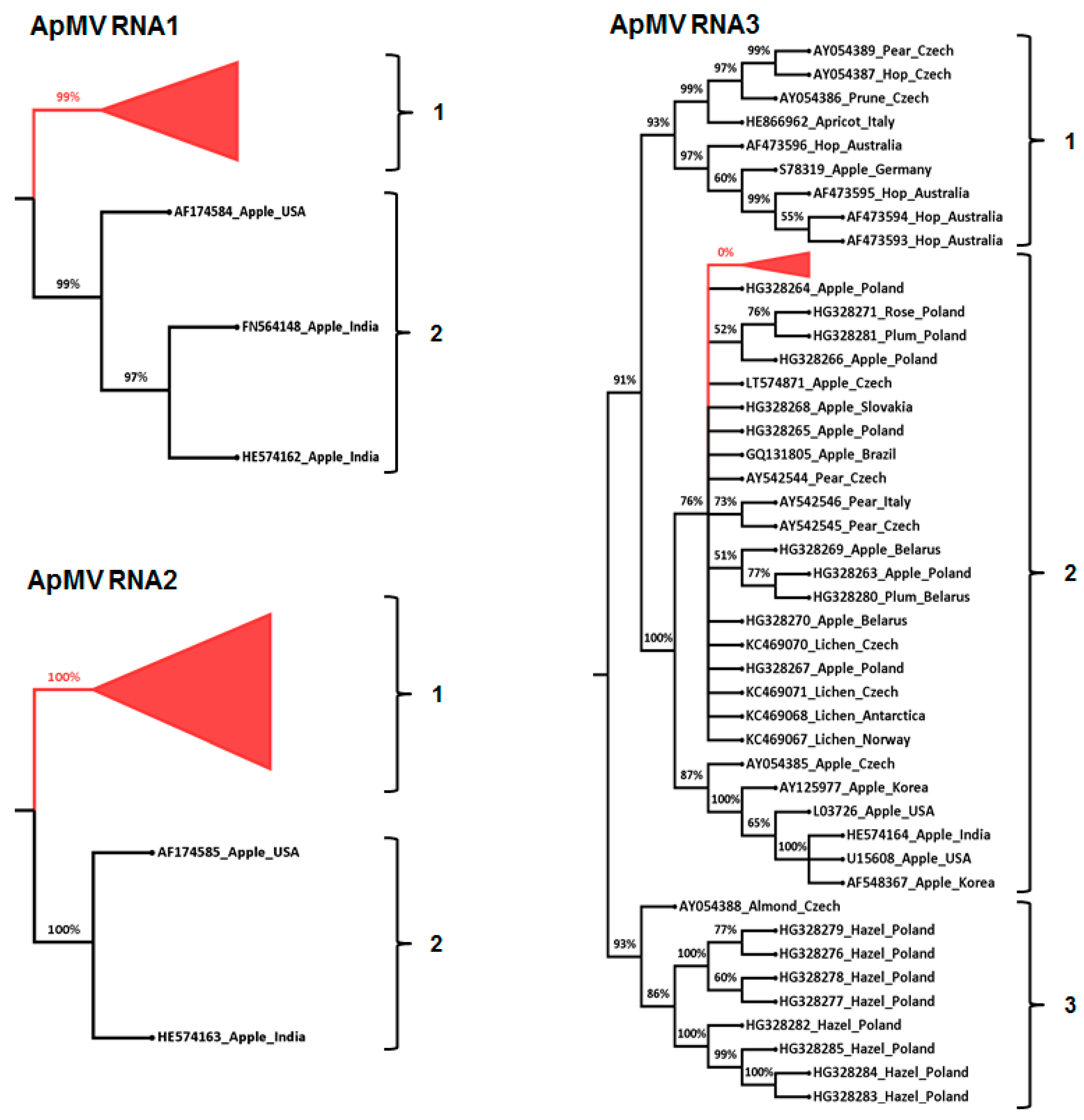
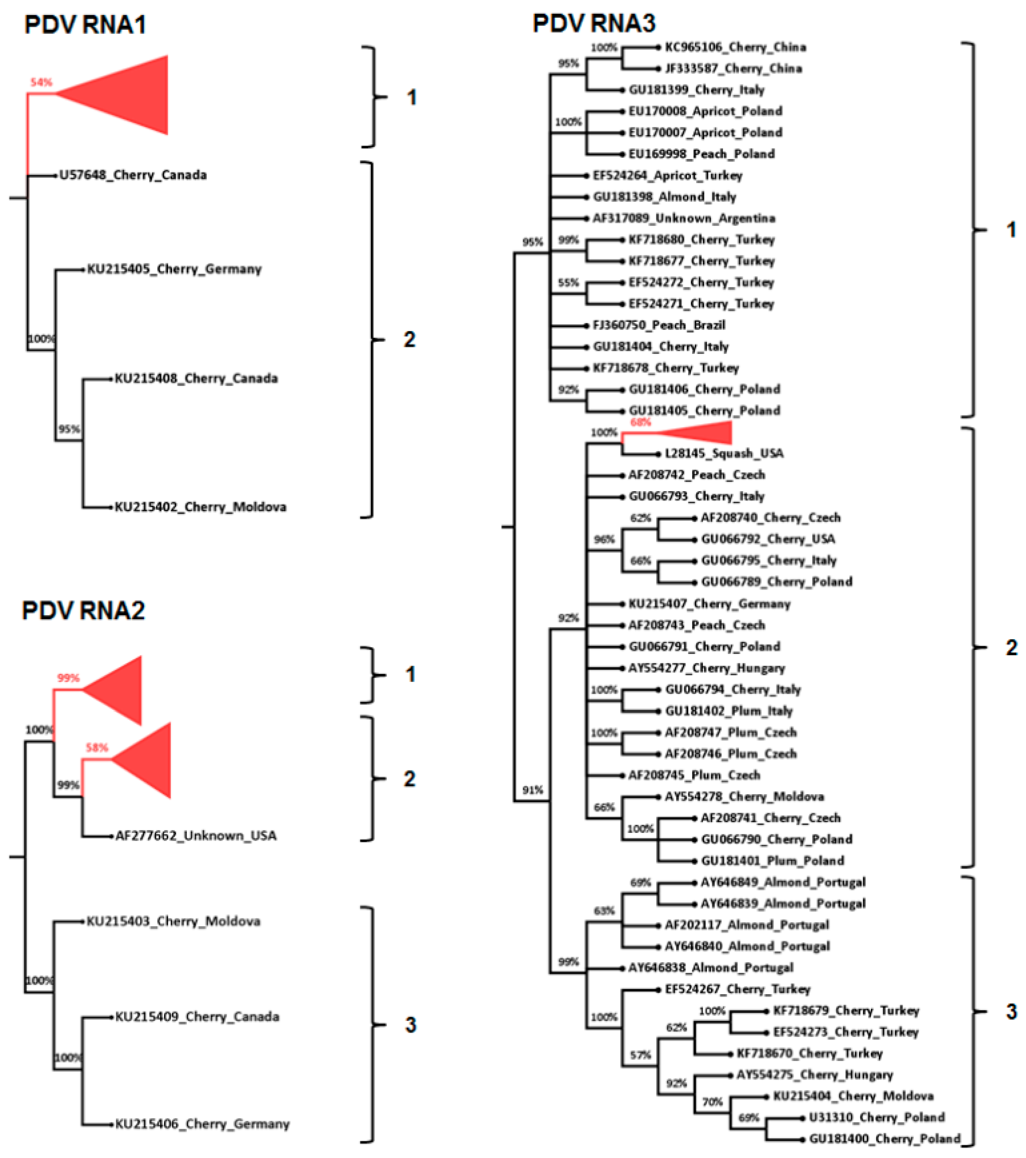
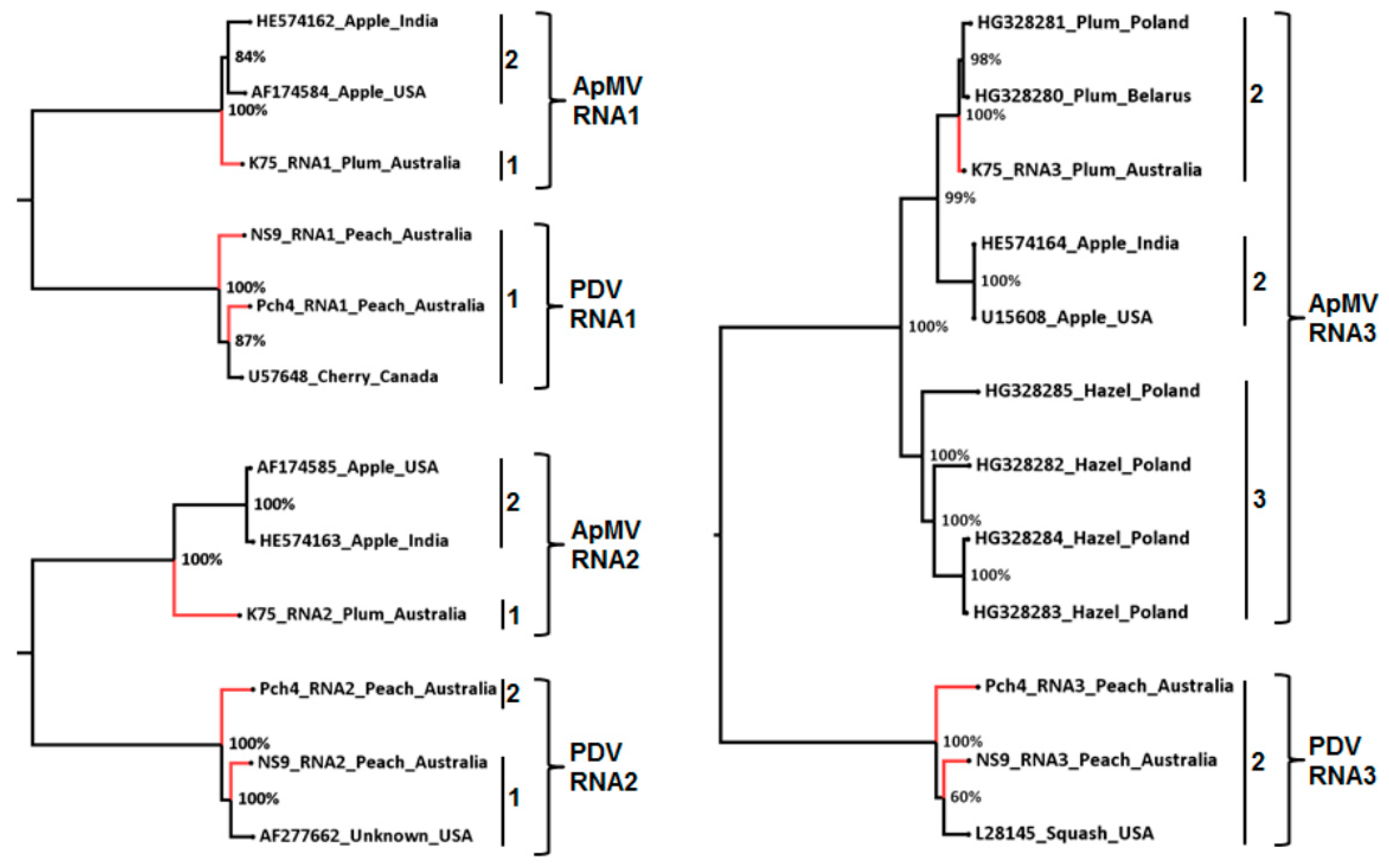
3.3. Phylogenetic and Sequence Identity Analysis

Phylogenetic analysis of each of the pooled ApMV sequence variants for each of RNA1, RNA2, and RNA3 and their corresponding published GenBank isolate sequences (Table 4; Table S5) provided >90% bootstrap support for two phylogenetic groups (phylo-groups 1 and 2) for each of RNA1 and RNA2 and three phylogenetic groups for RNA3 (phylo-groups 1, 2, and 3) (Figure 1). ApMV phylo-group 1 of RNA1 and phylo-group 1 of RNA2 each consisted of amplicon sequence variants from Australian Prunus trees only. Phylo-group 2 of RNA1 and phylo-group 2 of RNA2 each consisted of ApMV sequences occurring in apple plant hosts from other countries (Figure 1; Table S5). All Australian ApMV RNA3 amplicon sequence variants from this study occurred in phylo-group 2, which also consisted of sequences from 12 apples, 3 pears, 1 rose, and 4 lichen isolates occurring in other countries (Figure 1; Table S5). ApMV RNA3 phylo-group 1 consisted of sequences from four Australian hop isolates and also one isolate each from hop, prune, apricot, pear, and apple occurring in other countries. ApMV RNA3 phylo-group 3 contained sequences of four hazelnut isolates; two apple isolates and one isolate each from almond and strawberry (Figure 1; Table S5). None of the Australian ApMV isolates from this study occurred in RNA3 phylo-groups 1 or 3.
Phylogenetic analysis of PDV RNA1 sequences resulted in two phylo-groups with >90% bootstrap support and all Australian amplicon sequence variants from this study occurred in phylo-group 1 as well as a single cherry isolate from Canada. PDV RNA1 phylo-group 2 consisted of only three cherry isolates. PDV RNA2 had three phylo-groups and phylo-group 1 consisted only of Australian PDV sequence variants from this study, phylo-group 2 consisted primarily of Australian isolates but also included a single isolate from the USA. RNA2 sequence variants of Australian isolate CNS3 occurred in both phylo-group 1 and 2 (Figure 2; Table S5). Phylo-group 3 contained cherry isolates from various geographic origins, but none were from Australia. PDV RNA3 had three phylo-groups with sequence variants from this study only occurring in phylo-group 2 and which were most closely related to an experimental isolate maintained in squash from the USA. PDV RNA3 phylo-group 1 contained sequences from almond, apricot, cherry, and peach whereas phylo-group 3 contained isolates from only almond and cherry (Figure 2; Table S5).
SDT identity analysis indicated that amplicon variants of ApMV and PDV occurring within the same phylo-group for each of RNA1, RNA2, and RNA3 shared more than 97% sequence similarity except for PDV isolate CNS3. Amplicon sequence variants from this isolate had RNA2 variants occurring in two phylo-groups, 1 and 2. Each of the RNA 1 and 3 variants in this isolate and the RNA1, RNA2, and RNA3 variants of all other ApMV and PDV isolates only occurred in one phylogenetic group. (Table S6).
Phylogenetic analysis of the full-length ApMV RNA1 and RNA2 genome segments resulted in similar phylo-groupings to the phylogenetic analysis of their corresponding amplicon sequences. The Australian ApMV isolate K75 did not cluster with isolates from the USA and India, which clustered together. Three phylo-groups were observed from phylogenetic analysis of the full-length ApMV RNA3 which separated based on host and the Australian ApMV plum isolate occurred in plum group (Figure 3). The RNA1, RNA2, and RNA3 full-length genome sequences of both Australian PDV isolates occurred in the same phylogenetic groupings that were observed in the phylogenetic analysis of amplicon sequence variants (Figure 3).

4. Discussion

This study presents the first in-depth analysis of the incidence and genetic diversity of ApMV and PDV strains occurring in Australian Prunus trees. The low incidence of ApMV (3%) observed amongst the 127 Australian Prunus tree samples is similar to findings of several studies in other countries [2,35,36,37]. PDV only occurred in 10% of the Australian Prunus tree samples, which is in contrast to PDV survey studies from other countries that found a higher incidence, up to 40%, of PDV [2,35,36,37]. In Australia, many plantings of Prunus trees were established with virus-tested material, which would minimize virus transmission through vegetative propagation and pollen [12,38], and this could explain the low incidence of both ApMV and PDV.
Previous studies of ApMV and PDV diversity in other countries have focussed only on the MP and/or CP gene on RNA 3 [16,17,21,39]. This study provides a more comprehensive analysis of diversity of these viruses by using conserved gene regions of RNA1 and RNA2 in addition to RNA3 and also a comparison with full length genome sequences of these two viruses. Australian ApMV and PDV amplicon sequence variants and full length genome sequences of RNA1, RNA2, and RNA3 from Prunus trees clustered into distinct phylo-groups that were separate from previously published sequences from other hosts and/or geographic regions. The phylogenetic analysis identified a lower level of diversity within and between Australian isolates of each virus compared to the diversity observed amongst Australian PNRSV isolates that were also detected in Prunus trees [27]. Although only a limited number of full length sequences were available for RNA1, RNA2, and RNA3 of each virus, the phylogenetic analysis of these sequences also supported the phylo-groupings based on the amplicon sequences that were observed in this study.
A comparison of the amplicon sequences of Australian isolates with overseas isolates indicated that ApMV RNA1 and RNA2 each had two phylo-groups and ApMV RNA3 had three phylo-groups. Amongst early studies there was no clear consensus on the number of ApMV RNA3 phylogenetic groups and three or five phylo-groups were identified [15,16]. A recent phylogenetic study by Grimová, Winkowska, Ryšánek, Svoboda, and Petrzik [17] of ApMV isolates identified two major phylo-groups that were also observed by later studies [18]. In this study, these two phylo-groups were also observed, corresponding to phylo-groups 1 and 2, and a third phylo-group was also observed, consisting of recently published ApMV isolates from hazelnut that were not used in the earlier studies [40]. This study used representative sequences of phylogenetic clusters and sub-clusters from all these studies that had phylogenetically informative sites ensuring our study is the most current and inclusive analysis of ApMV RNA3 genetic diversity.
Similar to previous phylogeny studies on ApMV CP gene [17], there was no clear correlation between RNA3 phylo-groups to geographical origin in this study. However, the ApMV RNA3 phylo-groups identified in this study appeared to differentiate with host: phylo-groups 1 and 3 consisted mainly of, but were not limited to, isolates of hops and hazelnut respectively, whilst phylo-group 2 was associated with apple, lichen, pear, and plum. Similar RNA3 CP gene host based phylo-groupings have been previously reported and suggest that ApMV strains may have co-evolved with their plant host species resulting in CP gene diversity from host-imposed selective pressure [15]. This may be further supported by the observed host-based RNA3 phylogenetic groupings of full length genome segments of ApMV in this study, although only nine sequences were analysed.
Australian ApMV amplicon HTS sequence variants and full length genome sequences of RNA1 and RNA2 from Australian Prunus trees in this study were clustered into distinct phylo-groups and were separate from previously published sequences, which were mainly from apple trees and other geographic regions. It is difficult to ascertain if the separation of Australian isolates, which were all from Prunus species, from apple isolates occurring in other regions was based on host or geography due to limited availability sequences of ApMV isolates for comparative analysis.
The results of this study indicate that PDV had two RNA1 phylo-groups and three phylo-groups each for RNA2 and RNA3. RNA1 amplicon HTS sequence variants of the Australian PDV isolates occurred only in phylo-group 1 together with a Canadian PDV cherry isolate and this result was also supported by phylogenetic analysis of PDV RNA1 full length sequences. RNA1 phylo-group 2 comprised of three cherry isolates from diverse geographical origins. These results suggest that there may not be any correlation in PDV RNA1 phylo-groupings to host species or geographical origin. PDV RNA2 had three phylo-groups supported by phylogenetic analysis of partial and full length RNA2 sequences. Australian PDV isolates occurred in RNA2 phylo-groups 1 and 2, and a single Australian isolate (CNS3) had its sequence variants occurring in PDV RNA2 phylo-groups 1 and 2. This occurrence of virus sequence variants from a single isolate in multiple phylo-groups has been previously reported for the Ilarvirus PNRSV [27]. The biological implication of this occurrence of multiple distinct populations of PDV RNA2 in isolate CNS3 is not known, and it is possible that two separate infection events—either through grafting or by pollen—resulted in this dual infection.
The occurrence of the three PDV RNA3 phylo-groups in this study concurs with previous diversity analysis of PDV isolates from various host and geographical origin [2,19,21]. Previous studies reported that the PDV RNA3 CP gene groupings were based on host species or geographical origin [19,41], while other studies and this study could find no association [2,21,42]. In this study all Australian PDV isolates and all their sequence variants from different Prunus species occurred in phylo-group 3. The host or geographical origin specific groupings reported in other countries may have occurred due to the analysis of a high number of a single strain of PDV isolates sampled from multiple plants of a single host species in a particular geographic region. Similarly, no host or geographical origin specific groupings were observed from PDV RNA3 full length sequences phylogenetic analysis, although only three sequences were analysed. It is possible that greater PDV diversity exists in Australia, which might have been identified had more isolates been analysed.
The RNA1 and RNA2 phylo-groups of ApMV and PDV are based on limited sequences from other regions and very few hosts in Australia. More data is required to determine if these phylo-groups are correct. Similarly, the more data that becomes available for RNA3 of both viruses from different regions and hosts will further support the delineation of phylo-groups observed in this study. This information might then be used to study specific nucleic acid and protein motifs that are important in the interaction between hosts and viral strains.
The low genetic diversity of Australian ApMV and PDV strains observed in this study could indicate that they were derived from a single virus population or very few introductions on infected planting material into Australia, which may be associated with Australia’s strict quarantine regulations. However, only a few sequences from Prunus trees were available for comparison and analysis of a larger number of isolates from within Australia and in other countries from Prunus and other hosts is required to better understand the effect of host and geographic origin on strain differentiation and diversity.
Sequence variants occurring in the same phylo-group in each of the Australian ApMV and PDV RNA1, RNA2, and RNA3 had identities ranging from 97–99%. These findings are similar to the identity demarcation observed on a study of the Ilarvirus PNRSV by [27] who proposed that a genetic strain of PNRSV in a biological isolate (plant) must comprise of at least one variant of each RNA component that encodes the expected open reading frame (ORF); and may include sequence variants that are ≥97% similar. Extrapolating this definition to ApMV and PDV, this study identified a single Prunus infecting genetic strain of ApMV based on the occurrence of all ApMV isolates sequence variants occurring in a single phylo-group in each RNA: RNA1 phylo-group 1, RNA2 phylo-group 1 and RNA3 phylo-group 3. Based on the combination of RNA1, RNA2, and RNA3 phylo-groups, two genetic strains of PDV were identified to occur in Australia with one Prunus tree (CNS3) having both strains of PDV. The PDV strains were: RNA1 phylo-group 1, RNA2 phylo-group 1, and RNA3 phylo-group 2; and RNA1 phylo-group 1, RNA2 phylo-group 2, and RNA3 phylo-group 2.
The observed phylo-groups and proposed genetic strains of ApMV and PDV based on the amplicon HTS analysis was supported by the full genome phylogeny. This highlights the potential of amplicon HTS in achieving accurate phylogenetic inference based on analysis of a segment of a virus genome and also to detect mixed populations of virus strains. However, the use of complete genomes to define phylogenetic groupings may give a clearer insight on correlation of such groupings to host species or geographic origin due to the larger volume of sequence information.

Supplementary Materials

The following are available online at https://www.mdpi.com/1999-4915/10/3/136/s1, Table S1: The origin, host and the GenBank accession number of the apple mosaic virus (ApMV) and prune dwarf virus (PDV) RNA1, RNA2, and RNA3 sequences that were used for phylogenetic analysis in this study. Table S2: The origin, host, length of full genome sequence and the corresponding GenBank accession number of the apple mosaic virus (ApMV) and prune dwarf virus (PDV) RNA1, RNA2, and RNA3 genome segments that were used for phylogenetic analysis in this study. Table S3: The number of raw sequences reads generated from the next generation amplicon sequencing of apple mosaic virus (ApMV) methyltransferase (MT), RNA dependent RNA polymerase (RdRp) and coat protein (CP) partial gene segments on RNA1, RNA2, and RNA3 respectively and the number of reads remaining after trimming. Table S4: The number of raw sequences reads generated from the next generation amplicon sequencing of prune dwarf virus (PDV) methyltransferase (MT), RNA dependent RNA polymerase (RdRp) and coat protein (CP) partial gene segments on RNA1, RNA2, and RNA3 respectively and the number of reads remaining after trimming. Table S5: The phylo-groups identified from phylogenetic analysis of amplicon sequence variants from partial gene segments of methyltransferase (MT), RNA dependent RNA polymerase (RdRp) and coat protein (CP) genes on RNA1, RNA2 and RNA3 respectively of the Australian apple mosaic virus (ApMV) and prune dwarf virus (PDV) isolates and GenBank isolate sequences. Table S6: The % identity cut-off of apple mosaic virus (ApMV) and prune dwarf virus (PDV) amplicon sequence variants from partial gene segments of methyltransferase (MT), RNA dependent RNA polymerase (RdRp) and coat protein (CP) genes on RNA1, RNA2, and RNA3 respectively determined by sequence identity analysis.

Acknowledgments

We would like to acknowledge and thank Almond Board Australia, Horticulture Innovation Australia Limited, Agriculture Victoria and La Trobe University School of Life Science Postgraduate Publication Award and Postgraduate research scholarship for financial support. This research work was undertaken using the facilities of Agriculture Victoria, Australia. The funders had no role in study design, data collection and analysis, decision to publish, or preparation of the manuscript.

Author Contributions

Wycliff M. Kinoti participated in the design of the study, collected and screened the samples for virus infection, carried out the library preparation and sequencing, performed computational analysis, and drafted the manuscript. Fiona E. Constable participated in the design of the study, sample collection, data analysis, and contributed to drafting the manuscript. Narelle Nancarrow participated in collection and screening of samples for virus infection. Brendan Rodoni participated in the design of the study, data analysis, and drafting the manuscript. Kim M. Plummer participated in the design of the study, data analysis, and contributed to drafting the manuscript. All authors read and approved the final manuscript.

Conflicts of Interest

The authors declare that the research was conducted in the absence of any commercial or financial relationships that could be construed as a potential conflict of interest.

References

  1. Bujarski, J.; Figlerowicz, M.; Gallittelli, D.; Roossinck, M.; Scott, S. Family bromoviridae. In Virus Taxonomy, Ninth Report of the International Committee on Taxonomy of Viruses; King, A.M.Q., Adams, M.J., Carstens, E.B., Lefkowitz, E.J., Eds.; Elsevier-Academic Press: Amsterdam, The Netherlands, 2012; Volume 9, pp. 965–976. [Google Scholar]
  2. Pallas, V.; Aparicio, F.; Herranz, M.; Amari, K.; Sanchez-Pina, M.; Myrta, A.; Sanchez-Navarro, J. Ilarviruses of prunus spp.: A continued concern for fruit trees. Phytopathology 2012, 102, 1108–1120. [Google Scholar] [CrossRef] [PubMed]
  3. Codoner, F.M.; Elena, S.F. The promiscuous evolutionary history of the family bromoviridae. J. Gen. Virol. 2008, 89, 1739–1747. [Google Scholar] [CrossRef] [PubMed]
  4. Shiel, P.; Berger, P. The complete nucleotide sequence of apple mosaic virus (ApMV) RNA 1 and RNA 2: ApMV is more closely related to alfalfa mosaic virus than to other ilarviruses. J. Gen. Virol. 2000, 81, 273–278. [Google Scholar] [CrossRef] [PubMed]
  5. Rampitsch, C.; Eastwell, K. The complete nucleotide sequenceof prune dwarf ilarvirus RNA-1. Arch. Virol. 1997, 142, 1911–1918. [Google Scholar] [CrossRef] [PubMed]
  6. Shiel, P.; Alrefai, R.; Domier, L.; Korban, S.; Berger, P. The complete nucleotide sequence of apple mosaic virus RNA-3. Arch. Virol. 1995, 140, 1247–1256. [Google Scholar] [CrossRef] [PubMed]
  7. Nemeth, M.; Szalay-Marzsó, L.; Posnette, A. Virus, Mycoplasma and Rickettsia Diseases of Fruit Trees; Akademiai Kiado: Dordrecht, The Netherlands, 1986. [Google Scholar]
  8. Wood, G. Virus and Virus-Like Diseases of Pome Fruits and Stone Fruits in New Zealand; Department of Scientific and Industrial Research: Auckland, New Zealand, 1979; p. 87. [Google Scholar]
  9. Šutić, D.D.; Ford, R.E.; Tošić, M.T. Handbook of Plant Virus Diseases; CRC Press: Boca Raton, FL, USA, 1999. [Google Scholar]
  10. Martelli, G.; Savino, V. Infectious diseases of almond with special reference to the mediterranean area 1. Bull. OEPP 1997, 27, 525–534. [Google Scholar] [CrossRef]
  11. Çağlayan, K.; Ulubas-Serce, C.; Gazel, M.; Varveri, C. Prune dwarf virus. In Virus and Virus-Like Diseases of Pome and Stone Fruits; APS Press/American Phytopathological Society: St. Paul, MN, USA, 2011; pp. 199–205. [Google Scholar]
  12. Fulton, R. Apple Mosaic Virus. CMI/AAB Descriptions of Plant Viruses; Applied Biologists: Wellesbourne, UK, 1972; p. 83. [Google Scholar]
  13. Digiaro, M.; Savino, V.; Terlizzi, B.D. Ilarviruses in apricot and plum pollen. Acta Hortic. 1992, 309, 93–98. [Google Scholar]
  14. Mink, G. Pollen and seed-transmitted viruses and viroids. Annu. Rev. Phytopathol. 1993, 31, 375–402. [Google Scholar] [CrossRef] [PubMed]
  15. Lee, G.P.; Ryu, K.H.; Kim, H.R.; Kim, C.S.; Lee, D.W.; Kim, J.S.; Park, M.H.; Noh, Y.M.; Choi, S.H.; Han, D.H. Cloning and phylogenetic characterization of coat protein genes of two isolates of apple mosaic virus from ‘fuji’apple. Plant Pathol. J. 2002, 18, 259–265. [Google Scholar] [CrossRef]
  16. Lakshmi, V.; Hallan, V.; Ram, R.; Ahmed, N.; Zaidi, A.; Varma, A. Diversity of apple mosaic virus isolates in india based on coat protein and movement protein genes. Indian J. Virol. 2011, 22, 44–49. [Google Scholar] [CrossRef] [PubMed]
  17. Grimová, L.; Winkowska, L.; Ryšánek, P.; Svoboda, P.; Petrzik, K. Reflects the coat protein variability of apple mosaic virus host preference? Virus Genes 2013, 47, 119–125. [Google Scholar] [CrossRef] [PubMed]
  18. Valasevich, N.; Cieślińska, M.; Kolbanova, E. Molecular characterization of apple mosaic virus isolates from apple and rose. Eur. J. Plant Pathol. 2015, 141, 839–845. [Google Scholar] [CrossRef]
  19. Ulubaş Serçe, Ç.; Ertunç, F.; Oeztuerk, A. Identification and genomic variability of prune dwarf virus variants infecting stone fruit trees in turkey. J. Phytopathol. 2009, 157, 298–305. [Google Scholar] [CrossRef]
  20. Vašková, D.; Petrzik, K.; Špak, J. Molecular variability of the capsid protein of the prune dwarf virus. Eur. J. Plant Pathol. 2000, 106, 573–580. [Google Scholar] [CrossRef]
  21. Predajňa, L.; Sihelská, N.; Benediková, D.; Šoltys, K.; Candresse, T.; Glasa, M. Molecular characterization of prune dwarf virus cherry isolates from slovakia shows their substantial variability and reveals recombination events in pdv RNA3. Eur. J. Plant Pathol. 2017, 147, 877–888. [Google Scholar] [CrossRef]
  22. Constable, F.E.; Joyce, P.A.; Rodoni, B.C. A survey of key australian pome fruit growing districts for exotic and endemic pathogens. Australas. Plant Pathol. 2007, 36, 165–172. [Google Scholar] [CrossRef]
  23. Stubs, L.; Smith, P. The association of prunus ringspot, prune dwarf, and dark green sunken mottle viruses in the rosetting and decline disease of peach. Crop Pasture Sci. 1971, 22, 771–785. [Google Scholar] [CrossRef]
  24. Greber, R.; Teakle, D.; Mink, G. Thrips-facilitated transmission of prune dwarf and prunus necrotic ringspot viruses from cherry pollen to cucumber. Plant Dis. 1992, 76, 1039–1041. [Google Scholar] [CrossRef]
  25. Parakh, D.; Shamloul, A.; Hadidi, A.; Waterworth, H.; Scott, S.; Howell, H.; Mink, G. Detection of prune dwarf ilarvirus from infected stone fruits using reverse transcription-polymerase chain reaction. Acta Hortic. 1994, 386, 421–430. [Google Scholar] [CrossRef]
  26. Petrzik, K.; Svoboda, P. Screening of apple mosaic virus in hop cultivars in the czech republic by reverse transcription-polymerase chain reaction. Acta Virol. 1997, 41, 101–103. [Google Scholar] [PubMed]
  27. Kinoti, W.M.; Constable, F.E.; Nancarrow, N.; Plummer, K.M.; Rodoni, B. Analysis of intra-host genetic diversity of prunus necrotic ringspot virus (PNRSV) using amplicon next generation sequencing. PLoS ONE 2017, 12, e0179284. [Google Scholar] [CrossRef] [PubMed]
  28. Edgar, R.C. Muscle: Multiple sequence alignment with high accuracy and high throughput. Nucleic Acids Res. 2004, 32, 1792–1797. [Google Scholar] [CrossRef] [PubMed]
  29. Martin, M. Cutadapt removes adapter sequences from high-throughput sequencing reads. EMBnet J. 2011, 17, 10–12. [Google Scholar] [CrossRef]
  30. Stamatakis, A. Raxml version 8: A tool for phylogenetic analysis and post-analysis of large phylogenies. Bioinformatics 2014, 30, 1312–1313. [Google Scholar] [CrossRef] [PubMed]
  31. Andrew, R. Figtree. Available online: http://tree.bio.ed.ac.uk/software/ (accessed on 22 November 2017).
  32. Muhire, B.M.; Varsani, A.; Martin, D.P. Sdt: A virus classification tool based on pairwise sequence alignment and identity calculation. PLoS ONE 2014, 9, e108277. [Google Scholar] [CrossRef] [PubMed]
  33. Kinoti, W.M.; Constable, F.E.; Nancarrow, N.; Plummer, K.M.; Rodoni, B. Generic amplicon deep sequencing to determine ilarvirus species diversity in australian prunus. Front. Microbiol. 2017, 8, 1219. [Google Scholar] [CrossRef] [PubMed]
  34. Tamura, K.; Stecher, G.; Peterson, D.; Filipski, A.; Kumar, S. Mega6: Molecular evolutionary genetics analysis version 6.0. Mol. Biol. Evol. 2013, 30, 2725–2729. [Google Scholar] [CrossRef] [PubMed]
  35. Gümüs, M.; Paylan, I.; Matic, S.; Myrta, A.; Sipahioglu, H.; Erkan, S. Occurrence and distribution of stone fruit viruses and viroids in commercial plantings of prunus species in western anatolia, turkey. J. Plant Pathol. 2007, 89, 265–268. [Google Scholar]
  36. Myrta, A.; Di Terlizzi, B.; Savino, V.; Martelli, G. Virus diseases affecting the mediterranean stone fruit industry: A decade of surveys. In Virus and Virus-Like Diseases of Stone Fruits, with Particular Reference to the Mediterranean Region; Options Méditerranéennes, Série B; CIHEAM (Centre International de Hautes Etudes Agronomiques Méditerranéennes): Bari, Italy, 2003; pp. 15–23. [Google Scholar]
  37. Rouag, N.; Guechi, A.; Matic, S.; Myrta, A. Viruses and viroids of stone fruits in algeria. J. Plant Pathol. 2008, 90, 393–395. [Google Scholar]
  38. Petrzik, K. Capsid protein sequence gene analysis of apple mosaic virus infecting pears. Eur. J. Plant Pathol. 2005, 111, 355–360. [Google Scholar] [CrossRef]
  39. Kalinowska, E.; Mroczkowska, K.; Paduch-Cichal, E.; Chodorska, M. Genetic variability among coat protein of prune dwarf virus variants from different countries and different prunus species. Eur. J. Plant Pathol. 2014, 140, 863–868. [Google Scholar] [CrossRef]
  40. Cieślińska, M.; Valasevich, N. Characterization of apple mosaic virus isolates detected in hazelnut in poland. J. Plant Dis. Prot. 2016, 123, 187–192. [Google Scholar] [CrossRef]
  41. Boulila, M. Molecular characterization of an almond isolate of prune dwarf virus in tunisia: Putative recombination breakpoints in the partial sequences of the coat protein-encoding gene in isolates from different geographic origin. Phytopathol. Mediterr. 2010, 48, 411–421. [Google Scholar]
  42. Öztürk, Y.; Çevik, B. Genetic diversity in the coat protein genes of prune dwarf virus isolates from sweet cherry growing in turkey. Plant Pathol. J. 2015, 31, 41–49. [Google Scholar] [CrossRef] [PubMed]
Figure 1. Maximum likelihood phylogenetic relationship of 608, 1210, and 2047 apple mosaic virus ApMV sequence variants of partial methyltransferase (MT), RNA dependent RNA polymerase (RdRp) and coat protein (CP) genes segments on RNA1, RNA2 and RNA3 respectively, from Australian Prunus trees and corresponding ApMV sequences from GenBank (Table S1). Branches having less than 50% bootstrap support were collapsed and the Australian sequence variants from this study were also collapsed for ease of presentation (red colour). Each of ApMV RNA1, RNA2, and RNA3 phylo-groups were supported by branches with >90% bootstrap, as indicated on the phylogenetic tree.
Viruses 10 00136 g001
Figure 2. Maximum likelihood phylogenetic relationship of 1615, 2411, and 3232 prune dwarf virus (PDV) sequence variants of partial methyltransferase (MT), RNA dependent RNA polymerase (RdRp) and coat protein (CP) genes segments on RNA1, RNA2 and RNA3 respectively, from Australian Prunus trees and corresponding PDV sequences from GenBank (Table S1). Branches having less than 50% bootstrap support were collapsed and the Australian sequence variants from this study were also collapsed for ease of presentation (red colour). Each of PDV RNA1, RNA2, and RNA3 phylo-groups were supported by branches with >90% bootstrap, as indicated on the phylogenetic tree.
Viruses 10 00136 g002
Figure 3. Maximum likelihood phylogenetic relationship of full-length RNA1, RNA2, and RNA3 genome sequences of Australian apple mosaic virus (ApMV) and prune dwarf virus (PDV) isolates and the corresponding full-length sequences of ApMV and PDV from the GenBank (Table S2). The branch positions of the Australian ApMV and PDV isolates are indicated in red colour. Each phylo-group corresponding to phylo-groups determined from phylogenetic analysis of ApMV and PDV amplicon sequence variants (Figure 1 and Figure 2) are numbered.
Viruses 10 00136 g003
Table 1. The location and the number of each Prunus species used in this study.
LocationPrunus SpeciesNo. of Samples
New South WalesAlmond (P. dulcis)39
QueenslandApricot (P. armeniaca)4
Nectarine (P. persica var. nucipersica)1
Plum (P. domestica)6
Peach (P. persica)6
Sweet Cherry (P. avium)2
South AustraliaNectarine (P. persica var. nucipersica)3
Peach (P. persica)1
TasmaniaApricot (P. armeniaca)1
Almond (P. dulcis)1
Peach (P. persica)5
Plum (P. domestica)2
Sweet Cherry (P. avium)8
VictoriaAlmond (P. dulcis)30
Plum (P. domestica)4
Purple leaf plum (P. cerasifera)2
Peach (P. persica)12
Table 2. Primers used for the RT-PCR amplification of RNA1, RNA2, and RNA3 segments of the apple mosaic virus (ApMV) and prune dwarf virus (PDV) genomes.
PrimersPrimer Sequence (5’-3’)RNA (Gene Target) aAmplicon Length Annealing Temp.Reference
APMV-MT1AGTTTGTGTGATGTGAGATRNA1 (MT)222 bp53 °CThis study
APMV-MT2ATTTCTAAGGCGTAACTTC
APMV-Rd1TCATTGGATCCCTTTGCTTCRNA2 (RdRp)383 bp59 °CThis study
APMV-Rd2AAACTCGTCGTCCCTATCC
APMV-CP1TTGCGTTAATTGCAAGTGGRNA3 (CP)471 bp52 °CThis study
APMV-CP2TCAAAAGTTGTGTTTGGAG
PDV-MT1GCGCTGACGAGACTACTARNA1 (MT)205 bp55 °CThis study
PDV-MT2GCGAAACTGTGTGAGGAA
PDV-Rd1CGTTTCTGGAAGGAAGTGGRNA2 (RdRp)382 bp60 °CThis study
PDV-Rd2TTGCTTCGAAATTGAACAA
PDV-CP1TGTTAAGAAACAATTCCCARNA3 (CP)422 bp57 °CThis study
PDV-CP1GCTGAAAAGCGTTGTCATA
a Gene targets encoding: MT = methyltransferase; RdRp = RNA-dependent RNA polymerase; CP = Coat Protein.
Table 3. Prunus tree samples that tested positive for apple mosaic virus (ApMV) and prune dwarf virus (PDV) and their location (state) of origin in Australia.
IsolateHostLocationVirus Detected
K73Almond (P. dulcis)VictoriaApMV
K74Plum (P. domestica)VictoriaApMV
K75Plum (P. domestica)VictoriaApMV
M35Almond (P. dulcis)New South WalesApMV
M36Almond (P. dulcis)New South WalesApMV
CNS3Peach (P. persica)VictoriaPDV
CNS6Peach (P. persica)VictoriaPDV
K76Almond (P. dulcis)VictoriaPDV
NM20Almond (P. dulcis)New South WalesPDV
NM21Almond (P. dulcis)New South WalesPDV
NS3Peach (P. persica)VictoriaPDV
NS5Peach (P. persica)VictoriaPDV
NS7Peach (P. persica)VictoriaPDV
NS9Peach (P. persica)VictoriaPDV
PCH4Peach (P. persica)VictoriaPDV
Q1Apricot (P. armeniaca)QueenslandPDV
Q10Plum (P. domestica)QueenslandPDV
Tas6Peach (P. persica)TasmaniaPDV
Table 4. The number of reads generated from the next generation sequencing of the amplicons derived from the partial methyl transferase (MT) gene on RNA1, partial RNA dependent RNA polymerase (RdRp) gene on RNA2, and partial coat protein (CP) gene on RNA3 of apple mosaic virus (ApMV) and the number of sequence variants of each partial gene region before and after cluster analysis.
APMVRNA1 (Partial MT Gene: 173 bp)RNA2 (Partial RdRp Gene: 364 bp)RNA3 (Partial CP Gene: 452 bp)
Plant IDNo. of Reads after TrimmingNo. of Sequence VariantsNo. of Sequence Variants After Filter aNo. of Reads after TrimmingNo. of Sequence VariantsNo. of Sequence Variants after FilterNo. of Reads after TrimmingNo. of Sequence VariantsNo. of Sequence Variants after Filter
K7348,587104212320,384111418634,4541784298
K7414,74084310724,659153224262,8522816445
K7551,71181812619,328142222955,3052335376
M3542,301151614116,127168122718,2732585349
M3615,409123111121,249172532612,5893063579
Average34,550109012220,349149524236,6952517409
a Filtering of the non-coding and sequence variants with less than 10 reads.
Table 5. The number of reads generated from the next generation sequencing of the amplicons derived from the partial methyl transferase (MT) gene on RNA1, partial RNA dependent RNA polymerase (RdRp) gene on RNA2, and partial coat protein (CP) gene on RNA3 of prune dwarf virus (PDV) and the number of sequence variants of each partial gene region before and after cluster analysis.
PDVRNA1 (Partial MT Gene: 193 bp)RNA2 (Partial RdRp Gene: 358 bp)RNA3 (Partial CP Gene: 396 bp)
Plant IDNo. of Reads after TrimmingNo. of Sequence VariantsNo. of Sequence Variants after Filter aNo. of Reads after TrimmingNo. of Sequence VariantsNo. of Sequence Variants after FilterNo. of Reads after TrimmingNo. of Sequence VariantsNo. of Sequence Variants after Filter
CNS316,020116412848,112210127056,4402610308
CNS618,060108713721,083287038619,3771055134
K7620,460142616412,842140918516,6381890291
NM2012,33072510312,127123414225,2491197195
NM2115,387104312116,178102413611,8331640182
NS326,188102711617,450143616317,2052960373
NS542,242119113725,27298513585,1413866397
NS710,5426459813,794123715281,2292928342
NS927,752108012131,725109911247,6761916251
PCH447,346107511522,097114014779,2932805359
Q130,758108911012,866103813626,1501519231
Q1011,99888410750,068115913123,3491650193
Tas621,382136215831,649232431694,6293985366
Average23,113106112424,251146618544,9392309272
a Filtering of the non-coding and sequence variants with less than 10 reads.

