
Constraints on the Genetic and Antigenic Variability of Measles Virus


Icahn School of Medicine at Mount Sinai, New York, NY 10029, USA
*
Author to whom correspondence should be addressed.
Viruses 2016, 8(4), 109; https://doi.org/10.3390/v8040109
Submission received: 18 February 2016
/
Revised: 6 April 2016
/
Accepted: 14 April 2016
/
Published: 21 April 2016
(This article belongs to the Special Issue Recent Progress in Measles Virus Research)
Abstract
:Antigenic drift and genetic variation are significantly constrained in measles virus (MeV). Genetic stability of MeV is exceptionally high, both in the lab and in the field, and few regions of the genome allow for rapid genetic change. The regions of the genome that are more tolerant of mutations (i.e., the untranslated regions and certain domains within the N, C, V, P, and M proteins) indicate genetic plasticity or structural flexibility in the encoded proteins. Our analysis reveals that strong constraints in the envelope proteins (F and H) allow for a single serotype despite known antigenic differences among its 24 genotypes. This review describes some of the many variables that limit the evolutionary rate of MeV. The high genomic stability of MeV appears to be a shared property of the Paramyxovirinae, suggesting a common mechanism that biologically restricts the rate of mutation.
1. Introduction
Measles virus (MeV) is an enveloped virus with a nonsegmented, negative-sense RNA genome that is 15,894 nucleotides in length. It belongs to the genus Morbillivirus in the Paramyxovirinae sub-family. The organization of the MeV genome is similar to that of most members of the Paramyxovirinae: the genome contains six transcription units, which are separated by nontranscribed trinucleotide intergenic sequences and are flanked by 3′ leader and 5′ trailer sequences at the genome ends (Figure 2A). The MeV genome encodes a total of eight proteins. The six structural proteins are the nucleocapsid protein (N), phosphoprotein (P), matrix protein (M), fusion protein (F), attachment protein (H), and the large error-prone RNA-dependent RNA polymerase protein (L). Two additional nonstructural proteins (C and V) are encoded in the P transcription unit. While the C protein is translated from an overlapping reading frame within the P gene [1], the V protein is initiated from the same start codon as P, but a frameshift is created by mRNA editing. The outcome is that P and V share an N-terminal domain of 231 amino acids, but differ in their C-terminal domains (276 and 69 amino acids, respectively) [2].
2. Genotypic Heterogeneity in Measles Virus
Wildtype MeV isolates are genetically heterogeneous, with 24 genotypes recognized to date (A, B1–B3, C1–C2, D1–D11, E, F, G1–G3 and H1–H2) [3,4]. Only eight of these genotypes have been detected in recent years (B3, D3, D4, D6, D8, D9, G3, and H1), while five genotypes (B1, D1, E, F, and G1) are considered inactive since they have not been detected for more than 25 years [5,6]. The World Health Organization has established that measles genotyping requires a minimum sequence from the highly variable carboxy-terminal 450 nucleotides of N—a region of the gene termed N-450—or the entire protein-coding region of the H gene [3]. Some antigenic differences have been identified between viruses belonging to different genotypes [7,8] and specific genotypes have been causally linked with certain outbreaks or endemic transmission [4], but the clinical significance of the molecular and genetic differences between genotypes is not known [9,10].
An association has not yet been established between specific genotypes and differences in severity of disease, such as progression to measles inclusion body encephalitis (MIBE) and subacute sclerosing panencephalitis (SSPE), which are rare, fatal neurological conditions that result from persistent measles infection in the brain [11]. The genotype that is detected in persistently infected brain tissues is usually consistent with the genotype that was circulating at the time when the patient experienced acute measles infection. However, to our knowledge, viruses belonging to only 10 different genotypes have been isolated from SSPE patients (A, B, C1, C2, D1, D3, D4, D5, D7, E and F), while only three genotypes have been found in MIBE patients (A, B3 and E) [12,13,14,15,16,17,18,19]. That the latter occurs exclusively in immunocompromised subjects may explain that a vaccine strain (genotype A) [13] has been found to be the cause of at least one case of MIBE, whereas vaccine strains of measles have not been isolated in any cases of SSPE [20]. In summary, given the rarity of neurological complications of MeV infection (estimated to be between 1 in 1700 and 1 in 3300 cases of measles for children below five years of age [21]) and the lack of an appropriate animal model, a causal genotype-phenotype relationship might be impossible to establish even if the heterogeneity in the severity and clinical outcome of MeV infection might be influenced by the genotype of the virus at some level.
3. Serology, Genotypes, and Antigenic Diversity
Despite its genotypic diversity, measles has only one serotype, which implies a high degree of similarity in the surface antigens across all MeV strains. In vitro seroneutralization experiments have shown that a broad range of measles genotypes, likely all, can be neutralized by serum samples from vaccinees or individuals who have experienced a natural measles infection, although neutralizing titers can vary for different genotypes [7,22]. The single serotype nature of MeV is further evidenced by the fact that vaccination confers protective immunity against all known genotypes, even though all vaccine strains are members of a single genotype (genotype A). Furthermore, despite the presence of differing endemic genotypes around the world, effective mass vaccination campaigns have dramatically reduced the number of measles-related deaths globally and interrupted endemic transmission in large geographic areas [10,23]. In countries with high vaccination coverage and high seroprevalence rates, the overwhelming majority of measles cases occur in individuals that are unvaccinated or incompletely vaccinated [24]. The geographic variations in incidence are likely the result of varying degrees of vaccination coverage, and not the degree of protection that vaccination confers against various endemic genotypes.
In contrast with neutralization studies performed using serum from vaccinated individuals, vaccine-derived monoclonal antibodies have indeed revealed differences in their ability to neutralize different genotypes. A number of monoclonal antibody studies have sought to provide a structural and biochemical basis for the antigenic differences between genotypes, and perhaps more importantly, to elucidate the antigenic similarities that allow for a single measles serotype in spite of genotypic variation [25,26,27,28,29,30,31]. While vaccination elicits both F-specific and H-specific neutralizing antibodies, those directed against the H protein have a much larger contribution to virus neutralization [32], and as such have been the primary focus of antigenic studies. A recent study by Tahara et al. (2013) found that five distinct epitopes on the H protein are conserved across a virus panel spanning eight genotypes, and monoclonal antibodies that bind two of these epitopes, were highly effective for neutralizing viruses of all genotypes tested. One of these effective neutralizing epitopes is located in an epitope in the head domain involved in binding to the signaling lymphocytic activation molecule (SLAM) and antibody binding to this epitope was shown to inhibit SLAM binding. The other conserved neutralizing epitope is not involved in SLAM binding and antibody binding at this site is proposed to interfere with H-F interaction. Furthermore, an additional epitope was identified and characterized to be an effective neutralizing epitope in several, but not all, of the genotypes tested [33,34]. The existence of highly conserved epitopes that are targets for neutralizing antibodies is suggestive of structural and/or functional constraints on the measles H protein, which would prevent the emergence of escape mutations and contribute to the single serotype nature of MeV. A recent study by Lech et al. (2013) found that escape mutant viruses generated against a variety of neutralizing anti-H monoclonal antibodies were all neutralized effectively by polyclonal serum, indicating that MeV may need to carry escape mutations in multiple neutralizing epitopes of H for it to escape neutralization by polyclonal serum [35]. In vitro mutagenesis experiments support that the H protein is structurally constrained [36] and the long-lived immunity conferred by vaccination or natural infection suggests that MeV does not undergo any significant antigenic drift. In fact, there are surprisingly few regions across the entire MeV genome that allow for rapid change. These regions are discussed in the latter sections of this review.
4. High Genetic Stability of MeV in the Lab and in the Field
The genetic stability of measles is exceptionally high, and it has been observed that it undergoes remarkably little sequence variation over long periods of time, both in laboratory settings and in the field. In several studies on MeV genetic stability, comparison of genomic sequences obtained after passaging in vitro revealed either complete sequence identity with the seed stock [37,38] or a single nucleotide change between two working stocks that had a widely divergent passage history [39]. Even the most variable sequence in the MeV genome, N-450, appears to be very stable over the course of in vitro passaging, irrespective of the cell type used for growth [39]. As reviewed by Aktories, et al. (2009), a high level of genetic stability has also been observed in field isolates of MeV and there is very little variation in the N and H gene sequences of viruses isolated from the same chain of transmission [40]. Moreover, sequencing of viral isolates from the same genotype that were collected several years apart has revealed very little genetic change [41,42]. Rima et al. (1997) estimated the rate of mutation for MeV in the field to be 5 × 10⁻⁴ substitutions per base per year [41] and a similar estimate of 4 × 10⁻⁴ was made by Jenkins et al. (2002) [43] (Table 1). The substitution rate of MeV is significantly lower than estimates for many other RNA viruses such as human immunodeficiency virus type 1 (HIV-1), influenza virus A, foot-and-mouth disease, human enterovirus 71, which have substitution rates in excess of 1.6 × 10⁻³ substitutions per base per year [43,44,45,46,47].
Despite the multiple mutations that accumulate in strains of MeV that are associated with more severe neurological outcomes, these strains are no more genetically unstable than acute or vaccine strains of the virus. In fact, while high stability is seen in all strains of MeV, persistent viruses from SSPE and MIBE patients are perhaps even more genetically stable. Substitutions in SSPE associated viruses can be extensive, but taking into consideration the substantial latent period of ~7–10 years, the rate of molecular evolution for SSPE strains is estimated to be 3.4 × 10⁻⁴ substitutions per base per year [19]. This is similar to estimates for acute strains, while other estimates have calculated the substitution rate of SSPE strains to be even lower than in circulating viruses [9]; however, since these estimates do not take into account differences in the replication rates of acute and persistent viruses, it is difficult to directly compare their genetic stability. Nucleotide substitutions in SSPE-associated virus accumulate in particular regions of the genome, namely in the matrix and fusion genes, while the character of mutations are indicative of biased hypermutation [76]. Thus the difference between acute and persistent strains cannot be attributed to a higher overall rate of genetic change per se, but rather might be influenced by qualitative differences in the type of mutations that arise or variation in as yet unidentified host factors. The mechanistic underpinnings of viral persistence are currently an area of active investigation.
Interestingly, high genetic stability has been observed for many other paramyxoviruses in addition to MeV, and reported substitution rates are similar across the Paramyxovirinae subfamily [50] (Figure 1). This high genetic stability appears to be a property of the Paramyxovirinae subfamily only, and does not extend to the Pneumovirinae. A much higher substitution rate has been observed for both subgroups of human respiratory syncytial virus (HRSV) as well as human meptapneumovirus (HMPV), although the F and N genes of HMPV are notably more genetically stable than the attachment protein (G) [62,63,64,75]. Many paramyxoviruses have an extraordinarily low level of variation among strains. Rima et al. (2014) compared the sequences from 15 different strains of parainfluenza virus type 5 (PIV5), and found remarkably low diversity among them, in spite of the isolates being derived from a broad range of host species (humans, monkeys, pigs, and dogs) over a time period spanning several decades [77]. Similarly, mumps virus (MuV) also has very little diversity among strains of the same genotype [78]. Cui et al. (2013) compared the SH and HN gene sequences from 39 MuV strains belonging to seven different genotypes. In both genes, sequences from the same genotype were identical, regardless of being collected sporadically over several years from patients with or without neurological symptoms and without any epidemiological links between the cases [79]. Minimal genetic distance has been observed between different isolates of both Nipah (NiV) and Hendra (HeV) viruses, independent of host species or year isolated [80]. During the 1998 outbreak of NiV, sequences of isolates obtained from humans, pigs, and bats of the P. hypomelanus genus were found to be nearly identical, with only 56 nucleotide differences across the >18kb viral genome [81,82]. A bat isolate of NiV that was obtained six years later from a P. vampyrus bat was found to have only minor sequence variation from all known Malaysian isolates [61]. The exceptionally high genomic stability shared by most Paramyxovirinae suggests a common mechanism that biologically constrains the rate of mutation.
5. Mechanisms of Mutagenesis and Constraints on Mutation
MeV and other paramyxoviruses exhibit much higher genetic stability than most RNA viruses and there are numerous factors that could be contributing to this difference. For many RNA viruses, extensive studies have been done to determine mutation rates due to intrinsic factors, rather than selection. For RNA viruses, the intrinsically error-prone polymerase and lack of proofreading mechanisms result in a much higher rate of nucleotide misincorporation than that observed for DNA viruses, and in vitro measurements of the error rate for polymerases from RNA viruses ranges from 1 × 10⁻⁶ to 1 × 10⁻³ substitutions/site/replication [83,84,85]. The in vitro mutation rate of the MeV polymerase is similar to those of other RNA viruses, with estimates ranging from 1.8 × 10⁻⁶ to 9 × 10⁻⁵ substitutions/site/replication [86,87]. Thus, there is no evidence to suggest that there is a dramatic difference in the intrinsic error rate of the MeV RNA polymerase. Instead, the high error rates of the MeV polymerase likely produce large numbers of mutants during replication, but relatively few mutations are tolerated. This process of purifying selection, in which genetic diversity is narrowed through the elimination of genomes carrying deleterious mutations, is stronger in RNA viruses than DNA viruses [88]. However, the various selection pressures that drive this phenomenon in MeV are not well understood.
The reasons for such shallow genetic diversity in MeV and other Paramyxovirinae are not clear, and constraints potentially operate at many levels. Structural and biological pressures that limit change in specific regions of the genome are discussed in latter sections of this review. All viruses in the Paramyovirinae, but not the Pneumovirinae, are governed by the “rule of six”, in which their genomes must be an exact multiple of six nucleotides in length to replicate efficiently [89]. The rule of six restricts genomic tolerance for insertions or deletions, and these kind of mutations are rarely observed in MeV [90]. The genome is further constrained by hexameric phasing, which describes the periodic manner in which the N protein interacts with the genome. For each group of six nucleotides bound by the same N molecule, the position of a given nucleotide within that group corresponds to phases 1 to 6. The phase of some transcriptional elements is highly conserved in paramyxoviruses, which might allow these sequence elements to be better accessed by the viral polymerase complex [89,91]. For example, the phase of the transcriptional start site for the various mRNAs is remarkably conserved across every genus of the subfamily [89]. If phase is an important feature for viral transcription, it might function as a constraint on synonymous mutations that disrupt the conserved phasing pattern.
Recombination, which is an important mechanism for rapid genetic change in many viruses, might also be inhibited in part by the rule of six. Although recombination is common among positive-stranded RNA viruses that encode their own RNA polymerase [92], it rarely occurs in negative sense RNA viruses (NSVs). Infrequent recombination in NSVs might be attributed to the fact that neither genomic nor anti-genomic RNA is ever naked, to low rates of co-infection, or to selection against the low fitness of recombinants—although it is not clear why NSV recombinants should be less fit than recombinants in other lineages [93]. There is some evidence that recombination in MeV is possible [94], but studies on the recombination rate of viruses isolated from natural infections have failed to reveal any MeV recombinants [93,95]. Other factors that might constrain substitution rates are those that affect recognition by innate immune responses, such as avoiding the formation of double-stranded RNA structures with complementary cellular RNAs, including micro RNAs (miRNAs) [77]. Additionally, biases against specific dinucleotides [41] and codon usage [96,97] exert an effect on translational efficiency, and could provide a significant constraint on the occurrence of synonymous mutations.
RNA editing by adenosine deamination, resulting in biased hypermutation, may play a role in the generation of the sequence variation in the MeV genome. Members of an enzyme family known as adenosine deaminases that act on RNA (ADARs) catalyze conversion of adenosine to inosine in the negative-strand RNA genome, which then pairs preferentially with cytosine residues during template copying [98]. It has not been directly determined if ADARs are responsible for the sequence transitions observed in MeV substitution events, but their involvement in generating sequence diversity is supported by indirect evidence [98]. Analysis of variation in the MeV genome has revealed a pattern indicative of ADAR-mediated hypermutation, including a preponderance of U-to-C substitutions, mutations that appear in clusters, and a high ratio of nucleotide transitions to transversions [99,100]. It has been shown that ADAR1 acts as a proviral host factor for MeV infection, as viral growth in ADAR1-deficient cells is reduced while virus-induced apoptosis and interferon-β (IFN-β) induction is enhanced. However, the precise role of ADAR enzymes in the MeV lifecycle remain largely unknown [101,102]. The prevalence of hypermutation has been found in sequence comparisons between different strains of MuV, PIV5 and HRSV [77,103,104], suggesting that this mechanism of genetic change might be a common feature among all Paramyxoviridae.
Interestingly, it has been reported that the degree of hypermutation may vary between acute and persistent virus strains, particularly in the reading frame of the M protein [9,105]. Extensive U-to-C substitutions have been observed in sequences of M genes derived from patients with SSPE and MIBE [85,100,105,106,107,108]. In the M sequence of viral RNA isolated from the brain of an MIBE patient, ~50% of the U residues were changed to C [106]. Wong et al. (1991) found that 80% of the nucleotide differences between the M gene sequences of a persistent virus and a closely related acute strain involved U-to-C transitions, and biased hypermutations were determined to be responsible for all but one of the numerous missense genetic changes predicted to cause amino acid substitutions [108]. Differences in the type of nucleotide substitutions that arise in the evolution of acute and persistent virus could indicate distinct selection pressures or mutational mechanisms involved in the development of persistence. While the clustered distribution of mutations suggests an active mechanism of mutation, rather than mere accumulation of polymerase errors [9], the biological implications and exact mechanism behind biased hypermutation in MeV is not currently known. Henipaviruses (another genus of paramyxoviruses) can also establish persistent infections that result in central nervous system (CNS)-confined relapsing encephalitis that can occur up to 11 years after recovery from the primary infection [109,110]. No sequences from relapsing henipavirus encephalitis cases have been deposited in the database, but analysis of such sequences would help determine if the biased hypermutation seen in SSPE is a general property of persistent paramyxovirus infection in the brain or unique to the biology of MeV.
6. Variation in the Mutational Tolerance across Coding Regions
While the factors that constrain mutations and genetic variation in MeV are not well defined, the uneven distribution of mutational tolerance across the genome may provide some insight into the selective forces that drive its overall genomic stability. High tolerance for mutations in some regions of the genome is evidenced by the genetic variability observed in field isolates or passaged laboratory strains and a corresponding high tolerance for substitutional or insertional mutagenesis. In vitro transposon mutagenesis of the MeV genome by Fulton et al. (2015) found that viral mutants containing an insert in genomic locations that are known to be highly variable were frequently recovered and grew to higher titers than mutants with an insert in highly conserved regions [36] (Figure 1A,B). Taken together along with biochemical evidence, these findings highlight several regions of the genome that are inherently plastic and indicate structurally flexible domains of the encoded proteins. These regions are discussed below:
6.1. The N Protein
The C-terminal domain of the N protein, known as NTAIL, is one of the regions found to be most tolerant of insertional mutagenesis [36] and, with the exception of two conserved hydrophobic patches, NTAIL is extremely variable across MeV isolates [111]. Indeed, as previously discussed, genotyping of viral isolates is based partially on sequences from this region of the genome due to its extremely variable sequence. The structural flexibility implied by high mutational tolerance of NTAIL is supported by protein sequence analysis and biochemical evidence. NTAIL is an intrinsically disordered domain [112,113], and the disordered nature of the C-terminal domain of N appears to be a conserved feature within members of the Paramyxovirinae subfamily [114,115]. Biochemical evidence indicates that unbound NTAIL exists as a dynamic population of interconverting conformers and it undergoes α-helical folding upon binding to the P protein [111]. While the C-terminal domain of the N protein is hypervariable in wildtype strains, the N gene is one of the most conserved genes among MeV vaccine strains, displaying a much higher level of conservation than the P/V/C, M, and H genes. For wildtype isolates as well as vaccine strains, the N terminal domain of the N protein is much more conserved than NTAIL, and sequence comparison of all MeV vaccine strains found only four cumulative nucleotide changes in the region coding for the N-terminal 400 amino acids [116].
6.2. The P/V/C Proteins
High sequence variability is also frequently observed in the P gene [117,118]. Nucleotide substitutions are often observed in the P gene after passages of MeV in tissue culture, and in wildtype viruses, the P genes demonstrate a higher level of variation than the corresponding N genes [119,120,121]. The shared N-terminal domain of V and P was found to be more variable than the carboxyl termini in wildtype MeV isolates and vaccine strains [116,117], which indicates a higher degree of conformational flexibility in the common N-terminal domains of the P and V proteins. This is further supported by the finding that a green fluorescent protein (GFP) tag can be added to the N-terminus, but not the C-terminus, of the P and V proteins without significantly compromising function [122]. Furthermore, the poorly conserved N-terminal domain of P (PNT, amino acid (aa) 1–230) was found to be highly tolerant of transposon-mediated insertional mutagenesis [36] (Figure 2). On the other hand, the C-terminal domain of P (PCT, aa 231–507) is highly tolerant of insertions in the first 80 residues (aa 231–311), while it is relatively intolerant of insertions within the C-terminal 197 residues (aa 312–507). The latter includes the coiled-coiled domain essential for P oligomerization (aa 344–411), as well as the XD domain (aa 459–507) important for inducing the correct folding of the disordered NTAIL mentioned above [112,123,124]. The C-termini of both P and V are well conserved and relatively resistant to insertional mutagenesis, which is likely because this region regulates P-L, P-N, and P-P protein interactions as well as contain critical domains required for viral transcription and replication (P) or control of the innate immune response (V), respectively [116,125]. For several Paramyxovirinae genera, including Morbilliviruses, Henipaviruses, and Respiroviruses, a third protein (C) is encoded within the P gene from an overlapping reading frame. Like P and V, the C protein is also intrinsically disordered, highly variable among different strains, and substitutions in the C gene have a high frequency of coding changes [116,126].
6.3. The M Protein
The sequence diversity observed in sequences of the M gene from wildtype viruses is relatively low [127]. M gene sequences are well conserved across different strains for other paramyxoviruses as well, including NDV and MuV [128,129]. Considering the sequence conservation found in the M gene of wildtype strains, it is surprising that M is not required for viral replication and mutants that do not express the M protein are viable in vitro, albeit with lower replicative fitness [107,130]. Furthermore, the M gene was found to have a high tolerance for mutagenic insertions [36]. In vaccine strains, the M gene has a higher substitution rate and a higher proportion of coding changes than the N, F, or L genes [116]. In addition, viruses isolated from SSPE patients usually have a highly mutated M gene, with mutations that often create premature stop codons that completely eliminate M protein expression [9]. In contrast, mumps viruses isolated from cerebrospinal fluid of acute viral meningitis cases revealed almost no mutations in the M gene [129], indicating that the accumulation of mutations in M is not a generalizable property of paramyxovirus infections of the brain per se. Rather, M mutations may reflect the selective pressures involved in the establishment of persistent infections in the brain as discussed above (Section 5). Altogether, it appears that there is some fitness advantage to conservation of the MeV-M gene sequence, but the protein itself has a certain degree of structural plasticity and can change rapidly in response to different selection pressures.
6.4. The F and H Proteins
Variation might be expected in the viral fusion (F) and attachment (H) envelope glycoproteins due to immune selective pressures; however, both MeV envelope proteins are extremely stable. F is more conserved than the M or H genes, and the proportion of coding changes is lower. There are few differences between the F proteins of different wildtype or vaccine strains. Identical amino acid sequences of the F protein have been found between the attenuated Zagreb strain, the wildtype Edmonston strain, and the more recent wildtype IC-B isolate, despite a great disparity in the year they were isolated and a widely divergent passage history [116,121,131], which demonstrates a high level of conservation in F. Although the F protein is very highly conserved among wildtype and vaccine strains, particularly in important functional domains and motifs, such as the cytoplasmic tail, cleavage site, and fusion peptide [116], viruses isolated from SSPE patients usually display considerable mutations in the fusion F gene, usually restricted to the cytoplasmic tail domain [9]. For many viruses, including influenza A virus, sequence diversity readily develops in surface-exposed protein epitopes, facilitating evasion of the adaptive immune response. Although escape mutations that allow for evasion of seroneutralization can arise in both F and H proteins, antigenic evolution does not occur at an appreciable rate in either of the MeV envelope glycoproteins [51,132]. The intolerance for insertional mutation observed in the F and H genes suggests that this might be the result of structural constraints that prevent the development of antigenic diversity [36].
The level of variation observed in the H protein is extremely low [51,133]. As discussed above, measles H is quite antigenically stable over long periods of time, and this lack of antigenic drift makes the H gene sequence suitable for genotyping. As a whole, the H gene was found to be extremely resistant to insertional mutations, which is attributed to structural constrains on the protein itself [36]. Three proteinaceous cellular receptors have been identified for MeV—SLAM (or CD150), CD46, and Nectin-4 (or PVRL4)—and MeV-H has overlapping but distinct receptor binding sites for each of these, located on the side of its six-bladed beta-propeller head domain [134,135,136,137,138,139,140,141]. Amino acid residues that are involved in receptor binding are highly conserved among wildtype and vaccine strains that have common receptor usage [134,142,143,144,145]. All amino acid residues that are involved in SLAM- and Nectin-4 binding are conserved in Edmonston wildtype and all vaccine strains [116] and high conservation of these residues extends across the Morbillivirus genus [143]. Additionally, the cytoplasmic tails of the H proteins (aa 1−34) are completely conserved among all vaccine strains [116]. Comparison of vaccine strains with a wildtype strain showed that although the number of substitutions in the H gene is low, the ratio of coding changes is very high (92%) [116], which indicates that these mutations may be driven by selection pressure and could reflect attenuation or tissue culture adaptation. The use of a proteinaceous cellular receptor(s) for viral entry and the attendant larger receptor-envelope interaction interface might be thought to impose a greater constraint on the mutational tolerance of MeV-H compared with the attachment protein of other paramyxoviruses that use sialic acid as a receptor. However, a similar level of antigenic stability has been observed for the attachment proteins (HN) of sialic acid-using paramyxoviruses, such as mumps [78] and PIV5 [77,146]. Perhaps the complex molecular choreography that leads from receptor binding to fusion-protein triggering [147,148] might be the dominant constraint on the genetic stability of MeV-H (and paramyxovirus attachment proteins in general) regardless of whether the receptor is protein or glycan based. Future comparisons as to how tolerant HN-bearing paramyxoviruses are to insertional mutations in their HN protein will be highly informative.
6.5. The L Protein
The L open reading frame (ORF) stands out as the most conserved gene and it is highly refractory to insertional mutations [36,149]. The L gene is considerably more conserved among vaccine strains than any of the other genes, with only 75 cumulative changes across the 6549 nucleotide-long gene, the majority of which do not change the coding sequence [116,150]. Sequence conservation of the L gene appears to be a common feature of many other paramyxoviruses, including NDV, PIV5, and MuV, where a lower rate of substitution and a predominance of synonymous mutations has been observed [77,78,151]. In addition to the binding domains that allow L to interact with other MeV proteins, six discrete functional domains have been characterized for the L proteins of MeV and their sequences are well conserved across paramyxovirus L proteins [152,153]. The functions of these domains include all enzymatic activities required for transcription and replication. The functional domains in L are arranged within a larger protein structure that is comprised of at least two defined structural domains, which can be expressed as split protein fragments containing dimerization tags without compromising normal protein function [149]. Of the 16 positions in L that vary among the vaccine strains, the majority are located between the conserved domains [116]. The insertion of an epitope tag can be tolerated at two interdomain positions in L (residues 615 and 1708), but none of the highly conserved functional domains have been found to tolerate insertions [36,149]. The lack of transposon insertions in the interdomain regions [36] known to be tolerant to epitope tags [149] may reflect the fitness costs associated with such insertions. In summary, the extremely low tolerance for mutation in any of the functional domains of the L protein likely reflects some degree of structural inflexibility and underscores the interdependence of the various structural domains in mediating the critical role of L in viral transcription and replication.
7. Variation in Mutational Tolerance across Non-Coding Regions
Non-coding RNA comprises nearly 11% of the 16-kb MeV genome and exhibits some degree of conservation across wildtype and vaccine strains [116,154,155,156,157]. Many specialized sequence motifs known as cis-acting regulatory sequences have been identified within the non-coding regions of the MeV genome, and these elements have known or proposed functions in genome replication, genome packaging, translation, mRNA synthesis, processing, and editing [155]. Known cis-acting regulatory elements are extremely well conserved across all MeV strains [116,154,155,156]. The six transcription units are flanked by 3′ leader and 5′ trailer sequences located at the genomic termini. Comparison of viral sequences derived from various vaccine strains and wildtype viruses belonging to many different genotypes has revealed an extremely high level of conservation in the leader and trailer [116,155,157]. This conservation is likely driven in large part by the critical role of the genomic termini in viral replication; while both the leader and trailer contain critical regulatory elements such as encapsidation signals and promoters for genomic and antigenomic transcription, respectively, the 3′ leader also contains a promoter sequence for the initiation of mRNA synthesis [155]. Thus, considerable deviation from the consensus leader/trailer sequences could impair proper promoter recognition by the polymerase complex and severely attenuate replication. Indeed, for HPIV3, a related paramyxovirus in the Respirovirus genus, mutations in the leader sequences have been implicated in contributing to an attenuated phenotype [158].
In the intergenic sequences, the 3′ and 5′ untranslated regions (UTRs) that flank each transcription unit contain highly conserved gene-end (GE) and gene-start (GS) signals that mediate transcription termination and reinitiation during mRNA synthesis [154,155]. The UTRs have been shown to play an important role in the regulation of transcription and translation of viral genes in paramyxoviruses [159,160,161], and mutations within these regions could alter protein expression by affecting transcript initiation, translation efficiency or mRNA stability. Alteration of non-coding regions in MeV and canine distemper virus (CDV), a closely related morbillivirus, has been shown to modulate virulence through transcriptional control of gene expression [162,163]. In MeV, the intergenic region between the M and F genes is unusually long (~1 kb) compared with that of other paramyxoviruses (generally <0.5 bp), but data regarding the functional elements of the UTRs contained in this region are difficult to reconcile into a unifying hypotheses. The 3′UTR of M and the 5′UTR of F are thought to play important roles in viral replication and pathogenicity. In CDV, the M/F intergenic region was shown to potently modulate virulence through transcriptional regulation of F protein expression [162]. Furthermore, expression of the F protein is reduced in vaccine strains of MeV, which is thought to be due to changes in the 5′UTR of F and may reflect a mechanism of attenuation [164]. However, portions of the 5′UTR of MeV-F are dispensable for viral replication in cell culture [165], and while substitutions in the 5′UTR of CDV-F dramatically affected pathogenesis, deletions in this region had no effect on viral replication or the course of disease in vivo [162].
Interestingly, despite the critical role of the UTRs in viral replication and pathogenesis, they are surprisingly tolerant of insertional mutations. Insertional mutagenesis found the non-coding regions to be the most highly mutable regions in the genome, particularly the N/P, P/M, and M/F intergenic regions [36] (Figure 1). Furthermore, the 3′UTR of each gene is generally more tolerant of insertions than the corresponding 5′UTR. The biological significance of this difference awaits future investigations. Furthermore, the M/F intergenic region can be quite variable and has been found to contain the most substitutions in a comparison of non-coding regions from wildtype and vaccine strains [155,166], which is consistent with the finding that the M/F intergenic region is most accepting of transposon insertions [36]. In addition, the M/F intergenic region is often the most variable region of the genome viral sequences derived from persistent MIBE and SSPE strains and persistent isolates sometimes contain insertions or deletions in addition to substitutions [14,167]. This observation suggests that mutation of the non-coding regions could be mechanistically involved in the development of persistence, but this remains to be determined experimentally.
8. Conclusions
Genetic variation and antigenic drift are significantly constrained in MeV, and there are undoubtedly many variables that contribute to its limited evolutionary rate. This high genetic stability appears to be a shared property of the Paramyxovirinae, suggesting a common biological restriction on the rate of mutation that has not yet been defined. Genetic epistasis and the complex interactions between different viral proteins are likely responsible for some of the variation in mutational rates across different regions of genome. Structural, functional, and genetic constraints are particularly high in the envelope and polymerase proteins of MeV, the former of which probably contributes to its single serotype nature and the long-lived immunity conferred by vaccination or natural infection. In general, the mutational tolerance for insertions across the MeV genome is consistent with what is known about the genetic variability of MeV and the structure-function of each ORF. A revealing finding from the whole genome interrogation studies using insertional mutagenesis is that the 3′UTR of each gene is generally more tolerant of insertions than the corresponding 5′UTR, with the differences being most marked for the N, F, H, and L genes. This suggests a critical role for 5′UTRs—perhaps a regulatory function—that is ripe for future studies. Importantly, differences in the degree of tolerance to insertional mutagenesis revealed in the Fulton et al. (2015) study reflect the relative fitness costs compared with insertions in other nucleotide positions, as mutants were allowed to replicate in competition with each other during cell culture passages. Thus, a given genomic position might tolerate an insertion when examined in isolation, but may not be enriched relative to more fit mutants in this kind of whole-genome interrogation study. Future investigations are needed in order to address the more complex questions surrounding qualitative differences in mutations that arise under distinct growth conditions and how these differences relate to attenuation, virulence, or the development of viral persistence.
Acknowledgments
The literature that forms the basis of this review is vast. We apologize to colleagues whose work we were not able to cite due to space constraints. SMB was supported by the NIH Training grant T32 AI07647. B.L. acknowledges funding from NIH grant AI115226 and AI065359 for the genesis of ideas contained in this review.
Author Contributions
S.B. analyzed the data; S.B. and B.L. wrote the paper.
Conflicts of Interest
The authors declare no conflict of interest. The funding sponsors had no role in the design of the study; in the collection, analyses, or interpretation of data; in the writing of the manuscript; and in the decision to publish the results.
References
- Bellini, W.J.; Englund, G.; Rozenblatt, S.; Arnheiter, H.; Richardson, C.D. Measles virus P gene codes for two proteins. J. Virol. 1985, 53, 908–919. [Google Scholar] [PubMed]
- Cattaneo, R.; Kaelin, K.; Baczko, K.; Billeter, M.A. Measles virus editing provides an additional cysteine-rich protein. Cell 1989, 56, 759–764. [Google Scholar] [CrossRef]
- WHO Global Surveillance. Weekly Epidemiological Record Relevé Épidémiologique Hebdomadaire; World Health Organization: Geneva, Switzerland, 2012; Volume 204, pp. 73–80. [Google Scholar]
- Rota, P.A.; Brown, K.; Mankertz, A.; Santibanez, S.; Shulga, S.; Muller, C.P.; Hübschen, J.M.; Siqueira, M.; Beirnes, J.; Ahmed, H.; et al. Global Distribution of Measles Genotypes and Measles Molecular Epidemiology. J. Infect. Dis. 2011, 204, S514–S523. [Google Scholar] [CrossRef] [PubMed]
- Rota, P.A.; Bellini, W.J. Update on the global distribution of genotypes of wild type measles viruses. J. Infect. Dis. 2003, 187 (Suppl. S1), S270–S276. [Google Scholar] [CrossRef] [PubMed]
- World Health Organization. Immunization, Vaccines and Biologicals—Measles Surveillance Data. Regional Summary of Reported Measles Cases. Available online: http://www.who.int/immunization/monitoring_surveillance/burden/vpd/surveillance_type/active/measles_monthlydata/en/index1.html (accessed on 30 March 2016).
- Tamin, A.; Rota, P.A.; Wang, Z.D.; Heath, J.L.; Anderson, L.J.; Bellini, W.J. Antigenic analysis of current wild type and vaccine strains of measles virus. J. Infect. Dis. 1994, 170, 795–801. [Google Scholar] [CrossRef] [PubMed]
- Santibanez, S.; Niewiesk, S.; Heider, A.; Schneider-Schaulies, J.; Berbers, G.A.M.; Zimmermann, A.; Halenius, A.; Wolbert, A.; Deitemeier, I.; Tischer, A.; et al. Probing neutralizing-antibody responses against emerging measles viruses (MVs): Immune selection of MV by H protein-specific antibodies? J. Gen. Virol. 2005, 86, 365–374. [Google Scholar] [CrossRef] [PubMed]
- Kühne, M.; Brown, D.W.G.; Jin, L. Genetic variability of measles virus in acute and persistent infections. Infect. Genet. Evol. 2006, 6, 269–276. [Google Scholar] [CrossRef] [PubMed]
- Riddell, M.A.; Rota, J.S.; Rota, P.A. Review of the temporal and geographical distribution of measles virus genotypes in the prevaccine and postvaccine eras. Virol. J. 2005, 2. [Google Scholar] [CrossRef] [PubMed]
- Schneider-Schaulies, J.; Niewiesk, S.; Schneider-Schaulies, S.; ter Meulen, V. Measles virus in the CNS: The role of viral and host factors for the establishment and maintenance of a persistent infection. J. Neurovirol. 1999, 5, 613–622. [Google Scholar] [CrossRef] [PubMed]
- Bellini, W.J.; Rota, J.S.; Lowe, L.E.; Katz, R.S.; Dyken, P.R.; Zaki, S.R.; Shieh, W.-J.; Rota, P.A. Subacute sclerosing panencephalitis: More cases of this fatal disease are prevented by measles immunization than was previously recognized. J. Infect. Dis. 2005, 192, 1686–1693. [Google Scholar] [CrossRef] [PubMed]
- Bitnun, A.; Shannon, P.; Durward, A.; Rota, P.A.; Bellini, W.J.; Graham, C.; Wang, E.; Ford-Jones, E.L.; Cox, P.; Becker, L.; et al. Measles inclusion-body encephalitis caused by the vaccine strain of measles virus. Clin. Infect. Dis. 1999, 29, 855–861. [Google Scholar] [CrossRef] [PubMed]
- Hardie, D.R.; Albertyn, C.; Heckmann, J.M.; Smuts, H.E.M. Molecular characterisation of virus in the brains of patients with measles inclusion body encephalitis (MIBE). Virol. J. 2013, 10. [Google Scholar] [CrossRef] [PubMed] [Green Version]
- Ivancic-Jelecki, J.; Baricevic, M.; Santak, M.; Harcet, M.; Tešović, G.; Marusic Della Marina, B.; Forcic, D. The first genetic characterization of a D4 measles virus strain derived from a patient with subacute sclerosing panencephalitis. Infect. Genet. Evol. 2013, 17, 71–78. [Google Scholar] [PubMed]
- Jiang, D.-P.; Ide, Y.-H.; Nagano-Fujii, M.; Shoji, I.; Hotta, H. Single-point mutations of the M protein of a measles virus variant obtained from a patient with subacute sclerosing panencephalitis critically affect solubility and subcellular localization of the M protein and cell-free virus production. Microbes Infect. 2009, 11, 467–475. [Google Scholar] [CrossRef] [PubMed]
- Hotta, H.; Nihei, K.; Abe, Y.; Kato, S.; Jiang, D.-P.; Nagano-Fujii, M.; Sada, K. Full-length sequence analysis of subacute sclerosing panencephalitis (SSPE) virus, a mutant of measles virus, isolated from brain tissues of a patient shortly after onset of SSPE. Microbiol. Immunol. 2006, 50, 525–534. [Google Scholar] [CrossRef] [PubMed]
- Miki, K.; Komase, K.; Mgone, C.S.; Kawanishi, R.; Iijima, M.; Mgone, J.M.; Asuo, P.G.; Alpers, M.P.; Takasu, T.; Mizutani, T. Molecular analysis of measles virus genome derived from SSPE and acute measles patients in Papua, New Guinea. J. Med. Virol. 2002, 68, 105–112. [Google Scholar] [CrossRef] [PubMed]
- Woelk, C.H.; Pybus, O.G.; Jin, L.; Brown, D.W.G.; Holmes, E.C. Increased positive selection pressure in persistent (SSPE) versus acute measles virus infections. J. Gen. Virol. 2002, 83, 1419–1430. [Google Scholar] [CrossRef] [PubMed]
- Jin, L.; Beard, S.; Hunjan, R.; Brown, D.W.; Miller, E. Characterization of measles virus strains causing SSPE: A study of 11 cases. J. Neurovirol. 2002, 8, 335–344. [Google Scholar] [PubMed]
- Schönberger, K.; Ludwig, M.S.; Wildner, M.; Weissbrich, B. Epidemiology of Subacute Sclerosing Panencephalitis (SSPE) in Germany from 2003 to 2009: A Risk Estimation. PLoS ONE 2013, 8, e68909. [Google Scholar] [CrossRef] [PubMed]
- Klingele, M.; Hartter, H.K.; Adu, F.; Ammerlaan, W.; Ikusika, W.; Muller, C.P. Resistance of recent measles virus wild-type isolates to antibody-medicated neutralization by vaccinees with antibody. J. Med. Virol. 2000, 62, 91–98. [Google Scholar] [CrossRef]
- Perry, R.T.; Gacic-Dobo, M.; Dabbagh, A.; Mulders, M.N.; Strebel, P.M.; Okwo-Bele, J.-M.; Rota, P.A.; Goodson, J.L. Global control and regional elimination of measles, 2000–2012. MMWR Morb. Mortal. Wkly. Rep. 2014, 63, 103–107. [Google Scholar] [PubMed]
- Muscat, M.; Bang, H.; Wohlfahrt, J.; Glismann, S.; Mølbak, K. Measles in Europe: An epidemiological assessment. Lancet 2009, 373, 383–389. [Google Scholar] [CrossRef]
- Bouche, F.B.; Ertl, O.T.; Muller, C.P. Neutralizing B cell response in measles. Viral Immunol. 2002, 15, 451–471. [Google Scholar] [CrossRef] [PubMed]
- Ertl, O.T.; Wenz, D.C.; Bouche, F.B.; Berbers, G.A.M.; Muller, C.P. Immunodominant domains of the Measles virus hemagglutinin protein eliciting a neutralizing human B cell response. Arch. Virol. 2003, 148, 2195–2206. [Google Scholar] [CrossRef] [PubMed]
- Fournier, P.; Brons, N.H.C.; Berbers, G.A.M.; Wiesmüller, K.H.; Fleckenstein, B.T.; Schneider, F.; Jung, G.; Muller, C.P. Antibodies to a new linear site at the topographical or functional interface between the haemagglutinin and fusion proteins protect against measles encephalitis. J. Gen. Virol. 1997, 78, 1295–1302. [Google Scholar] [CrossRef] [PubMed]
- Ziegler, D.; Fournier, P.; Berbers, G.A.; Steuer, H.; Wiesmüller, K.H.; Fleckenstein, B.; Schneider, F.; Jung, G.; King, C.C.; Muller, C.P. Protection against measles virus encephalitis by monoclonal antibodies binding to a cystine loop domain of the H protein mimicked by peptides which are not recognized by maternal antibodies. J. Gen. Virol. 1996, 77, 2479–2489. [Google Scholar] [CrossRef] [PubMed]
- Liebert, U.G.; Flanagan, S.G.; Löffler, S.; Baczko, K.; ter Meulen, V.; Rima, B.K. Antigenic determinants of measles virus hemagglutinin associated with neurovirulence. J. Virol. 1994, 68, 1486–1493. [Google Scholar] [PubMed]
- Hu, A.; Sheshberadaran, H.; Norrby, E.; Kövamees, J. Molecular characterization of epitopes on the measles virus hemagglutinin protein. Virology 1993, 192, 351–354. [Google Scholar] [CrossRef] [PubMed]
- Carter, M.J.; Willcocks, M.M.; Löffler, S.; ter Meulen, V. Relationships between Monocional Antibody-binding Sites on the Measles Virus Haemagglutinin. J.Gen. Virol. 1982, 63, 113–120. [Google Scholar] [CrossRef] [PubMed]
- De Swart, R.L.; Yüksel, S.; Osterhaus, A.D.M.E. Relative contributions of measles virus hemagglutinin- and fusion protein-specific serum antibodies to virus neutralization. J. Virol. 2005, 79, 11547–11551. [Google Scholar] [CrossRef] [PubMed]
- Tahara, M.; Ito, Y.; Brindley, M.A.; Ma, X.; He, J.; Xu, S.; Fukuhara, H.; Sakai, K.; Komase, K.; Rota, P.A.; et al. Functional and structural characterization of neutralizing epitopes of measles virus hemagglutinin protein. J. Virol. 2013, 87, 666–675. [Google Scholar] [CrossRef] [PubMed]
- Tahara, M.; Ohno, S.; Sakai, K.; Ito, Y.; Fukuhara, H.; Komase, K.; Brindley, M.A.; Rota, P.A.; Plemper, R.K.; Maenaka, K.; et al. The receptor-binding site of the measles virus hemagglutinin protein itself constitutes a conserved neutralizing epitope. J. Virol. 2013, 87, 3583–3586. [Google Scholar] [CrossRef] [PubMed]
- Lech, P.J.; Tobin, G.J.; Bushnell, R.; Gutschenritter, E.; Pham, L.D.; Nace, R.; Verhoeyen, E.; Cosset, F.L.; Muller, C.P.; Russell, S.J.; et al. Epitope Dampening Monotypic Measles Virus Hemagglutinin Glycoprotein Results in Resistance to Cocktail of Monoclonal Antibodies. PLoS ONE 2013, 8, e52306. [Google Scholar] [CrossRef] [PubMed]
- Fulton, B.O.; Sachs, D.; Beaty, S.M.; Won, S.T.; Lee, B.; Palese, P.; Heaton, N.S. Mutational Analysis of Measles Virus Suggests Constraints on Antigenic Variation of the Glycoproteins. Cell Rep. 2015, 11, 1331–1338. [Google Scholar] [CrossRef] [PubMed]
- Borges, M.B.; Caride, E.; Jabor, A.V.; Malachias, J.M.N.; Freire, M.S.; Homma, A.; Galler, R. Study of the genetic stability of measles virus CAM-70 vaccine strain after serial passages in chicken embryo fibroblasts primary cultures. Virus Genes 2008, 36, 35–44. [Google Scholar] [CrossRef] [PubMed]
- Baricević, M.; Forcić, D.; Gulija, T.K.; Jug, R.; Mazuran, R. Determination of the coding and non-coding nucleotide sequences of genuine Edmonston-Zagreb master seed and current working seed lot. Vaccine 2005, 23, 1072–1078. [Google Scholar] [CrossRef] [PubMed]
- Rima, B.K.; Earle, J.A.; Yeo, R.P.; Herlihy, L.; Baczko, K.; ter Meulen, V.; Carabaña, J.; Caballero, M.; Celma, M.L.; Fernandez-Muñoz, R. Temporal and geographical distribution of measles virus genotypes. J. Gen. Virol. 1995, 76 Pt 5, 1173–1180. [Google Scholar] [CrossRef] [PubMed]
- Rota, P.; Featherstone, D.; Bellini, W. Molecular epidemiology of measles virus. Curr. Top. Microbiol. Immunol. 2009, 330, 129–150. [Google Scholar]
- Rima, B.K.; Mcferran, N.V. Dinucleotide and stop codon frequencies in single-stranded RNA viruses. J. Gen. Virol. 1997, 78, 2859–2870. [Google Scholar] [CrossRef] [PubMed]
- El Mubarak, H.S.; van de Bildt, M.W.G.; Mustafa, O.A.; Vos, H.W.; Mukhtar, M.M.; Ibrahim, S.A.; Andeweg, A.C.; El Hassan, A.M.; Osterhaus, A.D.M.E.; de Swart, R.L. Genetic characterization of wild-type measles viruses circulating in suburban Khartoum, 1997–2000. J. Gen. Virol. 2002, 83, 1437–1443. [Google Scholar] [CrossRef] [PubMed]
- Jenkins, G.M.; Rambaut, A.; Pybus, O.G.; Holmes, E.C. Rates of molecular evolution in RNA viruses: A quantitative phylogenetic analysis. J. Mol. Evol. 2002, 54, 156–165. [Google Scholar] [CrossRef] [PubMed]
- Gojobori, T.; Moriyama, E.N.; Kimura, M. Molecular clock of viral evolution, and the neutral theory. Proc. Natl. Acad. Sci. USA 1990, 87, 10015–10018. [Google Scholar] [CrossRef] [PubMed]
- Leitner, T.; Albert, J. The molecular clock of HIV-1 unveiled through analysis of a known transmission history. Proc. Natl. Acad. Sci. USA 1999, 96, 10752–10757. [Google Scholar] [CrossRef] [PubMed]
- Gorman, O.T.; Bean, W.J.; Kawaoka, Y.; Webster, R.G. Evolution of the nucleoprotein gene of influenza A virus. J. Virol. 1990, 64, 1487–1497. [Google Scholar] [PubMed]
- Martínez, M.A.; Dopazo, J.; Hernández, J.; Mateu, M.G.; Sobrino, F.; Domingo, E.; Knowles, N.J. Evolution of the capsid protein genes of foot-and-mouth disease virus: Antigenic variation without accumulation of amino acid substitutions over six decades. J. Virol. 1992, 66, 3557–3565. [Google Scholar] [PubMed]
- Rima, B.K.; Earle, J.A.; Baczko, K.; ter Meulen, V.; Liebert, U.G.; Carstens, C.; Carabaña, J.; Caballero, M.; Celma, M.L.; Fernandez-Muñoz, R. Sequence divergence of measles virus haemagglutinin during natural evolution and adaptation to cell culture. J. Gen. Virol. 1997, 78 Pt 1, 97–106. [Google Scholar] [CrossRef] [PubMed]
- Furuse, Y.; Suzuki, A.; Oshitani, H. Origin of measles virus: Divergence from rinderpest virus between the 11th and 12th centuries. Virol. J. 2010, 7. [Google Scholar] [CrossRef] [PubMed]
- Pomeroy, L.W.; Bjørnstad, O.N.; Holmes, E.C. The evolutionary and epidemiological dynamics of the paramyxoviridae. J. Mol. Evol. 2008, 66, 98–106. [Google Scholar] [CrossRef] [PubMed]
- Kimura, H.; Saitoh, M.; Kobayashi, M.; Ishii, H.; Saraya, T.; Kurai, D.; Tsukagoshi, H.; Shirabe, K.; Nishina, A.; Kozawa, K.; et al. Molecular evolution of haemagglutinin (H) gene in measles virus. Sci. Rep. 2015, 5, 11648. [Google Scholar] [CrossRef] [PubMed]
- Monne, I.; Fusaro, A.; Valastro, V.; Citterio, C.; Pozza, M.D.; Obber, F.; Trevisiol, K.; Cova, M.; De Benedictis, P.; Bregoli, M.; et al. A distinct CDV genotype causing a major epidemic in Alpine wildlife. Vet. Microbiol. 2011, 150, 63–69. [Google Scholar] [CrossRef] [PubMed]
- Padhi, A.; Ma, L. Genetic and epidemiological insights into the emergence of peste des petits ruminants virus (PPRV) across Asia and Africa. Sci. Rep. 2014, 4, 7040. [Google Scholar] [CrossRef] [PubMed]
- Miller, P.J.; Kim, L.M.; Ip, H.S.; Afonso, C.L. Evolutionary dynamics of Newcastle disease virus. Virology 2009, 391, 64–72. [Google Scholar] [CrossRef] [PubMed]
- Ramey, A.M.; Reeves, A.B.; Ogawa, H.; Ip, H.S.; Imai, K.; Bui, V.N.; Yamaguchi, E.; Silko, N.Y.; Afonso, C.L. Genetic diversity and mutation of avian paramyxovirus serotype 1 (Newcastle disease virus) in wild birds and evidence for intercontinental spread. Arch. Virol. 2013, 158, 2495–2503. [Google Scholar] [CrossRef] [PubMed]
- Chong, Y.L.; Padhi, A.; Hudson, P.J.; Poss, M. The effect of vaccination on the evolution and population dynamics of avian paramyxovirus-1. PLoS Pathog. 2010, 6, e1000872. [Google Scholar] [CrossRef] [PubMed]
- Beck, E.T.; He, J.; Nelson, M.I.; Bose, M.E.; Fan, J.; Kumar, S.; Henrickson, K.J. Genome sequencing and phylogenetic analysis of 39 human parainfluenza virus type 1 strains isolated from 1997–2010. PLoS ONE 2012, 7, e46048. [Google Scholar] [CrossRef] [PubMed]
- Mizuta, K.; Saitoh, M.; Kobayashi, M.; Tsukagoshi, H.; Aoki, Y.; Ikeda, T.; Abiko, C.; Katsushima, N.; Itagaki, T.; Noda, M.; et al. Detailed genetic analysis of hemagglutinin-neuraminidase glycoprotein gene in human parainfluenza virus type 1 isolates from patients with acute respiratory infection between 2002 and 2009 in Yamagata prefecture, Japan. Virol. J. 2011, 8. [Google Scholar] [CrossRef] [PubMed]
- Kiptinness, J.K.; Wurapa, E.K.; Wamunyokoli, F.; Bulimo, W.D. Molecular characterization of human parainfluenza virus type 1 in infants attending Mbagathi District Hospital, Nairobi, Kenya: A retrospective study. Virus Genes 2013, 47, 439–447. [Google Scholar] [CrossRef] [PubMed]
- Mizuta, K.; Tsukagoshi, H.; Ikeda, T.; Aoki, Y.; Abiko, C.; Itagaki, T.; Nagano, M.; Noda, M.; Kimura, H. Molecular evolution of the haemagglutinin-neuraminidase gene in human parainfluenza virus type 3 isolates from children with acute respiratory illness in Yamagata prefecture, Japan. J. Med. Microbiol. 2014, 63, 570–577. [Google Scholar] [CrossRef] [PubMed]
- Rahman, S.A.; Hassan, S.S.; Olival, K.J.; Mohamed, M.; Chang, L.Y.; Hassan, L.; Saad, N.M.; Shohaimi, S.A.; Mamat, Z.C.; Naim, M.S.; et al. Characterization of Nipah virus from naturally infected Pteropus vampyrus bats, Malaysia. Emerg. Infect. Dis. 2010, 16, 1990–1993. [Google Scholar] [CrossRef] [PubMed]
- Yang, C.-F.; Wang, C.K.; Tollefson, S.J.; Piyaratna, R.; Lintao, L.D.; Chu, M.; Liem, A.; Mark, M.; Spaete, R.R.; Crowe, J.E.; et al. Genetic diversity and evolution of human metapneumovirus fusion protein over twenty years. Virol. J. 2009, 6. [Google Scholar] [CrossRef] [PubMed]
- De Graaf, M.; Osterhaus, A.D.M.E.; Fouchier, R.A.M.; Holmes, E.C. Evolutionary dynamics of human and avian metapneumoviruses. J. Gen. Virol. 2008, 89, 2933–2942. [Google Scholar] [CrossRef] [PubMed]
- Padhi, A.; Verghese, B. Positive natural selection in the evolution of human metapneumovirus attachment glycoprotein. Virus Res. 2008, 131, 121–131. [Google Scholar] [CrossRef] [PubMed]
- Tan, L.; Coenjaerts, F.E.J.; Houspie, L.; Viveen, M.C.; van Bleek, G.M.; Wiertz, E.J.H.J.; Martin, D.P.; Lemey, P. The comparative genomics of human respiratory syncytial virus subgroups A and B: Genetic variability and molecular evolutionary dynamics. J. Virol. 2013, 87, 8213–8226. [Google Scholar] [CrossRef] [PubMed]
- Zlateva, K.T.; Lemey, P.; Vandamme, A.; Van Ranst, M. Molecular Evolution and Circulation Patterns of Human Respiratory Syncytial Virus Subgroup A: Positively Selected Sites in the Attachment G Glycoprotein Molecular Evolution and Circulation Patterns of Human Respiratory Syncytial Virus Subgroup A: Positi. J. Virol. 2004, 78, 4675–4683. [Google Scholar] [CrossRef] [PubMed]
- Yoshida, A.; Kiyota, N.; Kobayashi, M.; Nishimura, K.; Tsutsui, R.; Tsukagoshi, H.; Hirano, E.; Yamamoto, N.; Ryo, A.; Saitoh, M.; et al. Molecular epidemiology of the attachment glycoprotein (G) gene in respiratory syncytial virus in children with acute respiratory infection in Japan in 2009/2010. J. Med. Microbiol. 2012, 61, 820–829. [Google Scholar] [CrossRef] [PubMed]
- Matheson, J.W.; Rich, F.J.; Cohet, C.; Grimwood, K.; Huang, Q.S.; Penny, D.; Hendy, M.D.; Kirman, J.R.; Bobek, V.; Kolostova, K.; et al. Distinct Patterns of Evolution Between Respiratory Syncytial Virus Subgroups A and B From New Zealand Isolates Collected Over Thirty-Seven Years. Anticancer Res. 2006, 30, 4799–4804. [Google Scholar] [CrossRef] [PubMed]
- Tsukagoshi, H.; Yokoi, H.; Kobayashi, M.; Kushibuchi, I.; Okamoto-Nakagawa, R.; Yoshida, A.; Morita, Y.; Noda, M.; Yamamoto, N.; Sugai, K.; et al. Genetic analysis of attachment glycoprotein (G) gene in new genotype ON1 of human respiratory syncytial virus detected in Japan. Microbiol. Immunol. 2013, 57, 655–659. [Google Scholar] [CrossRef] [PubMed]
- Kushibuchi, I.; Kobayashi, M.; Kusaka, T.; Tsukagoshi, H.; Ryo, A.; Yoshida, A.; Ishii, H.; Saraya, T.; Kurai, D.; Yamamoto, N.; et al. Molecular evolution of attachment glycoprotein (G) gene in human respiratory syncytial virus detected in Japan 2008–2011. Infect. Genet. Evol. 2013, 18, 168–173. [Google Scholar] [CrossRef] [PubMed]
- Pretorius, M.A.; Van Niekerk, S.; Tempia, S.; Moyes, J.; Cohen, C.; Madhi, S.A.; Venter, M. Replacement and positive evolution of subtype A and B respiratory syncytial virus G-protein genotypes from 1997–2012 in South Africa. J. Infect. Dis. 2013, 208, S227–S237. [Google Scholar] [CrossRef] [PubMed]
- Malasao, R.; Okamoto, M.; Chaimongkol, N.; Imamura, T.; Tohma, K.; Dapat, I.; Dapat, C.; Suzuki, A.; Saito, M.; Saito, M.; et al. Molecular characterization of human respiratory syncytial virus in the Philippines, 2012–2013. PLoS ONE 2015, 10. [Google Scholar] [CrossRef] [PubMed]
- Trento, A.; Viegas, M.; Galiano, M.; Carballal, G.; Mistchenko, A.S.; Melero, A.; Videla, C. Natural History of Human Respiratory Syncytial Virus Inferred from Phylogenetic Analysis of the Attachment (G) Glycoprotein with a 60-Nucleotide Duplication. J. Viol. 2006, 80, 975–984. [Google Scholar] [CrossRef] [PubMed]
- Buonagurio Nakada, S.; PArvin, J.D.; Krystal, M.; Palese, P.; Fitch, W.M.D.A. Evolution of human influenza A viruses over 50 years: Rapid, uniform rate of change in NS gene. Science 1986, 232, 980–982. [Google Scholar] [CrossRef]
- Hanada, K.; Suzuki, Y.; Gojobori, T. A large variation in the rates of synonymous substitution for RNA viruses and its relationship to a diversity of viral infection and transmission modes. Mol. Biol. Evol. 2004, 21, 1074–1080. [Google Scholar] [CrossRef] [PubMed]
- Rima, B.K.; Duprex, W.P. Molecular mechanisms of measles virus persistence. Virus Res. 2005, 111, 132–147. [Google Scholar] [CrossRef] [PubMed] [Green Version]
- Rima, B.K.; Gatherer, D.; Young, D.F.; Norsted, H.; Randall, R.E.; Davison, A.J. Stability of the Parainfluenza Virus 5 Genome Revealed by Deep Sequencing of Strains Isolated from Different Hosts and following Passage in Cell Culture. J. Virol. 2014, 88, 3826–3836. [Google Scholar] [CrossRef] [PubMed] [Green Version]
- Jin, L.; Örvell, C.; Myers, R.; Rota, P.A.; Nakayama, T.; Forcic, D.; Hiebert, J.; Brown, K.E. Genomic diversity of mumps virus and global distribution of the 12 genotypes. Rev. Med. Virol. 2015, 25, 85–101. [Google Scholar] [CrossRef] [PubMed]
- Cui, A.; Brown, D.W.G.; Xu, W.; Jin, L. Genetic Variation in the HN and SH Genes of Mumps Viruses: A Comparison of Strains from Mumps Cases with and without Neurological Symptoms. PLoS ONE 2013, 8, e61791. [Google Scholar] [CrossRef] [PubMed]
- Halpin, K.; Young, P.L.; Field, H.E.; Mackenzie, J.S. Isolation of Hendra virus from pteropid bats: A natural reservoir of Hendra virus. J. Gen. Virol. 2000, 81, 1927–1932. [Google Scholar] [CrossRef] [PubMed]
- AbuBakar, S.; Chang, L.-Y.; Ali, A.R.M.; Sharifah, S.H.; Yusoff, K.; Zamrod, Z. Isolation and molecular identification of Nipah virus from pigs. Emerg. Infect. Dis. 2004, 10, 2228–2230. [Google Scholar] [CrossRef] [PubMed]
- Chua, K.B.; Lek Koh, C.; Hooi, P.S.; Wee, K.F.; Khong, J.H.; Chua, B.H.; Chan, Y.P.; Lim, M.E.; Lam, S.K. Isolation of Nipah virus from Malaysian Island flying-foxes. Microbes Infect. 2002, 4, 145–151. [Google Scholar] [CrossRef]
- Drake, J.W.; Holland, J.J. Mutation rates among RNA viruses. Proc. Natl. Acad. Sci. USA 1999, 96, 13910–13913. [Google Scholar] [CrossRef] [PubMed]
- Holland, J.J.; De La Torre, J.C.; Steinhauer, D.A. RNA virus populations as quasispecies. Curr. Top. Microbiol. Immunol. 1992, 176, 1–20. [Google Scholar] [PubMed]
- Cattaneo, R.; Billeter, M.A. Genetic Diversity of RNA Viruses; Holland, J.J., Ed.; Current Topics in Microbiology and Immunology; Springer: Berlin, Germany; Heidelberg, Germany, 1992; Volume 176. [Google Scholar]
- Zhang, X.; Rennick, L.J.; Duprex, W.P.; Rima, B.K. Determination of spontaneous mutation frequencies in measles virus under nonselective conditions. J. Virol. 2013, 87, 2686–2692. [Google Scholar] [CrossRef] [PubMed]
- Schrag, S.J.; Rota, P.A.; Bellini, W.J. Spontaneous mutation rate of measles virus: Direct estimation based on mutations conferring monoclonal antibody resistance. J. Virol. 1999, 73, 51–54. [Google Scholar] [PubMed]
- Hughes, A.L.; Hughes, M.A.K. More effective purifying selection on RNA viruses than in DNA viruses. Gene 2007, 404, 117–125. [Google Scholar] [CrossRef] [PubMed]
- Kolakofsky, D.; Pelet, T.; Garcin, D.; Hausmann, S.; Curran, J.; Roux, L. Paramyxovirus RNA synthesis and the requirement for hexamer genome length: The rule of six revisited. J. Virol. 1998, 72, 891–899. [Google Scholar] [PubMed]
- Bankamp, B.; Liu, C.; Rivailler, P.; Bera, J.; Shrivastava, S.; Kirkness, E.F.; Bellini, W.J.; Rota, P.A. Wild-type measles viruses with non-standard genome lengths. PLoS ONE 2014, 9, e95470. [Google Scholar] [CrossRef] [PubMed]
- Cordey, S.; Roux, L. Further characterization of a paramyxovirus transcription initiation signal: Search for required nucleotides upstream and importance of the N phase context. J. Gen. Virol. 2007, 88, 1555–1564. [Google Scholar] [CrossRef] [PubMed]
- Lai, M.M. RNA recombination in animal and plant viruses. Microbiol. Rev. 1992, 56, 61–79. [Google Scholar] [PubMed]
- Han, G.Z.; Worobey, M. Homologous recombination in negative sense RNA viruses. Viruses 2011, 3, 1358–1373. [Google Scholar] [CrossRef] [PubMed]
- Schierup, M.H.; Mordhorst, C.H.; Muller, C.P.; Christensen, L.S. Evidence of recombination among early-vaccination era measles virus strains. BMC Evol. Biol. 2005, 5. [Google Scholar] [CrossRef] [PubMed]
- Chare, E.R.; Gould, E.A.; Holmes, E.C. Phylogenetic analysis reveals a low rate of homologous recombination in negative-sense RNA viruses. J. Gen. Virol. 2003, 84, 2691–2703. [Google Scholar] [CrossRef] [PubMed]
- Li, M.; Kao, E.; Gao, X.; Sandig, H.; Limmer, K.; Pavon-Eternod, M.; Jones, T.E.; Landry, S.; Pan, T.; Weitzman, M.D.; et al. Codon-usage-based inhibition of HIV protein synthesis by human schlafen 11. Nature 2012, 491, 125–128. [Google Scholar] [CrossRef] [PubMed]
- Rima, B.K. Nucleotide sequence conservation in paramyxoviruses; the concept of codon constellation. J. Gen. Virol. 2015, 96, 939–955. [Google Scholar] [CrossRef] [PubMed]
- Bass, B.L. RNA editing by adenosine deaminases that act on RNA. Annu. Rev. Biochem. 2002, 71, 817–846. [Google Scholar] [CrossRef] [PubMed]
- Woelk, C.H.; Jin, L.; Holmes, E.C.; Brown, D.W. Immune and artificial selection in the haemagglutinin (H) glycoprotein of measles virus. J. Gen. Virol. 2001, 82, 2463–2474. [Google Scholar] [CrossRef] [PubMed]
- Cattaneo, R.; Schmid, A.; Rebmann, G.; Baczko, K.; ter Meulen, V.; Bellini, W.J.; Rozenblatt, S.; Billeter, M.A. Accumulated measles virus mutations in a case of subacute sclerosing panencephalitis: Interrupted matrix protein reading frame and transcription alteration. Virology 1986, 154, 97–107. [Google Scholar] [CrossRef]
- Li, Z.; Okonski, K.M.; Samuel, C.E. Adenosine Deaminase Acting on RNA 1 (ADAR1) Suppresses the Induction of Interferon by Measles Virus. J. Virol. 2012, 86, 3787–3794. [Google Scholar] [CrossRef] [PubMed]
- Toth, A.M.; Li, Z.; Cattaneo, R.; Samuel, C.E. RNA-specific adenosine deaminase ADAR1 suppresses measles virus-induced apoptosis and activation of protein kinase PKR. J. Biol. Chem. 2009, 284, 29350–29356. [Google Scholar] [CrossRef] [PubMed]
- Chambers, P.; Rima, B.K.; Duprex, W.P. Molecular differences between two Jeryl Lynn mumps virus vaccine component strains, JL5 and JL2. J. Gen. Virol. 2009, 90, 2973–2981. [Google Scholar] [CrossRef] [PubMed]
- Martínez, I.; Dopazo, J.; Melero, J.A. Antigenic structure of the human respiratory syncytial virus G glycoprotein and relevance of hypermutation events for the generation of antigenic variants. J. Gen. Virol. 1997, 78, 2419–2429. [Google Scholar] [CrossRef] [PubMed]
- Seki, F.; Yamada, K.; Nakatsu, Y.; Okamura, K.; Yanagi, Y.; Nakayama, T.; Komase, K.; Takeda, M. The SI Strain of Measles Virus Derived from a Patient with Subacute Sclerosing Panencephalitis Possesses Typical Genome Alterations and Unique Amino Acid Changes That Modulate Receptor Specificity and Reduce Membrane Fusion Activity. J. Virol. 2011, 85, 11871–11882. [Google Scholar] [CrossRef] [PubMed]
- Cattaneo, R.; Schmid, A.; Eschle, D.; Baczko, K.; ter Meulen, V.; Billeter, M.A. Biased hypermutation and other genetic changes in defective measles viruses in human brain infections. Cell 1988, 55, 255–265. [Google Scholar] [CrossRef]
- Baczko, K.; Lampe, J.; Liebert, U.G.; Brinckmann, U.; ter Meulen, V.; Pardowitz, I.; Budka, H.; Cosby, S.L.; Isserte, S.; Rima, B.K. Clonal expansion of hypermutated measles virus in a SSPE brain. Virology 1993, 197, 188–195. [Google Scholar] [CrossRef] [PubMed]
- Wong, T.C.; Ayata, M.; Ueda, S.; Hirano, A. Role of biased hypermutation in evolution of subacute sclerosing panencephalitis virus from progenitor acute measles virus. J. Virol. 1991, 65, 2191–2199. [Google Scholar] [PubMed]
- Ahmad, S.B.; Tan, C.T. Nipah encephalitis—An update. Med. J. Malays. 2014, 69, 103–111. [Google Scholar]
- Abdullah, S.; Chang, L.Y.; Rahmat, K.; Goh, K.J.; Tan, C.T. Late-onset Nipah virus encephalitis 11 years after the initial outbreak: A case report. Neurol. Asia 2012, 17, 71–74. [Google Scholar]
- Shu, Y.; Habchi, J.; Costanzo, S.; Padilla, A.; Brunel, J.; Gerlier, D.; Oglesbee, M.; Longhi, S. Plasticity in structural and functional interactions between the phosphoprotein and nucleoprotein of measles virus. J. Biol. Chem. 2012, 287, 11951–11967. [Google Scholar] [CrossRef] [PubMed]
- Longhi, S.; Receveur-Bréchot, V.; Karlin, D.; Johansson, K.; Darbon, H.; Bhella, D.; Yeo, R.; Finet, S.; Canard, B. The C-terminal domain of the measles virus nucleoprotein is intrinsically disordered and folds upon binding to the C-terminal moiety of the phosphoprotein. J. Biol. Chem. 2003, 278, 18638–18648. [Google Scholar] [CrossRef] [PubMed]
- Jensen, M.R.; Communie, G.; Ribeiro, E.A.; Martinez, N.; Desfosses, A.; Salmon, L.; Mollica, L.; Gabel, F.; Jamin, M.; Longhi, S.; et al. Intrinsic disorder in measles virus nucleocapsids. Proc. Natl. Acad. Sci. USA 2011, 108, 9839–9844. [Google Scholar] [CrossRef] [PubMed]
- Karlin, D.; Ferron, F.; Canard, B.; Longhi, S. Structural disorder and modular organization in Paramyxovirinae N and P. J. Gen. Virol. 2003, 84, 3239–3252. [Google Scholar] [CrossRef] [PubMed]
- Bourhis, J.M.; Canard, B.; Longhi, S. Structural disorder within the replicative complex of measles virus: Functional implications. Virology 2006, 344, 94–110. [Google Scholar] [CrossRef] [PubMed]
- Bankamp, B.; Takeda, M.; Zhang, Y.; Xu, W.; Rota, P.A. Genetic characterization of measles vaccine strains. J. Infect. Dis. 2011, 204, 533–548. [Google Scholar] [CrossRef] [PubMed]
- Bankamp, B.; Lopareva, E.N.; Kremer, J.R.; Tian, Y.; Clemens, M.S.; Patel, R.; Fowlkes, A.L.; Kessler, J.R.; Muller, C.P.; Bellini, W.J.; et al. Genetic variability and mRNA editing frequencies of the phosphoprotein genes of wild-type measles viruses. Virus Res. 2008, 135, 298–306. [Google Scholar] [CrossRef] [PubMed]
- Baczko, K.; Pardowitz, I.; Rima, B.K.; ter Meulen, V. Constant and variable regions of measles virus proteins encoded by the nucleocapsid and phosphoprotein genes derived from lytic and persistent viruses. Virology 1992, 190, 469–474. [Google Scholar] [CrossRef]
- Bankamp, B.; Fontana, J.M.; Bellini, W.J.; Rota, P.A. Adaptation to cell culture induces functional differences in measles virus proteins. Virol. J. 2008, 5. [Google Scholar] [CrossRef] [PubMed]
- Takeda, M.; Kato, A.; Kobune, F.; Sakata, H.; Li, Y.; Shioda, T.; Sakai, Y.; Asakawa, M.; Nagai, Y. Measles virus attenuation associated with transcriptional impediment and a few amino acid changes in the polymerase and accessory proteins. J. Virol. 1998, 72, 8690–8696. [Google Scholar] [PubMed]
- Takeda, M.; Sakaguchi, T.; Li, Y.; Kobune, F.; Kato, A.; Nagai, Y. The genome nucleotide sequence of a contemporary wild strain of measles virus and its comparison with the classical Edmonston strain genome. Virology 1999, 256, 340–350. [Google Scholar] [CrossRef] [PubMed]
- Devaux, P.; Cattaneo, R. Measles Virus Phosphoprotein Gene Products: Conformational Flexibility of the P/V Protein Amino-Terminal Domain and C Protein Infectivity Factor Function Measles Virus Phosphoprotein Gene Products: Conformational Flexibility of the P/V Protein Amino. J. Virol. 2004, 78, 11632–11640. [Google Scholar] [CrossRef] [PubMed]
- Rahaman, A.; Srinivasan, N.; Shamala, N.; Shaila, M.S.; Castagne, N.; Barbier, A.; Bernard, J.; Rezaei, H.; Huet, J.-C.; Henry, C.; et al. Paramyxovirus Phosphoproteins Form Homotrimers as Determined by an Epitope Dilution Assay, via Predicted Coiled Coils. Virology 2004, 279, 23606–23614. [Google Scholar]
- Gely, S.; Lowry, D.F.; Bernard, C.; Jensen, M.R.; Blackledge, M.; Costanzo, S.; Bourhis, J.M.; Darbon, H.; Daughdrill, G.; Longhi, S. Solution structure of the C-terminal X domain of the measles virus phosphoprotein and interaction with the intrinsically disordered C-terminal domain of the nucleoprotein. J. Mol. Recognit. 2010, 23, 435–447. [Google Scholar] [CrossRef] [PubMed]
- Melin, L.; Persson, R.; Andersson, A.; Bergstrm, A.; Rnnholrn, R.; Pettersson, R.F. Protein interactions entered into by the measles virus P, V, and C proteins. Virus Res. 1995, 36, 49–66. [Google Scholar] [CrossRef]
- Lo, M.K.; Søgaard, T.M.; Karlin, D.G. Evolution and structural organization of the C proteins of paramyxovirinae. PLoS ONE 2014, 9, e90003. [Google Scholar] [CrossRef] [PubMed]
- Kai, C.; Yamanouchi, K.; Sakata, H.; Miyashita, N.; Takahashi, H.; Fumio, K. Nucleotide Sequences of the M Gene of Prevailing Wild Measles Viruses and a Comparison with Subacute Sclerosing Panencephalitis Virus. Virus Genes 1996, 12, 85–87. [Google Scholar] [CrossRef] [PubMed]
- Seal, B.S. Analysis of matrix protein gene nucleotide sequence diversity among Newcastle disease virus isolates demonstrates that recent disease outbreaks are caused by viruses of psittacine origin. Virus Genes 1995, 11, 217–224. [Google Scholar] [CrossRef] [PubMed]
- Afzal, M.A.; Pickford, A.R.; Yates, P.J.; Forsey, T.; Minor, P.D. Matrix protein gene sequence of vaccine and vaccine-associated strains of mumps virus. J. Gen. Virol. 1994, 75, 1169–1172. [Google Scholar] [CrossRef] [PubMed]
- Cathomen, T.; Mrkic, B.; Spehner, D.; Drillien, R.; Naef, R.; Pavlovic, J.; Aguzzi, A.; Billeter, M.A.; Cattaneo, R. A matrix-less measles virus is infectious and elicits extensive cell fusion: Consequences for propagation in the brain. EMBO J. 1998, 17, 3899–3908. [Google Scholar] [CrossRef] [PubMed]
- Takeuchi, K.; Miyajima, N.; Kobune, F.; Tashiro, M. Comparative nucleotide sequence analyses of the entire genomes of B95a cell-isolated and Vero cell-isolated measles viruses from the same patient. Virus Genes 2000, 20, 253–257. [Google Scholar] [CrossRef] [PubMed]
- Kweder, H.; Ainouze, M.; Cosby, S.L.; Muller, C.P.; Lévy, C.; Verhoeyen, E.; Cosset, F.L.; Manet, E.; Buckland, R. Mutations in the H, F, or M proteins can facilitate resistance of measles virus to neutralizing human anti-MV sera. Adv. Virol. 2014, 2014, 205617. [Google Scholar] [CrossRef] [PubMed]
- Bellini, W.J.; Rota, P.A. Genetic diversity of wild-type measles viruses: Implications for global measles elimination programs. Emerg. Infect. Dis. 1998, 4, 29–35. [Google Scholar] [CrossRef] [PubMed]
- Santiago, C.; Celma, M.L.; Stehle, T.; Casasnovas, J.M. Structure of the measles virus hemagglutinin bound to the CD46 receptor. Nat. Struct. Mol. Biol. 2010, 17, 124–129. [Google Scholar] [CrossRef] [PubMed]
- Tatsuo, H.; Ono, N.; Tanaka, K.; Yanagi, Y. SLAM (CDw150) is a cellular receptor for measles virus. Nature 2000, 406, 893–897. [Google Scholar] [PubMed]
- Mühlebach, M.D.; Mateo, M.; Sinn, P.L.; Prüfer, S.; Uhlig, K.M.; Leonard, V.H.J.; Navaratnarajah, C.K.; Frenzke, M.; Wong, X.X.; Sawatsky, B.; et al. Adherens junction protein nectin-4 is the epithelial receptor for measles virus. Nature 2011, 480, 530–533. [Google Scholar] [CrossRef] [PubMed]
- Naniche, D.; Varior-krishnan, G.; Cervoni, F.; Wild, T.F.; Rossi, B.; Rabourdin-combe, C.; Gerlierl, D. Human Membrane Cofactor Protein (CD46) Acts as a Cellular Receptor for Measles Virus. J. Virol. 1993, 67, 6025–6032. [Google Scholar] [PubMed]
- Buchholz, C.J.; Koller, D.; Devaux, P.; Mumenthaler, C.; Schneider-Schaulies, J.; Braun, W.; Gerlier, D.; Cattaneo, R. Mapping of the primary binding sites of measles virus to its receptor CD46. J. Biol. Chem. 1997, 272, 22072–22079. [Google Scholar] [CrossRef] [PubMed]
- Noyce, R.S.; Bondre, D.G.; Ha, M.N.; Lin, L.T.; Sisson, G.; Tsao, M.S.; Richardson, C.D. Tumor cell marker pvrl4 (nectin 4) is an epithelial cell receptor for measles virus. PLoS Pathog. 2011, 7, e1002240. [Google Scholar] [CrossRef] [PubMed]
- Schneider, U.; Von Messling, V.; Devaux, P.; Cattaneo, R. Efficiency of Measles Virus Entry and Dissemination through Different Receptors Efficiency of Measles Virus Entry and Dissemination through Different Receptors. Society 2002, 76, 7460–7467. [Google Scholar]
- Santiago, C.; Björling, E.; Stehle, T.; Casasnovas, J.M. Distinct kinetics for binding of the CD46 and SLAM receptors to overlapping sites in the measles virus hemagglutinin protein. J. Biol. Chem. 2002, 277, 32294–32301. [Google Scholar] [CrossRef] [PubMed]
- Leonard, V.H.J.; Sinn, P.L.; Hodge, G.; Miest, T.; Devaux, P.; Oezguen, N.; Braun, W.; McCray, P.B.; McChesney, M.B.; Cattaneo, R. Measles virus blind to its epithelial cell receptor remains virulent in rhesus monkeys but cannot cross the airway epithelium and is not shed. J. Clin. Investig. 2008, 118, 2448–2458. [Google Scholar] [CrossRef] [PubMed]
- Tahara, M.; Takeda, M.; Shirogane, Y.; Hashiguchi, T.; Ohno, S.; Yanagi, Y. Measles virus infects both polarized epithelial and immune cells by using distinctive receptor-binding sites on its hemagglutinin. J. Virol. 2008, 82, 4630–4637. [Google Scholar] [CrossRef] [PubMed]
- Leonard, V.H.J.; Hodge, G.; Reyes-Del Valle, J.; McChesney, M.B.; Cattaneo, R. Measles virus selectively blind to signaling lymphocytic activation molecule (SLAM; CD150) is attenuated and induces strong adaptive immune responses in rhesus monkeys. J. Virol. 2010, 84, 3413–3420. [Google Scholar] [CrossRef] [PubMed]
- Zhang, X.; Lu, G.; Qi, J.; Li, Y.; He, Y.; Xu, X.; Shi, J.; Zhang, C.W.-H.; Yan, J.; Gao, G.F. Structure of measles virus hemagglutinin bound to its epithelial receptor nectin-4. Nat. Struct. Mol. Biol. 2013, 20, 67–72. [Google Scholar] [CrossRef] [PubMed]
- Chatziandreou, N.; Stock, N.; Young, D.; Andrejeva, J.; Hagmaier, K.; McGeoch, D.J.; Randall, R.E. Relationships and host range of human, canine, simian and porcine isolates of simian virus 5 (parainfluenza virus 5). J. Gen. Virol. 2004, 85, 3007–3016. [Google Scholar] [CrossRef] [PubMed]
- Bose, S.; Jardetzky, T.S.; Lamb, R.A. Timing is everything: Fine-tuned molecular machines orchestrate paramyxovirus entry. Virology 2015, 479–480, 518–531. [Google Scholar] [CrossRef] [PubMed]
- Plattet, P.; Plemper, R.K. Envelope protein dynamics in paramyxovirus entry. MBio 2013, 4, 16. [Google Scholar] [CrossRef] [PubMed]
- Dochow, M.; Krumm, S.A.; Crowe, J.E.; Moore, M.L.; Plemper, R.K. Independent structural domains in Paramyxovirus polymerase protein. J. Biol. Chem. 2012, 287, 6878–6891. [Google Scholar] [CrossRef] [PubMed]
- Parks, C.L.; Lerch, R.A.; Walpita, P.; Wang, H.P.; Sidhu, M.S.; Udem, S.A. Comparison of predicted amino acid sequences of measles virus strains in the Edmonston vaccine lineage. J. Virol. 2001, 75, 910–920. [Google Scholar] [CrossRef] [PubMed]
- Wise, M.G.; Sellers, H.S.; Alvarez, R.; Seal, B.S. RNA-dependent RNA polymerase gene analysis of worldwide Newcastle disease virus isolates representing different virulence types and their phylogenetic relationship with other members of the paramyxoviridae. Virus Res. 2004, 104, 71–80. [Google Scholar] [CrossRef] [PubMed]
- Horikami, S.M.; Smallwood, S.; Bankamp, B.; Moyer, S.A. An amino-proximal domain of the L protein binds to the P protein in the measles virus RNA polymerase complex. Virology 1994, 205, 540–545. [Google Scholar] [CrossRef] [PubMed]
- Sidhu, M.S.; Menonna, J.P.; Cook, S.D.; Dowling, P.C.; Udem, S.A. Canine distemper virus L gene: Sequence and comparison with related viruses. Virology 1993, 193, 50–65. [Google Scholar] [CrossRef] [PubMed]
- Hu, K.; Lin, C.; Wang, X. [Nucleotide sequence of the noncoding regions of measles virus stain CC-47 and comparison with other measles viruses]. Zhonghua Shi Yan He Lin Chuang Bing Du Xue Za Zhi 2001, 15, 327–331. [Google Scholar] [PubMed]
- Parks, C.L.; Lerch, R.A.; Walpita, P.; Wang, H.; Sidhu, M.S.; Udem, S.A. Analysis of the Noncoding Regions of Measles Virus Strains in the Edmonston Vaccine Lineage. Society 2001, 75, 921–933. [Google Scholar] [CrossRef] [PubMed]
- Bankamp, B.; Hodge, G.; McChesney, M.B.; Bellini, W.J.; Rota, P.A. Genetic changes that affect the virulence of measles virus in a rhesus macaque model. Virology 2008, 373, 39–50. [Google Scholar] [CrossRef] [PubMed]
- Liu, X.; Bankamp, B.; Xu, W.; Bellini, W.J.; Rota, P.A. The genomic termini of wild-type and vaccine strains of measles virus. Virus Res. 2006, 122, 78–84. [Google Scholar] [CrossRef] [PubMed]
- Skiadopoulos, M.H.; Surman, S.; Tatem, J.M.; Paschalis, M.; Wu, S.L.; Udem, S.A.; Durbin, A.P.; Collins, P.L.; Murphy, B.R. Identification of mutations contributing to the temperature-sensitive, cold-adapted, and attenuation phenotypes of the live-attenuated cold-passage 45 (cp45) human parainfluenza virus 3 candidate vaccine. J. Virol. 1999, 73, 1374–1381. [Google Scholar] [PubMed]
- Kim, S.-H.; Samal, S.K. Role of untranslated regions in regulation of gene expression, replication, and pathogenicity of Newcastle disease virus expressing green fluorescent protein. J. Virol. 2010, 84, 2629–2634. [Google Scholar] [CrossRef] [PubMed]
- Rennick, L.J.; Duprex, W.P.; Rima, B.K. Measles virus minigenomes encoding two autofluorescent proteins reveal cell-to-cell variation in reporter expression dependent on viral sequences between the transcription units. J. Gen. Virol. 2007, 88, 2710–2718. [Google Scholar] [CrossRef] [PubMed]
- Edworthy, N.L.; Easton, A.J. Mutational analysis of the avian pneumovirus conserved transcriptional gene start sequence identifying critical residues. J. Gen. Virol. 2005, 86, 3343–3347. [Google Scholar] [CrossRef] [PubMed]
- Anderson, D.E.; von Messling, V. Region between the canine distemper virus M and F genes modulates virulence by controlling fusion protein expression. J. Virol. 2008, 82, 10510–10518. [Google Scholar] [CrossRef] [PubMed]
- Takeda, M.; Ohno, S.; Seki, F.; Nakatsu, Y.; Tahara, M.; Yanagi, Y. Long untranslated regions of the measles virus M and F genes control virus replication and cytopathogenicity. J. Virol. 2005, 79, 14346–14354. [Google Scholar] [CrossRef] [PubMed]
- Sinitsyna, O.A.; Khudaverdyan, O.E.; Steinberg, L.L.; Nagieva, F.G.; Lotte, V.D.; Dorofeeva, L.V.; Rozina, E.E.; Boriskin, Y.S. Further-attenuated measles vaccine: Virus passages affect viral surface protein expression, immunogenicity and histopathology pattern in vivo. Res. Virol. 1990, 141, 517–531. [Google Scholar] [CrossRef]
- Valsamakis, A.; Schneider, H.; Auwaerter, P.G.; Kaneshima, H.; Billeter, M.A.; Griffin, D.E. Recombinant measles viruses with mutations in the C, V, or F gene have altered growth phenotypes in vivo. J. Virol. 1998, 72, 7754–7761. [Google Scholar] [PubMed]
- Heider, A.; Santibanez, S.; Tischer, A.; Gerike, E.; Tikhonova, N.; Ignatyev, G.; Mrazova, M.; Enders, G.; Schreier, E. Comparative investigation of the long non-coding M-F genome region of wild-type and vaccine measles viruses. Arch. Virol. 1997, 142, 2521–2528. [Google Scholar] [CrossRef] [PubMed]
- Baricevic, M.; Forcic, D.; Santak, M.; Mazuran, R. A comparison of complete untranslated regions of measles virus genomes derived from wild-type viruses and SSPE brain tissues. Virus Genes 2007, 35, 17–27. [Google Scholar] [CrossRef] [PubMed]
Figure 1.
Reported substitution rates for selected viruses with RNA genomes. Each point represents a reported estimate of the substitution rate for the indicated virus. Estimates include measurements made from one or more gene sequences (N, F or H/HN/G genes) and/or the entire genome sequence. Horizontal bars indicate the mean for each virus. ** Human immunodeficiency virus (HIV) is a retrovirus and is included in this figure for comparison.

Figure 2.
Tolerance for insertional mutagenesis in different regions of the MeV genome. The number of positions that tolerate transposon-mediated insertional mutagenesis in each region of the genome were normalized to the length of the region to determine insertional tolerance. (A) Schematic representation of the MeV genome organization, with each region colored according to its mutational tolerance. UTR = untranslated region. Grey inset box shows the mutational tolerance of different domains within N and P, with residue numbers indicated below each respective domain; PNT = N-terminal domain of P; PCT = C-terminal domain of P; (B) Quantification of tolerance for insertional mutation in different genomic regions. Data obtained from Fulton et al. (2015) [36].


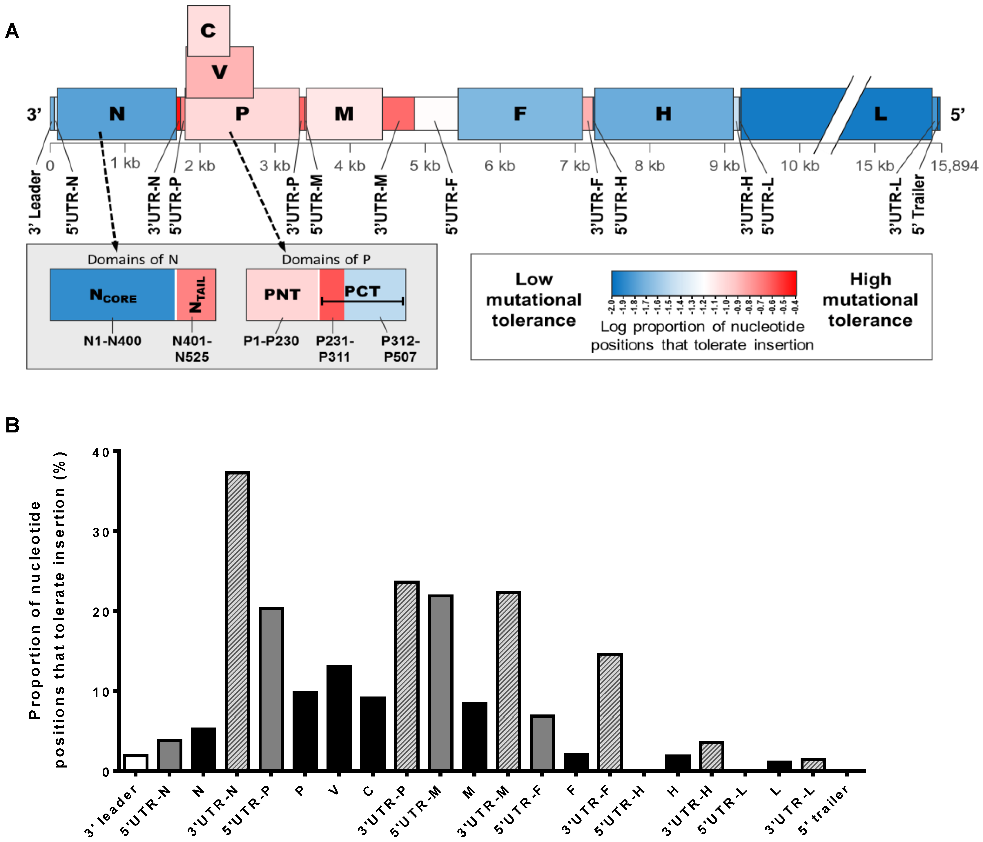
Table 1.
Reported substitution rates for RNA viruses. The range of substitution rates encompasses reported measurements made from any gene or entire genome sequence for the indicated virus. ** Human immunodeficiency virus (HIV) is a retrovirus and is included in this table for comparison.
| Family or Subfamily | Genus/Species | Substitution Rate Substitutions/Base/Year (× 10−4) | References |
|---|---|---|---|
| Paramyxovirinae | Morbillivirus | ||
| MeV | 3.4–9.02 | [19,43,48,49,50,51] | |
| CDV | 10.53–11.65 | [50,52] | |
| PPRV | 6.9–13.4 | [53] | |
| Rubulavirus | |||
| MuV | 2.5–9.168 | [43,50] | |
| Avulavirus | |||
| NDV | 5.04–13.5 | [54,55,56] | |
| Respirovirus | |||
| HPIV-1 | 2.2–13.7 | [43,57,58,59] | |
| HPIV-3 | 11.0 | [60] | |
| Henipavirus | |||
| NiV | 5 | [61] | |
| Pneumovirinae | Metapneumovirus | ||
| HMPV | 7.12–64.9 | [62,63,64] | |
| Pneumovirus | |||
| HRSV-A | 6.47–63.0 | [43,65,66,67,68,69,70,71,72] | |
| HRSV-B | 2.7–96.2 | [43,65,67,68,70,71,72,73] | |
| Orthomyxoviridae | IFVA | 13.9–131.0 | [43,44,46,74] |
| IFVB | 16–32.0 | [43,75] | |
| Picornaviridae | FMDV | 14 | [43,47] |
| EV71 | 34 | [43] | |
| Retroviridae ** | HIV-1 | 25–130.8 | [43,44,45] |
MeV: Measles virus; CDV: Canine distemper virus; PPRV: Peste-des-petits-ruminants virus; MuV: Mumps virus; NDV: Newcastle disease virus; HPIV: Human parainfluenza viruses; NiV: Nipah virus; HMPV: Human metapneumovirus; HRSV: Human respiratory syncytial virus; IFV: Infectious flacherie virus; FMDV: Foot-and-mouth disease virus; EV71: Enterovirus 71; HIV-1: Human immunodeficiency virus type 1.
© 2016 by the authors; licensee MDPI, Basel, Switzerland. This article is an open access article distributed under the terms and conditions of the Creative Commons Attribution (CC-BY) license (http://creativecommons.org/licenses/by/4.0/).
Share and Cite
MDPI and ACS Style
Beaty, S.M.; Lee, B. Constraints on the Genetic and Antigenic Variability of Measles Virus. Viruses 2016, 8, 109. https://doi.org/10.3390/v8040109
AMA Style
Beaty SM, Lee B. Constraints on the Genetic and Antigenic Variability of Measles Virus. Viruses. 2016; 8(4):109. https://doi.org/10.3390/v8040109
Chicago/Turabian StyleBeaty, Shannon M., and Benhur Lee. 2016. "Constraints on the Genetic and Antigenic Variability of Measles Virus" Viruses 8, no. 4: 109. https://doi.org/10.3390/v8040109
Note that from the first issue of 2016, this journal uses article numbers instead of page numbers. See further details here.