Article

Synergistic Effects of n-Hexane Fraction of Parkia biglobosa (Jacq.) Bark Extract and Selected Antibiotics on Bacterial Isolates

by
Oluwatayo E. Abioye
1,2,3,*,
David A. Akinpelu
2,3 and
Anthony I. Okoh
1,2
1
SAMRC Microbial Water Quality Monitoring Centre, University of Fort Hare, Private Bag X314, Alice 5700, South Africa
2
Applied and Environmental Microbiology Research Group, Department of Biochemistry and Microbiology, University of Fort Hare, Alice 5700, South Africa
3
Department of Microbiology, Obafemi Awolowo University, Ile-Ife 220, Osun State, Nigeria
*
Author to whom correspondence should be addressed.
Sustainability 2017, 9(2), 228; https://doi.org/10.3390/su9020228
Submission received: 21 December 2016 / Revised: 22 January 2017 / Accepted: 25 January 2017 / Published: 8 February 2017
(This article belongs to the Special Issue Sustainable Development Initiatives towards Poverty Alleviation)

Abstract

:
The incidence of resistance to commonly used antimicrobial agents by microbial pathogens demands increased effort in the development of effective ways of treating infections and diseases. The n-hexane fraction of lyophilized crude bark extract of Parkia biglobosa (Jacq.) was prepared and, in combination with selected antibiotics, assayed for antimicrobial activity against some selected bacterial pathogens using time-kill assay. Protein leakage analysis of the combined agents was performed using Bradford protein quantification method. Determination of active compounds present in the n-hexane fraction was done using Fourier Transform Infrared Spectroscopy (FTIR). While time-kill assay detected 43.33% synergy; 56.67% indifference and no antagonism at 1/2 × minimum inhibitory concentration (MIC), 1 × MIC exhibited 55% synergy, 45% indifference and no antagonism. Protein leakages from the cells of selected bacteria ranged from 1.20 µg/mL to 256.93 µg/mL. The presence of a phenyl group, an aromatic ring and phenolic compounds in the n-hexane fraction was confirmed at 2162 cm−1–2020 cm−1, 1605 cm−1–1533 cm−1 and 1438 cm−1–1444 cm−1 spectra peaks, respectively. The observed antibiotic−n-hexane fraction synergistic interaction revealed the improved antibacterial activity of the selected antibiotics. Hence, exploration of a combination of antibiotics with plant secondary metabolites is hereby advocated in the global quest for means of combating infectious diseases caused by multidrug-resistant pathogens.

1. Introduction

Infectious diseases cause a high level of morbidity and mortality of humankind, especially in developing countries [1]. The discovery of antibiotics was thought to be an absolute solution to the problem of infectious diseases until the emergence of bacterial strains with resistance to the antibiotics [2,3]. A number of new antibacterial drugs has been produced by pharmaceutical companies to combat the menace of infections caused by pathogens in the recent past, but resistance to these antibacterial drugs has been increasing and has thus become a global concern [1,4,5]. Although antimicrobial resistance may not be new, the increasing number of organisms that are resistant to a particular drug, geographical sites affected by such resistant pathogens, the unparalleled mounting and diverse resistance harbor by a single pathogen, as well as the commonness of antibiotic-resistant bacterial infections in clinical settings call for a quick discovery for new drugs or new ways of treating infections [2,6,7]. Immune-compromised and cancer patients often suffer serious medical complications due to multidrug-resistant bacteria, and such incidences have been on the rise for the last few years [5,7]. The resistance by pathogens to drugs always impaired the effectiveness of antimicrobial agents, leading to significant increase in illnesses and death rate [8]. The intricacies involved in developing and deploying a new drug for human use is enormous, but when such a drug finally is developed and deployed, the appearance of resistant microbial strains to this new drug within a few years after its first clinical use is also a big challenge to public health [9]. The problem of antibiotics resistance has led to re-emergence of infections which were thought to have been controlled by antibiotics in new forms of resistant dynamics [10,11,12]. Thus, a global search for new drugs and/or new ways of combating this menace caused by drug-resistant bacteria is imperative [13,14]. To tackle the challenge, the inhibitory effect of secondary metabolites on the multidrug-resistance efflux pump found in most resistant bacteria has been explored and the findings suggest that they could be an excellent enhancer of an antibacterial drug’s activity [15]. The World Health Organization (WHO) has also emphasised that medicinal plants are one of the best sources to obtain a variety of drugs [5]. Factors such as cost implication of some orthodox drugs, resistance of pathogens to available drugs, cultural beliefs in addition to the search for cures for some presently incurable diseases, have raised the global renaissance in the use of herbal preparations, with their gradual integration into the primary and secondary healthcare systems of some developing countries [16]. The success over the years in folk medicine as a means of keeping the human population healthy has led to the inauguration of traditional medicine strategy programs that will span from 2014 to 2023 by WHO [17].
Exploration of herbs and higher plants for antimicrobial biomolecules and formulation of new synergistic combinations using two commercially available antibiotics or to combine an antibiotic with natural antimicrobial plant products are two promising ways that have been proposed as a remedy to infections associated with multiple drug-resistant (MDR) bacterial strains [4,18,19,20]. The advantages of antimicrobial combination therapy involving biomolecules could include treatment of mixed infections, therapy for acute and critical infections caused by known etiological agents, augmentation of antibacterial activities, reduction of the time needed for developing long-term antimicrobial agents and prevention of resistant microbes [21,22]. Furthermore, this kind of therapy may involve protection of an active substance from degradation by enzymes, facilitate transport across barriers such as cell and organelle walls, overcome multidrug-resistance mechanisms or provide other signals to the host’s cells that result in higher efficacy of the crude drug when combined with a synthetic drug [23]. Although combination therapy has been reported to be beneficial, there is a need to investigate a combination of antimicrobial agents that will yield synergism, indifference or antagonism. This need is the result of non-synergistic responses observed in some combination therapies as seen in the case of penicillin−chlortetracycline combination therapy for pneumococcal meningitis which resulted in a lower response rate than that obtained with individual antibiotics [24].
Furthermore, a secondary metabolite from some plants has been found to potentiate the efficacy of synthetic antibiotics when they are combined [13]. Although combination therapy with existing antibiotics is not new, combination therapy between secondary metabolites and antibiotics can potentially reduce the multiple side effects of combining synthetic drugs. Toxicity assay has shown that antibiotics (especially synthetic ones) are toxic to humans. Although the toxicity of some of the antibiotics is within the permissible range for humans, the level of toxicity could go beyond the allowable threshold if two or more of these antibiotics are used in combination therapy [25,26]. This envisaged challenge of multiple toxic effects could be addressed by exploring combination therapy between orthodox drugs and bioactive compounds from medicinal plants [1,3,15]. Also, this could result in cheaper drug combination therapy that might solve the problem of non-completion of drug regimens by poor communities of patients who are suffering from diseases which require expensive drug-combination therapy [4]. Furthermore, antibiotic-bioactive compound combination therapies that are synergistic will proffer a new method of treating resistant bacterial infections as they are expected to (re-)enable the use of antibiotics that are no longer (or less) effective for therapeutic purposes [4]. The successful use of combinations of plant extracts has been observed in anti-infective therapy and treatment of cancer, HIV, inflammation, stress-induced insomnia, osteoarthritis and hypertension [3,27]. Nevertheless, studies on the synergy between plant extracts and synthetic drugs are limited [3,28,29], hence the need to explore and discover the synergistic potentials between natural bioactive agents and synthetic antibiotics in an attempt to combat most pathogens that have developed resistances against the available antibiotics. Such a discovery will improve the treatment of infectious diseases caused by multiple drug-resistant pathogens.
The plant Parkia biglobisa used in this study has been reported to possess various folk medicinal applications, and one such report is that of [30], wherein we demonstrated that bark crude extract and fractions from the plant exhibited varying degrees of antibacterial activities. This makes the plant a potential source that could be experimentally examined for combination therapy against infections. Based on the antibacterial potential of P. biglobosa and its fractions as earlier reported in our article, this work was designed to investigate the possible synergistic antimicrobial effects of the n-hexane fraction of Parkia biglobosa bark extract when combined with some selected standard antibiotics, on some pathogens that are resistant to some available synthetic antibiotics.

2. Materials and Methods

2.1. Plant Sample

The stem bark of Parkia biglobosa used for this study was collected from Ring Road Area Osogbo, Osun State, Nigeria. It was afterwards identified at the Herbarium of Botany Department, Obafemi Awolowo University Ile-Ife, Osun State, Nigeria, and a well-prepared voucher specimen was deposited at the Ife Herbarium under reference Number 16721.

2.2. Drying and Extraction of Crude Extract

The plant bark sample was processed following the method of [31] with little modification. The stem bark sample was expurgated into small bits, oven dried at 40 °C to constant weight and powdered. About 1500 g of the powder was soaked in 3:2 (v/v) solutions of methanol and sterile distilled water for four days with regular agitation. The concoction was filtered, concentrated in vacuo to semi-solid and then lyophilized to obtain 422 g of dried crude extract.

2.3. Solvent Partitioning of the Crude Extract

The stem bark crude extract (100 g) of P. biglobosa was reconstituted in sterile distilled water (200 mL) in a separatory funnel and extracted with n-hexane (4 × 200 mL). The n-hexane phase obtained was concentrated to dryness in vacuo and the resulting powder (28.0 g) was kept in an air-tight container for further use. This procedure was repeated until enough powder of n-hexane fraction for the synergy assay was obtained.

2.4. Sensitivity Testing of n-Hexane Fraction from Crude Bark Extract of Parkia biglobosa and Standard Antibiotics on Bacterial Isolates

The vulnerability of bacteria to n-hexane fraction and standard antibiotic was determined using the bioassay method of [32]. Solutions of a known concentration of selected antibiotics were prepared while solution of desired concentration of n-hexane fraction was prepared by dissolving its powder in 5% methanol which also served as control. Wells were made with a cork borer on a Mueller Hinton sterile agar plate that had been seeded with standardized inoculum (106 cfu/mL), and these were separately filled with 1 mL solution of n-hexane fraction and standard antibiotics. The resulting plates were allowed to stand at room temperature for 1 h before they were incubated at 37 °C for 24 h. Zones of inhibition were afterwards measured. The results obtained were as reported in [30].

2.5. Determination of Minimum Inhibitory Concentration (MIC) of n-Hexane Fraction and Standard Antibiotics

The MICs of the antibacterial agents were determined according to the procedure of [33]. Solutions of the antibacterial agents were prepared and 2 mL of these agents were added to 18 mL of pre-sterilized molten nutrient agar to give final desired concentrations. The medium that had been seeded with the antibacterial agent was afterwards poured into a sterile petri dish and allowed to set. After the surface was dry enough, 18 h-old standardised inoculum of the test bacterial isolates was streaked on it. The setup was incubated at 37 °C for 72 h and examined for the presence of growth or otherwise. The MIC was taken as the lowest concentration that inhibits the bacterial growth and the results were as reported in [30].

2.6. Antibiotic-Extract Combination Experiment

The potentiating power of the n-hexane fraction of P. biglobosa stem bark extract on selected antibiotics was assessed using the time-kill assay method as described by [34,35]. Conical flasks containing 50 mL of sterile nutrient broth dosed with n-hexane fraction, antibiotics and their combinations at 1/2 × MIC and 1 × MIC were inoculated with the standardised inoculum (~106 cfu/mL) to serve as test experimental setup. To ascertain the authenticity of the observations made in the course of the experiment, two controls were used. The first control involved the experimental setup in which diluents were used instead of the antibacterial agents that were tested in this study while the second control involved experimental setup without test organisms. The first control was set up to ensure that the experimental setup is devoid of any other antibacterial agents aside those intended while the second control ensures sterility before the introduction of test bacteria. Aliquots (100 μL) of the control flasks, as well as those of the test flasks, were taken, serially diluted, plated on nutrient agar incorporated with 0.5% 2,3,5-triphenyl tetrazolium chloride and incubated at 37 °C for 24 h in order to determine the zero hour counts. Also, the test and control flasks were immediately incubated at 37 °C with shaking at 120 rpm for 24 h. Afterwards, aliquots (100 μL) were taken from control and each test flask was transferred to recovery medium containing 3% antibacterial neutralizer (tween 80) and serially diluted in sterile physiological saline before being plated out in five replicates. The plates were incubated at 37 °C for 24 h after which the numbers of colonies were counted and the mean counts (cfu/mL) for each test and controls were calculated and expressed as log10. According to [36,37], interactions were considered synergistic when the antibacterial agent combinations achieved ≥2 log10 cfu/mL decrease in average viable colony counts when compared to average viable colony count achieved by most active single agents. They were considered additive or indifferent when the colony count decrease achieved is <2 log10 cfu/mL, and antagonistic when average colony count of the combination is ≥2 log10 cfu/mL as compared to that achieved by most active single agents.

2.7. Determination of Protein Leakage

Cells of Ps. aureginosa and E. faecalis from 18 h-old nutrient broth culture were discretely watched in normal saline (0.9% w/v NaCl). Then 0.5 McFarland standard-washed cell suspensions of the test bacteria were treated with 1/2 MIC and MIC concentrations of n-hexane fraction, antibiotic and their combination relative at various time intervals of 15 min for 2 h. Each suspension was then centrifuged at 7000 rpm and supernatant collected was assayed for protein using Bradford protein quantification method [38]. Bradford reagent (0.4 mL) was added to 1.6 mL of the supernatant solution (0.2 mL supernatant plus 1.4 mL sterile distilled water) to make 2 mL reaction volume. The optical density of each of the reaction mixes was taken at 595 nm after 5 min using spectrophotometer and corresponding protein concentration was extrapolated from the equation of best-fit linear regression line of the Bovine Serum Albumin (BSA) standard curve.

2.8. Fourier Transform Infrared Spectroscopy (FTIR)

FTIR was carried out on bulked n-hexane fraction of P. biglobosa stem bark extract showing strong activity against the test organism in order to identify possible compounds in the fraction following standard procedures as described by [39,40]. A 3 mm translucent sample disc of dried n-hexane fraction (10 mg) was prepared by encapsulating it in 100 mg of KBr salt pellet. The disc was immediately placed in the sample holder of the FTIR spectrophotometer (Perkin Elmer System 2000) and infrared (IR) spectra were recorded in the absorption range of 4000–450 cm−1.

3. Results

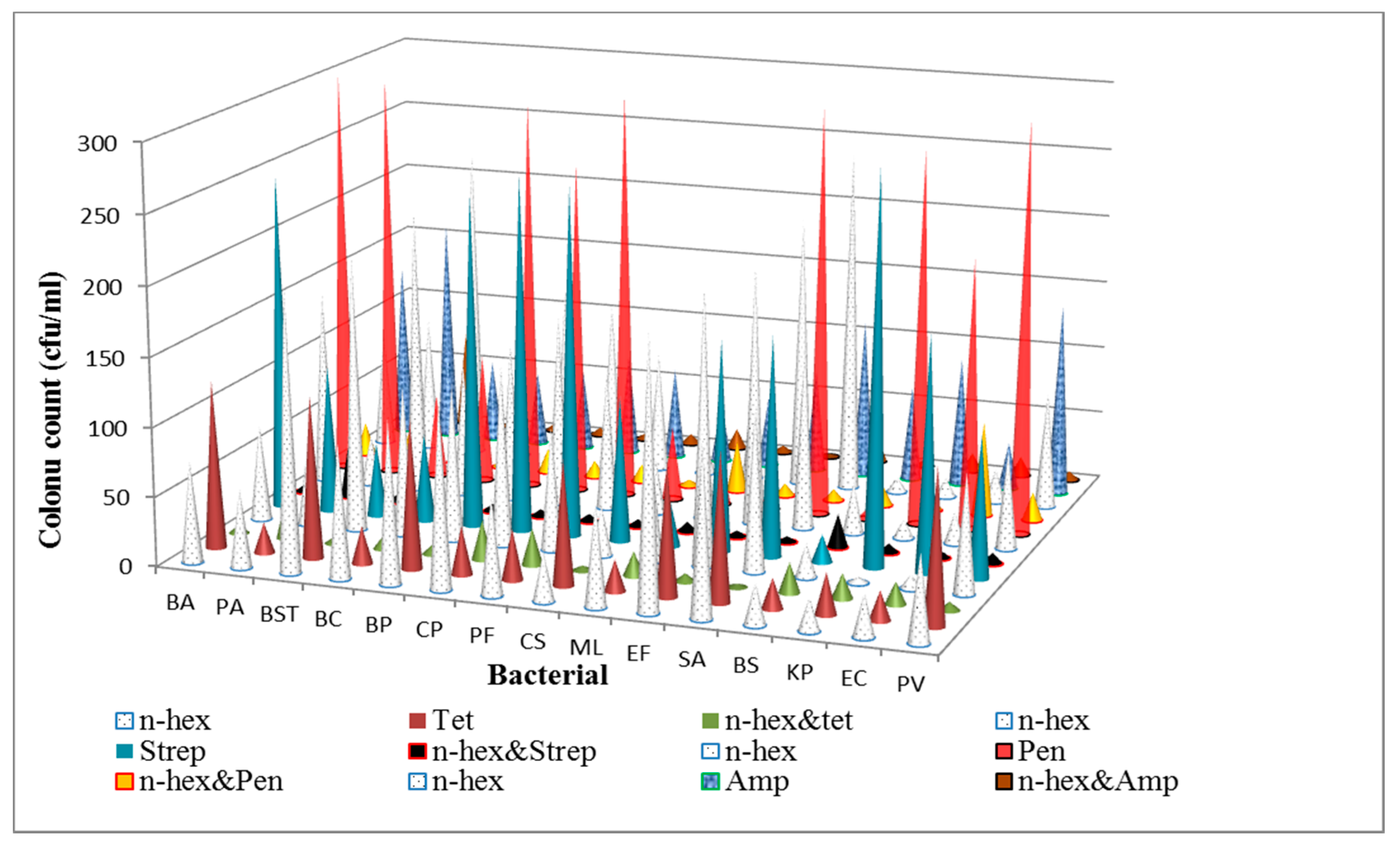
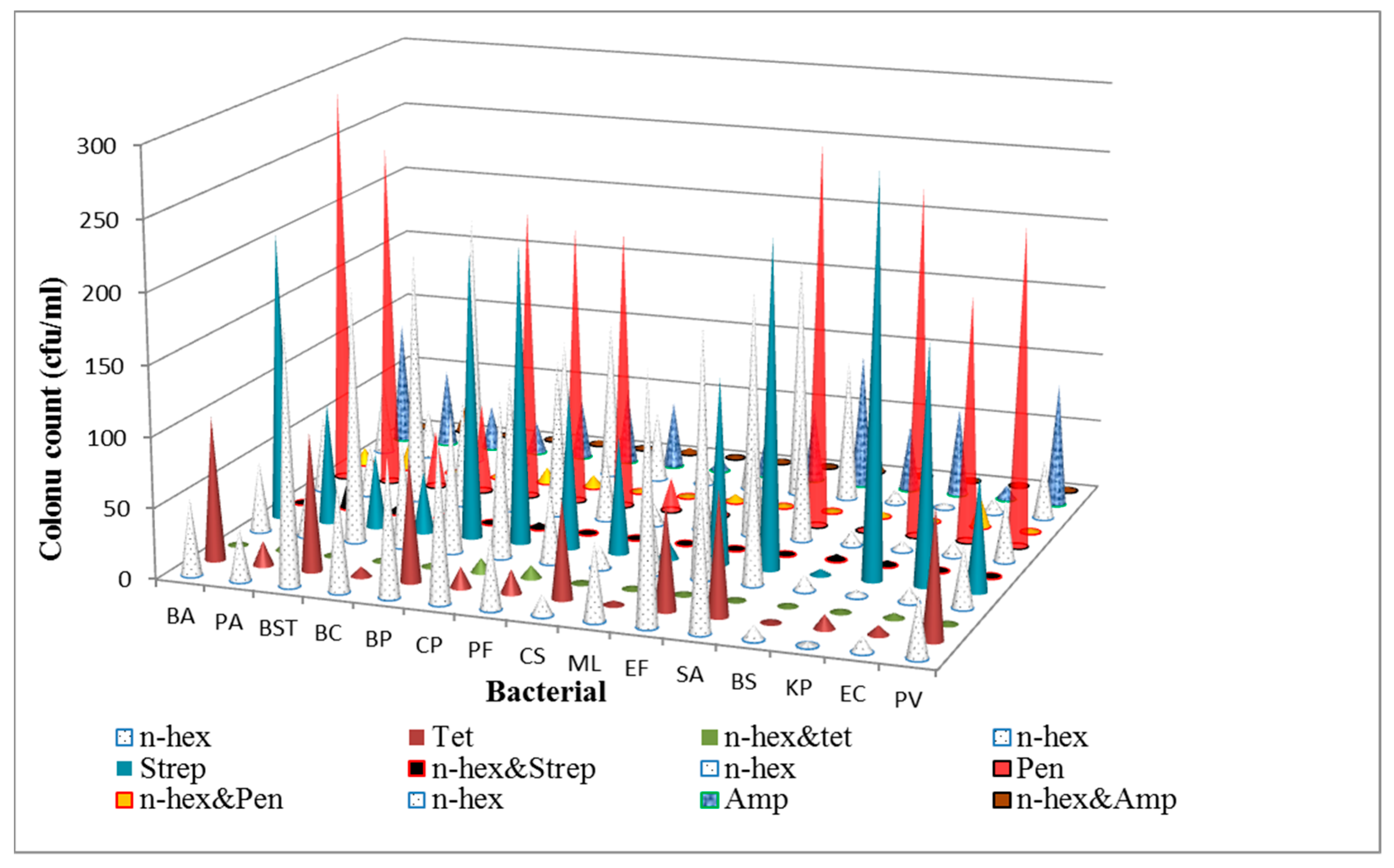
3.1. Determination of Antibacterial Activities and Minimum Inhibitory Concentrations (MICs) of the n-Hexane Fraction and Standard Antibiotics on Bacterial Isolates

The sensitivity testing and minimum inhibitory concentration (MICs) results of crude extracts, fractions and selected antibiotics has been published in our article [30] (Tables 3 and 4) with n-hexane fraction exhibiting strong antibacterial activities.

3.2. Synergy Experiment (The Time-Kill Assay)

Figure 1 and Figure 2 below show the trends observed as regards to colony counts after the expiration of time-kill assay experiment. One-way ANOVA test carried out shows that the mean colony counts differences observed after the exposure of the test organisms to the various antibacterial agents were significantly different at a p value of 0.05. The mean bacterial counts achieved by the antibacterial agents were further subjected to Tukey’s post hoc test at a p value of 0.05. A few of the bacterial counts were not significantly different when compared using the test: TET vs. N-HEX + TET against M. luteus and B. subtilis, N-HEX vs. TET + N-HEX against K. pneumoniae, STREP vs. N-HEX + STREP against B. subtilis, N-HEX vs. N-HEX + STREP against K. pneumonia, PEN vs. N-HEX + PEN against B. subtilis, N-HEX vs. N-HEX + PEN against K. pneumonia, N-HEX vs. N-HEX + AMP against K. pneumonia and AMP vs. N-HEX + AMP against E. coli at 1 × MIC. At 1/2 × MIC, the colony counts that were not significantly different when compared were TET vs. TET + N-HEX against B. subtilis, N-HEX vs. N-HEX + STREP against B. subtilis, N-HEX vs N-HEX + STREP against B. subtilis, N-HEX vs. N-HEX + PEN against K. pneumonia, and N-HEX vs. N-HEX + AMP against K. pneumonia. More than 91% of instances of non-significance in a mean difference of colony counts achieved by antibacterial agents was particular to K. pneumonia and B. subtilis, while less than 9% was for E. coli. This shows that one of the agents achieved mean colony counts similar to combinations of those instances, as is clearly shown in Figure 1 and Figure 2.
The time-kill effect of combinations of the n-hexane fraction of P. biglobosa stem bark extract and antibiotics is shown in Table 1 and Table 2. The fraction showed the ability to improve the antibacterial effect of the antibiotics on both Gram-positive and Gram-negative bacteria. The highest reduction in cell density of 3.98 ± 0.00 log10 was observed against B. stearothermophillus when n-hexane fraction and tetracycline were combined at 1 × MIC values. At the 1/2 × MIC level, 43.33% synergy, 56.67% indifference, and no antagonism were observed, whereas at the 1 × MIC level, 55% synergy, 45% indifference, and no antagonism were observed. Overall, synergistic response constituted about 49.17% of all forms of combinations of extract and antibiotics against all test isolates; antagonism was not detected among the 120 tests carried out.

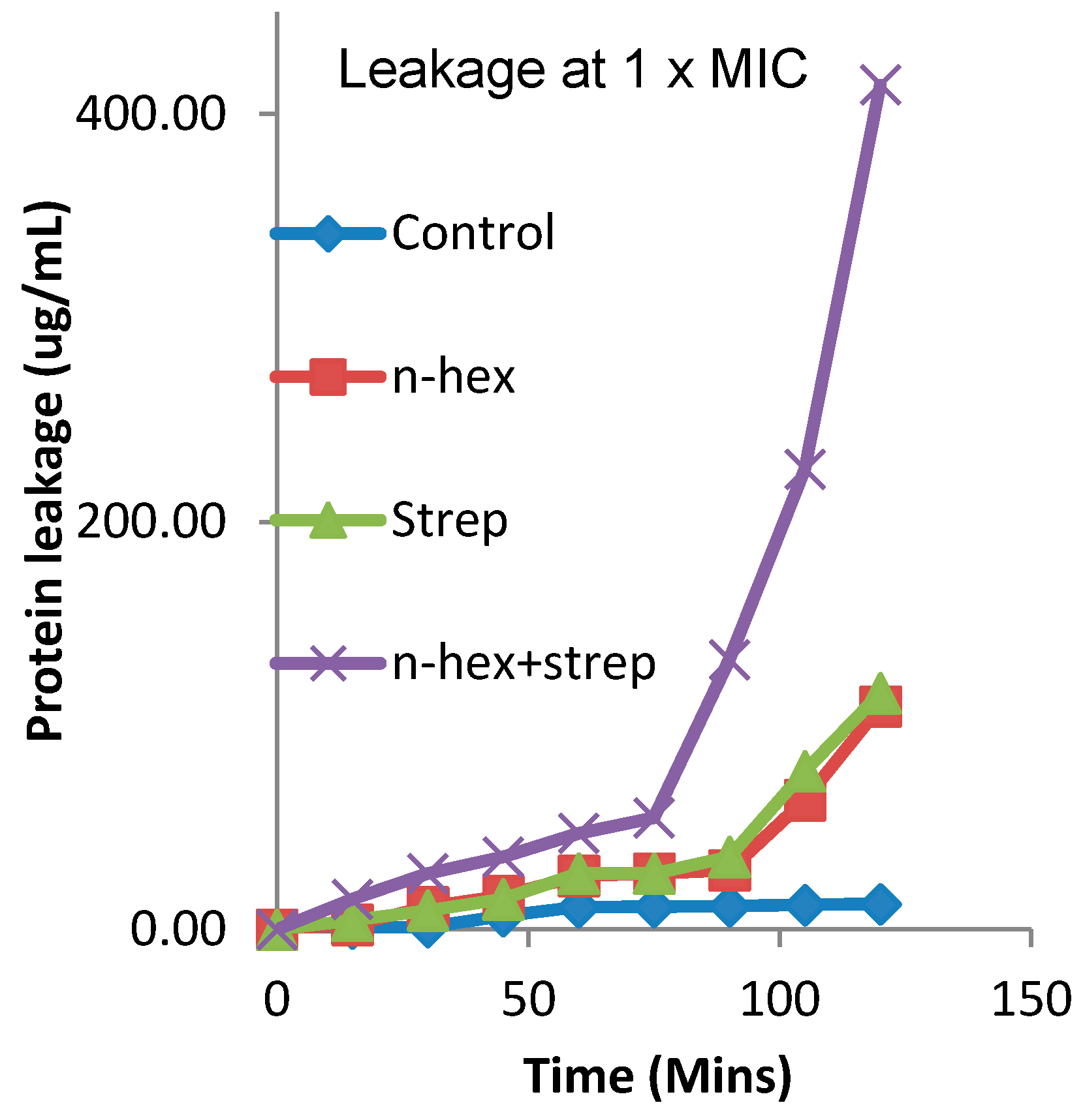
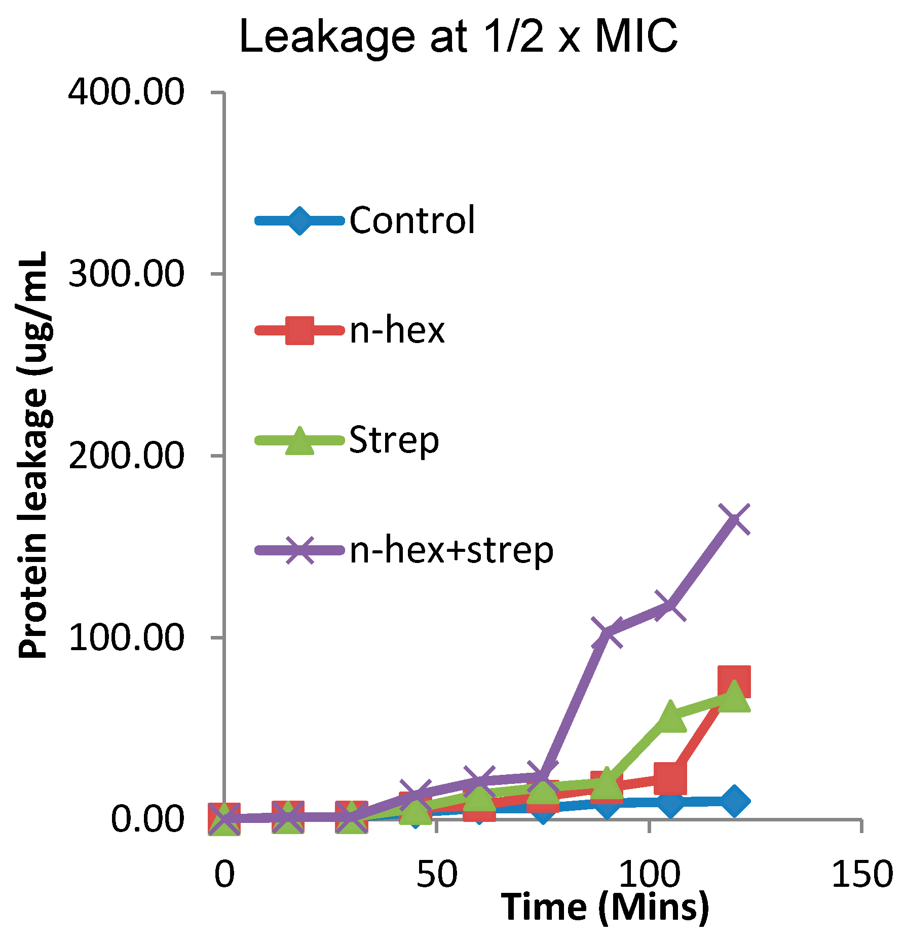
3.3. The Effect of the Combination of n-Hexane Fraction and Streptomycin on Protein Leakage from E. faecalis Cells

The protein leakage from E. faecalis cells due to the effect of n-hexane fraction, streptomycin and the combination of the two is shown in Figure 3 and Figure 4. The protein leakages after 15, 30, 45, 60, 75, 90, 105 and 120 min of contact time to E. faecalis with n-hexane fraction were 0.66 ± 0.01, 9.39 ± 0.01, 14.88 ± 0.02, 20.88 ± 0.02, 22.51 ± 0.00, 50.38 ± 0.00, 50.38 ± 0.02, and 99.56 ± 0.00 µg/mL respectively at 1/2 × MIC. The results of protein leakages by streptomycin for the same time intervals for the same organism at 1/2 × MIC were 0.66 ± 0.01, 1.21 ± 0.01, 14.86 ± 0.00, 19.23 ± 0.00, 22.51 ± 0.01, 50.38 ± 0.01, 66.78 ± 0.01 and 93.00 ± 0.05 µg/mL, respectively. When the two antimicrobial agents were combined, the protein leakages observed at the same order of time interval at concentration 1/2 × MIC were 0.67 ± 0.02, 14.33 ± 0.02, 20.89 ± 0.01, 29.08 ± 0.01, 32.35 ± 0.01, 42.18 ± 0.00, 89.73 ± 0.01 and 191.36 ± 0.01 µg/mL respectively.
Furthermore, the protein leakages from E. faecalis at 1 × MIC for n-hexane fraction at the same time intervals were 2.29 ± 0.01, 11.04 ± 0.01, 16.51 ± 0.01, 26.34 ± 0.01, 26.34 ± 0.01, 27.43 ± 0.01, 29.09 ± 0.01, 63.48 ± 0.01 and 109.39 ± 0.00 µg/mL, respectively. The protein leakages by streptomycin at 1 × MIC using the same time intervals were 4.47 ± 0.01, 9.26 ± 0.18, 14.87 ± 0.01, 27.44 ± 0.01, 27.45 ± 0.01, 35.62 ± 0.01, 77.71 ± 0.01, and 115.96 ± 0.01 µg/mL, while the protein leakages by the combination of n-hexane and streptomycin at 1 × MIC were 14.86 ± 0.00, 27.44 ± 0.01, 35.63 ± 0.01, 47.10 ± 0.00, 54.76 ± 0.01, 132.34 ± 0.00, 225.79 ± 0.00 and 414.33 ± 0.02 µg/mL respectively.

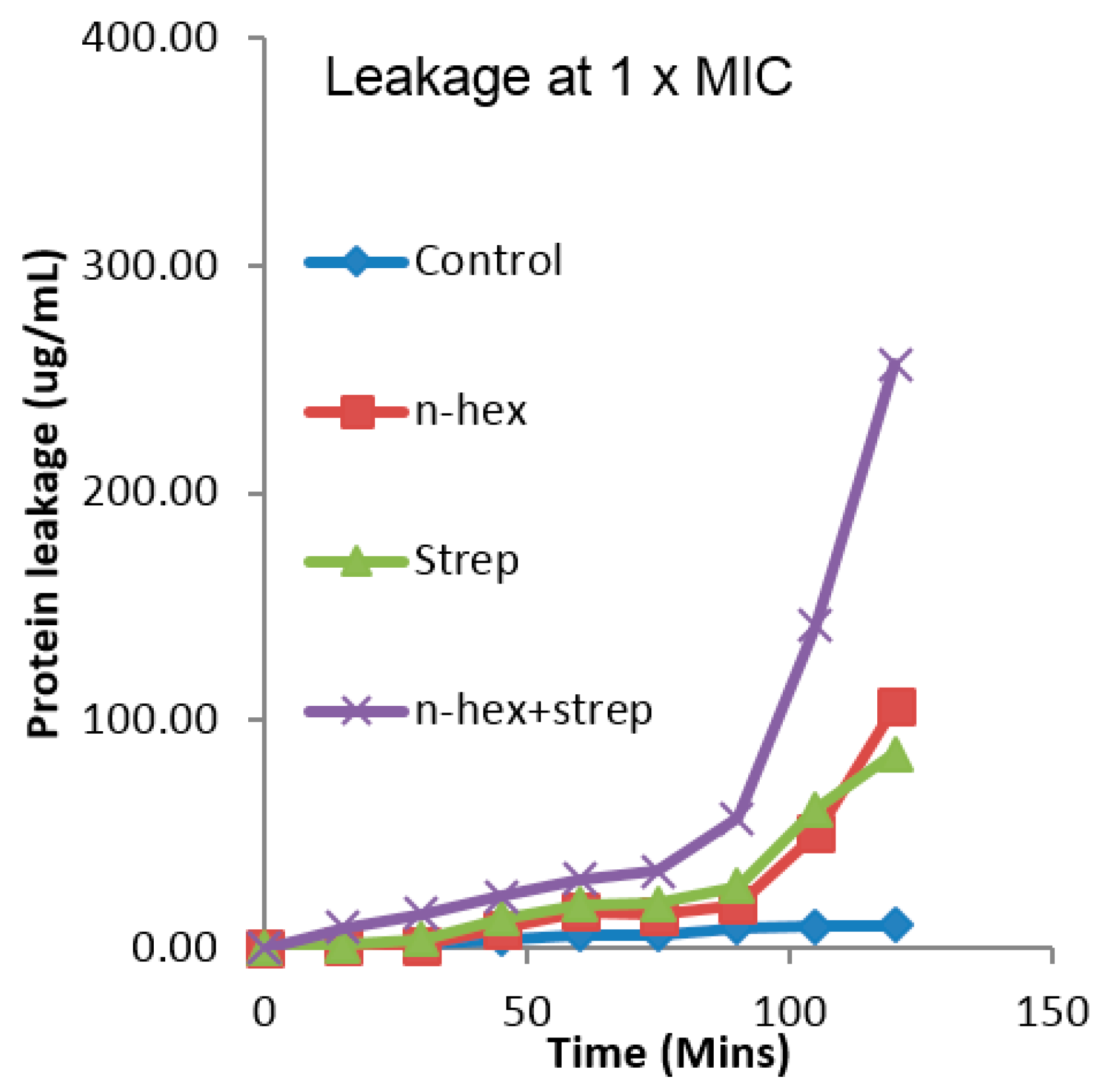
3.4. The Effect of the Combination of n-Hexane Fraction and Streptomycin on Protein Leakage from Pseudomonas aureginosa Cells

The protein leakage from Ps. aureginosa cells due to the effect of n-hexane fraction, streptomycin and the combination of the two is shown in Figure 5 and Figure 6. The protein leakages after 15, 30, 45, 60, 75, 90, 105 and 120 min of contact time of Ps. aureginosa with n-hexane fraction were 1.20 ± 0.00, 1.21 ± 0.00, 6.11 ± 0.00, 7.75 ± 0.00, 12.68 ± 0.00, 17.60 ± 0.01, 22.51 ± 0.00, 75.72 ± 0.00 µg/mL, respectively, at 1/2 × MIC. The results of protein leakages by streptomycin for the same time intervals, for the same organism at 1/2 × MIC, were 1.21 ± 0.01, 1.21 ± 0.01, 6.12 ± 0.01, 13.77 ± 0.00, 17.59 ± 0.01, 20.32 ± 0.01, 56.94 ± 0.01 and 67.90 ± 0.01 µg/mL, respectively. When the two antimicrobial agents were combined, the protein leakages observed for the same order of time intervals at concentration 1/2 × MIC were 1.21 ± 0.01, 1.22 ± 0.02, 13.23 ± 0.01, 20.88 ± 0.01, 23.61 ± 0.01, 102.84 ± 0.00, 117.60 ± 0.01 and 165.13 ± 0.00 µg/mL, respectively.
At 1 × MIC concentration, the protein leakages from Ps. aureginosa by n-hexane fraction at the same time intervals as earlier described were 1.21 ± 0.01, 1.22 ± 0.02, 8.30 ± 0.00, 15.97 ± 0.02, 14.31 ± 0.00, 19.23 ± 0.00, 50.38 ± 0.00 and 106.11 ± 0.00 µg/mL, respectively. At these same time intervals and at 1 × MIC, the protein leakages by streptomycin were 1.21 ± 0.01, 3.94 ± 0.01, 13.22 ± 0.00, 19.23 ± 0.00, 19.78 ± 0.01, 27.44 ± 0.01, 61.31 ± 0.01 and 85.36 ± 0.01 µg/mL, while the protein leakages by the combination of n-hexane and streptomycin at 1 × MIC were 8.86 ± 0.01, 14.87 ± 0.01, 22.51 ± 0.00, 30.16 ± 0.00, 33.98 ± 0.01, 56.93 ± 0.00, 142.19 ± 0.00 and 256.94 ± 0.01 µg/mL, respectively.
Statistically speaking, both one-way ANOVA test and Tukey’s post hoc test show that the amount of protein leakages from Ps. aureginosa and E. faecalis at 1 × MIC and 1/2 × MIC by various antibacterial agents used in this work are significantly different at a p value of 0.05.

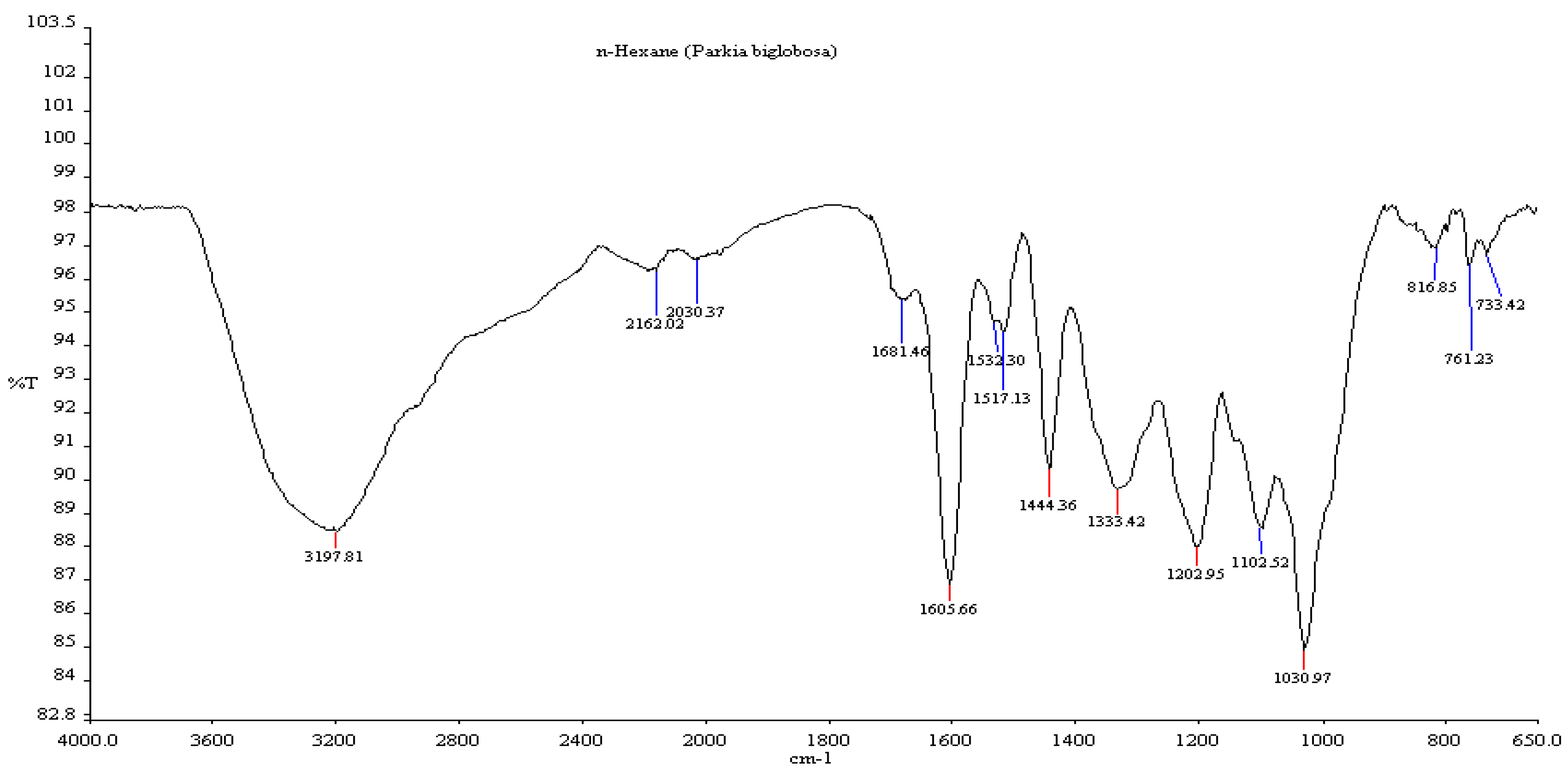
3.5. Fourier Transform Infrared Spectroscopy (FTIR)

The infrared absorption spectra of P. biglobosa n-hexane fraction contain characteristic features and strong vibrational bands in the region as shown in Figure 7. The various bands for the n-hexane fraction are 3197, 2162, 2030, 1681, 1605, 1532, 1517, 1444, 1333, 1202, 1102, 1030, 816, 761 and 733 cm−1.

4. Discussion

Table 1 and Table 2 show the results of the combination experiment between the n-hexane fraction and four selected antibiotics. The MIC values of the antibiotics, as well as those of the n-hexane fraction, were first determined to serve as reference point for defining the interactions. Going by the recommendation on MIC breakpoints in relation to the antibiotic used in this study [41,42], the various bacteria used fall into either resistant or slightly susceptible groups as reported in our article [30].
The combination of antibiotics with bioactive compounds has been reported to have enhanced the potency of synthetic antibiotics, especially first-line antibiotics [1,3]. The result of the combination of four antibiotics (ampicillin, tetracycline, streptomycin and penicillin G) with n-hexane fraction of P. biglobosa via time-kill assay method detected broad-spectrum synergy (43.33% at 1/2 × MIC, 55% at 1 × MIC), broad-spectrum, indifference/additive (56.67% at 1/2 × MIC, 45% at 1 × MIC) and no antagonistic interactions against tested organisms (Table 1 and Table 2). These results corroborate reports of [1,3] on the potentiating ability of plant extracts on standard antibiotics. The non-specificity of synergy detected in this investigation toward antibiotics type, as well as Gram-positive or -negative bacteria, suggests that the n-hexane fraction contains biocompounds that could enhance the antibacterial and resistance-modifying potentials of different classes of antibiotics as suggested by some studies [4,13,43,44,45]. This finding adds to the existing knowledge on the need to explore nature for novel biocompounds which can be used in standardised herbal-antibiotics combination therapy to treat and control infectious diseases, especially those caused by antibiotic-resistant pathogens.
The in vivo activities of antibiotics that attack the cell wall of bacteria could be enhanced by biocompounds from the plant by co-attacking the cell wall [2,8]. The presence of phenolic bioactive compounds as shown by FTIR analysis which has been reported to fight bacteria via membrane perturbation [46] in the n-hexane fraction coupled with peptidoglycan inhibitory ability of β-lactams (penicillin and ampicillin) used in this investigation explains in part the observed synergy in this study. On the other hand, the synergy observed for the combination of n-hexane fraction with either streptomycin and tetracycline in this study could be via inhibition of bacterial efflux pumps by a bioactive component of n-hexane fraction which in turn enhanced the protein synthesis inhibitory activity of the antibiotics. The potentiating power of some plant-derived biocompounds on antimicrobial agents by inhibiting multidrug-resistant (MDR) efflux systems, which is responsible for the substantial amount of antibiotic resistance in bacteria, has been reported by [47].
Broad-spectrum inhibitory potential of plant extracts and biocompounds from natural sources against bacteria have been reported [48,49,50,51], and efflux pump inhibitors such as 5′-methoxyhydnocarpin isolated from Berberis fremontii which inhibited NorA efflux pump in Staphylococcus aureus, have been found to enhance antibacterial properties of other antibacterial agents [23,43]. These efflux pump inhibitors if actually present in the n-hexane fraction used in this study (further work is ongoing on this) may have broad-spectrum activity considering the fact that synergy observed was non-specific to either Gram-positive or -negative bacteria. Biocompounds of plant origin that demonstrated broad-spectrum inhibitory activities against quinolone, tetracycline and erythromycin efflux pumps (NorA, TetK and MsrA) have been reported [3,52]. Therefore, the difference in the modes of action of the n-hexane fraction from that of the antibiotics used in this work may be an important factor in the synergy observed when they were combined.
The observed broad-spectrum activity of the n-hexane fraction of the bark extract and antibiotic combinations which were either synergistic or indifferent demonstrated the therapeutic potentials of P. biglobosa and suggested that this kind of combination therapy could be beneficial if explored in treating infectious diseases even at a concentration lower than MIC of an individual agent used in the combination experiment. It was observed that concentrations relative to MIC and time of exposure to antibacterial agents played a vital role in the outcomes of the combination experiment considering the results of various n-hexane fraction-antibiotics combinations (Table 1 and Table 2) and amount of protein leaked from representative Gram-negative and Gram-positive bacteria (Figure 3, Figure 4, Figure 5 and Figure 6) at 1/2 × MIC and 1 × MIC. The amount of protein leakages from Ps. aureginosa cells (Gram-negative bacterial representative) and E. faecalis cells (Gram-positive bacterial representative) further upheld the results of the combination. The protein component leakages by n-hexane fraction and streptomycin combination (256.94 ± 0.01 ug/mL from Ps. aureginosa cells and 414.33 ± 0.02 ug/mL from E. faecalis cells) were more than the addition of that by n-hexane fraction (106.11 ± 0.00 ug/mL from Ps. aureginosa cells and 109.39 ± 0.00 ug/mL from E. faecalis cells) and streptomycin (85.36 ± 0.01 ug/mL for Ps. aureginosa and 115.96 ± 0.01 ug/mL for E. faecalis) at 1 × MIC after 120 min of contact time. This same trend was observed when the MICs of the various antibacterial agents used were halved. The protein leakage results also showed that the antibacterial agents demonstrated a classic monophasic pattern of effect when used singly and in combination, thus supporting the earlier report of [53,54] on the ability of plant extracts and their fractions to leak intracellular material from bacterial cells in a classic monophasic pattern.
The results of protein leakage obtained in the synergy experiment suggest that the mechanisms of action of the various antimicrobial agents used in the combination experiment could include cell membrane disruption or damage which in turn leads to the leakage of the cell contents. The leakage of cell content due to the antibacterial effects of some plants’ bioactive compounds has been reported [55,56], while streptomycin has been reported to cause protein misread in the translation process, thereby causing incorporation of misread protein into the cell membrane leading to the formation of abnormal channels [57]. These abnormal channels could lead to the influx and accumulation of effective concentration of the antibacterial agents in bacterial cells and cause bactericidal or bacteriostatic effects. Therefore, the combination of cell membrane disruption ability of the n-hexane fraction as well as the formation of abnormal channels by streptomycin, also explain in part the observed synergistic effects of the combination of n-hexane fraction of P. biglobosa bark extract with streptomycin on test organisms. As much as the protein leakage experiment suggested membrane distribution as the mode of action of the n-hexane fraction, a proteomics analysis will give a more detailed knowledge and ascertain the origin of the protein leakage.
Fourier Transform Infrared Spectroscopy (FTIR), as one of the most widely used methods to identify the chemical constituents of plant extracts [58], was employed to elucidate the likely compounds present in the fraction used for this work. The infrared absorption spectra of the n-hexane fraction contain characteristic features and strong vibrational bands in the region 3200–3197 cm−1 which give an indication of the presence of the O–H and/or N–H group. The presence of a phenyl group was, however, confirmed in the fraction by the appearance of bands in the region of 2162–2020 cm−1. The band peaks at ~1605 and 1532 cm−1 were attributed to the presence of the aromatic ring. The peak at 1681 cm−1 was correlated with the stretching vibration of carbonyl (C=O) and/or (C–N, amide 1) [59,60]. However, a report by [61], showed that the carbonyl absorptions in flavones, isoflavone and their 5-hydroxy derivatives are not abnormal as suggested by earlier work. The infrared data can be explained by taking into account the C=C absorption which often overlaps with the C=O bands as occurred in the n-hexane fraction.
Other features of the fraction are the absorption bands at 1438 and 1444 cm−1, which are related to the first overtone of the –OH stretch of H2O. It has been reported that the –OH phenolic compounds do have vibrational bands at 1200 and 1350 cm−1 [62]. The close proximity of these bands to those found in the fraction suggests that the n-hexane fraction of P. biglobosa bark under investigation may contain flavonoid constituents. Ethereal functional groups, as found for C–O–C vibrations in the fraction, were supported by the bands at 1102–1030 cm−1, and the aromatic substitution patterns were confirmed by the presence of the vibration bands at 832–733 cm−1; this is attributed to both ortho- and meta-substitution of the hydroxyl groups on the phenyl ring. Phenol compounds with high concentration have been shown to have vibrational frequency characteristics at 3200–3400 cm−1, which are usually broad [62]. The FTIR assay confirmed the presence of aromatic, phenyl, carbonyl, amide, hydroxyl, phenolic, and ethereal chemical groups, thereby corroborating the findings of [63]. These identified chemical groups are important components of some bioactive phytochemicals such as flavonoids, saponins, tannins and triterpenes that are present in the bark of P. biglobosa [30,64,65]. A pure triterpene compound called lupeol was isolated by [65] who further found that this compound is one of the major bioactive components of P. biglobosa. This explains in part the compound that could possibly be responsible for the improved bioactivity that we observed when the n-hexane fraction of P. biglobosa bark extract was combined with the selected antibiotics. Although the FT-IR results provided clues to the type of compounds that may be present in this active fraction, work is ongoing to isolate chemical compounds from the fraction.

5. Conclusions

The combinations of n-hexane fraction from the crude stem bark extract of Parkia biglobosa with the standard antibiotics used in this study displayed only synergistic and additive interactions. These observations suggest that the bark of the plant possesses bioactive constituents that could be used in combination therapy with standard antibiotics against pathogens. This report further showed that the activity of antibiotics to which pathogens had developed resistances could be enhanced by bioactive compounds that are present in the bark of P. biglobosa. This study also showed that n-hexane is a very good solvent to be considered for the extraction of antibiotic-resistant modulating biomolecules present in the bark of P. biglobosa. Although we did not run an assay to investigate side effects of n-hexane fraction of P. biglobosa on eukaryote cells, experimental reports from researchers such as [11,66,67,68] and the use of this plant in herbal medicament in some African countries (e.g., Mali, Cote d’Ivoire and Nigeria) prove that the fraction has negligible toxicity to eukaryotic cells. This explains the reason for the successful and wide usage of the plant in folklore medicine and suggests that formulations should be prepared for in vivo assay in an animal model. If successful, clinical trials should follow in order to establish a scientifically based regime in terms of quality and quantity. This kind of scientific-based regime will prevent the abuse of herbal-antibiotic therapy that has become more common, especially in this era of increasing infections caused by antibiotic-resistant pathogens. Since the extract from the plant’s bark is non-toxic to humans and is readily available, the n-hexane fraction-antibiotic formulation of this kind is expected to be more effective, safer and cheaper than antibiotics−antibiotic combination therapy in combating the infections caused by the pathogens used in this study. Finally, absolute knowledge on the potentiating power of the n-hexane fraction from the bark of P. biglobosa on synthetic antibiotics used in this study cannot be claimed until compounds that are responsible for the observed interactions are isolated; however, work on these compounds is underway.

Supplementary Materials

The following are available online at www.mdpi.com/2071-1050/9/2/228/s1, Table S1: Colony count for tetracycline and n-hexane fraction combination at 1 × MIC; Table S2: Colony count for streptomycin and n-hexane fraction combination at 1 × MIC; Table S3: Colony count for penicillin and n-hexane fraction combination at 1 × MIC; Table S4: Colony count for ampicillin and n-hexane fraction combination at 1 × MIC; Table S5: Colony count for tetracycline and n-hexane fraction combination at 1/2 × MIC; Table S6: Colony count for streptomycin and n-hexane fraction combination at 1/2 × MIC; Table S7: Colony count for penicillin and n-hexane fraction combination at 1/2 × MIC; Table S8: Colony count for ampicillin and n-hexane fraction combination at 1/2 × MIC; Table S9: The effect of the combination of n-hexane fraction and streptomycin on protein leakage from E. faecalis; Table S10: The effect of the combination of n-hexane fraction and streptomycin on protein leakage from Ps. Aureginosa; Table S11: Standard curve for bovine serum albumin; Figure S1: Caption Standard curve graph for bovine serum albumin.

Acknowledgments

We acknowledge HC Illoh of Botany Department and AO Adeloye of Chemistry Department, Obafemi Awolowo University, Ile-Ife, Nigeria for their contribution towards plant sample identification and FTIR analysis respectively. Our gratitude also goes to AEMREG, University of Fort Hare and Medical Research Council, South Africa for their technical and financial support. Figure S1: Caption Standard curve graph for bovine serum albumin.

Authors Contributions

All authors contributed equally to this work. All authors read and approved the final manuscript.

Conflicts of Interests

All authors declare that there is no conflict of interests.

References

  1. Adwan, G.M.; Abu-Shanab, B.A.; Adwan, K.M. In vitro activity of certain drugs in Combination with plant extracts against Staphylococcus aureus infections. Pak. J. Med. Sci. 2008, 24, 541–544. [Google Scholar]
  2. Aiyegoro, O.A.; Afolayan, A.J.; Okoh, A.I. Synergistic interaction of Helichrysum pedunculatum leaf extracts with antibiotics against wound infection associated bacteria. Biol. Res. 2009, 42, 327–338. [Google Scholar] [CrossRef] [PubMed]
  3. Hemaiswarya, S.; Kruthiventi, A.K.; Doble, M. Synergism between natural products and antibiotics against infectious diseases. Phytomedicine 2008, 15, 639–652. [Google Scholar] [CrossRef] [PubMed]
  4. Ali, N.H.; Kazim, S.U.; Faizi, S. Activity of synergistic combination amoxy-cassia against salmonella. Pak. J. Pharma. Sci. 2007, 20, 140–145. [Google Scholar]
  5. Nascimento, G.G.F.; Juliana, L.; Paulo, C.F.; Giuliana, L.S. Antibacterial activity of plant extracts and phytochemicals on antibiotic-resistant bacteria. Braz. J. Microbiol. 2000, 31, 247–256. [Google Scholar] [CrossRef]
  6. Yang, Z.C.; Wang, B.C.; Yang, X.S.; Wang, Q.; Ran, L. The synergistic activity of antibiotics combined with eight traditional Chinese medicines against two different strains of Staphyloccus aureus. Coll. Surf. B Biointerfaces 2005, 41, 79–81. [Google Scholar] [CrossRef] [PubMed]
  7. Ahmad, I.; Aqil, F. In vitro efficacy of bioactive extract of 15 medicinal plants against ESβL-producing multidrug-resistant enteric bacteria. Microbiol. Res. 2007, 162, 264–275. [Google Scholar] [CrossRef] [PubMed]
  8. Center for Disease Dynamics, Economics & Policy (CDDEP). State of the World’s Antibiotics; CDDEP: Washington, DC, USA, 2015; Available online: https://cddep.org/sites/default/files/swa_2015_final.pdf (accessed on 9 November 2016).
  9. Sibanda, T.; Okoh, A.I. In vitro evaluation of the interactions between acetone extracts of Garcinia kola seeds and some antibiotics. Afr. J. Biotechnol. 2008, 7, 1672–1678. [Google Scholar]
  10. Levy, S.B.; Marshal, B. Antibacterial resistance worldwide: Causes, challenges and responses. Nat. Med. 2004, 10 (Suppl. 12), 122–129. [Google Scholar] [CrossRef] [PubMed]
  11. Udobi, C.E.; Onaolapo, J.A.; Agunu, A. Antibacterial activities and bioactive components of the aqueous fraction of the stem bark of Parkia biglobosa (Jacq) (mimosaceae). Niger. J. Pharm. Sci. 2008, 7, 49–55. [Google Scholar]
  12. Baylor College of Medicine. Emerging Infectious Diseases. Available online: https://www.bcm.edu/departments/molecular-virology-and-microbiology/emerging-infections-and-biodefense/emerging-infectious-diseases (accessed on 20 December 2016).
  13. Ghaleb, A.; Mohammad, M. Synergistic effects of plant extracts and antibiotics on Staphylococcus aureus strains isolated from clinical specimens. Middle-East J. Sci. Res. 2008, 3, 134–139. [Google Scholar]
  14. Salvat, A.; Antonnacci, L.; Fortunato, R.H.; Suarez, E.Y.; Godoy, H.M. Screening of some plants from Northern Argentina for their antimicrobial activity. Lett. Appl. Microbiol. 2001, 32, 293–297. [Google Scholar] [CrossRef] [PubMed]
  15. Aiyegoro, O.A.; Okoh, A.I. Use of bioactive plant products in combination with standard antibiotics: Implications in antimicrobial chemotherapy. J. Med. Plant. Res. 2009, 3, 1147–1152. [Google Scholar]
  16. El-Mahmood, A.M.; Ameh, J.M. In vitro antibacterial activity of Parkia biglobosa (Jacq.) root bark extract against some microorganisms associated with urinary tract infections. Afr. J. Biotechnol. 2007, 6, 1272–1275. [Google Scholar]
  17. World Health Organisation (WHO) Traditional Medicine Strategy: 2014–2023. Available online: http://apps.who.int/iris/bitstream/10665/92455/1/9789241506090_eng.pdf (accessed on 28 October 2016).
  18. Veloira, W.G.; Domenico, P.; Lipuma, J.J.; Davis, J.M.; Gurzenda, E.; Kazzaz, J. In vitro activity and synergy of bismuth thiols and tobramycin against Burkholderia cepacia complex. J. Antimicrob. Chemother. 2003, 52, 915–919. [Google Scholar] [CrossRef] [PubMed]
  19. Dawis, M.A.; Isenberg, H.D.; France, K.A.; Jenkinsin, S.G. In vitro activity of gatifloxacin alone and in combination with cefepime, meropenem, piperacillin and gentamicin against multi-drug resistant organisms. J. Antimicrob. Chemother. 2003, 51, 1203–1211. [Google Scholar] [CrossRef] [PubMed]
  20. Mandal, S.; Mandal, M.D.; Pal, N.K. Combination effects of ciprofloxacine and gentamicin against clinical isolates of Salmonella enterica Serover typhi with reduced susceptibility to ciproflocin. Jpn. J. Infect. Dis. 2003, 56, 156–157. [Google Scholar] [PubMed]
  21. Hugo, W.B.; Russel, A.D. Pharmaceutical Microbiology, 5th ed.; Blackwell Scientific Publishing: New York, NY, USA, 1993; pp. 119–120. [Google Scholar]
  22. Levinson, W.; Jawetz, E. Medical Microbiology and Immunology: Examination and Board Review, 7th ed.; Lange Medical Books; McGraw-Hill: New York, NY, USA, 2002; pp. 73–83. [Google Scholar]
  23. Gilbert, B.; Alves, L.F. Synergy in plant medicines. Curr. Med. Chem. 2003, 10, 13–20. [Google Scholar] [CrossRef] [PubMed]
  24. Young, L.S. Synergy between antimicrobial agents: Review of clinical significance of synergy in gram-negative infections at the University of California Los Angeles hospital. Infection 1978, 6 (Suppl. 1), S47. [Google Scholar] [CrossRef]
  25. Cunha, B.A. Antibiotic side effects. Medical Clinics of North America. Med. Clin. N. Am. 2001, 85, 149–185. [Google Scholar] [CrossRef]
  26. Wijsman, J.A.; Rieder, M.J.; Dekaban, G.A. Differential toxicity of reactive metabolites of clindamycin and sulphonmide in HIV-Infected cells. J. Clin. Pharmacol. 2005, 45, 346–351. [Google Scholar] [CrossRef] [PubMed]
  27. Williamson, E.M. Synergy and other interactions in phytomedicines. Phytomedicines 2001, 8, 401–409. [Google Scholar] [CrossRef] [PubMed]
  28. Ginsburg, H.; Deharo, E. A call for using natural compounds in the development of new antimalarial treatments an introduction. Malar. J. 2011, 10 (Suppl. S1). [Google Scholar] [CrossRef] [PubMed]
  29. Odunbaku, O.A.; Ilusanya, O.A.F. Synergistic effect of ethanol leaf extract of Senna alata and antimicrobial drugs on some pathogenic microbes. Adv. Environ. Biol. 2011, 5, 2162–2165. [Google Scholar]
  30. Abioye, O.E.; Akinpelu, D.A.; Aiyegoro, O.A.; Adegboye, M.F.; Oni, M.O.; Okoh, I.O. Preliminary Phytochemical Screening and Antibacterial Properties of Crude Stem Bark Extracts and Fractions of Parkia biglobosa (Jacq.). Molecules 2013, 18, 8485–8499. [Google Scholar] [CrossRef] [PubMed]
  31. Edeoga, H.O.; Okwu, D.E.; Mbaebie, B.O. Phytochemical constituents of some Nigeria medicinal plants. Afr. J. Biotechnol 2005, 4, 685–688. [Google Scholar] [CrossRef]
  32. Betoni, J.E.C.; Mantovani, R.P.; Barbosa, L.N.; Di Stasi, L.C.; Fernandes, A., Jr. Synergism between plant extract and antimicrobial drugs used on Staphylococcus aureus diseases. Mem. Inst. Oswaldo Cruz 2006, 101, 387–390. [Google Scholar] [CrossRef] [PubMed]
  33. Akinpelu, D.A.; Kolawole, D.O. Phytochemistry and antimicrobial activity of leaf extract of Piliostigma thonningii (Schum). Sci. Focus 2004, 7, 64–70. [Google Scholar]
  34. White, R.L.; Burgess, D.S.; Manduru, M.; Bosso, J.A. Comparison of three different In Vitro Methods of Detecting Synergy: Time-Kill, Checkerboard, and E test. Antimicrob. Agents Chemother. 1996, 40, 1914–1918. [Google Scholar] [PubMed]
  35. Pankey, G.; Ashcraft, D. In vitro synergy of ciprofloxacin and gatifloxacin against ciprofloxacin-resistant Pseudomonas aeruginosa. Antimicrob. Agents Chemother. 2005, 49, 2959–2964. [Google Scholar] [CrossRef] [PubMed]
  36. Lee, J.Y.; Oh, W.S.; Ko, K.S.; Heo, S.T.; Moon, C.S.; Ki, H.K.; Kiem, S.; Peck, K.R.; Song, J.H. Synergy of arbekacin-based combinations against vancomycin hetero-intermediate Staphylococcus aureus. J. Korean Med. Sci. 2006, 21, 188–192. [Google Scholar] [CrossRef] [PubMed]
  37. Sato, M.; Tanaka, H.; Yamaguchi, R.; Kato, K.; Etoh, H. Synergistic effects of mupirocin and an isoflavanone isolated from Erythrina variegata on growth and recovery of methicillin-resistant Staphylococcus aureus. Int. J. Antimicrob. Agents 2004, 24, 43–48. [Google Scholar] [CrossRef] [PubMed]
  38. Bradford, M.M. A rapid and sensitive method for quantification of microgram quantities of protein utilizing the principle of protein-dye binding. Annu. Rev. Plant Physiol. Plant Mol. Biol. 1976, 72, 245–254. [Google Scholar]
  39. Starlin, T.; Gopalakrishnan, V.K.; Arul, R.C.; Ragavendran, P. Phytochemical screening, functional groups and element analysis of Tylophora pauciflora Wight and Arn. IRJP 2012, 3, 180–183. [Google Scholar]
  40. Ashokkumar, R.; Ramaswamy, M. Phytochemical screening by FTIR spectroscopicanalysis of leaf extracts of selected Indian Medicinal plants. Int. J. Curr. Microbiol. Appl. Sci. 2014, 3, 395–406. [Google Scholar]
  41. European Committee for Antimicrobial Susceptibity Testing (EUCAST). Determination of minimum inhibitory concentrations (MICs) of antibacterial agents by agar dilution. Clin. Microbiol. Inf. 2000, 6, 509–515. [Google Scholar]
  42. Comite de L’antibiogramme de la Societe Francaise de Microbiologie Recommandations 2012. Available online: http://www.sfm-microbiologie.org/UserFiles/files/casfm/CASFM_2012.pdf (accessed on 20 December 2016). (In French)
  43. Frank, R.S.; Peter, L.; Jeanne, N.T.; Lauren, A.Z.; Kim, L. Synergy in a medicinal plant: Antimicrobial action of berberine potentiated by 5′-methoxyhydnocarpin, a multidrug pump inhibitor. Proc. Natl. Acad. Sci. USA 2000, 15, 1433–1437. [Google Scholar]
  44. Basri, D.F.; Zin, N.M.; Bakar, N.S.; Rahmat, F.; Mohtar, M. Synergistic effects of phytochemicals and oxacillin on laboratory passage-derived vancomycin-intermediate Staphylococcus aureus strain. J. Med. Sci. 2008, 8, 131–136. [Google Scholar]
  45. Wisatre, K.; Chintana, C.; Thitiporn, D.; Wipaphorn, J.; Sungwan, H.; Benjamas, S.F.; Patra, R.; Kaikwan, J. The potential co-treatment effects of three plant extracts and three antibiotics on multidrug-resistant bacteria. J. Sci. Technol. Humanit. 2007, 5, 17–27. [Google Scholar]
  46. Esimone, C.O.; Iroha, I.R.; Ibezim, E.C.; Okeh, C.O.; Okpana, E.M. In vitro evaluation of the interaction between tea extracts and penicillin G against Staphylococcus aureus. Afr. J. Biotechnol. 2006, 5, 1082–1086. [Google Scholar]
  47. Savoia, D. Plant-derived antimicrobial compounds. Future Microbiol. 2012, 7, 979–990. [Google Scholar] [CrossRef] [PubMed]
  48. Seasotiya, L.; Dalal, S. Screening of Indian medicinal plants as efflux pump inhibitors of fluoroquinolones. J. Pharmacogn. Phytochem. 2014, 3, 235–241. [Google Scholar]
  49. Fiamegos, Y.C.; Kastritis, P.L.; Exarchou, V.; Han, H.; Bonvin, A.M.J.J.; Vervoort, J.; Lewis, K.; Hamblin, M.R.; Tegos, G.P. Antimicrobial and Efflux Pump Inhibitory Activity of Caffeoylquinic Acids from Artemisia absinthium against GramPositive Pathogenic Bacteria. PLoS ONE 2011, 6, e18127. [Google Scholar] [CrossRef] [PubMed] [Green Version]
  50. Stavri, M.; Piddock, L.J.V.; Gibbons, S. Bacterial efflux pump inhibitors from natural sources. J. Antimicrob. Chemother. 2007, 59, 1247–1260. [Google Scholar] [CrossRef] [PubMed]
  51. Piddock, L.J.V. Clinically relevant chromosomally encoded multidrug resistance efflux pumps in bacteria. Clin. Microbiol. Rev. 2006, 19, 382–402. [Google Scholar] [CrossRef] [PubMed]
  52. Smith, E.C.J.; Williamson, E.M.; Wareham, N.; Kaatz, G.W.; Gibbons, S. Antibacterials and modulators of bacterial resistance from the immature cones of Chamaecyparis lawsoniana. Phytochemistry 2007, 68, 210–217. [Google Scholar] [CrossRef] [PubMed]
  53. Akinpelu, D.A.; Aiyegoro, O.A.; Okoh, A.I. In vitro antimicrobial and phytochemical properties of extract of stem bark of Afzelia africana (Smith). Afr. J. Biotechnol. 2008, 7, 3665–3670. [Google Scholar]
  54. Oloke, J.K. The Antibacterial and Antifungal Effect of the Volatile Oil and Partially Purified Extracts of Aframomum melegueta K. Schum. Ph.D. Thesis, Department of Microbiology, University of Ife, Ile-Ife, Nigeria, 1989. [Google Scholar]
  55. Akinpelu, D.A.; Alayande, K.A.; Aiyegoro, O.A.; Akinpelu, O.F.; Okoh, A.I. Probable mechanisms of biocidal action of Cocos nucifera Husk extract and fractions on bacteria isolates. BMC Complement. Altern. Med. 2015, 15, 116. [Google Scholar] [CrossRef] [PubMed]
  56. Aiyegoro, O.A.; Akinpelu, D.A.; Okoh, A.I. In vitro potentials of the stem bark of red water tree (Erythrophyleum suaveolens). J. Biol. Sci. 2007, 7, 1233–1238. [Google Scholar]
  57. Davis, B.D.; Chen, L.; Tai, P.C. Misread protein creates memnbrane channels: An essential steps in the bacterial action of aminoglycosides. Proc. Natl. Acad. Sci. USA 1986, 83, 6164–6168. [Google Scholar] [CrossRef] [PubMed]
  58. Druy, M.A. Applications for mid-IR Spectroscopy in pharmaceutical process environment. Spectroscopy 2004, 19, 60–63. [Google Scholar]
  59. Nakanishi, K.; Solomon, P.H. Infrared Absorption Spectroscopy, 2nd ed.; Holden-Day, Inc.: San Francisco, CA, USA, 1977. [Google Scholar]
  60. Koeppen, B.H.; Basson, D.S. The anthocyanin pigments of barlinka grapes. Phytochemistry 1966, 5, 183–187. [Google Scholar] [CrossRef]
  61. Jose, C.I.; Phadke, P.S.; Rama Rao, A.V. Infrared spectra of flavones and isoflavones: Effect of iodine and boron trifluoride on carbonyl frequencies. Spectrochim. Acta A Mol. Spectrosc. 1974, 30, 1199–1206. [Google Scholar] [CrossRef]
  62. Ficarra, R.; Tommasini, S.; Raneri, D.; Calabro, M.L.; Dibella, M.R.; Rusticherri, C.; Gamberini, M.C. Study of flavonoids/β-cyclodextrins inclusion complexes by NMR, FT-IR, DSC, X-ray investigation. J. Pharm. Biomed. Anal. 2002, 29, 1005–1014. [Google Scholar] [CrossRef]
  63. Udobi, C.E.; Onaolapo, J.A. Bioactive compounds of the bark of P. biglobosa. J. Appl. Pharm. Sci. 2012, 2, 133–137. [Google Scholar]
  64. Millogo-Kone, H.; Guissou, I.P.; Nacoulma, O.; Traore, A.S. Antimicrobial effects of the stem bark extracts of Parkia biglobosa (Jacq.) Benth. on shigellae. Afr. J. Tradit. Complement. Altern. Med. 2007, 4, 392–396. [Google Scholar] [CrossRef] [PubMed]
  65. Corrado, T.; Carmela, S.; Onofrio, D.L. Bioactive constituents of the bark of Parkia biglobosa. Fitoterapia 2000, 71, 118–125. [Google Scholar]
  66. Gernmah, D.I.; Atolagbe, M.O.; Echegwo, C.C. Nutritional composition of the African locust bean (Parkia biglobosa) fruit pulp. Niger. Food J. 2007, 25, 190–196. [Google Scholar] [CrossRef]
  67. Tijani, A.Y.; Okhale, S.E.; Salawu, T.A.; Onigbanjo, H.O.; Obianodo, L.A.; Akingbasote, J.A.; Salawu, O.A.; Okogun, J.I.; Kunle, F.O.; Emeje, M. Antidiarrheal and antibacterial properties of crude aqueous stem bark extract and fractions of P. biglobosa (Jacq.) R.Br Ex G. Don. Afr. J. Pharm. Pharmacol. 2009, 7, 347–353. [Google Scholar]
  68. Builders, M.I.; Isichie, C.O.; Aguiyi, J.C. Toxicity studies of the extracts of Parkia biglobosa stem bark in rats. Br. J. Pharm. Res. 2012, 2, 1–16. [Google Scholar] [CrossRef]
Figure 1. Trend of mean colony counts achieved by single and combined antibacterial agents at 1 × MIC (minimum inhibitory concentration). Key: BA = B. anthracis, PA = Ps. aureginosa, BST = B. stearothermophillus, BC = B. cereus, BP = B. polymyxa, CP = C. pyogenes, PF = Ps. fluorescence, CS = C. sporogenes, ML = M. luteus, EF = E. faecalis, SA = Staph. aureus, BS = B. subtilis, KP = K. pneumoniae, EC= E. coli, PV = P. vulgaris, n-hex = n-hexane fraction, Strep = streptomycin, Tet = tetracycline, AMP = ampicillin, Pen = penicillin.
Sustainability 09 00228 g001
Figure 2. Trend of mean colony counts achieved by single and combined antibacterial agents at 1/2 × MIC. Key: BA = B. anthracis, PA = Ps. aureginosa, BST = B. stearothermophillus, BC = B. cereus, BP = B. polymyxa, CP = C. pyogenes, PF = Ps. fluorescence, CS = C. sporogenes, ML = M. luteus, EF = E. faecalis, SA = Staph. aureus, BS = B. subtilis, KP = K. pneumoniae, EC= E. coli, PV = P. vulgaris, n-hex = n-hexane fraction, Strep = streptomycin, Tet = tetracycline, AMP = ampicillin, Pen = penicillin.
Sustainability 09 00228 g002
Figure 3. Caption. The effect of n-hexane fraction, streptomycin and their combination at 1 × MIC on protein leakage from Enterococcus faecalis cells. Each point represent the amount of protein leaked (µg/mL) from the cells at a particular time interval in the presence of the antibacterial agents. Key: n-hex = n-hexane fraction, strep = Streptomycin.
Sustainability 09 00228 g003
Figure 4. Caption. The effect of n-hexane fraction, streptomycin and their combination at 1/2 × MIC on protein leakage from Enterococcus faecalis cells. Each point represent the amount of protein leaked (µg/mL) from the cells at a particular time interval in the presence of the antibacterial agents. Key: n-hex = n-hexane fraction, strep = Streptomycin.
Sustainability 09 00228 g004
Figure 5. Caption. The effect of n-hexane fraction, streptomycin and their combination at 1 × MIC on protein leakage from Pseudomonas aureginosa cells. Each point represent the amount of protein leaked (µg/mL) from the cells at a particular time interval in the presence of the antibacterial agents. Key: n-hex = n-hexane fraction, strep = Streptomycin.
Sustainability 09 00228 g005
Figure 6. Caption. The effect of n-hexane fraction, streptomycin and their combination at 1/2 × MIC on protein leakage from Pseudomonas aureginosa cells. Each point represent the amount of protein leaked (µg/mL) from the cells at a particular time interval in the presence of the antibacterial agents. Key: n-hex = n-hexane fraction, strep = Streptomycin.
Sustainability 09 00228 g006
Figure 7. FTIR Spectral of n-hexane fraction of Parkia biglobosa stem bark extract.
Sustainability 09 00228 g007
Table 1. In vitro activity of n-hexane fraction-antibiotic combination at MIC level against test bacterial isolates. (Reduction in bacterial counts ((log10 CFU/mL ± SD) ** of combined agents compared with the two antimicrobial agents used alone).
Bacterial IsolatesTET + N-HEXSTREP + N-HEXPEN + N-HEXAMP + N-HEX
B. anthracis (LIO)−2.86 ± 0.01(S)−2.81 ± 0.02(S)−1.34 ± 0.10(I)−2.80 ± 0.17(S)
Ps. aureginosa (NCIB 950)−0.70 ± 0.05(I)−0.32 ± 0.05(I)−0.49 ± 0.05(I)−0.52 ± 0.05(I)
B. stearothermophillus (NCIB 8222)−3.98 ± 0.00(S)−2.07 ± 0.10(S)−2.91 ± 0.10(S)−3.01 ± 0.13(S)
B. cereus (NCIB 6349)−0.93 ± 0.17(I)−2.73 ± 0.09(S)−2.27 ± 0.16(S)−2.17 ± 0.11(S)
B. polymyxa (LIO)−2.96 ± 0.22(S)−3.57 ± 0.14(S)−1.90 ± 0.01(I)−2.27 ± 0.13(S)
C. pyogenes (LIO)−0.35 ± 0.04(I)−2.87 ± 0.10(S)−2.22 ± 0.06(S)−2.78 ± 0.17(S)
Ps. fluorescence (NCIB 3756)−0.56 ± 0.14(I)−3.34 ± 0.10(S)−2.90 ± 0.04(S)−2.13 ± 0.10(S)
C. sporogenes (NCIB 532)−2.11 ± 0.14(S)−2.29 ± 0.10(S)−2.15 ± 0.10(S)−1.45 ± 0.08(I)
M. luteus (NCIB 196)−0.45 ± 0.14(I)−1.68 ± 0.10(I)−0.28 ± 0.06(I)−2.29 ± 0.27(S)
E. faecalis (NCIB 775)−3.15 ± 0.01(S)−3.88 ± 0.11(S)−1.46 ± 0.14(I)−3.46 ± 0.17(S)
Staph. aureus (NCIB 8588)−3.65 ± 0.19(S)−3.55 ± 0.11(S)−3.83 ± 0.17(S)−1.82 ± 0.04(I)
B. subtilis (NCIB 3610)−0.05 ± 0.00(I)0.01 ± 0.04(I)0.26 ± 0.01(I)−0.81 ± 0.05(I)
K. pneumoniae (NCIB 418)−0.38 ± 0.38(I)−0.17 ± 0.17(I)−0.97 ± 0.97(I)−0.33 ± 0.33(I)
E. coli (NCIB 86)−0.53 ± 0.06(I)−1.08 ± 0.12(I)0.63 ± 0.04(I)−1.28 ± 0.06(I)
P. vulgaris (LIO)−2.76 ± 0.17(S)−2.87 ± 0.13(S)−2.61 ± 0.10(S)−2.46 ± 0.08(S)
Key: NCIB = National Collection of Industrial Bacteriology, LIO = Locally Isolated Organisms, TET = tetracycline, PEN = penicillin G, STREP = streptomycin AMP = ampicillin, N-HEX = n-hexane fraction; S = synergy, I = indifference, ** = Mean of five replicates.
Table 2. In vitro activity of n-hexane fraction-antibiotic combination at 1/2 × MIC level against test bacterial isolates. (Reduction in bacterial counts (log10 CFU/mL ± SD) ** of combined agents compared with the two antimicrobial agents used alone).
Bacterial IsolatesTET + N-HEXSTREP + N-HEXPEN + N-HEXAMP + N-HEX
B. anthracis (LIO)−2.18 ± 0.01(S)−2.21 ± 0.14(S)−1.55 ± 0.03(I)−2.08 ± 0.10(S)
Ps. aureginosa (NCIB 950)0.31 ± 0.05(I)−0.10 ± 0.03(I)−0.35 ± 0.04(I)−0.29 ± 0.02(I)
B. stearothermophillus (NCIB 8222)−2.75 ± 0.00(S)−2.00 ± 0.10(S)−2.15 ± 0.10(S)−2.28 ± 0.13(S)
B. cereus (NCIB 6349)−0.86 ± 0.04(I)−2.01 ± 0.09(S)−2.25 ± 0.11(S)−1.93 ± 0.11(I)
B. polymyxa (LIO)−2.38 ± 0.10(S)−2.63 ± 0.14(S)−1.64 ± 0.03(I)−2.00 ± 0.65(S)
C. pyogenes (LIO)−0.18 ± 0.03(I)−2.78 ± 0.09(S)−2.14 ± 0.08(S)−2.47 ± 0.11(S)
Ps. fluorescence (NCIB 3756)−0.29 ± 0.02(I)−2.41 ± 0.10(S)−1.80 ± 0.04(I)−1.95 ± 0.12(I)
C. sporogenes (NCIB 532)−1.98 ± 0.14(I)−1.67 ± 0.10(I)−1.86 ± 0.10(I)−0.48 ± 0.08(I)
M. luteus (NCIB 196)−0.22 ± 0.02(I)−1.34 ± 0.08(I)0.12 ± 0.05(I)−2.01 ± 0.09(S)
E. faecalis (NCIB 775)−2.40 ± 0.01(S)−3.06 ± 0.11(S)−0.53 ± 0.14(I)−3.10 ± 0.17(S)
Staph. aureus (NCIB 8588)−3.32 ± 0.19(S)−2.87 ± 0.11(S)−2.44 ± 0.17(S)−1.45 ± 0.04(I)
B. subtilis (NCIB 3610)0.00 ± 0.00(I)0.16 ± 0.04(I)0.41 ± 0.01(I)−0.77 ± 0.05(I)
K. pneumoniae (NCIB 418)−0.20 ± 0.05(I)0.42 ± 0.19(I)0.03 ± 0.15(I)−0.04 ± 0.11(I)
E. coli (NCIB 86)−0.24 ± 0.06(I)−0.52. ± 0.12(I)0.94 ± 0.04(I)−0.45 ± 0.06(I)
P. vulgaris (LIO)−2.05 ± 0.17(S)−2.10 ± 0.13(S)−1.07 ± 0.10(I)−2.23 ± 0.08(S)
Key: NCIB = National Collection of Industrial Bacteriology, LIO = Locally Isolated Organisms, TET = tetracycline, PEN = penicillin G, STREP = streptomycin, AMP = ampicillin, N-HEX = n-hexane fraction S = synergy, I = indifference, ** = Mean of five replicates.

