Understanding the Impact of Dietary Cholesterol on Chronic Metabolic Diseases through Studies in Rodent Models

1
INCLIVA Health Research Institute, 46010 Valencia, Spain
2
CIBER de Diabetes y Enfermedades Metabólicas asociadas (CIBERDEM), 28029 Madrid, Spain
*
Author to whom correspondence should be addressed.
†
These authors contributed equally to this work.
Nutrients 2018, 10(7), 939; https://doi.org/10.3390/nu10070939
Submission received: 22 June 2018
/
Revised: 17 July 2018
/
Accepted: 19 July 2018
/
Published: 21 July 2018
Abstract
:The development of certain chronic metabolic diseases has been attributed to elevated levels of dietary cholesterol. However, decades of research in animal models and humans have demonstrated a high complexity with respect to the impact of dietary cholesterol on the progression of these diseases. Thus, recent investigations in non-alcoholic fatty liver disease (NAFLD) point to dietary cholesterol as a key factor for the activation of inflammatory pathways underlying the transition from NAFLD to non-alcoholic steatohepatitis (NASH) and to hepatic carcinoma. Dietary cholesterol was initially thought to be the key factor for cardiovascular disease development, but its impact on the disease depends partly on the capacity to modulate plasmatic circulating low-density lipoprotein (LDL) cholesterol levels. These studies evidence a complex relationship between these chronic metabolic diseases and dietary cholesterol, which, in certain conditions, might promote metabolic complications. In this review, we summarize rodent studies that evaluate the impact of dietary cholesterol on these two prevalent chronic diseases and their relevance to human pathology.
1. Introduction
The role of dietary cholesterol has been studied extensively in humans and experimental animals. Studies in animal models have classically pointed to dietary cholesterol as responsible for several chronic diseases, including cardiovascular disease (CVD) and non-alcoholic fatty liver disease (NAFLD). However, recent clinical and epidemiological studies have changed perspectives on dietary cholesterol [1].
In humans, nearly 70% of overweight subjects and up to 90% of obese people will develop NAFLD [2], leading to the accepted view that the main risk factor for NAFLD development is overweight status or obesity. NAFLD, also referred to as fatty liver or hepatic steatosis, is asymptomatic per se, but progression toward hepatic fibrosis and inflammation induces non-alcoholic steatohepatitis (NASH), a dysfunctional hepatic state. In some cases, NASH can develop into hepatocarcinoma (HCC). However, NASH was initially described as developing via the “two-hit” model hypothesis, which included a first step of hepatic fat accumulation, followed by liver injury through activation of hepatic stellate cells, by oxidative stress or activation of cytokine-signaling pathways [3]. Currently, the hypothesis has evolved toward a “multiple-hit” hypothesis with many factors involved in the progression from simple hepatic steatosis to NASH [4]. Defective insulin signaling (such as in insulin resistance (IR) and type 2 diabetes (T2DM)), as well as genetic and environmental factors are some of the determinants in the transition from an asymptomatic status toward severe disease. Clinical data from NAFLD subjects with and without obesity led to the association between dietary cholesterol and progression of the disease [5,6,7]. Of note, studies in mouse models treated with the so-called “atherogenic diet” have shown that dietary cholesterol could be one of the main factors inducing the hepatic injury that leads to NASH [8]. Interestingly, increased cardiovascular risk in subjects with chronic NAFLD [9], especially in co-occurrence with diabetes, has also been described [10].
Atherosclerosis, the main cause of CVD, is considered a chronic inflammatory disease resulting from the interaction between cholesterol-containing particles (mainly low-density lipoproteins, LDLs) and elements of the arterial wall and immune cells. The process leads to initial fatty streak lesions consisting of lipid-loaded macrophages or foam cells in the subendothelial space [9]. Macrophages, which initially have a pro-resolving M2 phenotype, keep lesions small, but perpetuation of this process soon switches macrophages toward a proinflammatory M1 phenotype. T cells also recruited into the lesions play a key role modulating the inflammatory process by secreting pro and anti-inflammatory stimuli [10]. A deranged balance between proinflammatory T helper (Th1) and anti-atherogenic regulatory T (Treg) cells promotes a chronic unresolved inflammatory process that accelerates cell death events and plaque rupture, leading to thrombotic and ischemic events [11].
Because atherosclerosis is initiated by lipid particles, high circulating cholesterol levels, mostly LDL-cholesterol (C) particles, constitute the main risk factor for developing atherosclerosis. Reducing LDL-C is a primary goal in atherosclerosis prevention [12] and it was long believed that restricting dietary cholesterol intake would significantly reduce disease incidence. However, plasma cholesterol levels ultimately depend on the activation status of pathways that modulate hepatic function such as bile acid synthesis, cholesterol secretion into bile, and cholesterol pool storage [13]. In fact, many clinical and epidemiologic studies have shown inconsistency among results. In some studies the effect of dietary cholesterol on serum total circulating cholesterol was small, while in others the LDL:HDL ratio, an index for atherogenic risk assessment, remained unchanged [14]. In yet other studies, a discrete shift in the size of LDL particles was found, with no change to CVD risk [15]. Moreover, although dietary cholesterol might in some conditions aggravate human CVD, other dietary components such as the presence of saturated fats, lifestyle issues, genetics, and environmental factors (i.e., pollution) also modulate the effect of dietary cholesterol in LDL-C levels and therefore on vascular disease [13,15]. Indeed, a whole body of evidence from studies in humans has shown that it is dietary saturated fat which best correlates with circulating serum LDL-C levels and with incidence of CVD [12]. Thus, dietary studies limiting saturated fat intake showed efficiency in reducing LDL-C in plasma [12]. On the other hand, given that foods enriched in saturated fat also contain high amounts of cholesterol, it has been difficult to understand the specific contribution of dietary cholesterol.
CVD and NAFLD share hepatic homeostasis derangement, which might be driven by differential expression of key metabolic genes. The liver X receptors (LXRs) are nuclear receptors activated by oxysterols which are derivatives of cholesterol, therefore acting as cholesterol sensors [16,17]. Upon activation of LXRs, a number of main cholesterol metabolic genes are transcribed, which mainly modulate the cholesterol pool in the liver and avoid cholesterol overload in peripheral tissues. LXRs in macrophages enhance the expression of ABCA1, promoting reverse cholesterol transport by HDL. In the liver, LXR promotes cholesterol secretion into the bile by activating the expression of CYP7A1, a key enzyme in bile acid biosynthesis, and adenosine triphosphate-binding cassette transporters G5 and G8 (ABCG5 and ABCG8, respectively), which mediate efflux of hepatic cholesterol into the bile. Importantly, activation of ABCG5/G8 transporters in the intestine also facilitates fecal cholesterol excretion and diminishes dietary cholesterol absorption [17]. LXRs are also involved in plasma cholesterol uptake by the liver through modulation of low-density lipoprotein receptor (LDLr) expression by Idol, which targets LDLr for degradation. Given that LXRs modulate many steps of cholesterol homeostasis, alteration of their functionality affects endogenous cholesterol disposal and pools, as well as dietary cholesterol absorption. Of note, biliary acids such as cholic acid, which has been used in dietary cholesterol studies, might aggravate the effects of dietary cholesterol. By binding to the farnesoid X receptor (FXR), cholic acid increases the absorption of dietary cholesterol from the intestine and might repress CYP7A1, which facilitates cholesterol accumulation in the liver [18]. Alteration of these pathways has been observed in the progression of both CVD and NAFLD [16,17].
In this review, we summarize available evidence from rodent models describing the relevance of dietary cholesterol in the two abovementioned related diseases. There are a high number of studies in rodent models that routinely use cholesterol-containing diets. However, to include the studies in the present review, the selection criterion was to specifically address the contribution of dietary cholesterol using different concentrations and/or combinations with fat, with a comparison to diets without cholesterol. Of note, in these studies, dietary cholesterol corresponds roughly to 20,000 mg/day, which is 60-fold the amount of cholesterol consumed daily by humans. Therefore, the findings in these studies may not directly always translate into human pathology process.
2. Dietary Cholesterol in Non-Alcoholic Fatty Liver Disease in Rodent Models
The understanding of the mechanisms behind NAFLD/NASH progression has been stablished in studies examining animal models fed with different dietary combinations of fat, cholesterol, and cholate. In the following sections, we summarize rodent studies describing the impact of dietary cholesterol as one of the key determinants in NAFLD progression. These models are summarized in Table 1 and Table 2.
2.1. Studies in Rodent Models Using Different Cholesterol-Rich Diets
Described in this section are studies using different cholesterol and fat contents in rats and wild-type mice to analyze their impact in NAFLD.
2.1.1. Studies in Rats
Sprague–Dawley rats fed an atherogenic diet containing 40% fat and 1.25% cholesterol promoted hepatic accumulation of triglycerides and cholesterol compared with regular diet-fed rats [19]. An atherogenic diet was also associated with a decrease in the liver of the farnesoid X receptor (FXR), 3-hydroxy-3-methylglutaryl coenzyme A reductase (HMG-CoA-r), squalene synthase, and ABCG8, suggesting that dietary cholesterol could promote fatty liver disease by modulating expression of these genes. Also in Sprague–Dawley rats, a comparison of different diets containing fat alone or in combination with 1.25% or 2.5% cholesterol indicated that dietary cholesterol plays a role in the inflammatory process of hepatic steatosis and diminishes hepatic metabolic gene expression of microsomal triglyceride transfer proteins, carnitine palmitoyl transferase activity, and ABCG5 [20].
In a recently described stroke-prone, spontaneously hypersensitive SHRSP5/Dmcr rat model, the pro-inflammatory TNFα cytokine and p50/p65 nuclear factor kappa B (NFkB) pathway mediated inflammatory signals and oxidative stress characteristic of the hepatic fibrotic and inflammatory status of NASH [21] when fed a diet rich in cholesterol. Moreover, authors identified serum K18Asp396 levels, a neoepitope generated during cleavage of keratin 18 by caspases, as a potential biomarker of hepatic necrosis [22]. In another study, examination of SHRSP5/Dmcr rats fed a high-cholesterol diet showed steatosis as well as inflammatory and fibrotic status in a time-dependent manner, characterized by eosinophilic inclusion bodies and mega-mitochondria, which are signs of advanced NASH [23].
2.1.2. Studies in Mice
In wild-type (WT) C57Bl/6J mice, atherogenic diets containing 1.25% cholesterol and two different amounts of fat (7.5% and 60%) induced oxidative stress, fibrosis, steatohepatitis, and cellular ballooning, which is an important histological feature in human NASH, as compared with mice fed a control diet [8].
Savard and colleagues reported that diets containing either 15% of fat (high fat, HF) or 1% cholesterol (high-cholesterol, HC) produced hepatic fat accumulation without signs of inflammation, liver damage, or fibrosis [24]. However, dietary combination of 15% of fat and 1% cholesterol resulted in severe steatosis, inflammation, and fibrosis resembling that of human NASH, indicating that fat and cholesterol synergize to promote steatohepatitis [25].
DNA microarray technology was employed in C57BL/6J mice to compare the effect of different diets containing fat, cholate and/or cholesterol, or these nutrients in combination. The study revealed specific genes up- and down-regulated by cholesterol [26]. Interestingly, in this study mice developed hepatic steatohepatitis when fed cholesterol only, but hepatic stellate cell activation, a key event in the progression toward NASH, was only observed when cholic acid and cholesterol were combined in the diet. Among the activated genes were serum amyloid A (SAA) family genes, histocompatibility antigens, interleukin-2 receptor γ (Il2rg), small inducible cytokine B9 (Scyb9), and SAM domain and HD domain 1 (Samhd1). Interestingly, among downregulated genes were some that used the hepatic cholesterol pool, such as aquaporin-8, involved in bile secretion, Cyp17a1, a key enzyme in steroid biosynthesis, and apolipoprotein A-IV, a component of high-density lipoproteins. Thus, dietary cholesterol induces gene expression changes in the liver that facilitate cholesterol accumulation.
Other studies with long dietary treatments or using dietary combinations with cholate led to mouse models with hepatic tumors and identified key inflammatory genes. Treatment of mice with an atherogenic diet containing 1.25% cholesterol, 0.5% cholic acid, and 16% fat for 3 weeks induced mononuclear leukocyte infiltration in the liver, hepatic steatosis, and increased expression of inflammatory genes such as MCP1, RANTES, and MIP2. Of note, toll-like receptor (TLR)4-deficient mice displayed 30% attenuation in hepatic injury enzymes, 50% reduction in leukocyte infiltration, and a fourfold reduction in chemokine expression [27]. In another study, long-term treatment of mice with a diet composed of 15% milk fat, 1.5% cholesterol, and 0.1% cholic acid for 55 weeks resulted in hypercholesterolemia, hepatic steatosis, fibrosis, and tumor formation in the form of focal nodular hyperplasia. These were associated with elevated plasma monocyte chemotactic protein 1 (MCP1) levels and hepatic expression of platelet-derived growth factor b (PDGF-B) protein; thus these proteins influence the severity of disease [28]. Similarly, mice fed a high-fat, high-cholesterol diet with a high sugar supplement showed hepatic steatosis at the early stage of 4 weeks of treatment. This developed into fibrosis and characteristics of NASH at week 27 with enhanced levels of MCP1, TNFα, and IL-1β pro-inflammatory cytokines [29]. Tumor development occurred in 41% of mice, with increased p53 expression and a change in the macrophage polarization toward an M1 macrophage pro-inflammatory phenotype.
Interestingly, genetic inactivation of LXRα/β in mice increased the number of proinflammatory F4/80+CD11b+ macrophages in the liver, suggesting that deranged cholesterol homeostasis that affects LXR might directly activate inflammatory pathways and immune responses [43].
By using a sophisticated metabolomic approach, Tu and co-workers have recently shown that dietary cholesterol promotes NAFLD and NASH even in the absence of obesity or increase in abdominal fat [30]. Mice fed a high-fat high-cholesterol and cholate diet for three weeks displayed hepatic pathology similar to NAFLD and NASH. The lipidomic and metabolic profiling in the liver led to the conclusion that the mechanisms leading to disease were driven by elevated free cholesterol, cholesterol esters and cholic acid, as well as changes to metabolism of sphingomyelins and phosphatidylcholines.
2.2. Studies in Mice Deficient in Lipid Metabolism Genes in Combination with Cholesterol-Rich Diets
Studies with genetically modified mice deficient in lipid metabolism genes fed with diets containing different amounts of cholesterol have also shed light in the role of dietary cholesterol in NAFLD progression. In this section, investigations with mice deficient in lipases (ATGL and HL), LDLr, and apolipoprotein E (apoE), as well as Alms1 mutant (foz/foz) and diabetes NOD.B10 mouse models are summarized.
2.2.1. Studies in Lipase-Deficient Mice in Combination with Cholesterol-Rich Diets
Lipase deficiencies usually result in hypertriglyceridemia, which under certain conditions may promote hepatic lipid accumulation and steatosis. Thus, hepatic depletion of adipose triglyceride lipase (ATGL) in mice led to severe hepatic steatosis in several studies [44,31]. One of the mechanisms by which ATGL-deficiency diminished hepatic homeostasis was activation of inflammatory pathways [32] probably by a peroxisome proliferator-activated receptor α (PPARα)-mediated mechanism [33]. Consistent with these studies, hepatic ATGL overproduction decreased hepatic steatosis and improved liver insulin sensitivity [34].
Similarly, hepatic lipase (HL) deficiency, in combination with a high-fat high-cholesterol diet, also led to dyslipidemia which included hypertriglyceridemia, increased NEFA and hypercholesterolemia. These changes were accompanied by hepatic steatosis and liver inflammation shown as enhanced content in F4/80+ macrophages and increased MCP1 hepatic expression [35]. Significantly, these changes were also accompanied by augmented circulating levels of MCP1 and the Th17 T-cell subset. These findings are in line with previous research showing that pharmacological suppression of Th17 cell differentiation or genetic ablation of the IL-17A receptor in myeloid cells prevented NASH and hepatocarcinoma [45,46]. In this same study, when fed a regular chow diet, age-matched HL-deficient mice did not develop fatty liver or hepatic inflammation, pointing to dietary intervention with a fat content of 10.8% fat and 0.75% cholesterol as the cause of NAFLD [35]. In addition, activation of c-Jun N-terminal kinase (JNK) in liver was observed, which is one of the main drivers in NASH development [47,48]. Surprisingly, a previous study reported decreased BW gain and reduced hepatic steatosis in HL-deficient mice when fed an obesogenic diet containing 21% of fat and 0.15% of cholesterol [36]. The seeming discrepancy between these two studies, might be related to the different impact of the dietary components in the circulating lipid levels. Thus, in the study by Chiu et al. [36] dietary regimen did not change cholesterol or FFA levels while in the study by Andrés-Blasco and colleagues the diet, with less fat but higher levels of dietary cholesterol, produced an important impact on circulating lipid levels and therefore on hepatic fat accumulation.
Comparison of these two later studies therefore points toward the capacity of dietary cholesterol to modulate lipid levels and inflammation as a determinant in NASH development, rather than dietary lipid content alone.
2.2.2. Studies in Genetically Modified Mice in Combination with Cholesterol-Rich Diets
Studies performed in mouse models of hyperlipidemia have also contributed to the understanding of dietary cholesterol in diet-induced NASH.
Studies in LDLr-Deficient Mice
Hyperlipidemic low-density lipoprotein receptor (LDLr)-deficient and apoE2 knock-in mice display hepatic steatosis with inflammation when fed a high-fat diet with cholesterol compared with WT mice [3]. However, omission of cholesterol in the diet in these genetically-modified mice produced fatty liver but prevented inflammation. Foamy and bloated Kupffer cells were also reported in cholesterol-fed mice, while absence of cholesterol in the high-fat diet prevented hepatic foam cells, suggesting that cholesterol overload might induce stress in hepatic cells. These results are consistent with research describing that free cholesterol deposition within hepatocytes induces hepatocyte apoptosis and necrosis through activation JNK1 inflammatory pathway [49].
In another study, LDLr-deficient mice fed with a diet enriched in fat, carbohydrate, and cholesterol developed NASH characterized by macrovesicular steatosis, inflammatory cell foci, and fibrosis, while omitting cholesterol from the same diet prevented these characteristics [37]. Moreover, comparison between these two treatments showed that dietary cholesterol also promotes hepatic macrophage infiltration, apoptosis, and oxidative stress. This is consistent with the hypothesis that upon lipotoxic stimulus, such as high dietary cholesterol, Kupffer cells secrete cytokines and reactive oxygen species (ROS), inducing inflammation and oxidative stress [38].
Studies in NOD.B10 Mice
Similarly, in Alms1 mutant (foz/foz) and wild-type diabetes NOD.B10 mouse models of obesity, adding dietary cholesterol produced hepatic free cholesterol accumulation accompanied by increased macrophages, liver apoptosis, and fibrosis. These were prevented by removal of cholesterol from the diet [39].
Studies in apoE-Deficient Mice
In apolipoprotein E (apoE)-deficient mice, treatment with a Western-type diet containing 1.25% of cholesterol for 7 weeks led to a phenotype resembling that of human NASH. These included hepatic fibrosis, upregulation of cytokines such as transforming growth factor β (TGFβ), increased formation of hepatic collagen and activation of hepatic stellate cells [40].
Interestingly, a study showed a differential effect of Western-type diet (20% of fat and 0.25% of cholesterol) on hepatic steatosis in apoE-/- and LDLr-/- mice. Thus, while LDLr-/- mice developed hepatic steatosis and obesity, apoE-/- mice displayed enhanced macrophage content and inflammatory nodules, indicating differential response of the two genetically modified mice to high-fat high-cholesterol diets [41].
Double deficiency in apoE and LDLr has been used to study steatohepatitis and associated tumorigenesis. To this end in the study, apoE-/-LDLr-/- and WT mice were kept for 35 weeks on a Western diet containing 5% cholesterol and 21% or regular chow control diet [42]. Comparison among the groups demonstrated that double-deficient mice developed increased hepatic steatosis, macrophage and T cell infiltration, fibrosis and hepatocellular injury. Some animals also developed liver damage in the form of tumorigenesis. These changes were accompanied by hepatic ROS accumulation, JNK activation, and induction of peroxisome proliferator-activated receptor (PPAR)-α which is of relevance as previous studies have pointed to this as determinant in hepatic steatosis and inflammation.
3. The Role of Dietary Cholesterol in the Development of Atherosclerosis in Animal Models
The first assumption of dietary cholesterol contribution to CVD had its foundation in animal models which, unlike humans, display a dramatic increase in serum cholesterol levels when fed a high-cholesterol content diet [50]. Human atherosclerosis develops with moderate increases of LDL-C circulating levels, but in animal models, and especially in mice, atherogenic hypercholesterolemia has been achieved by using extreme amounts of cholesterol in the diet and/or in combination with cholic acid. Although this approach might not translate to human pathology cases, feeding animal models with cholesterol-enriched diets has been highly valuable for obtaining mechanistic insight into the molecular processes of the disease. Moreover, genetically-modified mice have been the most commonly used models to study cholesterol metabolism and atherosclerosis. The main studies directly relating dietary cholesterol and atherosclerosis are described in the next sections and are summarized in Table 3.
3.1. Impact of Dietary Cholesterol on Atherosclerosis in Mouse Models with an Historic Perspective
Mice are highly resistant to developing atherosclerosis mainly due to the absence of proatherogenic lipoproteins. In fact, adding fat and cholesterol is not sufficient to induce atherosclerosis, which can be only achieved by adding cholic acid to the diet in combination with high amounts of fat and cholesterol. Thus, in 1968 the Wissler laboratory reported the impact of dietary cholesterol on mouse atherosclerosis by using a diet containing 30% fat, 5% cholesterol, and 2% cholic acid. Feeding mice with the diet increased serum cholesterol levels and induced atheroma lesions but was highly toxic and produced extreme hepatic damage [51].
In 1987, a study by the Beverly Paigen laboratory introduced a less toxic atherogenic diet by combining 1.25% cholesterol, 15% fat, and 0.5% cholic acid. Mice fed with this diet for 14 weeks displayed a reduction in anti-atherogenic HDL-C, with discrete hypercholesterolemia and atheroma lesions in the vasculature in the C57BL/6 mouse strain [52]. These dietary studies underscored the resistance of mice to developing vascular pathology as only one of the three mouse strains tested, which included C57BL/6, BALB/c, and C3H, developed vascular lesions. Moreover, a comparison of these strains led to the identification of a number of genetic loci predisposing to atherosclerosis development such as Ath1, located in chromosome 1 [76].
To evaluate the specific contributions of the atherogenic diet components in WT C57BL mice, Vergnes and colleagues performed a DNA microarray study in the liver of mice treated with different diets. The diets used were a chow diet, a typical atherogenic diet containing 1.25% cholesterol, 0.5% sodium cholate, and 7.5% cocoa butter, and three other diets which omitted one of the three components of the mentioned atherogenic diet [26]. Gene expression analysis demonstrated that dietary cholesterol induces the expression of inflammatory genes, while cholate promotes the expression of extracellular matrix deposition genes such as collagen and the activation of stellate cells.
The resistance observed in mice to developing atherosclerosis rely on several metabolic differential characteristics. Thus, circulating levels of LDL in mice are not detectable due to the high LDL-clearance rate by the liver. Thus, most of the cholesterol is carried by HDL particles [77]. Another difference is the absence in mice of cholesterol-ester transport protein (CETP), which transfers cholesteryl esters from HDL to LDL and VLDL in exchange for triglycerides [78]. Given these differences between mice and humans, most of the cholesterol metabolism and atherosclerosis studies are currently performed in genetically-modified mice. These mouse models are mice deficient in key metabolic proteins such as LDLr [53] and apoE [79,63] or transgenic mice overexpressing atherogenic proteins such as the isoform ApoE3 Leiden, a apoE isoform with low affinity to hepatic receptors [80] or apoB100, an apolipoprotein present in all proatherogenic particles.
3.2. Impact of Dietary Cholesterol in Genetically-Modified Mouse Models of Atherosclerosis
Current knowledge of the mechanisms and risk factors for atherosclerosis development have been obtained from research on genetically-modified mice using different approaches including combination with different diets to induce hypercholesterolemia [81]. Some studies used dietary cholesterol to accelerate atherosclerosis formation thus linking dietary cholesterol and plaque development. Moreover, the combination of several genetic modifications has led to the identification of up to 827 mouse atherosclerotic genes. Some of these findings have been translated into human pathology and helped to develop therapeutic targets which have received excellent review elsewhere [81]. Below are shown studies using different dietary cholesterol contents in genetically-modified mouse models.
3.2.1. Studies in LDLr-Deficient Mice
Genetic inactivation of LDLr in mice delays LDL clearance and increases LDL-C levels in plasma up to 300 mg/dL [53]. This gene defect is analogous to familial hypercholesterolemia in humans. In the absence of dietary cholesterol, LDLr-deficient mice did not display atherosclerosis but the combination with a diet enriched with cholesterol containing 1.25% cholesterol, 7.5% cocoa butter, 7.5% casein, and 0.5% cholic acid resulted in severe hypercholesterolemia (1600–2200 mg/dL of plasma cholesterol) and atheroma lesions in the vascular wall [53]. Although it was initially thought that LDLr-/- mice needed cholate to develop atherosclerosis, later studies showed that dietary cholesterol was sufficient to induce atherogenesis [54]. Addition of 0.5% and 1.25% of cholesterol to a high-fat diet (40% kcal from lipid source) resulted in lesion plaque formation in a dose-dependent fashion in LDLr-/- mice. In another study, the effect of dietary cholesterol was evaluated in LDLr-/- mice fed either a Western-type diet, 0.06% cholesterol/21% milk fat, or a cholesterol-enriched diet (1% cholesterol/4.4% fat) for 28 weeks [55]. Atherosclerotic surface areas in the entire aorta were similar in both dietary regimens, but in aortic cross sections the lesions were larger in mice fed the Western-type diet. Surprisingly, lesion morphology was indistinguishable between mice treated either with a Western-type or cholesterol-enriched diet. Therefore, this study led to the conclusion that LDLr-/- mice can develop extensive atherosclerosis when fed a 1% cholesterol diet, without other added fat or cholate components which might add confounding metabolic changes such as IR and metabolic syndrome. Another study showed that feeding LDLr-/- mice a carbohydrate-rich diet with 0.15% cholesterol for 24 weeks produced an accumulation of macrophages in adipose tissue and acceleration of atherosclerosis, thus connecting obesity-mediated inflammation and CVD [56].
In order to better characterize the effect of dietary cholesterol and atherosclerosis, Teupser and colleagues performed a study in LDLr-/- mice with C57BL/6J and FVB/NJ backgrounds [57]. Feeding mice a diet containing 4.3% fat in combination with 0.00% or 0.02% cholesterol for 16 weeks induced hypercholesterolemia and lesions in the aortic root and brachiocephalic artery in LDLr-/- mice with a C57BL/6J background. However, addition of dietary cholesterol of 0.15%, 0.30%, or 0.50% resulted in extensive atherosclerosis in the aortic root, brachiocephalic artery, and whole aorta in LDLr-/- mice with a C57BL/6J background. LDLr-/- mice with a FVB/NJ background remain resistant to the development of atheroma lesions regardless of the cholesterol content in the diet.
In LDLr-/- mice added fat did not worsen atherosclerosis formation despite inducing obesity, hypertriglyceridemia, hyperglycemia, and IR, major risk factors for atherosclerosis. However, lesion size was bigger in mice fed 0.15% cholesterol versus 0.03% [58]. Thus, dietary cholesterol might be determinant in vascular lesion formation in LDLr-/- mice.
LDLr-deficient mice have been also used to investigate a time-course atherosclerotic lesion development. LDLr-/- mice subjected to a diet with 21% high-fat and 0.15% of cholesterol developed mild atherosclerosis in both the aortic root and innominate artery before 3 months. However, advanced lesions were observed at 3 months of feeding in the aortic sinus and in the innominate artery between 6 and 9 months [59].
Interestingly, adding 0.2% of cholesterol using a Western type diet induced accumulation of cholesterol in the liver and aggravated aortic root atherosclerosis in LDLr-/- mice that overexpressed ABCA1 in the liver [60]. The mechanisms of increased cholesterol hepatic content pointed to enhanced HDL-C reverse transport but a not proper removal of cholesterol despite upregulated hepatobiliary secretion. The ABCA1 overexpression accelerated atherosclerosis by a delayed catabolism of apoB-containing lipoproteins (VLDL and LDL). These studies pointed to a key ABCA1 in the modulation of hepatic cholesterol and its impact on atherosclerosis.
Interestingly, genetic inactivation of CD36, a major scavenger receptor in macrophage foam cell formation for which deficiency restrains atherosclerosis, did not confer atheroprotection in LDLr-deficient mice when fed a Western-type diet. However, atherogenesis was significantly diminished when LDLr-/-CD36-/- mice were fed a high-cholesterol diet, thus directly connecting dietary cholesterol and CD36-mediated macrophage foam cells [61]. Increased JNK activation in isolated macrophages, enhanced plasmatic proinflammatory cytokine levels, and reactive oxygen species secretion were among the differences between mice fed a cholesterol-rich diet and Western-type diet. Therefore, dietary cholesterol activates inflammatory pathways in LDLr-deficient mice [61].
Adding dietary cholesterol into a high-fat diet in LDLr-/- also deficient in the scavenger receptor class B type I (SRBI), the main HDL receptor, accelerates cardiac and coronary atherosclerosis complications and thrombosis by an inflammatory mechanism. Thus, treatment of LDLr-/-SRBI-/- mice, with a high-fat diet for 12 weeks induced the lowest proportion of occluded coronary arteries when compared with LDLr-/-SRBI-/- mice fed with cholesterol-containing diets. These diets, which included a high-cholesterol, high-fat high-cholesterol, and a high-fat high-cholesterol cholate diet [62] resulted in different rates of death in LDLr-/-SRBI-/- mice, but mice exhibited similar proportions of occluded coronary arteries. Of note, accumulation of platelets in coronary arteries (a sign of thrombotic events) and myocardial fibrosis were also observed in mice fed a high-cholesterol diet. Other changes included increased circulating Ly6Chi monocytes, signaling a more proinflammatory phenotype induced by the presence of dietary cholesterol [62].
3.2.2. Studies in apoE-Deficient Mice
The most important function of apoE is to allow the specific uptake of apoE-particles which include apoB-containing lipoproteins. Based on the evidence of human kindreds with inherited apoE deficiency who developed xanthomatosis and type III hyperlipoproteinemia with elevated cholesterol, two independent laboratories developed apoE-deficient mice. Unlike the above models, apoE-deficient mice develop spontaneous hypercholesterolemia due to accumulation of VLDL/LDL particles in plasma [79,82]. Atheroma lesions in these mice develop in the aortic root and through the whole aorta in regular chow diet [79,82] and lesion formation accelerates with a Western diet [79]. Mice partially deficient in apoE displayed atheroma lesions when challenged for 12 weeks with an atherogenic diet containing 15.8% fat, 1.25% cholesterol, and 0.5% cholate [63]. This latter study suggested that atherogenic diets containing cholesterol might promote atherosclerosis in genetically predisposed humans due to quantitative limitations of different apoE gene polymorphisms.
By treating apoE-deficient mice with a Western-type diet containing 21% fat, 0.15% cholesterol, and 19.5% casein without sodium cholate, Nakashima and colleagues were able to better characterize the time-line of atherosclerotic events which were highly similar to those observed in humans [64]. The lesions were localized along the aortic tree and had characteristics of more advanced plaques which were characterized by a fibrous cap, presence of smooth muscle cells, and adhesion of monocytes.
Because dietary cholesterol in the apoE-/- mouse model generates lesions that are comparable to those observed in humans, numerous studies have employed diet-induced atherosclerosis in apoE-deficient mice.
In the line of studies in LDLr-deficient mice, hepatic overexpression of ABCA1 increased apoB-lipoprotein and HDL levels in plasma as well as aortic root atherosclerosis in apoE-/- mice fed regular chow diet [65]. In contrast, C57Bl mice overexpressing hepatic ABCA1 and fed a cocoa butter diet with 1.25% cholesterol and 0.5% cholic acid for 15 weeks displayed decreased atherosclerosis compared with control mice. These studies indicate a key role of the ABCA1 gene in hepatic cholesterol homeostasis which is dependent on the genetic background.
ApoE-/- mice are also resistant to developing coronary occlusion and myocardial infarction. A long-term diet cholesterol-enriched by supplementation of 0.15% cholesterol into a high-fat (21% lard) diet for 14 months in apoE-/- mice resulted in plaque rupture in the brachiocephalic artery [66], with small lipid-rich plaque overlying more complex lesions with intraplaque hemorrhage. Of note, this type of lesion generated by long-term feeding of apoE-/- with a cholesterol-enriched diet has been proposed to mimic the human process [67].
ApoE-/- mice have been also used to explore dietary manipulation and to define the detrimental effects of dietary cholesterol. A study comparing different fat-enriched diets showed that adding up to 10% fat from unsaturated sources such as extra virgin olive oil (EVOO) reduced lesion formation, while 1% cholesterol in the diet enhanced lesion size. Moreover, combination in the diet of cholesterol and unsaturated fat from EVOO prevented the beneficial effects of the latter [68].
Recent research has shown that dietary cholesterol in mice might as well modulate inflammatory cells and responses. Feeding apoE-/- mice a cholesterol-rich diet increases the number of total circulating monocytes and induces a proinflammatory Ly6Chi phenotype in a dose-dependent manner [69]. High cholesterol levels in apoE-/- mice have been associated with increased proinflammatory Th1 cell response and T cell receptor signaling [70]. ApoE-/- mice that lack an LXR-β nuclear receptor display enhanced autoantibody and B cell production when challenged with a high-cholesterol diet [71]. Globally, these studies underscore the relevance of the immune system in the effect of cholesterol in the atherosclerosis process by modulating the number and responses of circulating immune cells.
3.2.3. Studies in “Humanized” ApoB-100 Transgenic Mice
To mimic the human lipoprotein profile, studies have been also performed in transgenic mice expressing human apolipoproteins [83] in combination with other genetic manipulations. Transgenic mice overexpressing human apoB100, the main apoB in human LDL, fed a diet containing 16% fat and 1.25% of cholesterol for 18 weeks developed severe hypercholesterolemia and atherosclerosis as compared with regular chow diet-fed non-transgenic mice [72]. In a more recent study, adding 0.2% cholesterol was sufficient to induce vascular damage and atherosclerosis in LDLr-/-TgApoB100 [73]. In the latter study, mice were fed either a standard chow diet, a 0.2% cholesterol diet, a high-fat diet, or a high-fat 0.2% cholesterol diet for 6 months. Comparison of all four diets used in the study indicated that while dietary triglyceride has a modest effect on atherosclerosis addition of dietary cholesterol accelerates lesion formation in LDLr-/-TgApoB100 [73].
3.2.4. Studies in “Humanized” ApoE3*Leiden Transgenic Mice
ApoE*3Leiden transgenic mice, which overexpress a variant of apoE with low affinity for LDLr, develop hypercholesterolemia and atherosclerotic lesions in the whole aorta and carotid arteries when fed a high cholesterol diet in a dose-dependent manner [74]. In these mice, comparing the transcriptomic profiles from mice fed 0%, 0.25%, and 1.25% cholesterol revealed that dietary cholesterol produces hypercholesterolemia and atherosclerosis by inducing a change in the liver from a resilient state into a proinflammatory and proatherosclerotic state [75]. Interestingly, the hepatic resilience induced was attributed to metabolic genes such as SREBP-1/-2, SP-1, RXR, and PPARα. However, the stress induced by high cholesterol levels was associated with pro-inflammatory and pro-atherosclerotic proteins including NFkB, STAT1/3/5, SMAD3, AP-1 (c-jun/c-fos), complement cascade components, chemoattractant factors (ccl6, ccl12, ccl19), chemoattractant receptors (CCR2, CCR5), cytokines (IFN-γ), adhesion regulators, transforming growth factor (TGF)-β, and proteases.
4. Conclusions: Impact of Dietary Cholesterol on Inflammatory Processes Associated with NAFLD and ATHEROSCLEROSIS
In animal studies dietary cholesterol might be a key factor in the progression from NAFLD to NASH. The main mechanisms related to this are shown in Figure 1. Hepatic triglyceride accumulation is asymptomatic, but under certain conditions dietary cholesterol might promote the transition from NAFLD to NASH. Dietary cholesterol might activate hepatic injury by increasing the M1 phenotype in Kupffer cells, with activation of hepatic stellate cells, oxidative stress, and inflammation (TNFα, MCP1, IL-1β, and TLR4). Deranged regulation of hepatic genes, such as ABCA1, ABCG5/8, and LXR by dietary cholesterol might also promote further hepatic cholesterol accumulation and hepatic stress.
Hypercholesterolemia (especially high LDL-C) promotes CVD progression. However, the impact of dietary cholesterol on this disease is highly dependent on the ability to dietary cholesterol to modulate plasmatic cholesterol levels [84]. Although dietary cholesterol studies in animal models may not directly translate to humans, they have been useful to better understand the mechanisms of the process. Thus, as pointed out above, manipulation of dietary components has been employed to induce atherosclerosis in LDLr-/- mice, to speed up atheroma plaque formation animal models, to “humanize” lipoprotein profiles, to stress hepatic cholesterol metabolism for wide metabolomic screenings, and to induce coronary artery atherosclerosis and vulnerable plaques. Figure 2 displays the main determinants by which cholesterol in the diet accelerates atheroma plaque formations. Dietary cholesterol produces hypercholesterolemia and may increase circulating levels of pro-inflammatory Ly6Chi monocytes. This environment facilitates foam cell formation by uptake of modified LDL particles by CD36 scavenger receptors, among others. Deranged balance between pro-inflammatory Th1 and Treg cells and polarization of macrophages toward a pro-inflammatory M1 phenotype also lead to secretion of inflammatory mediators inducing the formation of advanced plaques, which are vulnerable to rupture, hemorrhage and thrombus formation.
In some experimental settings the relevance of hypercholesterolemia might be overruled. Studies in genetically-modified mice, like those with tissue-specific deficiencies or gain-of-function, showed that high cholesterol diets might not be sufficient to induce atherosclerosis. This is the case for mice with genetic manipulation in relevant inflammatory genes and cells, which modulate atherosclerosis without changing plasmatic cholesterol levels. For example, deficiency in any of the components of the MCP1/CCR2 proinflammatory axis (a main inflammatory pathway in the recruitment of monocyte-derived macrophages in atheroma lesions) in LDLr-/- or apoE-/- or in transgenic human apoB mouse models decreases atherosclerosis development despite high-cholesterol feeding [85]. It is well-known that cholesterol accumulation in the subendothelial space and in plasma becomes toxic to immune cells by promoting inflammatory responses that might become chronic [86] and induce cell death processes [87]. In fact, in the last decade the discovery of novel inflammatory pathways has provided an understanding of the connection between cholesterol metabolism and immune cells [88]; however, discussion of this research is beyond the scope of this review. All this new information points to immune cell modulation as a primary target for many therapeutic strategies that are currently under development in preclinical and clinical studies [9,89,90].
In summary, studies in animal models suggest that in order to gain benefit from dietary cholesterol restriction several factors should be considered. One limitation of these studies to bear in mind is that the cholesterol dietary content used in animal studies is about 60-fold the cholesterol daily consumed by humans. Therefore, a main conclusion that can be drawn from these studies is the use of moderate cholesterol-content diets for future experimental studies in metabolic chronic diseases. Moreover, caution must be taken with the mechanisms described in these studies as they might not directly be translated into human pathology. For NAFLD, dietary cholesterol might directly induce inflammatory stress in liver and promote hepatic dysfunction, therefore, under certain conditions cholesterol restriction would be beneficial. On the contrary, for atherosclerosis and CVD, the benefits of dietary cholesterol restriction largely depend on hepatic metabolic regulation and on how this cholesterol translates into plasmatic cholesterol levels. Despite the use of lipid-lowering strategies, CVD acute events are observed in patients at risk, meaning that although cholesterol levels have to be kept as low as possible, other therapeutic strategies are needed to restrain the progression of this metabolic disease.
Author Contributions
A.V. and A.H.-C. contributed equally to the conception and design of the manuscript, reviewed the literature, designed figures and tables, and helped in the writing of the manuscript. H.G.-N. conceived and designed the manuscript, supervised the acquisition of the literature, interpreted the reviewed studies, and wrote the manuscript. All authors critically revised the manuscript to ensure the integrity and accuracy of the work and read and approved the final version of the manuscript.
Funding
This research was funded by Carlos III Health Institute, CIBERDEM and the European Regional Development Fund grant numbers PI13/00834, PI16/00091 and CB07/08/0043. AH-C and AV salary was funded by European Foundation for the Study of Diabetes (EASD/Novo Nordisk Programme) grant number NN 2016_3 and Proyecto Paula grant number PRV00060. HG-N salary was funded by ‘Miguel Servet’ programme of the Carlos III Health Institute grant number CP16/00013.
Conflicts of Interest
The authors declare no conflict of interest.
Abbreviations
| ABCG8: | adenosine triphosphate-binding cassette transporter G8 |
| ABCG5: | adenosine triphosphate-binding cassette transporter G5 |
| ApoE: | apolipoprotein E |
| ATGL: | adipose triglyceride lipase |
| CPT: | carnitine palmitoyltransferase, |
| FDFT1: | farnesyldiphosphate farnesyl-transferase 1 |
| FXR: | farnesoid X receptor |
| HMG-CoA-r: | 3-hydroxy-3-methylglutaryl coenzyme A reductase |
| Il2rγ: | interleukin-2 receptor γ |
| LDL: | low-density lipoprotein |
| LDL-C: | low-density lipoprotein cholesterol |
| LDLr: | low-density lipoprotein receptor |
| LXR: | liver X receptor |
| MCP1: | monocyte chemotactic protein 1 |
| MIP2: | macrophage inflammatory protein 2 |
| MTTP: | microsomal triglyceride transfer protein |
| NFkB: | nuclear factor kappa B |
| PDGF-B: | platelet-derived growth factor protein B |
| PPAR-α: | peroxisome proliferator-activated receptor |
| RANTES: | regulated on activation normal T cell expressed and secreted |
| ROS: | reactive oxygen species |
| SAA: | serum amyloid A |
References
- Fernandez, M.L. Rethinking dietary cholesterol. Curr. Opin. Clin. Nutr. Metab. Care 2012, 15, 117–121. [Google Scholar] [CrossRef] [PubMed]
- Anstee, Q.M.; Targher, G.; Day, C.P. Progression of NAFLD to diabetes mellitus, cardiovascular disease or cirrhosis. Nat. Rev. Gastroenterol. Hepatol. 2013, 10, 330–344. [Google Scholar] [CrossRef] [PubMed]
- Wouters, K.; van Gorp, P.J.; Bieghs, V.; Gijbels, M.J.; Duimel, H.; Lutjohann, D.; Kerksiek, A.; van Kruchten, R.; Maeda, N.; Staels, B.; et al. Dietary cholesterol, rather than liver steatosis, leads to hepatic inflammation in hyperlipidemic mouse models of nonalcoholic steatohepatitis. Hepatology 2008, 48, 474–486. [Google Scholar] [CrossRef] [PubMed]
- Buzzetti, E.; Pinzani, M.; Tsochatzis, E.A. The multiple-hit pathogenesis of non-alcoholic fatty liver disease (NAFLD). Metabolism 2016, 65, 1038–1048. [Google Scholar] [CrossRef] [PubMed] [Green Version]
- Ioannou, G.N.; Morrow, O.B.; Connole, M.L.; Lee, S.P. Association between dietary nutrient composition and the incidence of cirrhosis or liver cancer in the United States population. Hepatology 2009, 50, 175–184. [Google Scholar] [CrossRef] [PubMed]
- Yasutake, K.; Nakamuta, M.; Shima, Y.; Ohyama, A.; Masuda, K.; Haruta, N.; Fujino, T.; Aoyagi, Y.; Fukuizumi, K.; Yoshimoto, T.; et al. Nutritional investigation of non-obese patients with non-alcoholic fatty liver disease: The significance of dietary cholesterol. Scand. J. Gastroenterol. 2009, 44, 471–477. [Google Scholar] [CrossRef] [PubMed]
- Musso, G.; Gambino, R.; De Michieli, F.; Cassader, M.; Rizzetto, M.; Durazzo, M.; Faga, E.; Silli, B.; Pagano, G. Dietary habits and their relations to insulin resistance and postprandial lipemia in nonalcoholic steatohepatitis. Hepatology 2003, 37, 909–916. [Google Scholar] [CrossRef] [PubMed] [Green Version]
- Matsuzawa, N.; Takamura, T.; Kurita, S.; Misu, H.; Ota, T.; Ando, H.; Yokoyama, M.; Honda, M.; Zen, Y.; Nakanuma, Y.; et al. Lipid-induced oxidative stress causes steatohepatitis in mice fed an atherogenic diet. Hepatology 2007, 46, 1392–1403. [Google Scholar] [CrossRef] [PubMed] [Green Version]
- Weber, C.; Noels, H. Atherosclerosis: Current pathogenesis and therapeutic options. Nat. Med. 2011, 17, 1410–1422. [Google Scholar] [CrossRef] [PubMed]
- Hedrick, C.C. Lymphocytes in atherosclerosis. Arterioscler. Thromb. Vasc. Biol. 2015, 35, 253–257. [Google Scholar] [CrossRef] [PubMed]
- Arbab-Zadeh, A.; Fuster, V. The myth of the “vulnerable plaque”: Transitioning from a focus on individual lesions to atherosclerotic disease burden for coronary artery disease risk assessment. J. Am. Coll. Cardiol. 2015, 65, 846–855. [Google Scholar] [CrossRef] [PubMed]
- Sacks, F.M.; Lichtenstein, A.H.; Wu, J.H.Y.; Appel, L.J.; Creager, M.A.; Kris-Etherton, P.M.; Miller, M.; Rimm, E.B.; Rudel, L.L.; Robinson, J.G.; et al. Dietary Fats and Cardiovascular Disease: A Presidential Advisory From the American Heart Association. Circulation 2017, 136, e1–e23. [Google Scholar] [CrossRef] [PubMed]
- Grundy, S.M. Does Dietary Cholesterol Matter? Curr. Atheroscler. Rep. 2016, 18, 68. [Google Scholar] [CrossRef] [PubMed]
- McNamara, D.J. Dietary cholesterol, heart disease risk and cognitive dissonance. Proc. Nutr. Soc. 2014, 73, 161–166. [Google Scholar] [CrossRef] [PubMed] [Green Version]
- Barona, J.; Fernandez, M.L. Dietary cholesterol affects plasma lipid levels, the intravascular processing of lipoproteins and reverse cholesterol transport without increasing the risk for heart disease. Nutrients 2012, 4, 1015–1025. [Google Scholar] [CrossRef] [PubMed]
- Millatt, L.J.; Bocher, V.; Fruchart, J.C.; Staels, B. Liver X receptors and the control of cholesterol homeostasis: Potential therapeutic targets for the treatment of atherosclerosis. Biochim. Biophys. Acta 2003, 1631, 107–118. [Google Scholar] [CrossRef]
- Zhao, C.; Dahlman-Wright, K. Liver X receptor in cholesterol metabolism. J. Endocrinol. 2010, 204, 233–240. [Google Scholar] [CrossRef] [PubMed]
- de Aguiar Vallim, T.Q.; Tarling, E.J.; Edwards, P.A. Pleiotropic roles of bile acids in metabolism. Cell Metab. 2013, 17, 657–669. [Google Scholar] [CrossRef] [PubMed]
- Cote, I.; Ngo Sock, E.T.; Levy, E.; Lavoie, J.M. An atherogenic diet decreases liver FXR gene expression and causes severe hepatic steatosis and hepatic cholesterol accumulation: Effect of endurance training. Eur. J. Nutr. 2013, 52, 1523–1532. [Google Scholar] [CrossRef] [PubMed]
- Ichimura, M.; Kawase, M.; Masuzumi, M.; Sakaki, M.; Nagata, Y.; Tanaka, K.; Suruga, K.; Tamaru, S.; Kato, S.; Tsuneyama, K.; et al. High-fat and high-cholesterol diet rapidly induces non-alcoholic steatohepatitis with advanced fibrosis in Sprague-Dawley rats. Hepatol. Res. 2015, 45, 458–469. [Google Scholar] [CrossRef] [PubMed]
- Moriya, T.; Kitamori, K.; Naito, H.; Yanagiba, Y.; Ito, Y.; Yamagishi, N.; Tamada, H.; Jia, X.; Tsuchikura, S.; Ikeda, K.; et al. Simultaneous changes in high-fat and high-cholesterol diet-induced steatohepatitis and severe fibrosis and those underlying molecular mechanisms in novel SHRSP5/Dmcr rat. Environ. Health Prev. Med. 2012, 17, 444–456. [Google Scholar] [CrossRef] [PubMed]
- Yetti, H.; Naito, H.; Jia, X.; Shindo, M.; Taki, H.; Tamada, H.; Kitamori, K.; Hayashi, Y.; Ikeda, K.; Yamori, Y.; et al. High-fat-cholesterol diet mainly induced necrosis in fibrotic steatohepatitis rat by suppressing caspase activity. Life Sci. 2013, 93, 673–680. [Google Scholar] [CrossRef] [PubMed]
- Horai, Y.; Utsumi, H.; Ono, Y.; Kishimoto, T.; Ono, Y.; Fukunari, A. Pathological characterization and morphometric analysis of hepatic lesions in SHRSP5/Dmcr, an experimental non-alcoholic steatohepatitis model, induced by high-fat and high-cholesterol diet. Int. J. Exp. Pathol. 2016, 97, 75–85. [Google Scholar] [CrossRef] [PubMed] [Green Version]
- Savard, C.; Tartaglione, E.V.; Kuver, R.; Haigh, W.G.; Farrell, G.C.; Subramanian, S.; Chait, A.; Yeh, M.M.; Quinn, L.S.; Ioannou, G.N. Synergistic interaction of dietary cholesterol and dietary fat in inducing experimental steatohepatitis. Hepatology 2013, 57, 81–92. [Google Scholar] [CrossRef] [PubMed] [Green Version]
- Neuschwander-Tetri, B.A.; Wang, D.Q. Excess cholesterol and fat in the diet: A dangerous liaison for energy expenditure and the liver. Hepatology 2013, 57, 7–9. [Google Scholar] [CrossRef] [PubMed] [Green Version]
- Vergnes, L.; Phan, J.; Strauss, M.; Tafuri, S.; Reue, K. Cholesterol and cholate components of an atherogenic diet induce distinct stages of hepatic inflammatory gene expression. J. Biol. Chem. 2003, 278, 42774–42784. [Google Scholar] [CrossRef] [PubMed]
- Desai, M.S.; Mariscalco, M.M.; Tawil, A.; Vallejo, J.G.; Smith, C.W. Atherogenic diet-induced hepatitis is partially dependent on murine TLR4. J. Leukoc. Biol. 2008, 83, 1336–1344. [Google Scholar] [CrossRef] [PubMed] [Green Version]
- Sumiyoshi, M.; Sakanaka, M.; Kimura, Y. Chronic intake of a high-cholesterol diet resulted in hepatic steatosis, focal nodular hyperplasia and fibrosis in non-obese mice. Br. J. Nutr. 2010, 103, 378–385. [Google Scholar] [CrossRef] [PubMed]
- Ganz, M.; Bukong, T.N.; Csak, T.; Saha, B.; Park, J.K.; Ambade, A.; Kodys, K.; Szabo, G. Progression of non-alcoholic steatosis to steatohepatitis and fibrosis parallels cumulative accumulation of danger signals that promote inflammation and liver tumors in a high fat-cholesterol-sugar diet model in mice. J. Transl. Med. 2015, 13, 193. [Google Scholar] [CrossRef] [PubMed]
- Tu, L.N.; Showalter, M.R.; Cajka, T.; Fan, S.; Pillai, V.V.; Fiehn, O.; Selvaraj, V. Metabolomic characteristics of cholesterol-induced non-obese nonalcoholic fatty liver disease in mice. Sci. Rep. 2017, 7, 6120. [Google Scholar] [CrossRef] [PubMed]
- Wu, J.W.; Wang, S.P.; Alvarez, F.; Casavant, S.; Gauthier, N.; Abed, L.; Soni, K.G.; Yang, G.; Mitchell, G.A. Deficiency of liver adipose triglyceride lipase in mice causes progressive hepatic steatosis. Hepatology 2011, 54, 122–132. [Google Scholar] [CrossRef] [PubMed] [Green Version]
- Vegliante, R.; Di Leo, L.; Ciccarone, F.; Ciriolo, M.R. Hints on ATGL implications in cancer: Beyond bioenergetic clues. Cell Death Dis. 2018, 9, 316. [Google Scholar] [CrossRef] [PubMed]
- Jha, P.; Claudel, T.; Baghdasaryan, A.; Mueller, M.; Halilbasic, E.; Das, S.K.; Lass, A.; Zimmermann, R.; Zechner, R.; Hoefler, G.; et al. Role of adipose triglyceride lipase (PNPLA2) in protection from hepatic inflammation in mouse models of steatohepatitis and endotoxemia. Hepatology 2014, 59, 858–869. [Google Scholar] [CrossRef] [PubMed] [Green Version]
- Turpin, S.M.; Hoy, A.J.; Brown, R.D.; Rudaz, C.G.; Honeyman, J.; Matzaris, M.; Watt, M.J. Adipose triacylglycerol lipase is a major regulator of hepatic lipid metabolism but not insulin sensitivity in mice. Diabetologia 2011, 54, 146–156. [Google Scholar] [CrossRef] [PubMed]
- Andres-Blasco, I.; Herrero-Cervera, A.; Vinue, A.; Martinez-Hervas, S.; Piqueras, L.; Sanz, M.J.; Burks, D.J.; Gonzalez-Navarro, H. Hepatic lipase deficiency produces glucose intolerance, inflammation and hepatic steatosis. J. Endocrinol. 2015, 227, 179–191. [Google Scholar] [CrossRef] [PubMed] [Green Version]
- Chiu, H.K.; Qian, K.; Ogimoto, K.; Morton, G.J.; Wisse, B.E.; Agrawal, N.; McDonald, T.O.; Schwartz, M.W.; Dichek, H.L. Mice lacking hepatic lipase are lean and protected against diet-induced obesity and hepatic steatosis. Endocrinology 2010, 151, 993–1001. [Google Scholar] [CrossRef] [PubMed]
- Subramanian, S.; Goodspeed, L.; Wang, S.; Kim, J.; Zeng, L.; Ioannou, G.N.; Haigh, W.G.; Yeh, M.M.; Kowdley, K.V.; O’Brien, K.D.; et al. Dietary cholesterol exacerbates hepatic steatosis and inflammation in obese LDL receptor-deficient mice. J. Lipid Res. 2011, 52, 1626–1635. [Google Scholar] [CrossRef] [PubMed] [Green Version]
- Prieur, X.; Roszer, T.; Ricote, M. Lipotoxicity in macrophages: Evidence from diseases associated with the metabolic syndrome. Biochim. Biophys. Acta 2010, 1801, 327–337. [Google Scholar] [CrossRef] [PubMed]
- Van Rooyen, D.M.; Larter, C.Z.; Haigh, W.G.; Yeh, M.M.; Ioannou, G.; Kuver, R.; Lee, S.P.; Teoh, N.C.; Farrell, G.C. Hepatic free cholesterol accumulates in obese, diabetic mice and causes nonalcoholic steatohepatitis. Gastroenterology 2011, 141, 1393–1403. [Google Scholar] [CrossRef] [PubMed]
- Schierwagen, R.; Maybuchen, L.; Zimmer, S.; Hittatiya, K.; Back, C.; Klein, S.; Uschner, F.E.; Reul, W.; Boor, P.; Nickenig, G.; et al. Seven weeks of Western diet in apolipoprotein-E-deficient mice induce metabolic syndrome and non-alcoholic steatohepatitis with liver fibrosis. Sci. Rep. 2015, 5, 12931. [Google Scholar] [CrossRef] [PubMed] [Green Version]
- Rodriguez-Sanabria, F.; Rull, A.; Aragones, G.; Beltran-Debon, R.; Alonso-Villaverde, C.; Camps, J.; Joven, J. Differential response of two models of genetically modified mice fed with high fat and cholesterol diets: Relationship to the study of non-alcoholic steatohepatitis. Mol. Cell. Biochem. 2010, 343, 59–66. [Google Scholar] [CrossRef] [PubMed]
- Kampschulte, M.; Stockl, C.; Langheinrich, A.C.; Althohn, U.; Bohle, R.M.; Krombach, G.A.; Stieger, P.; Churin, Y.; Kremer, S.; Dierkes, C.; et al. Western diet in ApoE-LDLR double-deficient mouse model of atherosclerosis leads to hepatic steatosis, fibrosis, and tumorigenesis. Lab. Investig. 2014, 94, 1273–1282. [Google Scholar] [CrossRef] [PubMed] [Green Version]
- Endo-Umeda, K.; Nakashima, H.; Komine-Aizawa, S.; Umeda, N.; Seki, S.; Makishima, M. Liver X receptors regulate hepatic F4/80 (+) CD11b(+) Kupffer cells/macrophages and innate immune responses in mice. Sci. Rep. 2018, 8, 9281. [Google Scholar] [CrossRef] [PubMed]
- Ong, K.T.; Mashek, M.T.; Bu, S.Y.; Greenberg, A.S.; Mashek, D.G. Adipose triglyceride lipase is a major hepatic lipase that regulates triacylglycerol turnover and fatty acid signaling and partitioning. Hepatology 2011, 53, 116–126. [Google Scholar] [CrossRef] [PubMed]
- Gomes, A.L.; Teijeiro, A.; Buren, S.; Tummala, K.S.; Yilmaz, M.; Waisman, A.; Theurillat, J.P.; Perna, C.; Djouder, N. Metabolic Inflammation-Associated IL-17A Causes Non-alcoholic Steatohepatitis and Hepatocellular Carcinoma. Cancer Cell 2016, 30, 161–175. [Google Scholar] [CrossRef] [PubMed]
- Harley, I.T.; Stankiewicz, T.E.; Giles, D.A.; Softic, S.; Flick, L.M.; Cappelletti, M.; Sheridan, R.; Xanthakos, S.A.; Steinbrecher, K.A.; Sartor, R.B.; et al. IL-17 signaling accelerates the progression of nonalcoholic fatty liver disease in mice. Hepatology 2014, 59, 1830–1839. [Google Scholar] [CrossRef] [PubMed] [Green Version]
- Schattenberg, J.M.; Singh, R.; Wang, Y.; Lefkowitch, J.H.; Rigoli, R.M.; Scherer, P.E.; Czaja, M.J. JNK1 but not JNK2 promotes the development of steatohepatitis in mice. Hepatology 2006, 43, 163–172. [Google Scholar] [CrossRef] [PubMed]
- Singh, R.; Wang, Y.; Xiang, Y.; Tanaka, K.E.; Gaarde, W.A.; Czaja, M.J. Differential effects of JNK1 and JNK2 inhibition on murine steatohepatitis and insulin resistance. Hepatology 2009, 49, 87–96. [Google Scholar] [CrossRef] [PubMed]
- Gan, L.T.; Van Rooyen, D.M.; Koina, M.E.; McCuskey, R.S.; Teoh, N.C.; Farrell, G.C. Hepatocyte free cholesterol lipotoxicity results from JNK1-mediated mitochondrial injury and is HMGB1 and TLR4-dependent. J. Hepatol. 2014, 61, 1376–1384. [Google Scholar] [CrossRef] [PubMed]
- Steinberg, D. In celebration of the 100th anniversary of the lipid hypothesis of atherosclerosis. J. Lipid Res. 2013, 54, 2946–2949. [Google Scholar] [CrossRef] [PubMed] [Green Version]
- Lee, Y.T.; Lin, H.Y.; Chan, Y.W.; Li, K.H.; To, O.T.; Yan, B.P.; Liu, T.; Li, G.; Wong, W.T.; Keung, W.; et al. Mouse models of atherosclerosis: A historical perspective and recent advances. Lipids Health Dis. 2017, 16, 12. [Google Scholar] [CrossRef] [PubMed]
- Paigen, B.; Holmes, P.A.; Mitchell, D.; Albee, D. Comparison of atherosclerotic lesions and HDL-lipid levels in male, female, and testosterone-treated female mice from strains C57BL/6, BALB/c, and C3H. Atherosclerosis 1987, 64, 215–221. [Google Scholar] [CrossRef]
- Ishibashi, S.; Goldstein, J.L.; Brown, M.S.; Herz, J.; Burns, D.K. Massive xanthomatosis and atherosclerosis in cholesterol-fed low density lipoprotein receptor-negative mice. J. Clin. Investig. 1994, 93, 1885–1893. [Google Scholar] [CrossRef] [PubMed]
- Lichtman, A.H.; Clinton, S.K.; Iiyama, K.; Connelly, P.W.; Libby, P.; Cybulsky, M.I. Hyperlipidemia and atherosclerotic lesion development in LDL receptor-deficient mice fed defined semipurified diets with and without cholate. Arterioscler. Thromb. Vasc. Biol. 1999, 19, 1938–1944. [Google Scholar] [CrossRef] [PubMed]
- Hartvigsen, K.; Binder, C.J.; Hansen, L.F.; Rafia, A.; Juliano, J.; Horkko, S.; Steinberg, D.; Palinski, W.; Witztum, J.L.; Li, A.C. A diet-induced hypercholesterolemic murine model to study atherogenesis without obesity and metabolic syndrome. Arterioscler. Thromb. Vasc. Biol. 2007, 27, 878–885. [Google Scholar] [CrossRef] [PubMed]
- Subramanian, S.; Han, C.Y.; Chiba, T.; McMillen, T.S.; Wang, S.A.; Haw, A., 3rd; Kirk, E.A.; O’Brien, K.D.; Chait, A. Dietary cholesterol worsens adipose tissue macrophage accumulation and atherosclerosis in obese LDL receptor-deficient mice. Arterioscler. Thromb. Vasc. Biol. 2008, 28, 685–691. [Google Scholar] [CrossRef] [PubMed]
- Teupser, D.; Persky, A.D.; Breslow, J.L. Induction of atherosclerosis by low-fat, semisynthetic diets in LDL receptor-deficient C57BL/6J and FVB/NJ mice: Comparison of lesions of the aortic root, brachiocephalic artery, and whole aorta (en face measurement). Arterioscler. Thromb. Vasc. Biol. 2003, 23, 1907–1913. [Google Scholar] [CrossRef] [PubMed]
- Wu, L.; Vikramadithyan, R.; Yu, S.; Pau, C.; Hu, Y.; Goldberg, I.J.; Dansky, H.M. Addition of dietary fat to cholesterol in the diets of LDL receptor knockout mice: Effects on plasma insulin, lipoproteins, and atherosclerosis. J. Lipid Res. 2006, 47, 2215–2222. [Google Scholar] [CrossRef] [PubMed]
- Ma, Y.; Wang, W.; Zhang, J.; Lu, Y.; Wu, W.; Yan, H.; Wang, Y. Hyperlipidemia and atherosclerotic lesion development in Ldlr-deficient mice on a long-term high-fat diet. PLoS ONE 2012, 7, e35835. [Google Scholar] [CrossRef] [PubMed]
- Joyce, C.W.; Wagner, E.M.; Basso, F.; Amar, M.J.; Freeman, L.A.; Shamburek, R.D.; Knapper, C.L.; Syed, J.; Wu, J.; Vaisman, B.L.; et al. ABCA1 overexpression in the liver of LDLr-KO mice leads to accumulation of pro-atherogenic lipoproteins and enhanced atherosclerosis. J. Biol. Chem. 2006, 281, 33053–33065. [Google Scholar] [CrossRef] [PubMed]
- Kennedy, D.J.; Kuchibhotla, S.D.; Guy, E.; Park, Y.M.; Nimako, G.; Vanegas, D.; Morton, R.E.; Febbraio, M. Dietary cholesterol plays a role in CD36-mediated atherogenesis in LDLR-knockout mice. Arterioscler. Thromb. Vasc. Biol. 2009, 29, 1481–1487. [Google Scholar] [CrossRef] [PubMed]
- Fuller, M.; Dadoo, O.; Serkis, V.; Abutouk, D.; MacDonald, M.; Dhingani, N.; Macri, J.; Igdoura, S.A.; Trigatti, B.L. The effects of diet on occlusive coronary artery atherosclerosis and myocardial infarction in scavenger receptor class B, type 1/low-density lipoprotein receptor double knockout mice. Arterioscler. Thromb. Vasc. Biol. 2014, 34, 2394–2403. [Google Scholar] [CrossRef] [PubMed]
- Zhang, S.H.; Reddick, R.L.; Burkey, B.; Maeda, N. Diet-induced atherosclerosis in mice heterozygous and homozygous for apolipoprotein E gene disruption. J. Clin. Investig. 1994, 94, 937–945. [Google Scholar] [CrossRef] [PubMed]
- Nakashima, Y.; Plump, A.S.; Raines, E.W.; Breslow, J.L.; Ross, R. ApoE-deficient mice develop lesions of all phases of atherosclerosis throughout the arterial tree. Arterioscler. Thromb. 1994, 14, 133–140. [Google Scholar] [CrossRef] [PubMed]
- Joyce, C.W.; Amar, M.J.; Lambert, G.; Vaisman, B.L.; Paigen, B.; Najib-Fruchart, J.; Hoyt, R.F., Jr.; Neufeld, E.D.; Remaley, A.T.; Fredrickson, D.S.; et al. The ATP binding cassette transporter A1 (ABCA1) modulates the development of aortic atherosclerosis in C57BL/6 and apoE-knockout mice. Proc. Natl. Acad. Sci. USA 2002, 99, 407–412. [Google Scholar] [CrossRef] [PubMed]
- Johnson, J.L.; Jackson, C.L. Atherosclerotic plaque rupture in the apolipoprotein E knockout mouse. Atherosclerosis 2001, 154, 399–406. [Google Scholar] [CrossRef]
- Bond, A.R.; Jackson, C.L. The fat-fed apolipoprotein E knockout mouse brachiocephalic artery in the study of atherosclerotic plaque rupture. J. Biomed. Biotechnol. 2011, 2011, 379069. [Google Scholar] [CrossRef] [PubMed]
- Acin, S.; Navarro, M.A.; Carnicer, R.; Arbones-Mainar, J.M.; Guzman, M.A.; Arnal, C.; Beltran, G.; Uceda, M.; Maeda, N.; Osada, J. Dietary cholesterol suppresses the ability of olive oil to delay the development of atherosclerotic lesions in apolipoprotein E knockout mice. Atherosclerosis 2005, 182, 17–28. [Google Scholar] [CrossRef] [PubMed]
- Swirski, F.K.; Libby, P.; Aikawa, E.; Alcaide, P.; Luscinskas, F.W.; Weissleder, R.; Pittet, M.J. Ly-6Chi monocytes dominate hypercholesterolemia-associated monocytosis and give rise to macrophages in atheromata. J. Clin. Investig. 2007, 117, 195–205. [Google Scholar] [CrossRef] [PubMed] [Green Version]
- Mailer, R.K.W.; Gistera, A.; Polyzos, K.A.; Ketelhuth, D.F.J.; Hansson, G.K. Hypercholesterolemia Enhances T Cell Receptor Signaling and Increases the Regulatory T Cell Population. Sci. Rep. 2017, 7, 15655. [Google Scholar] [CrossRef] [PubMed] [Green Version]
- Ito, A.; Hong, C.; Oka, K.; Salazar, J.V.; Diehl, C.; Witztum, J.L.; Diaz, M.; Castrillo, A.; Bensinger, S.J.; Chan, L.; et al. Cholesterol Accumulation in CD11c(+) Immune Cells Is a Causal and Targetable Factor in Autoimmune Disease. Immunity 2016, 45, 1311–1326. [Google Scholar] [CrossRef] [PubMed]
- Purcell-Huynh, D.A.; Farese, R.V., Jr.; Johnson, D.F.; Flynn, L.M.; Pierotti, V.; Newland, D.L.; Linton, M.F.; Sanan, D.A.; Young, S.G. Transgenic mice expressing high levels of human apolipoprotein B develop severe atherosclerotic lesions in response to a high-fat diet. J. Clin. Investig. 1995, 95, 2246–2257. [Google Scholar] [CrossRef] [PubMed]
- Laplante, M.A.; Charbonneau, A.; Avramoglu, R.K.; Pelletier, P.; Fang, X.; Bachelard, H.; Yla-Herttuala, S.; Laakso, M.; Despres, J.P.; Deshaies, Y.; et al. Distinct metabolic and vascular effects of dietary triglycerides and cholesterol in atherosclerotic and diabetic mouse models. Am. J. Physiol. Endocrinol. Metab. 2013, 305, E573–E584. [Google Scholar] [CrossRef] [PubMed]
- van Vlijmen, B.J.; van den Maagdenberg, A.M.; Gijbels, M.J.; van der Boom, H.; HogenEsch, H.; Frants, R.R.; Hofker, M.H.; Havekes, L.M. Diet-induced hyperlipoproteinemia and atherosclerosis in apolipoprotein E3-Leiden transgenic mice. J. Clin. Investig. 1994, 93, 1403–1410. [Google Scholar] [CrossRef] [PubMed]
- Kleemann, R.; Verschuren, L.; van Erk, M.J.; Nikolsky, Y.; Cnubben, N.H.; Verheij, E.R.; Smilde, A.K.; Hendriks, H.F.; Zadelaar, S.; Smith, G.J.; et al. Atherosclerosis and liver inflammation induced by increased dietary cholesterol intake: A combined transcriptomics and metabolomics analysis. Genome Biol. 2007, 8, R200. [Google Scholar] [CrossRef] [PubMed]
- Paigen, B.; Mitchell, D.; Reue, K.; Morrow, A.; Lusis, A.J.; LeBoeuf, R.C. Ath-1, a gene determining atherosclerosis susceptibility and high density lipoprotein levels in mice. Proc. Natl. Acad. Sci. USA 1987, 84, 3763–3767. [Google Scholar] [CrossRef] [PubMed]
- van der Wulp, M.Y.; Verkade, H.J.; Groen, A.K. Regulation of cholesterol homeostasis. Mol. Cell. Endocrinol. 2013, 368, 1–16. [Google Scholar] [CrossRef] [PubMed]
- Tall, A.R. Functions of cholesterol ester transfer protein and relationship to coronary artery disease risk. J. Clin. Lipidol. 2010, 4, 389–393. [Google Scholar] [CrossRef] [PubMed] [Green Version]
- Plump, A.S.; Smith, J.D.; Hayek, T.; Aalto-Setala, K.; Walsh, A.; Verstuyft, J.G.; Rubin, E.M.; Breslow, J.L. Severe hypercholesterolemia and atherosclerosis in apolipoprotein E-deficient mice created by homologous recombination in ES cells. Cell 1992, 71, 343–353. [Google Scholar] [CrossRef]
- Gijbels, M.J.; van der Cammen, M.; van der Laan, L.J.; Emeis, J.J.; Havekes, L.M.; Hofker, M.H.; Kraal, G. Progression and regression of atherosclerosis in APOE3-Leiden transgenic mice: An immunohistochemical study. Atherosclerosis 1999, 143, 15–25. [Google Scholar] [CrossRef]
- von Scheidt, M.; Zhao, Y.; Kurt, Z.; Pan, C.; Zeng, L.; Yang, X.; Schunkert, H.; Lusis, A.J. Applications and Limitations of Mouse Models for Understanding Human Atherosclerosis. Cell Metab. 2016, 25, 248–261. [Google Scholar] [CrossRef] [PubMed]
- Zhang, S.H.; Reddick, R.L.; Piedrahita, J.A.; Maeda, N. Spontaneous hypercholesterolemia and arterial lesions in mice lacking apolipoprotein E. Science 1992, 258, 468–471. [Google Scholar] [CrossRef] [PubMed]
- Linton, M.F.; Farese, R.V., Jr.; Chiesa, G.; Grass, D.S.; Chin, P.; Hammer, R.E.; Hobbs, H.H.; Young, S.G. Transgenic mice expressing high plasma concentrations of human apolipoprotein B100 and lipoprotein(a). J. Clin. Investig. 1993, 92, 3029–3037. [Google Scholar] [CrossRef] [PubMed]
- Getz, G.S.; Reardon, C.A. Diet and murine atherosclerosis. Arterioscler. Thromb. Vasc. Biol. 2006, 26, 242–249. [Google Scholar] [CrossRef] [PubMed]
- Peters, W.; Charo, I.F. Involvement of chemokine receptor 2 and its ligand, monocyte chemoattractant protein-1, in the development of atherosclerosis: Lessons from knockout mice. Curr. Opin. Lipidol. 2001, 12, 175–180. [Google Scholar] [CrossRef] [PubMed]
- Tall, A.R.; Yvan-Charvet, L. Cholesterol, inflammation and innate immunity. Nat. Rev. Immunol. 2015, 15, 104–116. [Google Scholar] [CrossRef] [PubMed] [Green Version]
- Moore, K.J.; Tabas, I. Macrophages in the pathogenesis of atherosclerosis. Cell 2011, 145, 341–355. [Google Scholar] [CrossRef] [PubMed]
- Taleb, S.; Tedgui, A.; Mallat, Z. Adaptive T cell immune responses and atherogenesis. Curr. Opin. Pharmacol. 2010, 10, 197–202. [Google Scholar] [CrossRef] [PubMed]
- Ridker, P.M.; Luscher, T.F. Anti-inflammatory therapies for cardiovascular disease. Eur. Heart J. 2014, 35, 1782–1791. [Google Scholar] [CrossRef] [PubMed] [Green Version]
- Foks, A.C.; Lichtman, A.H.; Kuiper, J. Treating atherosclerosis with regulatory T cells. Arterioscler. Thromb. Vasc. Biol. 2015, 35, 280–287. [Google Scholar] [CrossRef] [PubMed]
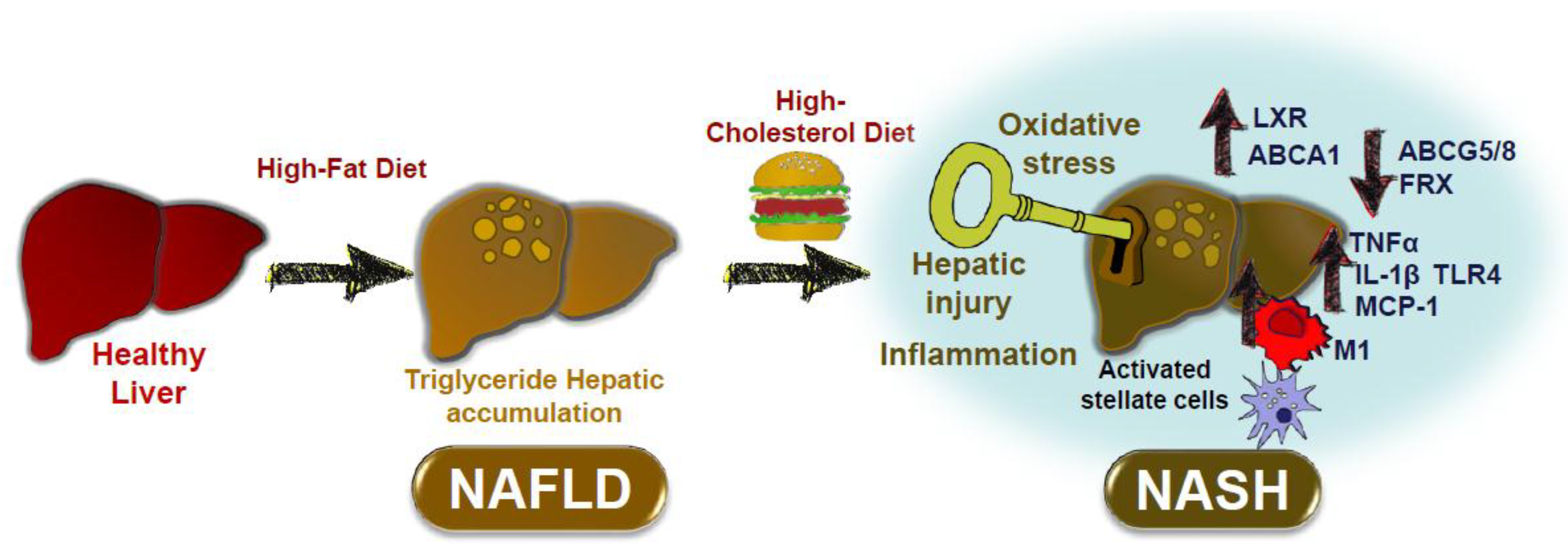
Figure 1.
Effect of dietary cholesterol on the progression of NAFLD to NASH. Non-alcoholic fatty liver disease (NAFLD) is characterized by hepatic lipid accumulation. A high-cholesterol diet promotes the progression toward non-alcoholic steatohepatitis (NASH) by several key mechanisms including hepatic stellate cell activation, oxidative stress, activation of inflammatory pathways and hepatocyte death. Dietary cholesterol promotes NASH by modulating the expression of hepatic metabolic genes such as FXR and ABCG5/8, as well as the expression of cytokines like TNFα, IL-1β, and MCP1. A change in macrophage phenotype in resident macrophages or Kupffer cells toward a M1 phenotype has been suggested. Other proposed mechanisms include the activation of TLR4-dependent pathways and the upregulation of ABCA1 by LXR nuclear receptors.

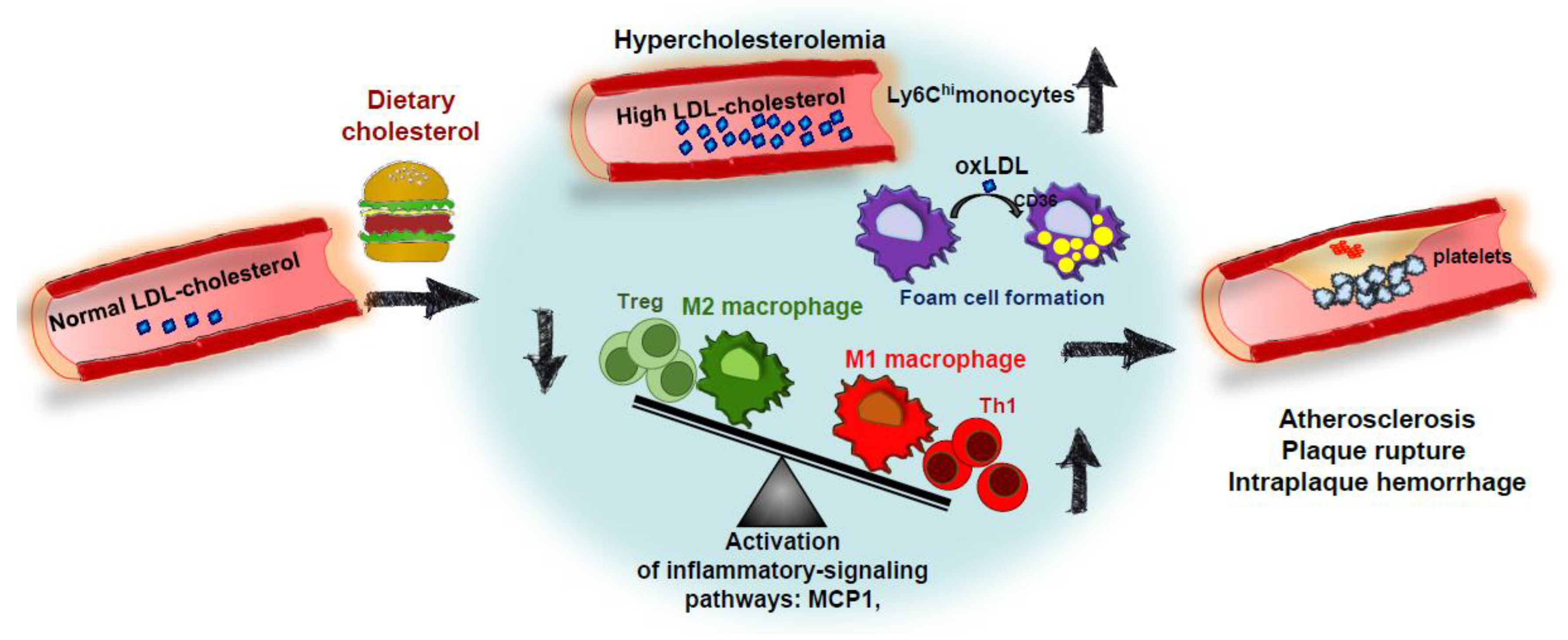
Figure 2.
Impact of dietary cholesterol on atherosclerosis. High circulating cholesterol levels (mostly cholesterol carried by LDL-C particles) are the main risk factor for developing atherosclerosis. Under certain conditions, dietary cholesterol induces hypercholesterolemia and enhanced levels of proinflammatory Ly6Chi monocytes. This might facilitate macrophage foam cell formation through the uptake of modified LDL particles by scavenger receptors (CD36). Atherosclerosis development involves an imbalance between M1 macrophages/M2 macrophages and Th1/Treg cells which can be facilitated by hypercholesterolemia. These events lead to the activation of inflammatory pathways and the progression of disease toward more advanced plaques and, in some cases, plaque rupture, intraplaque hemorrhage, and thrombosis. LDL-C: low-density lipoprotein cholesterol


Table 1.
Impact of dietary cholesterol in rat models of non-alcoholic fatty liver disease (NAFLD) and non-alcoholic steatohepatitis (NASH).
Table 1.
Impact of dietary cholesterol in rat models of non-alcoholic fatty liver disease (NAFLD) and non-alcoholic steatohepatitis (NASH).
| Study/Animal Model | Diet | Age | Stage Disease | Mechanism |
|---|---|---|---|---|
| Cote el al., 2013 [19] Sprague–Dawley rats | 40% fat and 1.25% cholesterol | 8-week-old female | Fatty liver disease | Hepatic accumulation triglycerides and cholesterol Decreased FXRs Lower expression of HMG-CoA-r, FDFT1, and ABCG8 |
| Ichimura et al., 2015 [20] Sprague–Dawley rats | Fat alone or in combination with 1.25% or 2.5% cholesterol | 9-week-old male | Hepatic steatosis | Diminished CPT activity and ABCG5 |
| Moriya et al., 2012 [21] SHRSP5/Dmcr rats | High-fat diet | 10-week-old male | Hepatic fibrotic and inflammatory status of NASH | Altered TNFα proinflammatory cytokine and NFkB pathways |
| Yeti et al., 2013 [22] SHRSP5/Dmcr rats | Fat and cholesterol High-fat high-cholesterol diet | Male SHRSP5/Dmcr rats at 10 weeks old | Phenotype similar to NASH in humans Inflammatory fibrotic liver disease Hepatocyte necrosis | Downregulation of caspase activity |
| Horai et al., 2016 [23] SHRSP5/Dmcr rats | High cholesterol | 6-week-old male rats | Hepatic steatosis, inflammation, and fibrosis | Eosinophilic inclusion bodies and mega-mitochondria |
HMG-CoA-r: 3-hydroxy-3-methylglutaryl coenzyme A reductase; FXR: farnesoid X receptor; FDFT1: farnesyldiphosphate farnesyl-transferase 1; ABCG8: adenosine triphosphate-binding cassette transporter G8; NFkB: nuclear factor kappa B; CPT: carnitine palmitoyltransferase.
Table 2.
Impact of dietary cholesterol in mouse models of non-alcoholic fatty liver disease (NAFLD) and non-alcoholic steatohepatitis.
Table 2.
Impact of dietary cholesterol in mouse models of non-alcoholic fatty liver disease (NAFLD) and non-alcoholic steatohepatitis.
| Study/Mouse Model | Diet | Age | Stage Disease | Mechanism |
|---|---|---|---|---|
| C57BL/6J | ||||
| Matsuzawa et al., 2007 [8] C57BL/6J mice | 1.25% cholesterol and two different amounts fat (7.5% and 60%) | Males at 6 weeks of age | Insulin resistance Fibrosis Steatohepatitis | Down-regulation of antioxidant enzymes |
| Savard et al., 2013 [24] C57BL/6J | 15% fat or 1% cholesterol | 30 weeks | Hepatic fat accumulation | |
| Neuschwander-Tetri et al., 2013 [25] C57BL/6J mice | 15% fat and 1% cholesterol | 30 weeks | Severe steatosis Inflammation Fibrosis | Inappropriate suppression of fatty acid β-oxidation |
| Vergnes et al., 2003 [26] C57BL/6J | Fat, cholate, and/or cholesterol | At 3 months of age, mice were fed with the specified diet for 3 weeks | Hepatic steatohepatitis | Activation of hepatic stellate cells, SAA family genes, histocompatibility antigens, Il2rγ, Scyb9, Samhd1 |
| Desai et al., 2008 [27] C57BL/6J | 1.25% cholesterol, 0.5% cholic acid, and 16% fat | Males at 8–10 weeks of age were fed for 3 weeks with the diet | Hepatic steatohepatitis | Mononuclear leukocyte infiltration in liver Enhanced MCP1, RANTES, MIP2 |
| Sumiyoshi et al., 2010 [28] C57BL/6 mice | 15% milk fat, 1.5% cholesterol and 0.1% cholic acid | Males at 4 weeks old were fed with diet for 25 or 55 weeks | Hepatic steatosis Fibrosis Tumor formation (focal nodular hyperplasia) | Elevated levels of MCP1 levels and PDGF-B protein |
| Ganz et al., 2015 [29] C57BL/6 | High fat, high cholesterol and high sugar supplement | Males aged 8–10 weeks old were fed with diet for 8, 27, or 49 weeks | Hepatic steatosis at early stage Fibrosis and characteristics of NASH at a late stage | Enhanced levels of MCP1, TNFα, and IL-1β Macrophage polarization toward an M1 |
| Tu et al., 2017 [30] C57BL/6J | High-fat high-cholesterol and cholate diet | Males and females at 8 weeks of age fed their respective diets for 3 weeks | Hepatic pathology similar to NAFLD and NASH | Elevated free cholesterol, cholesterol esters, and cholic acid Changes to metabolism of sphingomyelins and phosphatidylcholines |
| Studies in lipase-deficient mice | ||||
| ATGL-/- [31,32,33,34] | High-fat high-cholesterol diet | 2–12 months old | Severe hepatic steatosis | Activation of inflammatory pathways |
| Andres-Blasco et al., 2015 [35] HL-/- | High-fat high-cholesterol diet | At two months of age, mice were fed for 16 weeks with diet | Hepatic steatosis and liver inflammation | Dyslipidemia Increased NEFA Enhanced macrophages Circulating levels of MCP1 and Th17 T-cell subset |
| Chiu et al., HL-/-, 2010 [36] | High-fat 21% diet and 0.15% cholesterol | Females 21–23 weeks old | Decreased hepatic steatosis | No dyslipidemia and IR |
| Studies in low-density lipoprotein receptor and apolipoprotein E-deficient mice | ||||
| Wouters et al., 2008 [3] LDLr-deficient and apoE2 knock-in | High-fat diet with cholesterol | Males or/and females were fed for 2, 4, 7, and 21 days or for 7 days according to experiments | NASH Hepatic steatosis with inflammation | |
| Subramanian et al., 2011 [37] LDLr-deficient | Fat, carbohydrate and cholesterol | 10-week-old males were fed for 24 weeks with diet | NASH | Macrovesicular steatosis, inflammatory cell foci and fibrosis |
| Prieur et al., 2010 [38] LDLr-deficient mice | Diet enriched in fat, carbohydrate and cholesterol | Males at 10 weeks of age were fed for 24 weeks with diet | Hepatic inflammation | Hepatic macrophage infiltration, apoptosis, and oxidative stress. |
| Van Rooyen et al., 2011 [39]. Alms1 mutant (foz/foz) and wild-type diabetes NOD.B10, | Dietary cholesterol | Females at 8 weeks of age were fed for 12 or 24 weeks with diet | Hepatic free cholesterol accumulation | Increased macrophage, liver apoptosis and fibrosis |
| Schierwagen et al., 2015 [40]. apoE-/- | Western-type diet containing 1.25% of cholesterol | 12 weeks age + 7 weeks diet | Phenotype resembling that of human NASH. | Hepatic fibrosis Upregulation of TGFβ Increased hepatic collagen Activation of hepatic stellate cells |
| Rodríguez Sanabria et al., 2010 [41] apoE-/- vs LDLr-/- | 20% fat and 0.25% cholesterol | Males 10 weeks age + 6 weeks diet | Inflammation vs. fatty liver | Increased macrophage and inflammatory nodules (apolipoprotein E, apoE-/-) vs. hepatic steatosis (LDLr-/-) |
| Kampschulte et al., 2014 [42] apoE-/-LDLr-/- | Western diet containing 5% cholesterol and 21% or regular chow control diet | Males at 4 weeks of age were fed for 35 weeks with diet | Hepatic steatosis Fibrosis Hepatocellular injury | Macrophage and T cell infiltration, hepatic ROS accumulation, JNK activation Induction of PPAR-α |
TGF: transforming growth factor; Il2rγ: interleukin-2 receptor γ; MCP1: monocyte chemotactic protein 1; RANTES: regulated on activation normal T cell expressed and secreted; MIP2: macrophage inflammatory protein 2; SAA: serum amyloid A; Scyb9: small inducible cytokine B9; Samhd1: SAM domain and HD domain 1; PDGF-B: platelet-derived growth factor B; ROS: reactive oxygen species; JNK: c-Jun N-terminal kinase; PPAR-α: peroxisome proliferator-activated receptor α; LDLr: low-density lipoprotein receptor.
Table 3.
Impact of dietary cholesterol in mouse models of atherosclerosis.
| Study/Mouse Model | Diet | Age | Stage of Disease |
|---|---|---|---|
| C57BL/6J | |||
| Lee et al., 2017 [51] | 30% fat, 5% cholesterol and 2% cholic acid | Increased serum cholesterol levels Atheroma lesions and extreme hepatic damage | |
| Paigen et al., 1987 [52] C57BL/6 mouse | 1.25% cholesterol, 15% fat, and 0.5% cholic acid (toxic atherogenic diet) | Diet for 14 weeks | Atheroma lesions Discrete hypercholesterolemia |
| Vergnes et al., 2003 [26]. C57BL mice | Different diets (1.25% cholesterol, 0.5% sodium cholate, and 7.5% cocoa butter, and three other diets which omitted one of the three components of the atherogenic diet) | Males at 3 months of age were fed with the diet for 3 weeks | Dietary cholesterol induces the expression of inflammatory genes Cholate induces the expression of extracellular matrix deposition genes such as collagen |
| Studies in LDLr-deficient mice | |||
| Ishibashi et al., 1994 [53]. | 1.25% cholesterol, 7.5% cocoa butter, 7.5% casein, and 0.5% cholic acid | Diet for 6, 7, or 8 months | Severe hypercholesterolemia Atheroma lesions in the vascular wall |
| Lichtman et al., 1999 [54] | 0.5% and 1.25% of cholesterol to a high-fat diet | Males at 8 to 12 weeks of age were fed with the diet for 12 weeks | Lesion plaque formation in a dose-dependent manner |
| Hartvigsen et al., 2007 [55] | Western-type diet, 0.06% cholesterol/21% milk fat, or a cholesterol-enriched diet, 1% cholesterol/4.4% fat | Males were fed with the diet for 28 weeks | Atherosclerotic lesions |
| Subramanian et al., 2008 [56] | Carbohydrate-rich diet with 0.15% cholesterol | Males at 8 weeks old were fed with diet for 24 weeks | Accumulation of macrophages in adipose tissue Acceleration of atherosclerosis |
| Teupser et al. 2003 [57] LDLr-/- mice in C57BL/6J | 4.3% fat in combination with 0.02% 0.15%, 0.30%, or 0.50% cholesterol | Mice at 28 days of age were fed the diet for 16 weeks (20 weeks of age) | Atherosclerosis in aortic root, brachiocephalic artery and whole aorta |
| Wu et al., 2006 [58] | 0.15% cholesterol versus 0.03% cholesterol with high fat in each diet | Diet 20 or 40 weeks | Lesion size was bigger in mice fed 0.15% cholesterol versus 0.03% cholesterol Addition of fat to a cholesterol-rich diet did not increase atherosclerotic lesion hypertriglyceridemia |
| Ma et al., 2012 [59] | 21% fat, 0.15% cholesterol | Males at 8 weeks were fed with the diet for 1, 3, 6, 9, 12 months | Before 3 months: slight atherosclerotic lesions in aortic roots and innominate artery At 3 months: advanced lesions in the aortic sinus At 6–9 months: advanced lesions in the innominate artery |
| Joyce et al., 2006 [60] | 0.02% cholesterol and 4% fat or 0.2% cholesterol and 21.2% fat | Diet 4, 9, or 12 weeks prior to sacrifice | Increased hepatic content of cholesterol and aggravated aortic root atherosclerosis in LDLr-/- mice that overexpressed ABCA1 in the liver |
| Kennedy et al., 2009 [61] LDLr-/-CD36-/- mice | High-cholesterol diet | Diet for 12 weeks | Atherosclerotic lesions |
| Fuller et al., 2014 [62]. LDLr-/- SRBI-/- | High-cholesterol high-fat high-cholesterol high-fat high-cholesterol cholate diet | Females at 10–12 weeks of age were fed 12 weeks with the atherogenic diet | Occluded coronary arteries Accumulation of platelets in atherosclerotic plaque in coronary arteries (thrombosis) Myocardial fibrosis Increased circulating Ly6Chi monocytes |
| Studies in apoE-deficient mice | |||
| Zhang et al., 1994 [63] Partially deficient in apoE | 15.8% fat, 1.25% cholesterol, and 0.5% cholate | Beginning at 8 weeks of age, mice were fed 6 or 12 weeks with the diet | Atheroma lesions |
| Nakashima et al., 1994 [64] | Western-type diet, containing 21% fat, 0.15% cholesterol and 19.5% casein without sodium cholate | Males at 5 weeks of age were fed the diet until 6, 8, 10, 15, 20, 30, and 40 weeks of age | Advanced plaques Development of fibrous cap, presence of smooth muscle cell and adhesion of monocytes |
| Joyce et al., 2002 [65] ABCA1-Tg apoE-/- | 1.25% cholesterol and 0.5% cholic acid | Mice at 2–3 months of age were maintained on the diet for 15 weeks before sacrifice | Overexpression of ABCA1 increased apoB-lipoprotein and HDL levels in plasma and reduced atherosclerosis in vivo |
| Johnson et al., 2001 [66] | 0.15% cholesterol into a high-fat diet | Seven-week-old mice were fed the diet for 14 months | Plaque rupture in the brachiocephalic artery Lesions with intraplaque hemorrhage |
| Bond et al., 2011 [67]. | Cholesterol diet | Lesions in the brachiocephalic artery | |
| Acin et al., 2005 [68] | Different fat-enriched diet and cholesterol with or no unsaturated sources such as extra virgin olive oil (EVOO) | 2-month-old mice were fed with different diets for 10 weeks | Reduced lesion formation with EVOO Cholesterol prevented the beneficial effects of unsaturated fat from EVOO |
| Swirski et al., 2007 [69] | Cholesterol diet | Beginning at 10 weeks of age, mice were fed 20–25 weeks with the diet | Accelerated atherosclerosis by increasing Ly6Chi |
| Mailer et al., 2017 [70] | Cholesterol diet | Mice at 8–10 weeks of age were fed for 4, 12, or 24 weeks with the diet | Accelerated atherosclerosis by activating T cell receptor signaling |
| Ito et al., 2016 (in combination with LXRβ-/-) [71] | Cholesterol diet | Diet for 8, 12, or 16 weeks | Accelerated atherosclerosis and production of autoantibodies and B cell expansion |
| Studies in “humanized” ApoB-100 and ApoE3*Leiden transgenic mice | |||
| Purcell-Huynh et al., 1998 [72] | 16% fat and 1.25% cholesterol | Males and females at 5 weeks of age were fed the diet for 5 or 8 weeks | Severe hypercholesterolemia and atherosclerosis |
| Laplante et al. 2013 [73] LDLr-/-TgApoB100 and LDLr-/TgApoB100IGFII | Standard chow, 0.2% cholesterol diet, high-fat diet or high-fat 0.2% cholesterol diet | Males aged 6 weeks were fed the diet for 6 months | Cholesterol accelerates lesion formation in both LDLr-/-TgApoB100 and LDLr-/-TgApoB100IGFII. |
| Van Vlijmen et al., 1994 [74] ApoE*3Leiden transgenic mice | High cholesterol | At 8–10 weeks old age, mice were fed for 6 weeks with the diet | Hypercholesterolemia and atherosclerotic lesions in the whole aorta and carotid arteries |
| Kleemann et al., 2007 [75] ApoE*3Leiden transgenic mice | 0%, 0.25%, and 1.25% cholesterol | Female E3L mice at 12 weeks old were treated with diet for 10 weeks | Hypercholesterolemia and atherosclerosis |
© 2018 by the authors. Licensee MDPI, Basel, Switzerland. This article is an open access article distributed under the terms and conditions of the Creative Commons Attribution (CC BY) license (http://creativecommons.org/licenses/by/4.0/).
Share and Cite
MDPI and ACS Style
Vinué, Á.; Herrero-Cervera, A.; González-Navarro, H. Understanding the Impact of Dietary Cholesterol on Chronic Metabolic Diseases through Studies in Rodent Models. Nutrients 2018, 10, 939. https://doi.org/10.3390/nu10070939
AMA Style
Vinué Á, Herrero-Cervera A, González-Navarro H. Understanding the Impact of Dietary Cholesterol on Chronic Metabolic Diseases through Studies in Rodent Models. Nutrients. 2018; 10(7):939. https://doi.org/10.3390/nu10070939
Chicago/Turabian StyleVinué, Ángela, Andrea Herrero-Cervera, and Herminia González-Navarro. 2018. "Understanding the Impact of Dietary Cholesterol on Chronic Metabolic Diseases through Studies in Rodent Models" Nutrients 10, no. 7: 939. https://doi.org/10.3390/nu10070939
Note that from the first issue of 2016, this journal uses article numbers instead of page numbers. See further details here.