Article

Effects of Long-Term Oral Administration of Arachidonic Acid and Docosahexaenoic Acid on the Immune Functions of Young Rats

1
School of Pharmacy and Pharmaceutical Sciences, Mukogawa Women's University, 11-68 Koshien Kyuban-cho, Nishinomiya 663-8179, Japan
2
Department of Environmental Physiology, Shimane University Faculty of Medicine, 89-1 Enya-cho, Izumo 693-8501, Japan
3
Department of Health Chemistry, Graduate School of Pharmaceutical Sciences, University of Tokyo, 7-3-1 Hongo, Bunkyo-ku, Tokyo 113-0033, Japan
*
Author to whom correspondence should be addressed.
Nutrients 2013, 5(6), 1949-1961; https://doi.org/10.3390/nu5061949
Submission received: 21 March 2013 / Revised: 8 May 2013 / Accepted: 20 May 2013 / Published: 29 May 2013
(This article belongs to the Special Issue Nutrients and Immune Function)

Abstract

:
Natural killer (NK) cells have many functional activities, including cytotoxicity and the capacity to produce cytokines and chemokines. NK cell activity is regulated partly by eicosanoids, which are produced from arachidonic acid (ARA) and eicosapentaenoic (EPA) acid. In this study, we investigated the effects of long-term therapy with ARA or docosahexaenoic acid (DHA) on the cytotoxic effects of the NK cells of young rats, which were fed on a nonfish oil diet for two generations. Control oil, ARA (240 mg/kg BW/day) or DHA (240 mg/kg BW/day) were orally administrated to the rats for 13 weeks before determining the cytotoxic activity of NK cells from the spleen against YAC-1 mouse lymphoma cell line, as well as the plasma levels of docosanoids or eicosanoids and inflammatory cytokines. Long-term ARA administration significantly suppressed the cytotoxic activity of NK cells. Moreover, ARA administration significantly increased the plasma levels of ARA, prostaglandin (PG) E2, and PGD2. However, DHA administration did not produce any different effects compared with those in the control rats. Furthermore, the inflammatory cytokine levels were not affected by the administration of ARA or DHA. These results suggest that long-term ARA administration has an inhibitory effect on the tumor cytotoxicity of NK cells in rat spleen lymphocytes owing to the enhanced synthesis of PGE2 and PGD2 from ARA because of the elevated plasma ARA levels in young rats.

1. Introduction

Natural killer (NK) cells are a type of lymphocyte that participates in innate immunity. The function of NK cells in innate immunity is crucial for combating viral infection and for destroying cancer cells [1,2]. Recent studies have demonstrated that NK cells have a direct cytotoxic activity against cancer cells, and they can regulate the adaptive immune response via cytokine production [3,4,5]. Thus, NK cells have important roles during the initial responses to viruses and cancer cells. A low NK cell activity is associated with an increased risk of carcinogenesis [6]. Therefore, the focus of recent cancer therapies has been in enhancing the NK cell activity through the development of chemical compounds. However, inflammation is a critical component of tumor progression and tumor microenvironments are controlled by inflammatory cells. Tumor cells activate specific signaling molecules in the innate immune system, including cytokines, chemokines, and their receptors during invasion, migration, and metastasis [7,8,9]. Therefore, recent cancer therapies have targeted immune modulation [10,11].
Polyunsaturated fatty acids (PUFAs) such as arachidonic acid (ARA), docosahexaenoic acid (DHA), and eicosapentaenoic acid (EPA), are involved in the inflammatory process. Inflammatory cytokines and tumor cells induce PUFA metabolizing enzymes such as secreted phospholipase A2 (sPLA2) and cyclooxygenase (COX)-2 [12]. ARA, a long-chain omega-6 fatty acid, is stored in the cell membrane phospholipids. ARA is released from the membrane phospholipids by sPLA2 and cytoplasmic phospholipase A2 (cPLA2), and this released free ARA can form eicosanoids via COX-1 and -2, lipoxygenases (LOXs), or cytochrome P450 monooxygenases [13,14]. ARA-derived eicosanoids such as prostaglandins (PGE2, PGF, PGD2), prostacyclin (PGI2), or thromboxane A2 and hydroxyeicosatetraenoic acid (HETE) have well-recognized roles in inflammation [15]. In fact, prolonged administration of non-steroidal anti-inflammatory drugs that can inhibit COX are associated with a reduced risk of developing several types of cancers [16]. In contrast, long-term ARA administration is associated with the development of cancer or the stimulation of inflammatory conditions [17,18,19].
DHA and EPA, which are omega-3 fatty acids, have a variety of anti-inflammatory and immune-modulating effects. In addition, these fatty acids are oxidized by COX, LOXs, or cytochrome P450 monooxygenases to produce docosanoids and EPA-derived eicosanoids, which have anti-inflammatory effects [20]. Various biological effects of omega-3 fatty acids have been demonstrated in several feeding studies with humans and animals using fish or fish oil supplements. In humans, omega-3 fatty acids inhibit the production of cytokines involved with chronic inflammatory and autoimmune diseases [21]. Dietary omega-3 fatty acids are considered to prevent inflammation through a variety of activities linked to the inhibition of ARA-derived eicosanoid-mediated effects, anti-inflammatory properties, and the inhibition of cytokines and ARA-derived eicosanoid synthetic enzymes by competitive inhibition. The typical ARA-derived eicosanoid, PGE2 is a potent regulator of inflammation as well as innate and adaptive immune responses, which contributes to immune evasion during malignancy. It is known that PGE2 downregulates interleukin (IL)-2-activated lymphokine-activated killer (LAK) cell cytotoxicity through EP2 receptors [22,23]. Moreover, PGE2 substantially inhibits IFN-γ production as well; it reduces the amount of IFN-γ and TNF-α secretion by NK cells. Similarly, IFN-γ secretion is suppressed by PGE2 in human NK cells in a dose-dependent manner [24].
The aim of the present study was to determine the relationships among the plasma levels of eicosanoid or docosanoid, inflammatory cytokines, and NK cell activity in long-term ARA- and DHA-administered young rats. In this study, we measured the plasma levels of eicosanoids and docosanoids using liquid chromatography tandem mass spectrometry (LC-ESI-MS/MS) because long-term administration of ARA and DHA might affect the levels of eicosanoids and docosanoids. Changes in the levels of eicosanoids and docosanoids modulate the inflammatory response, as described above; thus, we also investigated whether NK cell activity was affected by long-term oral administration of ARA and DHA. We used YAC-1 as a target cancer cell to evaluate NK cell activity. YAC-1 is a murine T-lymphoma cell line, which was established from a tumor induced by Moloney sarcoma virus in A/Sn mice. YAC-1 cells do not express major histocompatibility complex (MHC) class I molecules, and they are efficiently killed by NK cells. Moreover, YAC-1 has been used to study the cytotoxic activities of NK cells and LAKcells, which are induced by IL-2 from NK cells, along with factors that affect the cytotoxic activities of these and other lymphocytes [25,26].

2. Materials and Methods

2.1. Animals Used in the Experiments

All animal experiments were performed in accordance with the procedures outlined in the Guidelines for Animal Experimentation of Shimane University compiled from the Guidelines for Animal Experimentation of the Japanese Association for Laboratory Animal Science.
The animal room was maintained at 23 °C with a relative humidity 50%–55%. The light/dark cycle was maintained at 12 h intervals. Young Wistar rats (5 weeks old) were used in the study, which were the offspring of rats maintained for two generations on a nonfish oil diet (Funabashi Farm, Japan). The mean initial weight of rats was 214.6 ± 18.5 g. These rats were divided into three groups: control group, ARA group (the triglyceride form of ARA rich oil: 240 mg/kg BW/day), and DHA group (the triglyceride form of DHA rich oil: 240 mg/kg BW/day). Control rats were administrated a basic mixture of oil (beef fat:soybean oil:rape seed oil = 2:1:1). Each oil mixture was orally administered to the rats (n = 8 in each group) for 13 weeks. The fatty acid compositions of the oils administered are summarized in Table 1.
Table 1. Composition of fatty acids in control, ARA, DHA oil.
(mol%)ControlARADHA
PLA13.8 ± 0.016.95 ± 0.0029.8 ± 0.03
STA13.8 ± 0.015.91 ± 0.008.10 ± 0.04
OLA42.5 ± 0.035.31 ± 0.0016.3 ± 0.01
LA20.0 ± 0.029.38 ± 0.011.96 ± 0.01
ARAND45.1 ± 0.042.49 ± 0.02
EPA0.13 ± 0.010.52 ± 0.006.61 ± 0.00
DPA n-3NDND1.17 ± 0.01
DHANDND32.6 ± 0.03
Note: PLA, palmitic acid; STA, stearic acid, OLA, oleic acid; LA, linolenic acid; ARA, arachidonic acid; EPA, eicosapentaenoic acid; DPA, docosapentaenoic acid; DHA, docosahexaenoic acid; ND, not detected.

2.2. Cell Culture

The YAC-1 mouse lymphoma cell line was provided by Riken Bio Resource Center through the National Bio-Resource Project of MEXT (Japan). Cells were cultured at 37 °C and 5% CO2 in RPMI-1640 medium with 10% FBS, 100 U/mL penicillin, and 100 µg/mL streptomycin. The cells were maintained in continuous suspension and cultured in the complete culture medium. YAC-1 cells were used to test the NK cytotoxic activity because of their notable sensitivity to NK cells.

2.3. Isolation of Rat Spleen Lymphocytes

The spleens were collected from rats who were administered the control, ARA, or DHA oils. These spleen samples were mashed through a strainer and the erythrocytes were removed using Lympholyte-Rat (Cedarlane, Burlington, Canada). Spleen lymphocytes were washed twice with Hank’s balanced salt solution and suspended in RPMI-1640 medium with 1% FBS. The number of lymphocytes in each sample was determined using a blood-cell counting chamber. The viability of lymphocytes was determined by trypan blue exclusion.

2.4. Cytotoxic Activity of NK Cell Assay

The NK cell activity was detected using freshly isolated spleen lymphocytes. The target cells used to determine the NK cell cytotoxicity were YAC-1 cells. Mixtures of spleen lymphocytes (1.5 × 10⁵, 7.5 × 10⁴, 3.75 × 10⁴/well/100 μL) and YAC-1 cells at 3 × 10⁴ cells/well/100 μL (lymphocytes:YAC-1 = 5:1, 2.5:1, 1.25:1) in RPMI-1640 with 1% FBS were incubated at 37 °C in a 5% CO2 incubator for 24 h. Subsequently, the cytotoxic activity levels of the NK cells in spleen lymphocytes were measured using a lactate dehydrogenase (LDH) Cytotoxicity Detection Kit (TaKaRa, Otsu, Japan). The cytotoxicity was measured on the basis of the amount of LDH released by damaged cells into the cell culture supernatant. LDH present in the culture supernatant participates in a coupled reaction that converts yellow tetrazolium salt into a red formazan product. The enzyme activity levels, which were measured as the absorbance at 490/620 nm using a microplate reader, were correlated with the number of damaged cells in the culture. The maximum release was the lysis achieved after adding Triton X-100 (final concentration 1%). The spontaneous release of LDH was determined by incubating labeled target cells in the absence of effector cells.
Nutrients 05 01949 i001

2.5. Sample Preparation for the Analysis of Fatty Acid Metabolites

Plasma samples were adjusted to 67% methanol and stored at −30 °C. The samples were centrifuged at 5,000 × g for 10 min at 4 °C to remove precipitated proteins. Thereafter, the supernatants were diluted with ice-cold distilled water and adjusted to 10% (v/v) methanol. Internal standards (5 ng of PGE2-d4, PGD2-d4, 5-HETE-d8, and AA-d8) were added to each sample. Samples were acidified to pH 4.0 using 0.1 M HCl and placed immediately in preconditioned solid phase extraction cartridges (Sep-Pak C18, Waters) to extract the fatty acid metabolites. Sep-Pak cartridges were washed with 20 mL MilliQ water and 20 mL n-hexane, and subsequently, the finally fatty acid metabolites were eluted with 10 mL methyl formate.

2.6. LC-ESI-MS/MS-Based Analysis

The fatty acid metabolites in the plasma were measured as described previously with a slight modification [27]. HPLC was combined with ESI-MS in a TSQ Quantum mass spectrometer (Thermo Fisher Scientific K.K., Japan). HPLC was conducted at 30 °C using a Luna 5u C18 (2) 100 Å LC column (150 × 2.0 mm, Phenomenex, USA). Samples were eluted with a mobile phase that comprised acetonitrile:methanol (4:1, v/v) and water:acetic acid (100:0.1, v/v) in a 27:73 ratio for 5 min, which was ramped up to a 100:0 ratio after 25 min, and subsequently held for 10 min at a flow rate of 0.1 mL/min. MS/MS analyses were conducted in the negative ion mode, and the fatty acid metabolites were detected and quantified by selected reaction monitoring (SRM). The conditions used for the detection of each compound by SRM are listed in Table 2. The peaks were selected and their areas were calculated using the Xcalibur 2.1 (Thermo Fisher Scientific K.K.).
Table 2. Selected reaction monitoring (SRM) transitions of fatty acid metabolites.
CompoundSRM Transition (m/z)CompoundSRM Transition (m/z)CompoundSRM Transition (m/z)
ARA303 > 259EPA301 > 257DHA327 > 283
PGE2351 > 2715-HEPE317 > 1157-HDoHE343 > 141
PGD2351 > 27112-HEPE317 > 17910-HDoHE343 > 153
PGF353 > 19315-HEPE317 > 21914-HDoHE343 > 193
5-HETE319 > 11518-HEPE317 > 25917-HDoHE343 > 245
12-HETE319 > 179RvE2333 > 115PD1359 > 153
15-HETE319 > 219AA-d8311 > 267PGE2-d4355 > 275
PGD2-d4355 > 275PGF-d4357 > 1975-HETE-d8327 > 116
Note: PG, prostaglandin; HETE, hydroxyeicosatetraenoic acid; HEPE, hydroxyeicosapentaenoic acids; RvE2, ResolvinE2; HDoHE, hydroxydocosahexaenoic acid; PD1, Protectin D1.

2.7. Determination of Cytokine Levels

The plasma concentrations of IL-1β, IL-4, IL-6, IL10, IL-13 and tumor necrosis factor (TNF)-α were measured using the Bio-Plex system, which combines a sandwich immunoassay with Luminex fluorescent bead-based technology (Bio-Rad Laboratories, Inc. Hercules, CA, USA).

2.8. Analysis

The results obtained using at least 6–8 animals were expressed as the mean ± standard error (S.E.). Statistical differences in the NK cell activity were determined using a one-way ANOVA with Dunnett’s post hoc test, whereas the statistical differences in fatty acid metabolites were tested using a Bonferroni test. Differences were considered statistically significant at p < 0.05.

3. Results

3.1. Measurement of Cytotoxic Activity of NK Cells

After 13 weeks of oral administration of control, ARA, or DHA oils, we measured the effects of the cytotoxic activity of NK cells from the spleen on YAC-1 cells. The NK cell activity in the ARA group of lymphocytes versus YAC-1 cells (5:1) was significantly suppressed compared with that in the control group. Moreover, the NK cell activity in the DHA group was slightly increased, but not significantly compared with that in the control group (Figure 1).
Figure 1. Cytotoxicity of natural killer (NK) cells in rat spleen lymphocytes against YAC-1 cells. The cytotoxicity levels in YAC-1 cells caused by NK cells in spleen lymphocytes after 24 h coculture in the y-axis. The ratio of the mixture of lymphocytes and YAC-1 is presented on the x-axis. The relative cytotoxicity levels are expressed as the mean ± S.E. (n = 6). * significant at p < 0.05 vs. rat lymphocytes in the control group using a one-way ANOVA followed by Dunnett’s test.
Nutrients 05 01949 g001

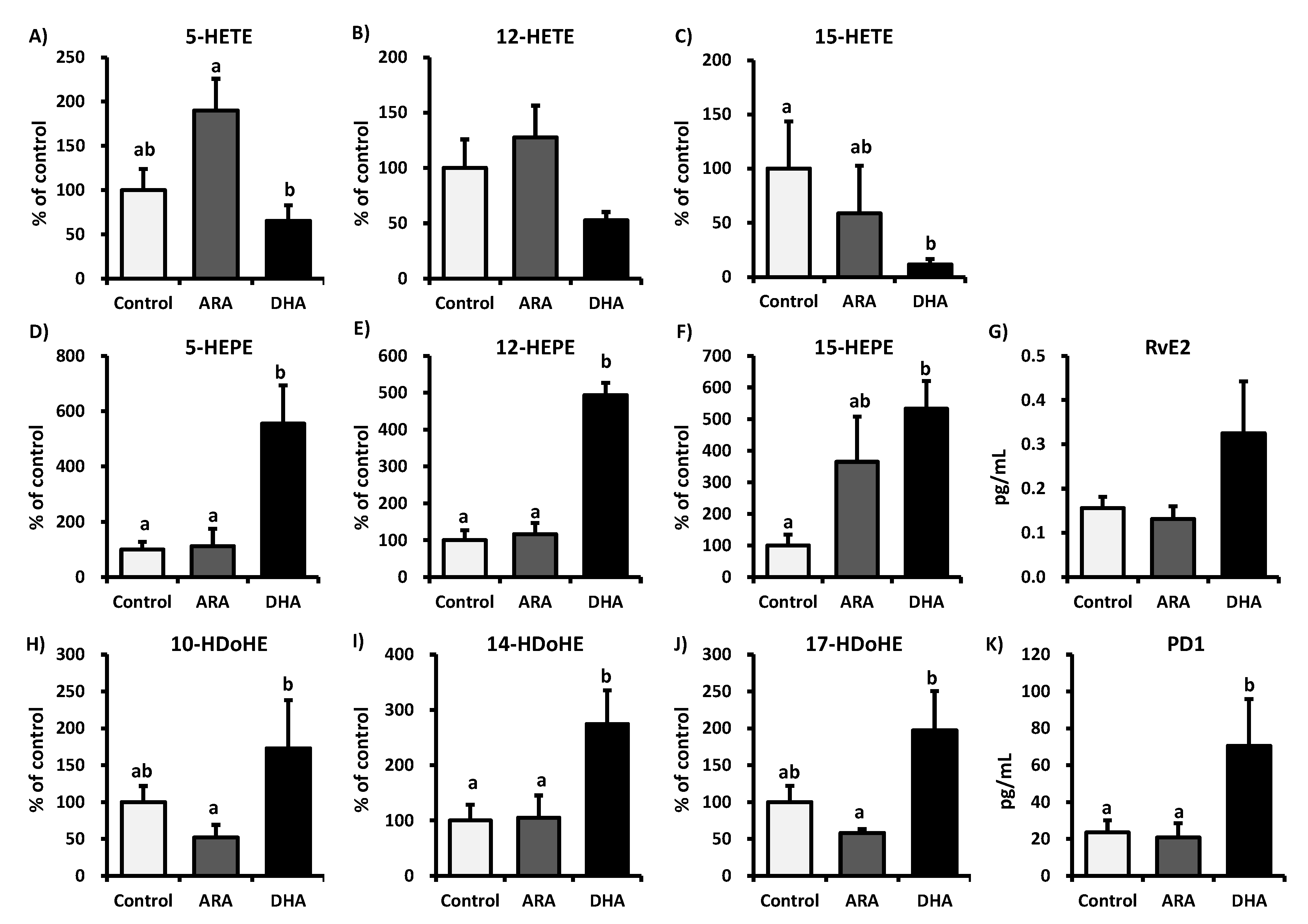
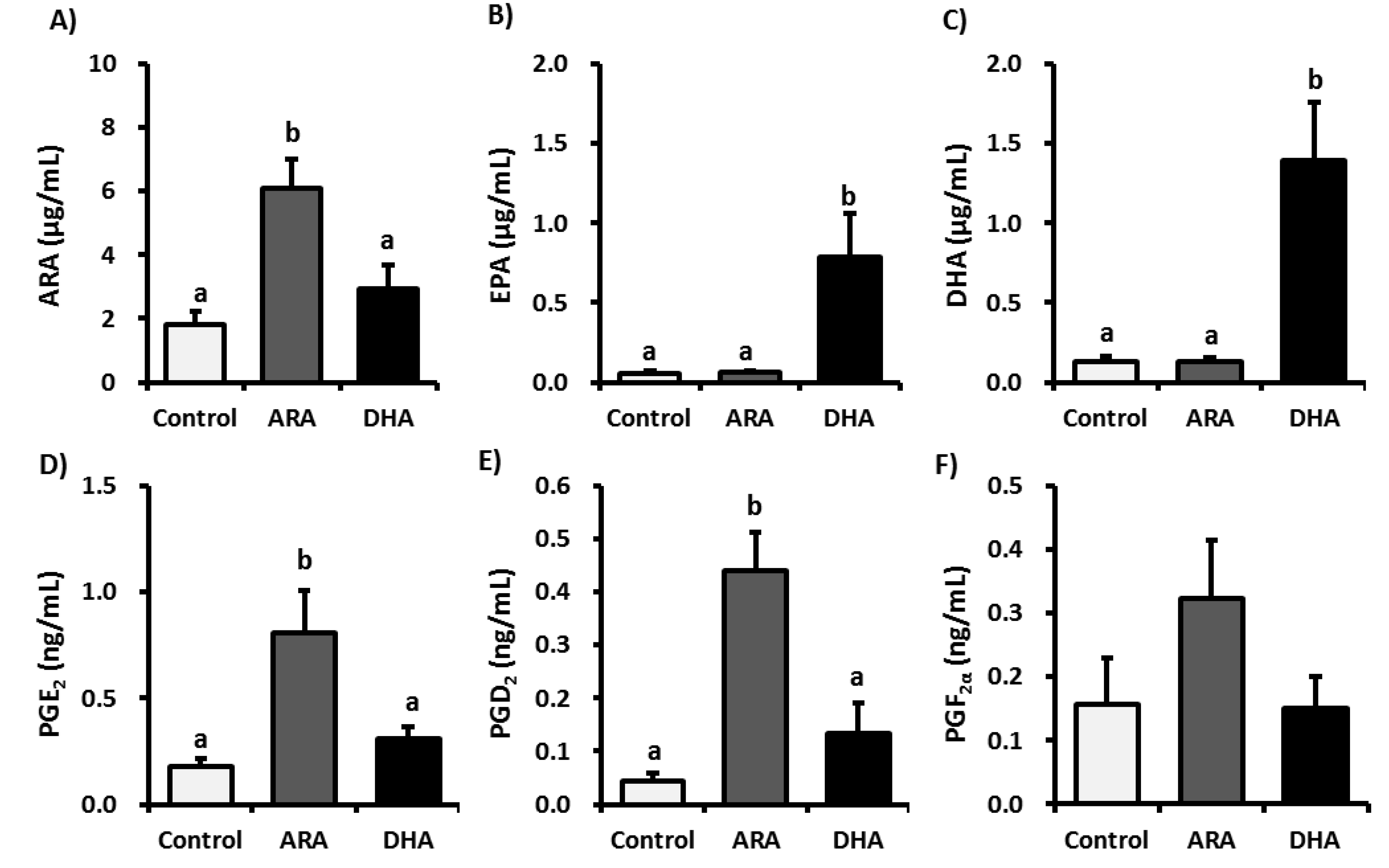
3.2. Analysis of Fatty Acid Metabolites

Figure 2 lists the free form ARA, EPA, DHA, and ARA-derived eicosanoids detected in the plasma. The plasma levels of ARA, PGE2, and PGD2 were significantly higher in the ARA group than in the control and DHA groups. However, DHA administration had no significant effect on the levels of ARA-derived eicosanoids. In contrast, the free form EPA and DHA levels in the plasma were significantly higher in the DHA group than in the control and ARA groups. Figure 3 presents the plasma levels of the metabolites produced from ARA, EPA, and DHA by LOXs. ARA administration tended to increase the plasma levels of 5-HETE but not 12-HETE and 15-HETE, whereas there were no significant effects on the EPA- and DHA-derived metabolite levels. However, DHA administration significantly decreased the plasma levels of 15-HETE but not 5-HETE and 12-HETE. In addition, DHA administration significantly increased the plasma levels of 5-HEPE, 12-HEPE, 15-HEPE, 14-HDoHE, and PD1, whereas the plasma levels of RvE2, 10-HDoHE, and 17-HDoHE also tended to increase, although not significantly, compared with those in the control group. These results suggest that the administration of ARA or DHA does not induce LOXs.
Figure 2. Plasma levels of free PUFAs and PGs in control, ARA, or DHA oil-treated rats. Plasma samples were subjected to LC-MS/MS lipidomic analysis. The graph presents the concentrations of: (A) ARA, (B) EPA, (C) DHA, (D) PGE2, (E) PGD2, and (F) PGF in the rat plasma. The values are expressed as the mean ± S.E. (n = 8). Samples were analyzed using a one-way ANOVA followed by a Bonferroni post hoc test. a, b Different letters indicate statistical differences at p < 0.05.
Nutrients 05 01949 g002
Figure 3. Plasma levels of ARA, EPA, and DHA metabolites generated by LOXs. Plasma samples were subjected to LC-MS/MS lipidomic analysis. The values are expressed as the mean ± S.E. (n = 8) percentages relative to the control. The graph presents the plasma levels of the metabolites from ARA, EPA, and DHA: (A) 5-HETE, (B) 12-HETE, (C) 15-HETE, (D) 5-HEPE, (E) 12-HEPE, (F) 15-HEPE, (G) RvE2, (H) 10-HDoHE, (I) 14-HDoHE, (J) 17-HDoHE, and K) PD1. Samples were analyzed using a one-way ANOVA followed by a Bonferroni post hoc test. a, b Different letters indicate statistical differences at p < 0.05.
Nutrients 05 01949 g003

3.3. Analysis of Inflammatory Cytokines in Plasma

After 13 weeks of oral administration of ARA or DHA, we measured the plasma levels of the inflammatory cytokines. IL-1β, IL-4, IL-6, IL-10, IL-13, and TNF-α were not affected by the administration of ARA and DHA in young rats (Table 3).
Table 3. Inflammatory cytokine levels in plasma.
(ng/mL)ControlARADHA
IL-1β107.4 ± 6.08146.9 ± 31.191.26 ± 8.42
IL-642.07 ± 11.627.87 ± 9.4726.69 ± 8.74
TNF-α20.84 ± 2.9620.99 ± 2.1721.65 ± 2.23
IL-46.99 ± 0.606.99 ± 0.626.65 ± 0.79
IL-10129.4 ± 16.9112.2 ± 8.25112.8 ± 16.6
IL-1314.63 ± 0.8017.54 ± 1.2513.20 ± 2.02
Note: IL, interleukin; TNF, tumor necrosis factor. Values are the means ± S.E. (n = 8).

4. Discussion

NK cell activity is important for suppressing cancer through the innate immune system. NK cells are activated by macrophages and various pro-inflammatory cytokines such as IL-2, IL-12, and TNF-α. NK cell activity is associated with innate immune reactions and inflammatory reactions [28,29]. In this study, we investigated the effects of long-term administration of ARA and DHA on the NK cell activity levels and inflammatory process in young rats. Long-term ARA administration in young rats suppressed the activity of NK cells from spleen lymphocytes, suggesting that ARA has the potential to inhibit the cancer cell cytotoxic activity of NK cells and reduce the resistance to cancer.
ARA is converted to pro-inflammatory PGs, which induce inflammation. In the present study, long-term ARA administration had no effect on pro-inflammatory cytokines in the plasma. In contrast, ARA administration significantly increased the plasma levels of ARA, PGD2, and PGE2, which suggests that ARA itself might not induce inflammation. PGE2 has an initial pro-inflammatory effect, which is followed by a role in the resolution of inflammation by the inhibition of 5-LOX and the induction of lipoxin production [30,31,32,33]. PGE2 mediates active inflammation, promotes local vasodilatation and local attraction, and activates neutrophils, macrophages, and mast cells in the early stages of inflammation. In contrast, PGE2 can promote the induction of suppressive IL-10 and directly suppresses the production of multiple pro-inflammatory cytokines, which allow it to limit nonspecific inflammation, thereby promoting the immune suppression associated with chronic inflammation and cancer. Recent studies have revealed that PGE2 down-regulates IL-2-activated LAK cell cytotoxicity via the PGE2 receptor, EP2. PGE2 inhibits lymphocyte proliferation, NK cell activity, and the production of Th1 cytokines (IL-2, IFN-γ) [34,35,36]. PGE2 also reduced the amount of IFN-γ and TNF-α secretion by NK cells, in human NK cells, IFN-γ secretion was suppressed by PGE2 in a dose-dependent manner [24]. In addition, PGE2 inhibits MHC II expression and the production of TNF-α and IL-1β by monocytes and macrophages. The presence of PGE2 inhibits the NK cell-mediated cytotoxicity against YAC-1 tumor targets [37,38]. Moreover, PGD2 regulates the immune function via D prostanoid receptors and chemoattractant receptor-like molecules on Th2 cells, which inhibit IL-12 production by dendritic cells and INF-γ production by T cells. It was reported that PGD2 inhibits the cytotoxic function of human NK cells directly via D prostanoid receptors [39,40,41,42,43]. In the present study, long-term ARA administration inhibited NK cell activity and increased the plasma levels of PGE2 and PGD2; this suggests that the immune response might be suppressed during chronic inflammation or cancer to maintain high levels of PGE2 and PGD2 in the plasma. The anti-inflammatory properties of EPA and its metabolites are beneficial during chronic inflammation, but they could suppress the inflammatory response required to attack acute viral infections [44]. EPA but not DHA has been reported to decrease the NK cell activity in rat lymphocytes, a mixture of NK cells and macrophages, and in human peripheral blood mononuclear cells, which are a mixture of lymphocytes such as NK cells and monocytes [45,46,47,48,49]. In this study, long-term DHA administration increased the plasma free EPA levels slightly but not significantly (Figure 2), which in turn increased the NK cell activity from spleen lymphocytes, particularly in cases with a low lymphocyte versus YAC-1 ratio. The differences between these groups might be because of the low EPA dose administered in our study.
PUFAs are present in dietary supplements for babies and adults. ARA is an essential fatty acid, which is stored in the cell membrane, and it is present in high levels in the brain and other tissues. Supplementation with ARA is considered to have beneficial effects on cognitive function and communication ability. However, long-term ARA administration has been associated with cancer development or the stimulation of inflammatory conditions [17,18,19]. In a previous study, ARA supplementation for four weeks in elderly Japanese subjects did not increase the levels of ARA metabolites, which suggests that it does not induce inflammation [50]. In the present study, we demonstrated that long-term ARA administration did not induce inflammation, but our results suggested that it might disrupt the immune system, thereby making it more vulnerable to infection or cancer. DHA and EPA, which are present in fish oil, are also essential fatty acids and they can suppress the serum triglyceride levels as well as improve cognitive function. In contrast to ARA, long-term DHA administration did not affect the plasma levels of free ARA, PGE2, PGD2, and PGF. Moreover, DHA had no effect on the plasma levels of inflammatory cytokines, and it tended to increase NK cell activity, thereby indicating that DHA does not disrupt the immune system. There were some limitations in the present study because we used normal young rats to test the effects of ARA or DHA administration. It is necessary to test an inflammatory model or cancer model animals to confirm the effects of long-term administration of ARA or DHA on the immune system, particularly NK cell activity in the spleen.

5. Conclusions

In conclusion, long-term ARA administration tended to decrease the innate immunity to cancer, although the inflammatory cytokine levels were unaltered in the plasma. The higher plasma levels of PGE2 and PGD2 might explain the decreased NK cell activity in the spleen. In future, we plan to elucidate the mechanism by which long-term ARA therapy controls the NK activity. Furthermore, we also plan to test a dosage range of ARA and a supplementary time period.

Acknowledgements

This study was supported in part by a grant from a Grant-in-Aid for Scientific Research (C) from the Ministry of Education Culture, Sports, Science and Technology (#23500955) and a Health and Labour Sciences Research Grants (#H22-Shokuhin-Ippan-002) from Japan.
The authors would like to thank Enago (www.enago.jp) for the English language review.

Conflict of Interest

The authors declare that there are no conflicts of interests.

References

  1. Chávez-Galán, L.; Arenas-Del Angel, M.C.; Zenteno, E.; Chávez, R.; Lascurain, R. Cell death mechanisms induced by cytotoxic lymphocytes. Cell. Mol. Immunol. 2009, 6, 15–25. [Google Scholar] [CrossRef]
  2. Seaman, W.E. Natural killer cells and natural killer T cells. Arthritis Rheum. 2000, 43, 1204–1217. [Google Scholar] [CrossRef]
  3. Hwang, Y.J.; Kim, J.; Park, D.S.; Hwang, K.A. Study on the immunomodulation effect of isodon japonicus extract via splenocyte function and NK anti-tumor activity. Int. J. Mol. Sci. 2012, 13, 4880–4888. [Google Scholar]
  4. Grivennikov, S.I.; Greten, F.R.; Karin, M. Immunity, inflammation, and cancer. Cell 2010, 140, 883–899. [Google Scholar] [CrossRef]
  5. O’Leary, J.G.; Goodarzi, M.; Drayton, D.L.; von Andrian, U.H. T cell- and B cell-independent adaptive immunity mediated by natural killer cells. Nat. Immunol. 2006, 7, 507–516. [Google Scholar] [CrossRef]
  6. Lu, L.M.; Zavitz, C.C.; Chen, B.; Kianpour, S.; Wan, Y.; Stämpfli, M.R. Cigarette smoke impairs NK cell-dependent tumor immune surveillance. J. Immunol. 2007, 178, 936–943. [Google Scholar]
  7. Cao, L.; Kulmburg, P.; Veelken, H.; Mackensen, A.; Mézes, B.; Lindemann, A.; Mertelsmann, R.; Rosenthal, F.M. Cytokine gene transfer in cancer therapy. Stem Cells 1998, 16, 251–260. [Google Scholar]
  8. Evans, C.; Dalgleish, A.G.; Kumar, D. Review article: Immune suppression and colorectal cancer. Aliment. Pharmacol. Ther. 2006, 24, 1163–1177. [Google Scholar]
  9. Budhu, A.; Wang, X.W. The role of cytokines in hepatocellular carcinoma. J. Leukoc. Biol. 2006, 80, 1197–1213. [Google Scholar] [CrossRef]
  10. Demaria, S.; Pikarsky, E.; Karin, M.; Coussens, L.M.; Chen, Y.C.; El-Omar, E.M.; Trinchieri, G.; Dubinett, S.M.; Mao, J.T.; Szabo, E.; et al. Cancer and inflammation: Promise for biologic therapy. J. Immunother. 2010, 33, 335–351. [Google Scholar] [CrossRef]
  11. Kundu, N.; Walser, T.C.; Ma, X.; Fulton, A.M. Cyclooxygenase inhibitors modulate NK activities that control metastatic disease. Cancer Immunol. Immunother. 2005, 54, 981–987. [Google Scholar] [CrossRef]
  12. Abrahao, A.C.; Castilho, R.M.; Squarize, C.H.; Molinolo, A.A.; dos Santos-Pinto, D., Jr.; Gutkind, J.S. A role for COX2-derived PGE2 and PGE2-receptor subtypes in head and neck squamous carcinoma cell proliferation. Oral Oncol. 2010, 46, 880–887. [Google Scholar] [CrossRef]
  13. Anwar-Mohamed, A.; El-Sherbeni, A.; Kim, S.H.; Elshenawy, O.H.; Althurwi, H.N.; Zordoky, B.N.; El-Kadi, A.O. Acute arsenic treatment alters cytochrome P450 expression and arachidonic acid metabolism in lung, liver and kidney of C57Bl/6 mice. Xenobiotica 2013, in press. [Google Scholar]
  14. Naraba, H.; Murakami, M.; Matsumoto, H.; Shimbara, S.; Ueno, A.; Kudo, I.; Oh-ishi, S. Segregated coupling of phospholipases A2, cyclooxygenases, and terminal prostanoid synthases in different phases of prostanoid biosynthesis in rat peritoneal macrophages. J. Immunol. 1998, 160, 2974–2982. [Google Scholar]
  15. Tilley, S.L.; Coffman, T.M.; Koller, B.H. Mixed messages: Modulation of inflammation and immune responses by prostaglandins and thromboxanes SourceDivision of Pulmonary and Critical Care Medicine. J. Clin. Invest. 2001, 108, 15–23. [Google Scholar]
  16. Vendramini-Costa, D.B.; Carvalho, J.E. Molecular link mechanisms between inflammation and cancer. Curr. Pharm. Des. 2012, 18, 3831–3852. [Google Scholar] [CrossRef]
  17. Murff, H.J.; Shu, X.O.; Li, H.; Yang, G.; Wu, X.; Cai, H.; Wen, W.; Gao, Y.T.; Zheng, W. Dietary polyunsaturated fatty acids and breast cancer risk in Chinese women: A prospective cohort study. Int. J. Cancer 2011, 128, 1434–1441. [Google Scholar] [CrossRef]
  18. Bougnoux, P.; Menanteau, J. Dietary fatty acids and experimental carcinogenesis. Bull. Cancer 2005, 92, 685–696. [Google Scholar]
  19. Greene, E.R.; Huang, S.; Serhan, C.N.; Panigrahy, D. Regulation of inflammation in cancer by eicosanoids. Prostaglandins Other Lipid Mediat. 2011, 96, 27–36. [Google Scholar]
  20. Giudetti, A.M.; Cagnazzo, R. Beneficial effects of n-3 PUFA on chronic airway inflammatory diseases. Prostaglandins Other Lipid Mediat. 2012, 99, 57–67. [Google Scholar] [CrossRef]
  21. Hata, A.N.; Breyer, R.M. Pharmacology and signaling of prostaglandin receptors: Multiple roles in inflammation and immune modulation. Pharmacol. Ther. 2004, 103, 147–166. [Google Scholar] [CrossRef]
  22. Fujino, H.; Salvi, S.; Regan, J.W. Differential regulation of phosphorylation of the cAMP response element-binding protein after activation of EP2 and EP4 prostanoid receptors by prostaglandin E2. Mol. Pharmacol. 2005, 68, 251–259. [Google Scholar]
  23. Joshi, P.C.; Zhou, X.; Cuchens, M.; Jones, Q. Prostaglandin E2 suppressed IL-15-mediated human NK cell function through down-regulation of common gamma-chain. J. Immunol. 2001, 166, 885–891. [Google Scholar]
  24. Simopoulos, A.P. Omega-3 fatty acids in inflammation and autoimmune diseases. J. Am. Coll. Nutr. 2002, 21, 495–505. [Google Scholar]
  25. Stewart, I.J.; Peel, S. Natural killer cytotoxicity and antibody-dependent cytotoxicity of cells of rat metrial glands. J. Reprod. Fertil. 1993, 98, 489–494. [Google Scholar] [CrossRef]
  26. Gardiner, C.M.; Meara, A.O.; Reen, D.J. Differential cytotoxicity of cord blood and bone marrow-derived natural killer cells. Blood 1998, 91, 207–213. [Google Scholar]
  27. Arita, M.; Iwamoto, R.; Isobe, Y. Lipidomics; Wiley-VCH: Weinheim, Germany, 2012; pp. 219–232. [Google Scholar]
  28. Ye, J.; Ortaldo, J.R.; Conlon, K.; Winkler-Pickett, R.; Young, H.A. Cellular and molecular mechanisms of IFN-gamma production induced by IL-2 and IL-12 in a human NK cell line. J. Leukoc. Biol. 1995, 58, 225–233. [Google Scholar]
  29. Zwirner, N.W.; Domaica, C.I. Cytokine regulation of natural killer cell effector functions. Biofactors 2010, 36, 274–288. [Google Scholar]
  30. Yu, Y.; Chadee, K. Prostaglandin E2 stimulates IL-8 gene expression in human colonic epithelial cells by a posttranscriptional mechanism. J. Immunol. 1998, 161, 3746–3752. [Google Scholar]
  31. Nakayama, T.; Mutsuga, N.; Yao, L.; Tosato, G. Prostaglandin E2 promotes degranulation-independent release of MCP-1 from mast cells. J. Leukoc. Biol. 2006, 79, 95–104. [Google Scholar] [CrossRef]
  32. Wang, X.S.; Lau, H.Y. Prostaglandin E potentiates the immunologically stimulated histamine release from human peripheral blood-derived mast cells through EP1/EP3 receptors. Allergy 2006, 61, 503–506. [Google Scholar] [CrossRef]
  33. Weller, C.L.; Collington, S.J.; Hartnell, A.; Conroy, D.M.; Kaise, T.; Barker, J.E.; Wilson, M.S.; Taylor, G.W.; Jose, P.J.; Williams, T.J. Chemotactic action of prostaglandin E2 on mouse mast cells acting via the PGE2 receptor 3. Proc. Natl. Acad. Sci. USA 2007, 104, 11712–11717. [Google Scholar]
  34. Kalinski, P. Regulation of immune responses by prostaglandin E2. J. Immunol. 2012, 188, 21–28. [Google Scholar] [CrossRef]
  35. Roper, R.L.; Ludlow, J.W.; Phipps, R.P. Prostaglandin E2 inhibits B lymphocyte activation by a cAMP-dependent mechanism: PGE-inducible regulatory proteins. Cell. Immunol. 1994, 154, 296–308. [Google Scholar] [CrossRef]
  36. Su, Y.; Huang, X.; Raskovalova, T.; Zacharia, L.; Lokshin, A.; Jackson, E.; Gorelik, E. Cooperation of adenosine and prostaglandin E2 (PGE2) in amplification of cAMP-PKA signaling and immunosuppression. Cancer Immunol. Immunother. 2008, 57, 1611–1623. [Google Scholar] [CrossRef]
  37. Holt, D.; Ma, X.; Kundu, N.; Fulton, A. Prostaglandin E (2) (PGE (2)) suppresses natural killer cell function primarily through the PGE (2) receptor EP4. Cancer Immunol. Immunother. 2011, 60, 1577–1586. [Google Scholar]
  38. Hirai, H.; Tanaka, K.; Yoshie, O.; Ogawa, K.; Kenmotsu, K.; Takamori, Y.; Ichimasa, M.; Sugamura, K.; Nakamura, M.; Takano, S.; Nagata, K. Prostaglandin D2 selectively induces chemotaxis in T helper type 2 cells, eosinophils, and basophils via seven-transmembrane receptor CRTH2. J. Exp. Med. 2001, 193, 255–261. [Google Scholar]
  39. Monneret, G.; Gravel, S.; Diamond, M.; Rokach, J.; Powell, W.S. Prostaglandin D2 is a potent chemoattractant for human eosinophils that acts via a novel DP receptor. Blood 2001, 98, 1942–1948. [Google Scholar]
  40. Gosset, P.; Bureau, F.; Angeli, V.; Pichavant, M.; Faveeuw, C.; Tonnel, A.B.; Trottein, F. Prostaglandin D2 affects the maturation of human monocyte-derived dendritic cells: Consequence on the polarization of naive Th cells. J. Immunol. 2003, 170, 4943–4952. [Google Scholar]
  41. Boie, Y.; Sawyer, N.; Slipetz, D.M.; Metters, K.M.; Abramovitz, M. Molecular cloning and characterization of the human prostanoid DP receptor. J. Biol. Chem. 1995, 270, 18910–18916. [Google Scholar]
  42. Faveeuw, C.; Gosset, P.; Bureau, F.; Angeli, V.; Hirai, H.; Maruyama, T.; Narumiya, S.; Capron, M.; Trottein, F. Prostaglandin D2 inhibits the production of interleukin-12 in murine dendritic cells through multiple signaling pathways. Eur. J. Immunol. 2003, 33, 889–898. [Google Scholar] [CrossRef]
  43. Chen, Y.; Perussia, B.; Campbell, K.S. Prostaglandin D2 suppresses human NK cell function via signaling through D prostanoid receptor. J. Immunol. 2007, 179, 2766–2773. [Google Scholar]
  44. Schwerbrock, N.M.; Karlsson, E.A.; Shi, Q.; Sheridan, P.A.; Beck, M.A. Fish oil-fed mice have impaired resistance to influenza infection. J. Nutr. 2009, 139, 1588–1594. [Google Scholar] [CrossRef]
  45. Yamashita, N.; Sugiyama, E.; Hamazaki, T.; Yano, S. Inhibition of natural killer cell activity by eicosapentaenoic acid in vivo and in vitro. Biochem. Biophys. Res. Commun. 1988, 150, 497–505. [Google Scholar] [CrossRef]
  46. Peterson, L.D.; Jeffery, N.M.; Thies, F.; Sanderson, P.; Newsholme, E.A.; Calder, P.C. Eicosapentaenoic and docosahexaenoic acids alter rat spleen leukocyte fatty acid composition and prostaglandin E2 production but have different effects on lymphocyte functions and cell-mediated immunity. Lipids 1998, 33, 171–180. [Google Scholar] [CrossRef]
  47. Chang, K.J.; Saito, H.; Tamura, Y.; Watanabe, K.; Yoshida, S. Effect of oral ingestion of eicosapentaenoic acid-ethyl ester on natural killer cell activity in rat spleen cells. Prostaglandins Leukot. Essent. Fatty Acid 1989, 37, 31–35. [Google Scholar] [CrossRef]
  48. Brouard, C.; Pascaud, M. Modulation of rat and human lymphocyte function by n-6 and n-3 polyunsaturated fatty acids and acetylsalicylic acid. Ann. Nutr. Metab. 1993, 37, 146–159. [Google Scholar] [CrossRef]
  49. Thies, F.; Nebe-von-Caron, G.; Powell, J.R.; Yaqoob, P.; Newsholme, E.A.; Calder, P.C. Dietary supplementation with eicosapentaenoic acid, but not with other long-chain n-3 or n-6 polyunsaturated fatty acids, decreases natural killer cell activity in healthy subjects aged > 55 y. Am. J. Clin. Nutr. 2001, 73, 539–548. [Google Scholar]
  50. Kakutani, S.; Ishikura, Y.; Tateishi, N.; Horikawa, C.; Tokuda, H.; Kontani, M.; Kawashima, H.; Sakakibara, Y.; Kiso, Y.; Shibata, H.; Morita, I. Supplementation of arachidonic acid-enriched oil increases arachidonic acid contents in plasma phospholipids, but does not increase their metabolites and clinical parameters in Japanese healthy elderly individuals: A randomized controlled study. Lipids Health Dis. 2011, 10, 241. [Google Scholar] [CrossRef]

