Article

Plasma Homocysteine and Serum Folate and Vitamin B12 Levels in Mild Cognitive Impairment and Alzheimer’s Disease: A Case-Control Study

1
Department of Epidemiology and Biostatistics, School of Public Health, Tianjin Medical University, Tianjin 300070, China
2
Department of Nutrition and Food Science, School of Public Health, Tianjin Medical University, Tianjin 300070, China
3
Community Health Service Center, Sanhuailu Street, Binhai New District, Tianjin 3000450, China
*
Author to whom correspondence should be addressed.
These authors contributed equally to this work.
Nutrients 2017, 9(7), 725; https://doi.org/10.3390/nu9070725
Submission received: 20 May 2017 / Revised: 13 June 2017 / Accepted: 4 July 2017 / Published: 8 July 2017

Abstract

:
Homocysteine (Hcy) is a risk factor for brain atrophy, cognitive impairment, and dementia. Vitamin B12 and folate are cofactors necessary for the methylation of Hcy. However, there is some debate regarding the differing levels of plasma Hcy and serum folate and vitamin B12 among healthy controls, patients with mild cognitive impairment (MCI), and patients with Alzheimer’s disease (AD). This study aimed to evaluate how the levels of plasma Hcy and its biological determinants, folate and vitamin B12, are related to MCI and AD in older Chinese adults. This is a case-control study including 112 subjects with MCI, 89 AD patients and 115 healthy controls. Diagnosis of AD was made according to the NINCDS-ADRDA and MCI with modified Petersen’s criteria. Serum folate and vitamin B12 concentrations were analyzed by radioimmunoassay, and plasma Hcy was assessed by a high-performance liquid chromatography-fluorescence method. Multivariate analysis of regression was used to examine the odds ratio (OR) of MCI or AD with Hcy or vitamin levels. Results have shown that serum folate and vitamin B12 levels were significantly lower, but the plasma Hcy level was higher, in patients with MCI and AD than in healthy controls. Multivariate regression analyses showed that subjects in the lowest folate tertile had significantly higher adjusted ORs for MCI (OR: 3.07; 95% confidence interval [CI]: 1.12, 8.07) and AD (3.42; 95% CI: 1.15, 8.34) compared to subjects in the highest tertile. The highest Hcy tertile was significantly associated with MCI (adjusted OR: 2.81; 95% CI: 1.15, 4.73) and AD (adjusted OR: 3.64; 95% CI: 1.13, 9.04) compared to the lowest tertile. No association existed between low vitamin B12 levels and AD or MCI (p > 0.05). Low blood levels of folate and vitamin B12 and elevated Hcy levels were associated with MCI and AD in older Chinese adults, and the association was stronger for AD.

1. Introduction

The prevalence and economic costs of Alzheimer’s disease (AD) are increasing along with the increasing number of older adults in the population [1]. Therefore, it is important to identify modifiable risk factors for this disease. Mild cognitive impairment (MCI) is an intermediate stage in the continuum from normal aging to dementia. Elderly individuals with MCI are at high risk of developing dementia, including AD. Subjects with a diagnosis of MCI appear to constitute a clinical entity that can be characterized for treatment interventions [2].
B vitamins, including folate, vitamin B2, vitamin B6, and vitamin B12 are involved in one-carbon transfer reactions such as methylation, which is necessary for the production of monoamine neurotransmitters, phospholipids, and nucleotides in the brain [3]. Low levels of these B vitamins have been associated with increased homocysteine (Hcy) [4], which is known to have a direct neurotoxic effect [5]. Moreover, several cross-sectional [6,7,8,9] and longitudinal [10,11,12,13] studies have proposed that elevated Hcy levels may be an independent risk factor for impaired cognitive function or AD, although other studies found no significant association between Hcy and cognitive function [14,15,16,17]. Variations in folate, vitamin B6, and vitamin B12 may explain the relationship between Hcy and cognitive performance. However, whether high Hcy levels and low B-vitamin concentrations play a causal role in the pathogenesis of cognitive disease or are the consequences of an inadequate dietary intake secondary to the illness remains an open issue. Findings on the risk of MCI or AD in relation to elevated Hcy levels in the Chinese population is limited, as a folic acid fortification policy has not yet been mandated.
In the present study, we used a case-control design to examine the associations of B vitamins and Hcy concentrations with older Chinese healthy controls, patients with MCI, and patients with AD.

2. Materials and Methods

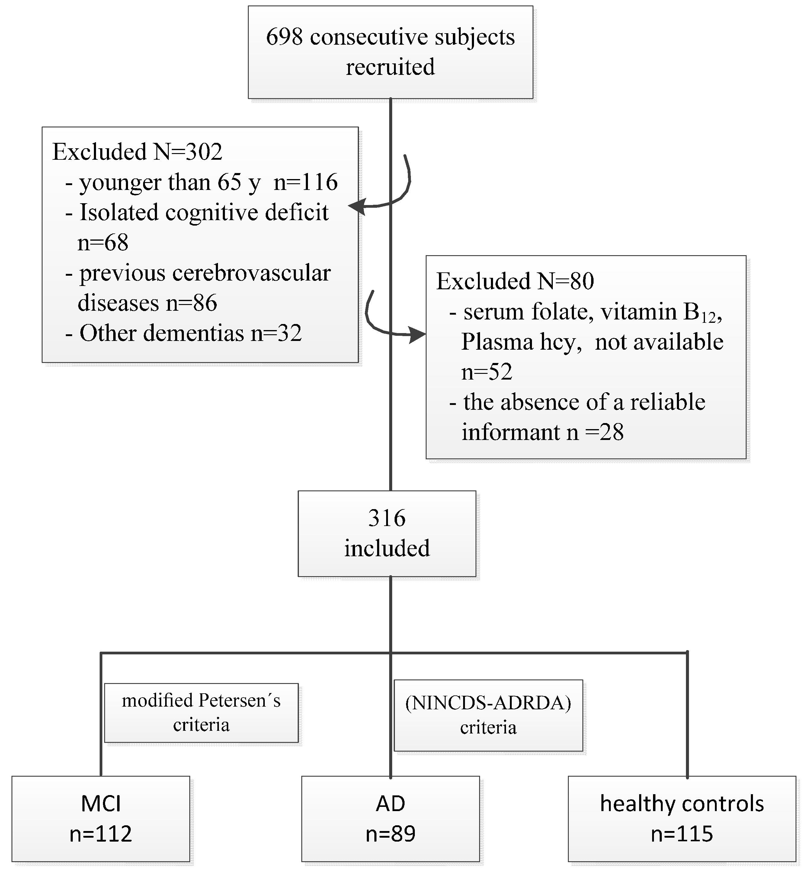
2.1. Study Design and Participants

This was a case-control study designed to evaluate the association of plasma Hcy and its biological determinants, folate and vitamin B12, with MCI and AD in elderly Chinese individuals. The recruitment, selection, and classification of patients were performed from April 2014 to June 2014, and a flow chart that outlines this process is shown in Figure 1. A series of 698 consecutive subjects (>65 years of age) were recruited at the neurology departments of several hospitals (Huanhu Hospital, The Second Hospital of Tianjin Medical University and The Sanhuailu Community Health Service Center in Binhai New District) in Tianjin, North China. Of these, 382 individuals were excluded from the analyses because they were younger than 65 years of age (n = 116) or because they had an isolated cognitive deficit (n = 68) or dementia other than AD (n = 32); previous cerebrovascular diseases (transient ischaemic attacks, stroke, or intracranial haemorrhage) (n = 86); plasma Hcy, serum folate, and vitamin B12 concentrations that were not available (n = 52); or the absence of a reliable informant (n = 28). The remaining 316 subjects were included in the analysis. All subjects were examined by neurologists and psychiatrists (see “Diagnosis of MCI and AD” for details), and according to their clinical diagnosis, were divided into the following three groups: older healthy control subjects who were free of cognitive impairments (n = 115), patients with MCI (n = 112), and patients with AD (n = 89).
The study was conducted in compliance with the ethical principles of the Declaration of Helsinki. All participants were informed of the objectives of the study and their consent to participate in the study was obtained. The research protocol was approved by the medical ethics committee of Tianjin Medical University, China.

2.2. Data Collection

All subjects were interviewed with their caregivers present by trained interviewers. The questionnaire was designed to obtain the following information regarding the patients’ general characteristics: age (in years), sex, education (in years), marital status, smoking status (whether they smoked, the number of packs smoked per year), alcohol use, medical history, medications taken, regular vitamin supplement uptake, and lifestyle habits. As part of the medication use questionnaire, all participants were asked to also report if they were taking any supplements, i.e., vitamins, fish-oils, omega-3 etc., and the frequency, per day. To obtain the amount of folate in each of the reported supplements/vitamins, we collected the ingredients of the supplements (in mg) either from the manufacturer’s website or by contacting the manufacturer. These values were then added to the participants’ nutritional intake data to give the total nutrient intake. The patients’ height and weight were measured, and their body mass index (BMI) (kg/m2) was calculated as their weight in kilograms divided by the square of the height in meters (kg/m2). Disease duration was defined as the time in years between the onset of the first symptoms (by history) and the clinical diagnosis. Dementia severity was assessed by the clinical dementia rating and the Mini Mental State Examination (MMSE).

2.3. Diagnosis of MCI and AD

Dementia was defined based on the clinical criteria of the Diagnostic and Statistical Manual of Mental Disorders, 4th edition (DSM-IV) [18]. The diagnosis of AD was based on the National Institute of Neurological and Communicative Disorders and Stroke and Alzheimer Disease and Related Disorders Association (NINCDS-ADRDA) criteria [19].
Only those individuals who were not diagnosed with dementia were considered for a diagnosis of MCI. The diagnosis of MCI was made by a panel of specialists who reviewed all of the existing information and used the modified Petersen’s criteria [20] as follows: subjective memory complaints with at least a 2-week duration; symptoms were not severe enough to fulfill the DSM-IV criteria for dementia; the cognitive performance was 1.5 standard deviations (SD) below the age-corrected (and education, where available) norms on at least one test in the neuropsychological battery; and activities of daily living (measured by a score < 26) were essentially preserved. The control subjects had no active medical therapy and no personal or family history of neurological and psychiatric disorders, as determined by clinical interviews. They performed within the normative range and did not meet the criteria for MCI or dementia. Based on the results of these evaluations, the participants were classified into the three groups mentioned above.

2.4. Blood Sampling and Laboratory Tests

Following overnight fasting (12–14 h), blood samples were collected from each participant. The samples were drawn by venipuncture into 5-mL plain evacuated tubes and then centrifuged at 2000× g for 10 min. Serum was used for the analysis of folate and vitamin B12 levels, and plasma was used for the analysis of Hcy. All specimens were collected and analyzed within 1 h or stored at −80 °C until use.
The concentrations of folate and vitamin B12 were determined on the same day using the Abbott Architect-i2000SR automated chemiluminescence immunoassay system and its supporting kit (Abbott, Washington, NJ,USA). In this assay, folate was quantified by measuring the population of unoccupied folate binding protein sites bound to the matrix using a conjugate of pteroic acid (folate analog) and alkaline phosphatase, as the signal-generating molecule, and a substrate, 4-methylumbelliferyl phosphate. Similar to folate, the serum level of vitamin B12 was measured using an Abbott kit based on a microparticle enzyme immunoassay. The concentrations of plasma Hcy were determined by a Hitachi 7180 automatic biochemistry analyzer (Hitachi, Tokyo, Japan), using the enzymatic conversion method. The kit was supplied by Beijing Strong Biotechnologies, Inc. (Beijing, China).
Folate concentrations <3 ng/mL in the serum have traditionally been considered a sign of inadequate folate [21,22]. According to the instruction of Abbott Laboratories, the reference range for serum folate was 7.0–31.4 ng/mL, suggesting that the desirable range for blood folate in the elderly may need to be changed in order to reduce the incidence of neurodegeneration. Other research set the normal and low reference values for folate at >7.2 and <7.2 ng/mL [23], respectively; therefore, we divided the serum folate levels into the following groups: <3 ng/mL, 3–7 ng/mL, and >7 ng/mL. The normal, borderline, and low reference values for vitamin B12 were >271, 208–271, and <208 pg/mL, respectively [24]. The normal, moderate, and severe Hcy values were classified as <30, 30–100, and >100 µmol/L, respectively [25].

2.5. Statistical Analysis
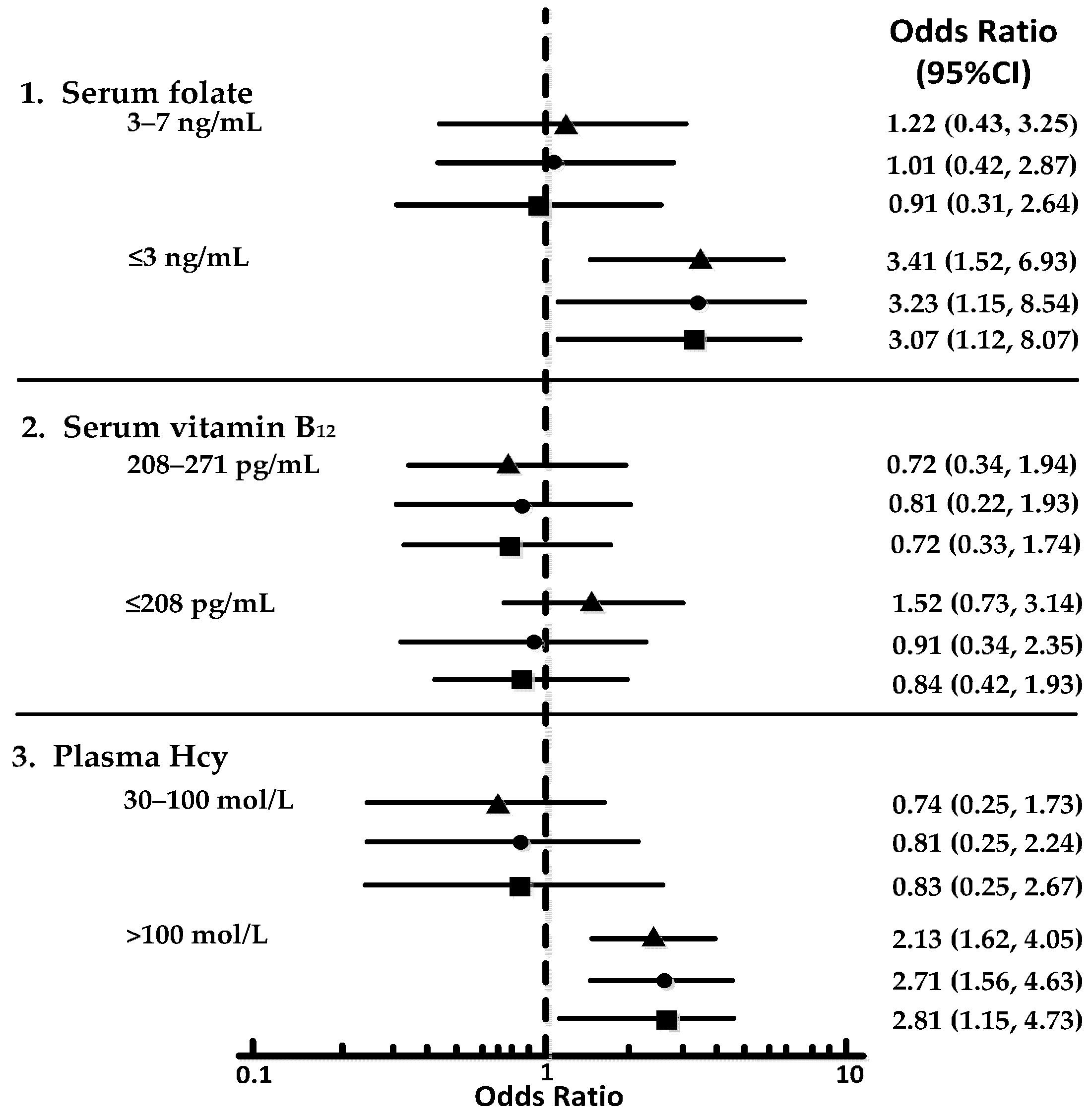
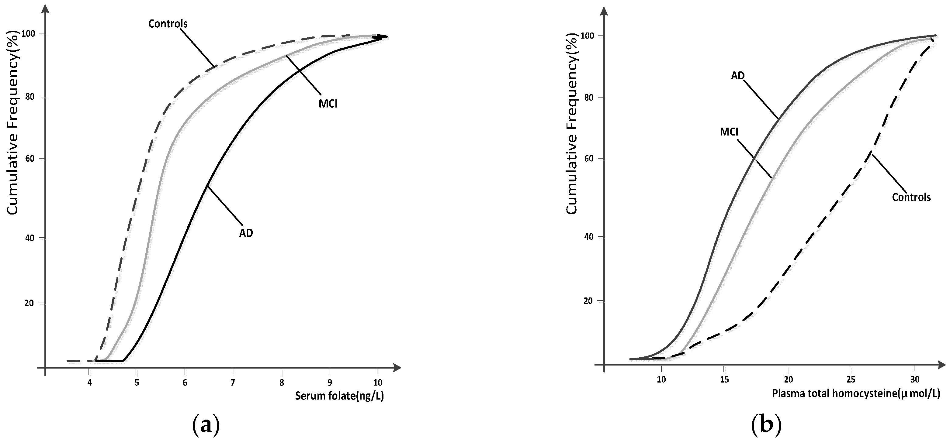
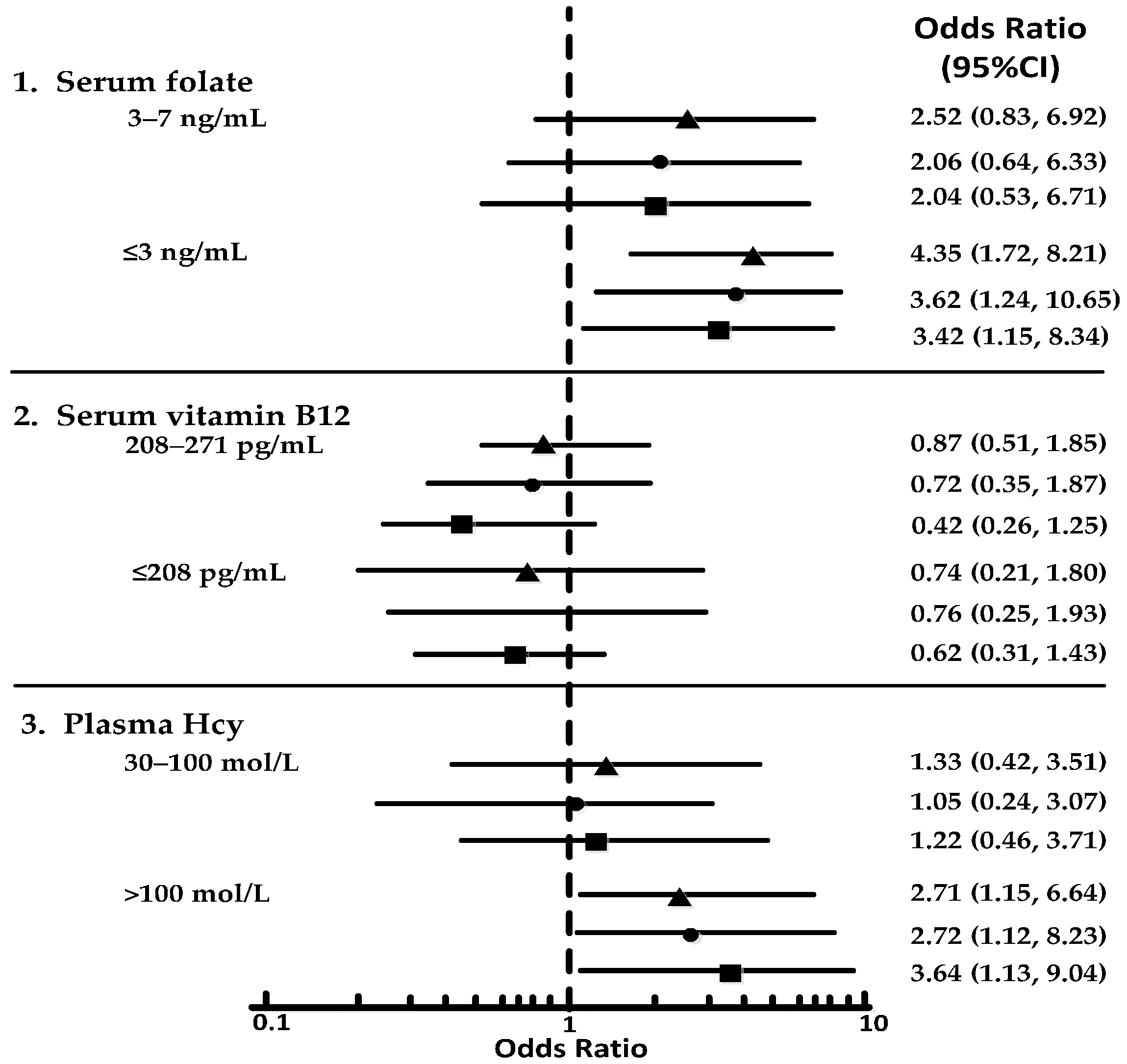
Data are expressed as the mean and standard deviation. Baseline values were compared among the healthy control, MCI, and AD groups using an analysis of variance (ANOVA) followed by Dunnett’s test or with a chi-squared test followed by a Bonferroni-corrected pairwise comparison test. The vitamin B12, folate, and Hcy concentrations were log-transformed before the analyses, as their distributions were slightly skewed. The cumulative frequency distributions of the Hcy and folate concentrations in patients with MCI and AD were compared with those in healthy control subjects by using the Kolmogorov–Smirnov test. Multiple logistic regression analyses were used to estimate the associations between quintiles of plasma Hcy, serum folate, and serum vitamin B12 concentrations and MCI and AD. Reference tertiles were the top tertiles for folate and vitamin B12 and the bottom tertile for plasma Hcy. The MCI and AD groups were each compared against the healthy control group in all analyses involving odds ratios (ORs). The crude ORs of MCI and AD were estimated using univariate logistic regression analyses in the three tertile levels of vitamin B12, folate, and Hcy. Folate, vitamin B12, and Hcy, together with age, sex, and education, were entered as covariates into multivariate logistic regression analyses to estimate the adjusted ORs of MCI and AD in the three tertiles of folate, vitamin B12, and Hcy. All analyses were performed using SPSS PASW Statistics for Windows, version 18.0 (SPSS Inc., Released 2009, Chicago, IL, USA).

3. Results

The demographic, clinical, and biochemical characteristics of the three groups (healthy controls, MCI, and AD) are shown in Table 1. The groups were well balanced in terms of their demographic and clinical information. The mean serum folate and vitamin B12 levels were significantly lower in the MCI and AD groups than they were in the healthy control group (p < 0.05). The mean plasma Hcy levels were significantly higher in patients with MCI and AD than they were in healthy controls (p < 0.01). The AD group had significantly lower mean serum folate and vitamin B12 levels and higher mean Hcy concentrations than did the MCI group (p < 0.05).
The cumulative frequency distributions of the folate and Hcy concentrations in the three groups are shown in Figure 2. The cumulative frequency of both folate and Hcy in the MCI group grew together with that in the AD group, which appeared distinctly separate in the healthy control group. The cumulative frequency plots show a shift in the distribution of the Hcy concentrations to higher values in patients with MCI and AD compared with healthy controls. There was a marked shift in the distribution of the folate concentrations to lower values in both patients with MCI and AD compared with healthy controls.
The crude (unadjusted) and adjusted ORs for the MCI and AD groups according to the tertile concentrations of serum folate, serum vitamin B12, and plasma Hcy are shown in forest plots (Figure 3 and Figure 4). MCI and AD were each significantly associated with the lowest tertile of folate and the highest tertile of Hcy. However, no association emerged between low vitamin B12 levels and AD or MCI. The lowest folate tertile, compared to the highest tertile, was associated with both MCI (crude OR: 3.41; 95% confidence interval [CI]: 1.52, 6.93) and AD (crude OR: 4.35; 95% CI: 1.72, 8.21). Compared to the lowest tertile, the highest Hcy tertile was associated with MCI (crude OR: 2.13; 95% CI: 1.62, 4.05) and AD (crude OR: 2.71; 95% CI: 1.15, 6.64). These ORs did not vary significantly when adjusted for age, sex, and education or when folate and vitamin B12 or Hcy concentrations were included as covariates in the model: subjects in the lowest folate tertile had an adjusted OR of 3.07 (95% CI: 1.12, 8.07) for MCI and of 3.42 (95% CI: 1.15, 8.34) for AD. Subjects in the highest Hcy tertile had an adjusted OR of 2.81 (95% CI: 1.15, 4.73) for MCI and of 3.64 (95% CI: 1.13, 9.04) for AD. In the MCI group, the chi-squared tests that were conducted before the individual OR comparisons showed nearly significant differences among the three folate tertiles in the crude analysis (p = 0.042), adjusted model 1 (p = 0.031), and adjusted model 2 (p = 0.037). In the AD group, all of the comparisons were significant among the folate and Hcy tertiles based on the chi-squared test.
To assess whether the prior duration of AD could explain the observed biochemical changes, 89 patients with AD with available data were classified by tertiles of duration of memory impairment (as reported by an informant) before the blood samples were taken (Table 2). The disease severity (MMSE score) was substantially greater in those with a longer duration of memory impairment, but there was no significant trend in the mean levels of any of the biochemical variables with the increasing duration of symptoms. The biochemical findings were also unaltered by the illness duration among patients with MCI (data not shown).

4. Discussion

In the present study, we retrospectively identified that MCI and AD were significantly associated with high plasma Hcy concentrations and low serum folate concentrations. In the highest Hcy tertile and the lowest folate tertile, the risk of MCI or AD was two to three times greater than the risk in subjects in the lowest and highest tertiles, respectively. A stronger association was shown in patients with AD compared to in patients with MCI.
Hcy may exert its neurotoxic effects by activating the N-methyl-d-aspartate receptor, leading to cell death [26], or by being converted into homocysteic acid, which also has an excitotoxic effect on neurons [27]. An Hcy-lowering effect in patients with AD has also been shown in an open-label trial using a folic acid, vitamin B12, and vitamin B6 regimen [28]. The association of low folate and vitamin B12 levels with AD may be mediated by the effects that folate and vitamin B12 have on Hcy levels [29], or may be related to their effects on methylation reactions in the brain [30]. Future randomized clinical trials with Hcy-reducing therapies should be conducted to provide further evidence of the relationships among Hcy, B vitamins, and cognition.
In agreement with the findings of the Kungsholmen population-based study [31] and the Bronx Longitudinal Aging Study [32], we did not find any significant association between the serum vitamin B12 concentrations and MCI and AD. The associations among vitamin B12, cognitive dysfunction, and AD in older individuals are weaker than are the folate associations [6,33,34,35], and often, the folate associations are absent or nonsignificant [8,31,32,33,34,35,36,37]. A better measure of vitamin B12 status may be the level of holotranscobalamin, which is an early marker of vitamin B12 deficiency. We hope to test this in a future prospective study.
Subjects with MCI have a high risk of developing AD in the short term or are already in a preclinical phase of dementia [38]. Thus, by comparing the biochemical profile of subjects with MCI with the profiles of the healthy control and AD groups, we tried to shed light on the possible role of folate, vitamin B12, and Hcy in the prodromal phases of the disease. The results of the current study suggest that the MCI group had both a lower mean folate concentration and a higher mean Hcy concentration compared to the healthy control group. These associations were independent of known or putative risk factors and were not modified by further adjustments for vitamin B12 and folate or vitamin B12 and Hcy.
The cumulative frequency plots showed that the frequency distribution of vitamin B12 in the MCI group was very similar to the distribution observed in the AD group. Interestingly, as was found in the study by Clarke et al. [7], the cumulative frequency distributions of the folate concentrations were more markedly separated than were the cumulative frequency distributions of the Hcy concentrations. However, we did identify a significant decrease in the serum folate levels in AD < MCI < healthy controls and a significant increase in the Hcy concentrations in AD > MCI > healthy controls (Figure 2). Additional studies are needed to establish the significance of these associations.
Some limitations of our study need to be addressed. First, this was a case-control study, and thus it cannot prove causality. It could, for example, be argued that AD leads to reduced serum folate and vitamin B12 concentrations, causing an elevation in Hcy levels. We cannot refute this possibility in this case-control study. Therefore, our findings need to be confirmed in further longitudinal studies. Second, as a random sample was not utilized, it is unlikely that our study population is representative of the general elderly population. Thus, our results may be difficult to interpret and apply to the general population. A further limitation of this study is the lack of data on the recent dietary intake and vitamin supplements in patients compared to in healthy controls.
Despite the above limitations, the chief strengths of the present study are as follows: (1) this was a relatively large, rigorous, population-based study designed to evaluate the association of plasma Hcy and its biological determinants, folate and vitamin B12, with MCI and AD in China; (2) the comprehensive clinical evaluations and the consensus approach provided reliable diagnoses of MCI and AD; and (3) the availability of pre-study plasma Hcy levels and baseline values for serum B vitamins and other covariates.

5. Conclusions

Based on the findings of the present study, it can be concluded that the low levels of folate and high levels of Hcy may be associated with a higher risk of MCI and AD. Because associations are not proof of a causal relationship, large, randomized, controlled clinical trials on B vitamin supplementation (a combination of folate, vitamin B12, and vitamin B6) and the onset and course of MCI and AD are under way.

Acknowledgments

The authors thank all of the subjects for their participation. This study was also supported by grants from Tianjin Science and Technology Support Program (grant number: 15ZCZDSY01040), the National Natural Science Foundation of China (grant number: 81130053).

Author Contributions

The authors’ contributions to manuscript were as follows—G.H.: project conception and development of overall research plan; A.S., J.Z. and T.W.: field survey and data collection; L.J. and M.Z.: hands-on conduct of the experiments and data collection; F.M.: analyzed data or performed statistical analysis; F.M.: wrote paper; G.H., F.M.: had primary responsibility for the final content. Final approval of the article: F.M., T.W., A.S., J.Z., L.J., M.Z. and G.H.

Conflicts of Interest

No potential conflicts of interest relevant to this article were reported.

Abbreviations

The following abbreviations are used in this manuscript:
ADAlzheimer Disease
ANOVAAnalysis of Variance
BMIBody Mass Index
DSM-IVDiagnostic and Statistical Manual of Mental Disorders, 4th edition
HcyHomocysteine
IQIntelligence Quotient
MCIMild Cognitive Impairment
MMSEMini Mental State Examination
NINCDS-ADRDANational Institute of Neurological and Communicative Disorders and Stroke and Alzheimer Disease and Related Disorders Association
ORsOdds Ratios
SDStandard Deviations
WAIS-RCChinese version of the Wechsler Adult Intelligence Scale-Revised

References

  1. Alzheimer’s Association. 2014 Alzheimer’s disease facts and figures. Alzheimers Dement. 2014, 10, e47–e92. [Google Scholar]
  2. Petersen, R.C.; Caracciolo, B.; Brayne, C.; Gauthier, S.; Jelic, V.; Fratiglioni, L. Mild cognitive impairment: A concept in evolution. J. Intern. Med. 2014, 275, 214–228. [Google Scholar] [CrossRef] [PubMed]
  3. Penninx, B.W.; Guralnik, J.M.; Ferrucci, L.; Fried, L.P.; Allen, R.H.; Stabler, S.P. Vitamin B12 deficiency and depression in physically disabled older women: Epidemiologic evidence from the Women’s Health and Aging Study. Am. J. Psychiatry 2000, 157, 715–721. [Google Scholar] [CrossRef] [PubMed]
  4. Hutto, B.R. Folate and cobalamin in psychiatric illness. Compr. Psychiatry 1997, 38, 305–314. [Google Scholar] [CrossRef]
  5. Ho, P.I.; Collins, S.C.; Dhitavat, S.; Ortiz, D.; Ashline, D.; Rogers, E.; Shea, T.B. Homocysteine potentiates beta-amyloid neurotoxicity: Role of oxidative stress. J. Neurochem. 2001, 78, 249–253. [Google Scholar] [CrossRef] [PubMed]
  6. Clarke, R.; Smith, A.D.; Jobst, K.A.; Refsum, H.; Sutton, L.; Ueland, P.M. Folate, vitamin B12, and serum total homocysteine levels in confirmed Alzheimer disease. Arch. Neurol. 1998, 55, 1449–1455. [Google Scholar] [CrossRef] [PubMed]
  7. Quadri, P.; Fragiacomo, C.; Pezzati, R.; Zanda, E.; Forloni, G.; Tettamanti, M.; Lucca, U. Homocysteine, folate, and vitamin B-12 in mild cognitive impairment, Alzheimer disease, and vascular dementia. Am. J. Clin. Nutr. 2004, 80, 114–122. [Google Scholar] [PubMed]
  8. Ravaglia, G.; Forti, P.; Maioli, F.; Muscari, A.; Sacchetti, L.; Arnone, G.; Nativio, V.; Talerico, T.; Mariani, E. Homocysteine and cognitive function in healthy elderly community dwellers in Italy. Am. J. Clin. Nutr. 2003, 77, 668–673. [Google Scholar] [PubMed]
  9. Mooijaart, S.P.; Gussekloo, J.; Frolich, M.; Jolles, J.; Stott, D.J.; Westendorp, R.G.; de Craen, A.J. Homocysteine, vitamin B-12, and folic acid and the risk of cognitive decline in old age: The Leiden 85-Plus study. Am. J. Clin. Nutr. 2005, 82, 866–871. [Google Scholar] [PubMed]
  10. Haan, M.N.; Miller, J.W.; Aiello, A.E.; Whitmer, R.A.; Jagust, W.J.; Mungas, D.M.; Allen, L.H.; Green, R. Homocysteine, B vitamins, and the incidence of dementia and cognitive impairment: Results from the Sacramento Area Latino Study on Aging. Am. J. Clin. Nutr. 2007, 85, 511–517. [Google Scholar] [PubMed]
  11. Ravaglia, G.; Forti, P.; Maioli, F.; Martelli, M.; Servadei, L.; Brunetti, N.; Porcellini, E.; Licastro, F. Homocysteine and folate as risk factors for dementia and Alzheimer disease. Am. J. Clin. Nutr. 2005, 82, 636–643. [Google Scholar] [PubMed]
  12. Seshadri, S.; Beiser, A.; Selhub, J.; Jacques, P.F.; Rosenberg, I.H.; D’Agostino, R.B.; Wilson, P.W.; Wolf, P.A. Plasma homocysteine as a risk factor for dementia and Alzheimer’s disease. N. Engl. J. Med. 2002, 346, 476–483. [Google Scholar] [CrossRef] [PubMed]
  13. Beydoun, M.A.; Beydoun, H.A.; Gamaldo, A.A.; Teel, A.; Zonderman, A.B.; Wang, Y. Epidemiologic studies of modifiable factors associated with cognition and dementia: Systematic review and meta-analysis. BMC Public Health 2014, 14, 643. [Google Scholar] [CrossRef] [PubMed]
  14. Fekkes, D.; van der Cammen, T.J.M.; van Loon, C.P.M.; Verschoor, C.; van Harskamp, F.; de Koning, I.; Schudel, W.J.; Pepplinkhuize, L. Abnormal amino acid metabolism in patients with early stage Alzheimer dementia. J. Neural Transm. 1998, 105, 287–294. [Google Scholar] [CrossRef] [PubMed]
  15. Dresner Pollak, R.; Pollak, A.; Idelson, M.; Bejarano-Achache, I.; Doron, D.; Blumenfeld, A. The C677T mutation in the methylenetetrahydrofolate reductase (MTHFR) gene and vascular dementia. J. Am. Geriatr. Soc. 2000, 48, 664–668. [Google Scholar] [CrossRef]
  16. Ravaglia, G.; Forti, P.; Maioli, F.; Vettori, C.; Grossi, G.; Bargossi, A.M.; Caldarera, M.; Franceschi, C.; Facchini, A.; Mariani, E.; et al. Elevated plasma homocysteine levels in centenarians are not associated with cognitive impairment. Mech. Ageing Dev. 2000, 121, 251–261. [Google Scholar] [CrossRef]
  17. Miller, J.W.; Green, R.; Mungas, D.M.; Reed, B.R.; Jagust, W.J. Homocysteine, vitamin B-6, and vascular disease in AD patients. Neurology 2002, 58, 1471–1475. [Google Scholar] [CrossRef] [PubMed]
  18. American Psychiatric Association. Diagnostic and Statistical Manual of Mental Disorders, 4th ed.; American Psychiatric Association: Washington, DC, USA, 1994. [Google Scholar]
  19. McKhann, G.; Drachman, D.; Folstein, M.; Katzman, R.; Price, D.; Stadlan, E.M. Clinical diagnosis of Alzheimer’s disease: Report of the NINCDS-ADRDA Work Group under the auspices of Department of Health and Human Services Task Force on Alzhimer’s Disease. Neurology 1984, 34, 939–944. [Google Scholar] [CrossRef] [PubMed]
  20. Petersen, R.C. Mild cognitive impairment as a diagnostic entity. J. Intern. Med. 2004, 256, 183–194. [Google Scholar] [CrossRef] [PubMed]
  21. Institute of Medicine (US). Dietary Reference Intakes for Thiamin, Riboflavin, Niacin, Vitamin B6, Folate, Vitamin B12, Pantothenic acid, Biotin, and Choline; National Academies Press: Washington, DC, USA, 1998; pp. 196–305. [Google Scholar]
  22. Han-Byul, J.; Young-Hee, H.; Chandrika, J.P.; Heon, K.; Tawasun, H. Intake and blood concentrations of folate and their association with health-related behaviors in Korean college students. Nutr. Res. Pract. 2013, 7, 216–223. [Google Scholar]
  23. Mohammad, Y.G.; Ridha, A.G.; Omar, F.K.; Mahmoud, A.A. Hyperhomocysteinemia, Low Folate, and Vitamin B12 Deficiency in Elderly Living at Home and Cwere Residences: A Comparative Study. Labmedicine 2010, 41, 410–414. [Google Scholar]
  24. Susan, J.T.; Alan, H.; Sheena, B.; Anne, L.; Malcolm, H.; Sean, O.; Bryant, C.N.; Christine, P. International Standard for serum vitamin B(12) and serum folate: International collaborative study to evaluate a batch of lyophilwased serum for B(12) and folate content. Clin. Chem. Lab. Med. 2007, 45, 380–386. [Google Scholar]
  25. Pitla, S.; Nagalla, B. Gender-related differences in the relationship between plasma homocysteine, anthropometric and conventional biochemical coronary heart disease risk factors in middle-aged Indians. Ann. Nutr. Metab. 2009, 54, 1–6. [Google Scholar] [CrossRef] [PubMed]
  26. Stuart, A.L.; Won-Ki, K.; Yun-Beom, C.; Shanta, K.; Danielle, M.D.; Posina, V.R.; Derrick, R.A.; Jonathan, S.S. Neurotoxicity associated with dual actions of homocysteine at the N-methyl-d-aspartate receptor. Proc. Natl. Acad. Sci. USA 1997, 94, 5923–5928. [Google Scholar]
  27. Beal, M.F.; Swartz, K.J.; Finn, S.F.; Mazurek, M.F.; Kowall, N.W. Neurochemical characterization of excitoxin lesions in the cerebral cortex. J. Neurosci. 1991, 11, 147–158. [Google Scholar] [PubMed]
  28. Paul, S.A.; Susan, E.; Howard, A.; Ramon, D.A.; Myron, W.; Charles, D.; William, J.; Joshua, W.M.; Ralph, G.; Karen, B.; et al. A pilot study of vitamins to lower plasma homocysteine levels in Alzheimer disease. Am. J. Geriatr. Psychiatry 2003, 1, 246–249. [Google Scholar]
  29. Selhub, J.; Jacques, P.F.; Wilson, P.W.F.; Rush, D.; Rosenberg, I.H. Vitamin status and intake as primary determinants of homocysteinemia in an elderly population. JAMA 1993, 270, 2693–2698. [Google Scholar] [CrossRef] [PubMed]
  30. Bottiglieri, T.; Hyland, K.; Reynolds, E.H. The clinical potential of adometionine (S-adenosylmethionine) in neurological disorders. Drugs 1994, 48, 137–152. [Google Scholar] [CrossRef] [PubMed]
  31. Basun, H.; Fratiglioni, L.; Winbland, B. Cobalamin levels are not reduced in Alzheimer’s disease: Results from a population-based study. J. Am. Geriatr. Soc. 1994, 42, 132–136. [Google Scholar] [CrossRef] [PubMed]
  32. Crystal, H.A.; Ortof, E.; Frishman, W.H.; Gruber, A.; Hershman, D.; Aronson, M. Serum vitamin B-12 levels and incidence of dementia in a healthy elderly population: A report from the Bronx Longitudinal Aging Study. J. Am. Geriatr. Soc. 1994, 42, 933–936. [Google Scholar] [CrossRef] [PubMed]
  33. Teunissen, C.E.; Blom, A.H.; Van Boxtel, M.P.; Bosma, H.; de Bruijn, C.; Jolles, J.; Wauters, B.A.; Steinbusch, H.W.; de Vente, J. Homocysteine: A marker for cognitive performance? A longitudinal follow-up study. J. Nutr. Health Aging 2003, 7, 153–159. [Google Scholar] [PubMed]
  34. David, A.S.; Christine, L.T.; Charles, D.S.; Kathryn, P.R.; William, R.M. Serum folate and the severity of atrophy of the neocortex in Alzheimer disease: Findings from the Nun Study. Am. J. Clin. Nutr. 2000, 71, 993–998. [Google Scholar]
  35. Wang, H.-X.; Wahlin, A.; Basun, H.; Fastbom, J.; Winblad, B.; Fratiglioni, L. Vitamin B12 and folate in relation to the development of Alzheimer’s disease. Neurology 2001, 56, 1188–1194. [Google Scholar] [CrossRef] [PubMed]
  36. Joosten, E.; Lesaffre, E.; Riezler, R.; Ghekiere, V.; Dereymaeker, L.; Pelemans, W.; Dejaeger, E. Is metabolic evidence for vitamin B-12 and folate deficiency more frequent in elderly patients with Alzheimer’s disease? J. Gerontol. 1997, 52, M76–M79. [Google Scholar] [CrossRef]
  37. Lehmann, M.; Gottfries, C.G.; Regland, B. Identification of cognitive impairment in the elderly: Homocysteine is an early marker. Dement. Geriatr. Cogn. Disord. 1999, 10, 12–20. [Google Scholar] [CrossRef] [PubMed]
  38. Morris, J.C.; Storandt, M.; Miller, J.P.; McKeel, D.W.; Price, J.L.; Rubin, E.H.; Berg, L. Mild cognitive impairment represents early-stage Alzheimer disease. Arch. Neurol. 2001, 58, 397–405. [Google Scholar] [CrossRef] [PubMed]
Figure 1. Flow chart detailing the derivation of the study sample.
Nutrients 09 00725 g001
Figure 2. Cumulative frequency distributions of serum folate (a) and plasma homocysteine (Hcy) (b) levels in the healthy controls, MCI and AD subjects.
Nutrients 09 00725 g002
Figure 3. Forest plots showing ORs for tertiles of the vitamin and Hcy levels in MCI. 1 indicates comparing to tertile of >7 ng/mL; 2 indicates comparing to tertile of >271 pg/mL; 3 indicates comparing to tertile of ≤30 mol/L. This figure illustrates unadjusted model (▲), adjusted model for age, sex, and education (●); and adjusted model for age, sex, education, folate, vitamin B12, and Hcy (■).
Nutrients 09 00725 g003
Figure 4. Forest plots showing ORs for tertiles of the vitamin and Hcy levels in AD. 1 indicates comparing to tertile of >7 ng/mL; 2 indicates comparing to tertile of >271 pg/mL; 3 indicates comparing to tertile of ≤30 mol/L. This figure illustrates unadjusted model (▲), adjusted model for age, sex, and education (●); and adjusted model for age, sex, education, folate, vitamin B12, and Hcy (■).
Nutrients 09 00725 g004
Table 1. Demographic, clinical, and biochemical characteristics of the three groups.
ProfileControl Group (n = 115)MCI Group (n = 112)AD Group (n = 89)
Demographics
Age (years)72.82 ± 8.8773.23 ± 8.6774.62 ± 8.01
Female69 (60.00)68 (60.71)56 (62.92)
Total education (years)10.92 ± 1.5310.42 ± 2.0110.33 ± 2.37
Clinical characteristics
Disease duration (months) 27.67 ± 22.24
MMSE score 28.53 ± 1.7416.23 ± 8.07 12.83 ± 8.13
BMI (kg/m2)25.87 ± 5.1325.14 ± 3.9624.73 ± 5.54
Current smokers24 (20.87)24 (21.43)20 (22.47)
Alcohol (units/week)9.12 ± 4.538.14 ± 3.228.57 ± 3.21
Total cholesterol (mmol/L)5.83 ± 1.356.02 ± 1.365.74 ± 1.43
Systolic blood pressure (mmHg)143.33 ± 25.20143.31 ± 25.21144.44 ± 26.47
Hypertension50 (43.48)49 (43.75)38 (42.70)
Diabetes20 (17.39)21 (18.75)16 (17.98)
Biochemical measures
Plasma Hcy (μmol/L)13.21 ± 4.0515.35 ± 8.44 16.37 ± 7.46
Serum folate (ng/mL)7.03 ± 3.685.74 ± 2.63 5.13 ± 3.57
Serum vitamin B12 (pg/mL)573.17 ± 75.41538.82 ± 84.06 531.21 ± 44.33
B vitamin supplements, n (%) *31 (26.96)31 (27.68)23 (25.84)
Use of fish-oils, omega-3, n (%)16 (13.91)16 (14.28)12 (13.48)
Results are shown as n (%) for the chi-squared or Fisher’s exact tests, and as the mean ± the standard deviation for independent t-tests (two-tailed) or analyses of variance. The vitamin B12, folate, and Hcy concentration values were log-transformed before the analyses. MCI, mild cognitive impairment; AD, Alzheimer’s disease; BMI, body mass index; MMSE, Mini-Mental State Examination; Hcy, homocysteine. p < 0.05 vs. healthy controls; p < 0.01 vs. healthy controls. * Intake of multivitamin supplements or any type of B vitamin supplements.
Table 2. Clinical and biochemical variables in patients with AD by duration of memory impairment at presentation.
Tertiles of Duration of Memory Impairment, YearsMMSE ScoreBiochemical Variables
Hcy, μmol/LFolate, ng/mLVitamin B12, pg/mL
I <217.80 ± 7.9917.25 ± 6.825.67 ± 4.11525.31 ± 37.21
II 2–414.22 ± 8.4416.47 ± 5.755.11 ± 5.17528.47 ± 63.41
III >47.55 ± 6.4916.79 ± 4.155.05 ± 7.39517.45 ± 43.49
p value0.0070.4520.3210.367
Results are shown as the mean ± the standard deviation for analyses of variance. AD, Alzheimer’s disease; MMSE, Mini-Mental State Examination; Hcy, homocysteine.

