Next Issue
Volume 10, May
Previous Issue
Volume 10, March


Toxins, Volume 10, Issue 4 (April 2018) – 41 articles

Cover Story (view full-size image): 
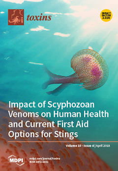
Male specimen of the mauve stinger Pelagia noctiluca (Cnidaria: Scyphozoa) photographed in the sea offshore Capo Milazzo (Sicily, Italy) during summer 2016. A detail of the umbrella with the stinging nematocyst buttons is shown (lower left). P. noctiluca is the most venomous autochthonous jellyfish of the Mediterranean Sea, and is responsible for the majority of jellyfish stings along the south Italian coasts. (Photo by Dr. Carmelo Isgró) View the paper here.
  • Issues are regarded as officially published after their release is announced to the table of contents alert mailing list.
  • You may sign up for e-mail alerts to receive table of contents of newly released issues.
  • PDF is the official format for papers published in both, html and pdf forms. To view the papers in pdf format, click on the "PDF Full-text" link, and use the free Adobe Reader to open them.
Order results
Result details
Section
Select all
Export citation of selected articles as:
24 pages, 457 KiB  
Review
Snakebite: When the Human Touch Becomes a Bad Touch
by Bryan G. Fry
Toxins 2018, 10(4), 170; https://doi.org/10.3390/toxins10040170 - 21 Apr 2018
Cited by 74 | Viewed by 14734
Abstract
Many issues and complications in treating snakebite are a result of poor human social, economic and clinical intervention and management. As such, there is scope for significant improvements for reducing incidence and increasing patient outcomes. Snakes do not target humans as prey, but [...] Read more.
Many issues and complications in treating snakebite are a result of poor human social, economic and clinical intervention and management. As such, there is scope for significant improvements for reducing incidence and increasing patient outcomes. Snakes do not target humans as prey, but as our dwellings and farms expand ever farther and climate change increases snake activity periods, accidental encounters with snakes seeking water and prey increase drastically. Despite its long history, the snakebite crisis is neglected, ignored, underestimated and fundamentally misunderstood. Tens of thousands of lives are lost to snakebites each year and hundreds of thousands of people will survive with some form of permanent damage and reduced work capacity. These numbers are well recognized as being gross underestimations due to poor to non-existent record keeping in some of the most affected areas. These underestimations complicate achieving the proper recognition of snakebite’s socioeconomic impact and thus securing foreign aid to help alleviate this global crisis. Antivenoms are expensive and hospitals are few and far between, leaving people to seek help from traditional healers or use other forms of ineffective treatment. In some cases, cheaper, inappropriately manufactured antivenom from other regions is used despite no evidence for their efficacy, with often robust data demonstrating they are woefully ineffective in neutralizing many venoms for which they are marketed for. Inappropriate first-aid and treatments include cutting the wound, tourniquets, electrical shock, immersion in ice water, and use of ineffective herbal remedies by traditional healers. Even in the developed world, there are fundamental controversies including fasciotomy, pressure bandages, antivenom dosage, premedication such as adrenalin, and lack of antivenom for exotic snakebites in the pet trade. This review explores the myriad of human-origin factors that influence the trajectory of global snakebite causes and treatment failures and illustrate that snakebite is as much a sociological and economic problem as it is a medical one. Reducing the incidence and frequency of such controllable factors are therefore realistic targets to help alleviate the global snakebite burden as incremental improvements across several areas will have a strong cumulative effect. Full article
(This article belongs to the Special Issue Toxins:10th Anniversary)
29 pages, 19193 KiB  
Review
Therapeutic Approaches of Botulinum Toxin in Gynecology
by Marius Alexandru Moga, Oana Gabriela Dimienescu, Andreea Bălan, Ioan Scârneciu, Barna Barabaș and Liana Pleș
Toxins 2018, 10(4), 169; https://doi.org/10.3390/toxins10040169 - 21 Apr 2018
Cited by 19 | Viewed by 7746
Abstract
Botulinum toxins (BoNTs) are produced by several anaerobic species of the genus Clostridium and, although they were originally considered lethal toxins, today they find their usefulness in the treatment of a wide range of pathologies in various medical specialties. Botulinum neurotoxin has been [...] Read more.
Botulinum toxins (BoNTs) are produced by several anaerobic species of the genus Clostridium and, although they were originally considered lethal toxins, today they find their usefulness in the treatment of a wide range of pathologies in various medical specialties. Botulinum neurotoxin has been identified in seven different isoforms (BoNT-A, BoNT-B, BoNT-C, BoNT-D, BoNT-E, BoNT-F, and BoNT-G). Neurotoxigenic Clostridia can produce more than 40 different BoNT subtypes and, recently, a new BoNT serotype (BoNT-X) has been reported in some studies. BoNT-X has not been shown to actually be an active neurotoxin despite its catalytically active LC, so it should be described as a putative eighth serotype. The mechanism of action of the serotypes is similar: they inhibit the release of acetylcholine from the nerve endings but their therapeutically potency varies. Botulinum toxin type A (BoNT-A) is the most studied serotype for therapeutic purposes. Regarding the gynecological pathology, a series of studies based on the efficiency of its use in the treatment of refractory myofascial pelvic pain, vaginism, dyspareunia, vulvodynia and overactive bladder or urinary incontinence have been reported. The current study is a review of the literature regarding the efficiency of BoNT-A in the gynecological pathology and on the long and short-term effects of its administration. Full article
Show Figures

Figure 1

16 pages, 2715 KiB  
Article
Analysis of the Relationship between Alternative Respiration and Sterigmatocystin Formation in Aspergillus nidulans
by Ákos P. Molnár, Zoltán Németh, Erzsébet Fekete, Michel Flipphi, Nancy P. Keller and Levente Karaffa
Toxins 2018, 10(4), 168; https://doi.org/10.3390/toxins10040168 - 20 Apr 2018
Cited by 12 | Viewed by 5945
Abstract
Aspergillus nidulans has one gene for alternative oxidase (EC 1.10.3.11). To investigate the relationship between this mitochondrial terminal oxidase and the formation of the mycotoxin sterigmatocystin, the encoding aodA gene was both deleted and overexpressed. Relative to the wild-type, the cyanide-resistant fraction of [...] Read more.
Aspergillus nidulans has one gene for alternative oxidase (EC 1.10.3.11). To investigate the relationship between this mitochondrial terminal oxidase and the formation of the mycotoxin sterigmatocystin, the encoding aodA gene was both deleted and overexpressed. Relative to the wild-type, the cyanide-resistant fraction of respiration in the late stationary stage—when sterigmatocystin production occurs—doubled in the overexpressing mutant carrying three aodA gene copies, but decreased to 10% in the deletant. Essentially identical results were obtained regardless whether the cultures were illuminated or protected from light. In contrast, sterigmatocystin yield in the aodA deletant was about half of that in the control when grown in the dark, while aodA overexpression resulted in up to 70% more sterigmatocystin formed, the yield increasing with alternative oxidase activity. Results were quite different when cultures were illuminated: under those conditions, sterigmatocystin volumetric yields were considerably lower, and statistically unvarying, regardless of the presence, absence, or the copy number of aodA. We conclude that the copy number of aodA, and hence, the balance between alternative- and cytochrome C-mediated respiration, appears to correlate with sterigmatocystin production in A. nidulans, albeit only in the absence of light. Full article
(This article belongs to the Collection Aflatoxins)
Show Figures

Figure 1

28 pages, 2329 KiB  
Review
Solid Phase Adsorption Toxin Tracking (SPATT) Technology for the Monitoring of Aquatic Toxins: A Review
by Mélanie Roué, Hélène Taiana Darius and Mireille Chinain
Toxins 2018, 10(4), 167; https://doi.org/10.3390/toxins10040167 - 20 Apr 2018
Cited by 34 | Viewed by 8498
Abstract
The Solid Phase Adsorption Toxin Tracking (SPATT) technology, first introduced in 2004, uses porous synthetic resins capable of passively adsorbing toxins produced by harmful microalgae or cyanobacteria and dissolved in the water. This method allows for the detection of toxic compounds directly in [...] Read more.
The Solid Phase Adsorption Toxin Tracking (SPATT) technology, first introduced in 2004, uses porous synthetic resins capable of passively adsorbing toxins produced by harmful microalgae or cyanobacteria and dissolved in the water. This method allows for the detection of toxic compounds directly in the water column and offers numerous advantages over current monitoring techniques (e.g., shellfish or fish testing and microalgae/cyanobacteria cell detection), despite some limitations. Numerous laboratory and field studies, testing different adsorbent substrates of which Diaion® HP20 resin appears to be the most versatile substrate, have been carried out worldwide to assess the applicability of these passive monitoring devices to the detection of toxins produced by a variety of marine and freshwater microorganisms. SPATT technology has been shown to provide reliable, sensitive and time-integrated sampling of various aquatic toxins, and also has the potential to provide an early warning system for both the occurrence of toxic microalgae or cyanobacteria and bioaccumulation of toxins in foodstuffs. This review describes the wide range of lipophilic and hydrophilic toxins associated with toxin-producing harmful algal blooms (HABs) that are successfully detected by SPATT devices. Implications in terms of monitoring of emerging toxic risks and reinforcement of current risk assessment programs are also discussed. Full article
(This article belongs to the Special Issue Public Health Outreach to Prevention of Aquatic Toxin Exposure)
Show Figures

Figure 1

15 pages, 3010 KiB  
Article
Electromyographic and Joint Kinematic Patterns in Runner’s Dystonia
by Omar F. Ahmad, Pritha Ghosh, Christopher Stanley, Barbara Karp, Mark Hallett, Codrin Lungu and Katharine Alter
Toxins 2018, 10(4), 166; https://doi.org/10.3390/toxins10040166 - 20 Apr 2018
Cited by 12 | Viewed by 6448
Abstract
Runner’s dystonia (RD) is a task-specific focal dystonia of the lower limbs that occurs when running. In this retrospective case series, we present surface electromyography (EMG) and joint kinematic data from thirteen patients with RD who underwent instrumented gait analysis (IGA) at the [...] Read more.
Runner’s dystonia (RD) is a task-specific focal dystonia of the lower limbs that occurs when running. In this retrospective case series, we present surface electromyography (EMG) and joint kinematic data from thirteen patients with RD who underwent instrumented gait analysis (IGA) at the Functional and Biomechanics Laboratory at the National Institutes of Health. Four cases of RD are described in greater detail to demonstrate the potential utility of EMG with kinematic studies to identify dystonic muscle groups in RD. In these cases, the methodology for muscle selection for botulinum toxin therapy and the therapeutic response is discussed. Lateral heel whip, a proposed novel presentation of lower-limb dystonia, is also described. Full article
(This article belongs to the Special Issue Muscle Selection for BoNT)
Show Figures

Figure 1

15 pages, 5886 KiB  
Article
Visual Non-Instrumental On-Site Detection of Fumonisin B1, B2, and B3 in Cereal Samples Using a Clean-Up Combined with Gel-Based Immunoaffinity Test Column Assay
by Wei Sheng, Hesen Wu, Weihong Ji, Zhi Li, Fangyu Chu and Shuo Wang
Toxins 2018, 10(4), 165; https://doi.org/10.3390/toxins10040165 - 19 Apr 2018
Cited by 8 | Viewed by 4136
Abstract
A visual immunoaffinity test column (IATC) assay was developed to detect fumonisins in cereal samples for spot tests without the need for special instruments. The developed IATC assay had equivalent recognition capability for fumonisin B1 (FB1), fumonisin B2 (FB [...] Read more.
A visual immunoaffinity test column (IATC) assay was developed to detect fumonisins in cereal samples for spot tests without the need for special instruments. The developed IATC assay had equivalent recognition capability for fumonisin B1 (FB1), fumonisin B2 (FB2), or fumonisin B3 (FB3), and exhibited no cross-reactivity with aflatoxin B1, ochratoxin A, zearalenone, or the T-2 toxin. The sample pretreatment was accomplished more rapidly and with greater ease, the entire assay procedure was completed in approximately 10 min, including sample pretreatment and testing. The limits of detection (LODs) of the IATC assay to detect fumonisins in the maize, barley, oat, and millet samples were 20 μg kg−1. The results of the spiked maize, barley, oat, and millet and real maize samples by the IATC assay agreed well with the results obtained by the commercial fumonisin enzyme-linked immunosorbent assay (ELISA) test kit and liquid chromatography-tandem mass spectrometry (LC-MS/MS), respectively. The developed IATC assay can serve as a useful screening tool for the rapid, qualitative, and semi-quantitative detection of the total content of fumonisins (sum of FB1, FB2, and FB3) in cereal samples on-site. Full article
(This article belongs to the Collection Biorecognition Assays for Mycotoxins)
Show Figures

Figure 1

8 pages, 3674 KiB  
Article
Lumbar Sympathetic Block with Botulinum Toxin Type A and Type B for the Complex Regional Pain Syndrome
by Yongki Lee, Chul Joong Lee, Eunjoo Choi, Pyung Bok Lee, Ho-Jin Lee and Francis Sahngun Nahm
Toxins 2018, 10(4), 164; https://doi.org/10.3390/toxins10040164 - 19 Apr 2018
Cited by 31 | Viewed by 6299
Abstract
A lumbar sympathetic ganglion block (LSB) is a therapeutic method for complex regional pain syndrome (CRPS) affecting the lower limbs. Recently, LSB with botulinum toxin type A and B was introduced as a novel method to achieve longer duration of analgesia. In this [...] Read more.
A lumbar sympathetic ganglion block (LSB) is a therapeutic method for complex regional pain syndrome (CRPS) affecting the lower limbs. Recently, LSB with botulinum toxin type A and B was introduced as a novel method to achieve longer duration of analgesia. In this study, we compared the botulinum toxin type A (BTA) with botulinum toxin type B (BTB) in performing LSB on patients with CRPS. LSB was performed with either BTA or BTB on patients with CRPS in their lower extremities. The length of time taken for patients to return to the pre-LSB pain score and the adverse effect of LSB with BTA/BTB were investigated. The median length of time taken for the patients to return to the pre-LSB pain score was 15 days for the BTA group and 69 days for the BTB group (P = 0.002). Scores on a visual analogue scale decreased in the patients of both groups, and no significant adverse effects were experienced. In conclusion, the administration of either BTA or BTB for LSB is a safe method to prolong the sympathetic blocking effect in patients with CRPS. BTB is more effective than BTA to prolong the sympathetic blocking effect in CRPS patients. Full article
Show Figures

Figure 1

20 pages, 25436 KiB  
Review
Epidemiology of Helicobacter pylori and CagA-Positive Infections and Global Variations in Gastric Cancer
by Jin Young Park, David Forman, Langgeng Agung Waskito, Yoshio Yamaoka and Jean E. Crabtree
Toxins 2018, 10(4), 163; https://doi.org/10.3390/toxins10040163 - 19 Apr 2018
Cited by 146 | Viewed by 13396
Abstract
Gastric cancer is a major health burden and is the fifth most common malignancy and the third most common cause of death from cancer worldwide. Development of gastric cancer involves several aspects, including host genetics, environmental factors, and Helicobacter pylori infection. There is [...] Read more.
Gastric cancer is a major health burden and is the fifth most common malignancy and the third most common cause of death from cancer worldwide. Development of gastric cancer involves several aspects, including host genetics, environmental factors, and Helicobacter pylori infection. There is increasing evidence from epidemiological studies of the association of H. pylori infection and specific virulence factors with gastric cancer. Studies in animal models indicate H. pylori is a primary factor in the development of gastric cancer. One major virulence factor in H. pylori is the cytotoxin-associated gene A (cagA), which encodes the CagA protein in the cag pathogenicity island (cag PAI). Meta-analysis of studies investigating CagA seropositivity irrespective of H. pylori status identified that CagA seropositivity increases the risk of gastric cancer (OR = 2.87, 95% CI: 1.95–4.22) relative to the risk of H. pylori infection alone (OR = 2.31, 95% CI: 1.58–3.39). Eradicating H. pylori is a strategy for reducing gastric cancer incidence. A meta-analysis of six randomised controlled trials (RCTs) suggests that searching for and eradicating H. pylori infection reduces the subsequent incidence of gastric cancer with a pooled relative risk of 0.66 (95% CI: 0.46–0.95). The introduction in regions of high gastric cancer incidence of population-based H. pylori screening and treatment programmes, with a scientifically valid assessment of programme processes, feasibility, effectiveness and possible adverse consequences, would impact the incidence of H. pylori-induced gastric cancer. Given the recent molecular understanding of the oncogenic role of CagA, targeting H. pylori screening and treatment programmes in populations with a high prevalence of H. pylori CagA-positive strains, particularly the more oncogenic East Asian H. pylori CagA strains, may be worth further investigation to optimise the benefits of such strategies. Full article
(This article belongs to the Special Issue H. pylori Virulence Factors in the Induction of Gastric Cancer)
Show Figures

Figure 1

11 pages, 283 KiB  
Review
Analgesic Effects of Botulinum Toxin in Children with CP
by Josephine Sandahl Michelsen, Gitte Normann and Christian Wong
Toxins 2018, 10(4), 162; https://doi.org/10.3390/toxins10040162 - 19 Apr 2018
Cited by 17 | Viewed by 4250
Abstract
Experiencing pain is the greatest contributor to a reduced quality of life in children with cerebral palsy (CP). The presence of pain is quite common (~60%) and increases with age. This leads to missed school days, less participation, and reduced ambulation. Despite these [...] Read more.
Experiencing pain is the greatest contributor to a reduced quality of life in children with cerebral palsy (CP). The presence of pain is quite common (~60%) and increases with age. This leads to missed school days, less participation, and reduced ambulation. Despite these alarming consequences, strategies to relieve the pain are absent and poorly studied. Moreover, it is difficult to evaluate pain in this group of children, especially in cases of children with cognitive deficits, and tools for pain evaluation are often inadequate. Botulinum toxin has been shown to alleviate pain in a variety of disorders and could potentially have an analgesic effect in children with CP as well. Even though most of the studies presented here show promising results, many also have limitations in their methodology as it is unlikely to capture all dimensions of pain in this heterogeneous group using only one assessment tool. In this review, we present a new way of examining the analgesic effect of botulinum toxin in children with CP using a variety of pain scores. Full article
16 pages, 10656 KiB  
Article
Analogs of the Scorpion Venom Peptide Stigmurin: Structural Assessment, Toxicity, and Increased Antimicrobial Activity
by Adriana M. S. Parente, Alessandra Daniele-Silva, Allanny A. Furtado, Menilla A. Melo, Ariane F. Lacerda, Moacir Queiroz, Cláudia Moreno, Elizabeth Santos, Hugo A. O. Rocha, Euzébio G. Barbosa, Eneas Carvalho, Arnobio A. Silva-Júnior, Marcelo S. Silva and Matheus De F. Fernandes-Pedrosa
Toxins 2018, 10(4), 161; https://doi.org/10.3390/toxins10040161 - 18 Apr 2018
Cited by 28 | Viewed by 5906
Abstract
Scorpion venom is a rich source of biologically active components and various peptides with high-potential therapeutic use that have been characterized for their antimicrobial and antiproliferative activities. Stigmurin is a peptide identified from the Tityus stigmurus venom gland with high antibacterial and antiproliferative [...] Read more.
Scorpion venom is a rich source of biologically active components and various peptides with high-potential therapeutic use that have been characterized for their antimicrobial and antiproliferative activities. Stigmurin is a peptide identified from the Tityus stigmurus venom gland with high antibacterial and antiproliferative activities and low toxicity. Amino acid substitutions in peptides without a disulfide bridge sequence have been made with the aim of reducing their toxicity and increasing their biological activities. The purpose of this study was to evaluate the structural conformation and structural stability, as well as antimicrobial, antiproliferative, and hemolytic activities of two peptide analogs to Stigmurin, denominated StigA6 and StigA16. In silico analysis revealed the α-helix structure for both analog peptides, which was confirmed by circular dichroism. Data showed that the net charge and hydrophobic moment of the analog peptides were higher than those for Stigmurin, which can explain the increase in antimicrobial activity presented by them. Both analog peptides exhibited activity on cancerous cells similar to the native peptide; however, they were less toxic when tested on the normal cell line. These results reveal a potential biotechnological application of the analog peptides StigA6 and StigA16 as prototypes to new therapeutic agents. Full article
(This article belongs to the Special Issue Scorpion Toxins)
Show Figures

Figure 1

4 pages, 225 KiB  
Editorial
Ribosome Inactivating Proteins: From Plant Defense to Treatments against Human Misuse or Diseases
by Julien Barbier and Daniel Gillet
Toxins 2018, 10(4), 160; https://doi.org/10.3390/toxins10040160 - 18 Apr 2018
Cited by 8 | Viewed by 3450
Abstract
Ribosome inactivating proteins (RIPs) form a vast family of hundreds of toxins from plants, fungi, algae, and bacteria. RIP activities have also been detected in animal tissues. They exert an N-glycosydase catalytic activity that is targeted to a single adenine of a ribosomal [...] Read more.
Ribosome inactivating proteins (RIPs) form a vast family of hundreds of toxins from plants, fungi, algae, and bacteria. RIP activities have also been detected in animal tissues. They exert an N-glycosydase catalytic activity that is targeted to a single adenine of a ribosomal RNA, thereby blocking protein synthesis and leading intoxicated cells to apoptosis. In many cases, they have additional depurinating activities that act against other nucleic acids, such as viral RNA and DNA, or genomic DNA. Although their role remains only partially understood, their functions may be related to plant defense against predators and viruses, plant senescence, or bacterial pathogenesis. Full article
(This article belongs to the Special Issue Ribosome Inactivating Toxins)
16 pages, 43089 KiB  
Article
Rice Phyllosphere Bacillus Species and Their Secreted Metabolites Suppress Aspergillus flavus Growth and Aflatoxin Production In Vitro and in Maize Seeds
by Subbaiah Chalivendra, Catherine DeRobertis, Jorge Reyes Pineda, Jong Hyun Ham and Kenneth Damann
Toxins 2018, 10(4), 159; https://doi.org/10.3390/toxins10040159 - 16 Apr 2018
Cited by 7 | Viewed by 4998
Abstract
The emergence of super-toxigenic strains by recombination is a risk from an intensive use of intraspecific aflatoxin (AF) biocontrol agents (BCAs). Periodical alternation with interspecific-BCAs will be safer since they preclude recombination. We are developing an AF-biocontrol system using rice-associated Bacilli reported previously [...] Read more.
The emergence of super-toxigenic strains by recombination is a risk from an intensive use of intraspecific aflatoxin (AF) biocontrol agents (BCAs). Periodical alternation with interspecific-BCAs will be safer since they preclude recombination. We are developing an AF-biocontrol system using rice-associated Bacilli reported previously (RABs). More than 50% of RABs inhibited the growth of multiple A. flavus strains, with RAB4R being the most inhibitory and RAB1 among the least. The fungistatic activity of RAB4R is associated with the lysis of A. flavus hyphal tips. In field trails with the top five fungistatic RABs, RAB4R consistently inhibited AF contamination of maize by Tox4, a highly toxigenic A. flavus strain from Louisiana corn fields. RAB1 did not suppress A. flavus growth, but strongly inhibited AF production. Total and HPLC-fractionated lipopeptides (LPs) isolated from culture filtrates of RAB1 and RAB4R also inhibited AF accumulation. LPs were stable in vitro with little loss of activity even after autoclaving, indicating their potential field efficacy as a tank-mix application. A. flavus colonization and AF were suppressed in RAB1- or RAB4R-coated maize seeds. Since RAB4R provided both fungistatic and strong anti-mycotoxigenic activities in the laboratory and field, it can be a potent alternative to atoxigenic A. flavus strains. On the other hand, RAB1 may serve as an environmentally safe helper BCA with atoxigenic A. flavus strains, due its lack of strong fungistatic and hemolytic activities. Full article
Show Figures

Figure 1

15 pages, 15530 KiB  
Article
Antivenom Production against Bothrops jararaca and Bothrops erythromelas Snake Venoms Using Cross-Linked Chitosan Nanoparticles as an Immunoadjuvant
by Karla Samara Rocha Soares, Fiamma Gláucia-Silva, Alessandra Daniele-Silva, Manoela Torres-Rêgo, Nathália Kelly de Araújo, Yamara Arruda Silva de Menezes, Igor Zumba Damasceno, Denise Vilarinho Tambourgi, Arnóbio Antônio Da Silva-Júnior and Matheus De Freitas Fernandes-Pedrosa
Toxins 2018, 10(4), 158; https://doi.org/10.3390/toxins10040158 - 16 Apr 2018
Cited by 17 | Viewed by 5238
Abstract
In Brazil, envenomation by snakes of the genus Bothrops is clinically relevant, particularly for the species Bothrops jararaca and B. erythromelas. The most effective treatment for envenomation by snakes is the administration of antivenoms associated with adjuvants. Novel adjuvants are required to [...] Read more.
In Brazil, envenomation by snakes of the genus Bothrops is clinically relevant, particularly for the species Bothrops jararaca and B. erythromelas. The most effective treatment for envenomation by snakes is the administration of antivenoms associated with adjuvants. Novel adjuvants are required to reduce side effects and maximize the efficiency of conventional serum and vaccine formulations. The polymer chitosan has been shown to have immunoadjuvant properties, and it has been used as a platform for delivery systems. In this context, we evaluated the potential immunoadjuvant properties of chitosan nanoparticles (CNPs) loaded with B. jararaca and B. erythromelas venoms in the production of sera against these venoms. Stable CNPs were obtained by ionic gelation, and mice were immunized subcutaneously for 6 weeks with 100 µL of each snake venom at concentrations of 5.0 or 10.0% (w/w), encapsulated in CNPs or associated with aluminium hydroxide (AH). The evaluation of protein interactions with the CNPs revealed their ability to induce antibody levels equivalent to those of AH, even with smaller doses of antigen. In addition, the CNPs were less inflammatory due to their modified release of proteins. CNPs provide a promising approach for peptide/protein delivery from snake venom and will be useful for new vaccines. Full article
(This article belongs to the Special Issue Discovery of Antibodies and Novel Antivenoms against Envenoming)
Show Figures

Figure 1

15 pages, 2772 KiB  
Article
Alpha-Toxin Contributes to Biofilm Formation among Staphylococcus aureus Wound Isolates
by Michele J. Anderson, Emily Schaaf, Laura M. Breshears, Heidi W. Wallis, James R. Johnson, Christine Tkaczyk, Bret R. Sellman, Jisun Sun and Marnie L. Peterson
Toxins 2018, 10(4), 157; https://doi.org/10.3390/toxins10040157 - 16 Apr 2018
Cited by 30 | Viewed by 6452
Abstract
Biofilms complicate treatment of Staphylococcus aureus (SA) wound infections. Previously, we determined alpha-toxin (AT)-promoted SA biofilm formation on mucosal tissue. Therefore, we evaluated SA wound isolates for AT production and biofilm formation on epithelium and assessed the role of AT in biofilm formation. [...] Read more.
Biofilms complicate treatment of Staphylococcus aureus (SA) wound infections. Previously, we determined alpha-toxin (AT)-promoted SA biofilm formation on mucosal tissue. Therefore, we evaluated SA wound isolates for AT production and biofilm formation on epithelium and assessed the role of AT in biofilm formation. Thirty-eight wound isolates were molecularly typed by pulsed-field gel electrophoresis (PFGE), multilocus sequence typing (ST), and spa typing. We measured biofilm formation of these SA isolates in vitro and ex vivo and quantified ex vivo AT production. We also investigated the effect of an anti-AT monoclonal antibody (MEDI4893*) on ex vivo biofilm formation by methicillin-resistant SA (USA 300 LAC) and tested whether purified AT rescued the biofilm defect of hla mutant SA strains. The predominant PFGE/ST combinations were USA100/ST5 (50%) and USA300/ST8 (33%) for methicillin-resistant SA (MRSA, n = 18), and USA200/ST30 (20%) for methicillin-susceptible SA (MSSA, n = 20). Ex vivo AT production correlated significantly with ex vivo SA wound isolate biofilm formation. Anti-alpha-toxin monoclonal antibody (MEDI4893*) prevented ex vivo biofilm formation by MRSA USA300 strain LAC. Wild-type AT rescued the ex vivo biofilm defect of non-AT producing SA strains. These findings provide evidence that AT plays a role in SA biofilm formation on epithelial surfaces and suggest that neutralization of AT may be useful in preventing and treating SA infections. Full article
(This article belongs to the Collection Staphylococcus aureus Toxins)
Show Figures

Graphical abstract

24 pages, 16409 KiB  
Article
Temperature Effects Explain Continental Scale Distribution of Cyanobacterial Toxins
by Evanthia Mantzouki, Miquel Lürling, Jutta Fastner, Lisette De Senerpont Domis, Elżbieta Wilk-Woźniak, Judita Koreivienė, Laura Seelen, Sven Teurlincx, Yvon Verstijnen, Wojciech Krztoń, Edward Walusiak, Jūratė Karosienė, Jūratė Kasperovičienė, Ksenija Savadova, Irma Vitonytė, Carmen Cillero-Castro, Agnieszka Budzyńska, Ryszard Goldyn, Anna Kozak, Joanna Rosińska, Elżbieta Szeląg-Wasielewska, Piotr Domek, Natalia Jakubowska-Krepska, Kinga Kwasizur, Beata Messyasz, Aleksandra Pełechata, Mariusz Pełechaty, Mikolaj Kokocinski, Ana García-Murcia, Monserrat Real, Elvira Romans, Jordi Noguero-Ribes, David Parreño Duque, Elísabeth Fernández-Morán, Nusret Karakaya, Kerstin Häggqvist, Nilsun Demir, Meryem Beklioğlu, Nur Filiz, Eti E. Levi, Uğur Iskin, Gizem Bezirci, Ülkü Nihan Tavşanoğlu, Koray Özhan, Spyros Gkelis, Manthos Panou, Özden Fakioglu, Christos Avagianos, Triantafyllos Kaloudis, Kemal Çelik, Mete Yilmaz, Rafael Marcé, Nuria Catalán, Andrea G. Bravo, Moritz Buck, William Colom-Montero, Kristiina Mustonen, Don Pierson, Yang Yang, Pedro M. Raposeiro, Vítor Gonçalves, Maria G. Antoniou, Nikoletta Tsiarta, Valerie McCarthy, Victor C. Perello, Tõnu Feldmann, Alo Laas, Kristel Panksep, Lea Tuvikene, Ilona Gagala, Joana Mankiewicz-Boczek, Meral Apaydın Yağcı, Şakir Çınar, Kadir Çapkın, Abdulkadir Yağcı, Mehmet Cesur, Fuat Bilgin, Cafer Bulut, Rahmi Uysal, Ulrike Obertegger, Adriano Boscaini, Giovanna Flaim, Nico Salmaso, Leonardo Cerasino, Jessica Richardson, Petra M. Visser, Jolanda M. H. Verspagen, Tünay Karan, Elif Neyran Soylu, Faruk Maraşlıoğlu, Agnieszka Napiórkowska-Krzebietke, Agnieszka Ochocka, Agnieszka Pasztaleniec, Ana M. Antão-Geraldes, Vitor Vasconcelos, João Morais, Micaela Vale, Latife Köker, Reyhan Akçaalan, Meriç Albay, Dubravka Špoljarić Maronić, Filip Stević, Tanja Žuna Pfeiffer, Jeremy Fonvielle, Dietmar Straile, Karl-Otto Rothhaupt, Lars-Anders Hansson, Pablo Urrutia-Cordero, Luděk Bláha, Rodan Geriš, Markéta Fránková, Mehmet Ali Turan Koçer, Mehmet Tahir Alp, Spela Remec-Rekar, Tina Elersek, Theodoros Triantis, Sevasti-Kiriaki Zervou, Anastasia Hiskia, Sigrid Haande, Birger Skjelbred, Beata Madrecka, Hana Nemova, Iveta Drastichova, Lucia Chomova, Christine Edwards, Tuğba Ongun Sevindik, Hatice Tunca, Burçin Önem, Boris Aleksovski, Svetislav Krstić, Itana Bokan Vucelić, Lidia Nawrocka, Pauliina Salmi, Danielle Machado-Vieira, Alinne Gurjão De Oliveira, Jordi Delgado-Martín, David García, Jose Luís Cereijo, Joan Gomà, Mari Carmen Trapote, Teresa Vegas-Vilarrúbia, Biel Obrador, Magdalena Grabowska, Maciej Karpowicz, Damian Chmura, Bárbara Úbeda, José Ángel Gálvez, Arda Özen, Kirsten Seestern Christoffersen, Trine Perlt Warming, Justyna Kobos, Hanna Mazur-Marzec, Carmen Pérez-Martínez, Eloísa Ramos-Rodríguez, Lauri Arvola, Pablo Alcaraz-Párraga, Magdalena Toporowska, Barbara Pawlik-Skowronska, Michał Niedźwiecki, Wojciech Pęczuła, Manel Leira, Armand Hernández, Enrique Moreno-Ostos, José María Blanco, Valeriano Rodríguez, Jorge Juan Montes-Pérez, Roberto L. Palomino, Estela Rodríguez-Pérez, Rafael Carballeira, Antonio Camacho, Antonio Picazo, Carlos Rochera, Anna C. Santamans, Carmen Ferriol, Susana Romo, Juan Miguel Soria, Julita Dunalska, Justyna Sieńska, Daniel Szymański, Marek Kruk, Iwona Kostrzewska-Szlakowska, Iwona Jasser, Petar Žutinić, Marija Gligora Udovič, Anđelka Plenković-Moraj, Magdalena Frąk, Agnieszka Bańkowska-Sobczak, Michał Wasilewicz, Korhan Özkan, Valentini Maliaka, Kersti Kangro, Hans-Peter Grossart, Hans W. Paerl, Cayelan C. Carey and Bas W. Ibelingsadd Show full author list remove Hide full author list
Toxins 2018, 10(4), 156; https://doi.org/10.3390/toxins10040156 - 13 Apr 2018
Cited by 146 | Viewed by 23466
Abstract
Insight into how environmental change determines the production and distribution of cyanobacterial toxins is necessary for risk assessment. Management guidelines currently focus on hepatotoxins (microcystins). Increasing attention is given to other classes, such as neurotoxins (e.g., anatoxin-a) and cytotoxins (e.g., cylindrospermopsin) due to [...] Read more.
Insight into how environmental change determines the production and distribution of cyanobacterial toxins is necessary for risk assessment. Management guidelines currently focus on hepatotoxins (microcystins). Increasing attention is given to other classes, such as neurotoxins (e.g., anatoxin-a) and cytotoxins (e.g., cylindrospermopsin) due to their potency. Most studies examine the relationship between individual toxin variants and environmental factors, such as nutrients, temperature and light. In summer 2015, we collected samples across Europe to investigate the effect of nutrient and temperature gradients on the variability of toxin production at a continental scale. Direct and indirect effects of temperature were the main drivers of the spatial distribution in the toxins produced by the cyanobacterial community, the toxin concentrations and toxin quota. Generalized linear models showed that a Toxin Diversity Index (TDI) increased with latitude, while it decreased with water stability. Increases in TDI were explained through a significant increase in toxin variants such as MC-YR, anatoxin and cylindrospermopsin, accompanied by a decreasing presence of MC-LR. While global warming continues, the direct and indirect effects of increased lake temperatures will drive changes in the distribution of cyanobacterial toxins in Europe, potentially promoting selection of a few highly toxic species or strains. Full article
Show Figures

Figure 1

12 pages, 671 KiB  
Review
The Role of Gut Microbiota and Diet on Uremic Retention Solutes Production in the Context of Chronic Kidney Disease
by Laetitia Koppe, Denis Fouque and Christophe O. Soulage
Toxins 2018, 10(4), 155; https://doi.org/10.3390/toxins10040155 - 13 Apr 2018
Cited by 59 | Viewed by 7681
Abstract
Uremic retention solutes (URS) are associated with cardiovascular complications and poor survival in chronic kidney disease. The better understanding of the origin of a certain number of these toxins enabled the development of new strategies to reduce their production. URS can be classified [...] Read more.
Uremic retention solutes (URS) are associated with cardiovascular complications and poor survival in chronic kidney disease. The better understanding of the origin of a certain number of these toxins enabled the development of new strategies to reduce their production. URS can be classified according to their origins (i.e., host, microbial, or exogenous). The discovery of the fundamental role that the intestinal microbiota plays in the production of many URS has reinstated nutrition at the heart of therapeutics to prevent the accumulation of URS and their deleterious effects. The intestinal microbiota is personalized and is strongly influenced by dietary habits, such as the quantity and the quality of dietary protein and fibers. Herein, this review out lines the role of intestinal microbiota on URS production and the recent discoveries on the effect of diet composition on the microbial balance in the host with a focus on the effect on URS production. Full article
(This article belongs to the Special Issue The Intestine and Uremia)
Show Figures

Figure 1

11 pages, 17894 KiB  
Article
Bavachinin Induces Oxidative Damage in HepaRG Cells through p38/JNK MAPK Pathways
by Shan Wang, Min Wang, Min Wang, Yu Tian, Xiao Sun, Guibo Sun and Xiaobo Sun
Toxins 2018, 10(4), 154; https://doi.org/10.3390/toxins10040154 - 12 Apr 2018
Cited by 29 | Viewed by 4652
Abstract
Drug-induced liver injury is one of the main causes of drug non-approval and drug withdrawal by the Food and Drug Administration (FDA). Bavachinin (BVC) is a natural product derived from the fruit of the traditional Chinese herb Fructus Psoraleae (FP). There have been [...] Read more.
Drug-induced liver injury is one of the main causes of drug non-approval and drug withdrawal by the Food and Drug Administration (FDA). Bavachinin (BVC) is a natural product derived from the fruit of the traditional Chinese herb Fructus Psoraleae (FP). There have been reports of acute liver injury following the administration of FP and its related proprietary medicines. To explore BVC hepatotoxicity and its mechanisms, we used the HepaRG cell line. In our recent research, we showed that BVC induces HepaRG cell death, mainly via BVC-induced oxidative damage. The formation of ROS is closely related to the activation of the stress-activated kinases, JNK and p38, while SP600125 (SP, JNK inhibitor) and SB203580 (SB, p38 inhibitor) pretreatment inhibited the generation of ROS. On the other hand, N-acetylcysteine (NAC) pretreatment prevented the phosphorylation of p38 but not that of JNK. Taken together, these data reveal that BVC induces HepaRG cell death via ROS and the JNK/p38 signaling pathways. Full article
(This article belongs to the Special Issue Toxicity of Plant Toxins in Medical Herbs)
Show Figures

Figure 1

16 pages, 31537 KiB  
Article
Crystal Structure of Botulinum Neurotoxin A2 in Complex with the Human Protein Receptor SV2C Reveals Plasticity in Receptor Binding
by Robert Gustafsson, Sicai Zhang, Geoffrey Masuyer, Min Dong and Pål Stenmark
Toxins 2018, 10(4), 153; https://doi.org/10.3390/toxins10040153 - 12 Apr 2018
Cited by 14 | Viewed by 7454
Abstract
Botulinum neurotoxins (BoNTs) are a family of highly dangerous bacterial toxins, with seven major serotypes (BoNT/A-G). Members of BoNTs, BoNT/A1 and BoNT/B1, have been utilized to treat an increasing number of medical conditions. The clinical trials are ongoing for BoNT/A2, another subtype of [...] Read more.
Botulinum neurotoxins (BoNTs) are a family of highly dangerous bacterial toxins, with seven major serotypes (BoNT/A-G). Members of BoNTs, BoNT/A1 and BoNT/B1, have been utilized to treat an increasing number of medical conditions. The clinical trials are ongoing for BoNT/A2, another subtype of BoNT/A, which showed promising therapeutic properties. Both BoNT/A1 and BoNT/A2 utilize three isoforms of synaptic vesicle protein SV2 (SV2A, B, and C) as their protein receptors. We here present a high resolution (2.0 Å) co-crystal structure of the BoNT/A2 receptor-binding domain in complex with the human SV2C luminal domain. The structure is similar to previously reported BoNT/A-SV2C complexes, but a shift of the receptor-binding segment in BoNT/A2 rotates SV2C in two dimensions giving insight into the dynamic behavior of the interaction. Small differences in key residues at the binding interface may influence the binding to different SV2 isoforms, which may contribute to the differences between BoNT/A1 and BoNT/A2 observed in the clinic. Full article
(This article belongs to the Special Issue Bacterial Toxins: Structure–Function Relationship)
Show Figures

Graphical abstract

14 pages, 9681 KiB  
Article
Bovine Peripheral Blood Mononuclear Cells Are More Sensitive to Deoxynivalenol Than Those Derived from Poultry and Swine
by Barbara Novak, Eleni Vatzia, Alexandra Springler, Alix Pierron, Wilhelm Gerner, Nicole Reisinger, Sabine Hessenberger, Gerd Schatzmayr and Elisabeth Mayer
Toxins 2018, 10(4), 152; https://doi.org/10.3390/toxins10040152 - 11 Apr 2018
Cited by 17 | Viewed by 5792
Abstract
Deoxynivalenol (DON) is one of the most prevalent mycotoxins, contaminating cereals and cereal-derived products. Its derivative deepoxy-deoxynivalenol (DOM-1) is produced by certain bacteria, which either occur naturally or are supplemented in feed additive. DON-induced impairments in protein synthesis are particularly problematic for highly [...] Read more.
Deoxynivalenol (DON) is one of the most prevalent mycotoxins, contaminating cereals and cereal-derived products. Its derivative deepoxy-deoxynivalenol (DOM-1) is produced by certain bacteria, which either occur naturally or are supplemented in feed additive. DON-induced impairments in protein synthesis are particularly problematic for highly proliferating immune cells. This study provides the first comparison of the effects of DON and DOM-1 on the concanavalin A-induced proliferation of porcine, chicken, and bovine peripheral blood mononuclear cells (PBMCs). Therefore, isolated PBMCs were treated with DON (0.01–3.37 µM) and DOM-1 (1.39–357 µM) separately, and proliferation was measured using a bromodeoxyuridine (BrdU) assay. Although pigs are considered highly sensitive to DON, the present study revealed a substantially higher sensitivity of bovine (IC50 = 0.314 µM) PBMCs compared to chicken (IC50 = 0.691 µM) and porcine (IC50 = 0.693 µM) PBMCs. Analyses on the proliferation of bovine T-cell subsets showed that all major subsets, namely, CD4+, CD8β+, and γδ T cells, were affected to a similar extent. In contrast, DOM-1 did not affect bovine PBMCs, but reduced the proliferation of chicken and porcine PBMCs at the highest tested concentration (357 µM). Results confirm the necessity of feed additives containing DON-to-DOM-1-transforming bacteria and highlights species-specific differences in the DON sensitivity of immune cells. Full article
(This article belongs to the Collection Fusarium Toxins – Relevance for Human and Animal Health)
Show Figures

Figure 1

16 pages, 11796 KiB  
Review
Colibactin: More Than a New Bacterial Toxin
by Tiphanie Faïs, Julien Delmas, Nicolas Barnich, Richard Bonnet and Guillaume Dalmasso
Toxins 2018, 10(4), 151; https://doi.org/10.3390/toxins10040151 - 10 Apr 2018
Cited by 133 | Viewed by 13637
Abstract
Cyclomodulins are bacterial toxins that interfere with the eukaryotic cell cycle. A new cyclomodulin called colibactin, which is synthetized by the pks genomic island, was discovered in 2006. Despite many efforts, colibactin has not yet been purified, and its structure remains elusive. Interestingly, [...] Read more.
Cyclomodulins are bacterial toxins that interfere with the eukaryotic cell cycle. A new cyclomodulin called colibactin, which is synthetized by the pks genomic island, was discovered in 2006. Despite many efforts, colibactin has not yet been purified, and its structure remains elusive. Interestingly, the pks island is found in members of the family Enterobacteriaceae (mainly Escherichia coli and Klebsiella pneumoniae) isolated from different origins, including from intestinal microbiota, septicaemia, newborn meningitis, and urinary tract infections. Colibactin-producing bacteria induce chromosomal instability and DNA damage in eukaryotic cells, which leads to senescence of epithelial cells and apoptosis of immune cells. The pks island is mainly observed in B2 phylogroup E. coli strains, which include extra-intestinal pathogenic E. coli strains, and pks E. coli are over-represented in biopsies isolated from colorectal cancer. In addition, pks E. coli bacteria increase the number of tumours in diverse colorectal cancer mouse models. Thus, colibactin could have a major impact on human health. In the present review, we will focus on the biological effects of colibactin, the distribution of the pks island, and summarize what is currently known about its synthesis and its structure. Full article
(This article belongs to the Section Bacterial Toxins)
Show Figures

Figure 1

23 pages, 1226 KiB  
Article
Oral and Intravenous Fumonisin Exposure in Pigs—A Single-Dose Treatment Experiment Evaluating Toxicokinetics and Detoxification
by Hanna Schertz, Jeannette Kluess, Jana Frahm, Dian Schatzmayr, Ilse Dohnal, Gerlinde Bichl, Heidi Schwartz-Zimmermann, Gerhard Breves and Sven Dänicke
Toxins 2018, 10(4), 150; https://doi.org/10.3390/toxins10040150 - 05 Apr 2018
Cited by 14 | Viewed by 4894
Abstract
We examined the toxicokinetics of fumonisin B1 (FB1) and its main metabolites after single dose application intravenously (iv) of 139 nmol FB1 or hydrolyzed FB1 (HFB1)/kg bodyweight (BW) in barrows (BW: 34.4 kg ± 2.7 kg), as well as the [...] Read more.
We examined the toxicokinetics of fumonisin B1 (FB1) and its main metabolites after single dose application intravenously (iv) of 139 nmol FB1 or hydrolyzed FB1 (HFB1)/kg bodyweight (BW) in barrows (BW: 34.4 kg ± 2.7 kg), as well as the toxicokinetics of FB1, FB2, FB3 and FB1 bioavailability from oral exposure (3425 nmol FB1/kg BW, on top of ration). Additionally, detoxification efficacy of FumD (240 U/kg feed; 3321 nmol FB1/kg BW), a fumonisin esterase, was examined for oral fumonisin application. Urine and feces were collected quantitatively and serum samples were taken over a period of 120 h. Serum toxicokinetics of FB1iv showed a short distribution half-life of 6 min followed by a longer elimination half-life of 36 min. After HFB1iv administration, serum clearance was three times higher compared to FB1iv group (5.6 and 1.8 L/kg/h respectively) which together with a 5-times higher volume of distribution indicates that HFB1 is more rapidly cleared from systemic circulation but distributed more extensively into the extravasal space than FB1. The bioavailability of FB1 in orally exposed pigs was 5.2% (incl. metabolites). Moreover, we found a significant reduction of FB1 bioavailability by 90% caused by the action of fumonisin esterase in the gastrointestinal tract, clearly demonstrating the efficacy of FumD. Full article
(This article belongs to the Collection Fusarium Toxins – Relevance for Human and Animal Health)
Show Figures

Figure 1

17 pages, 355 KiB  
Opinion
The Mycotox Charter: Increasing Awareness of, and Concerted Action for, Minimizing Mycotoxin Exposure Worldwide
by Antonio F. Logrieco, J. David Miller, Mari Eskola, Rudolf Krska, Amare Ayalew, Ranajit Bandyopadhyay, Paola Battilani, Deepak Bhatnagar, Sofia Chulze, Sarah De Saeger, Peiwu Li, Giancarlo Perrone, Amnart Poapolathep, Endang S. Rahayu, Gordon S. Shephard, François Stepman, Hao Zhang and John F. Leslie
Toxins 2018, 10(4), 149; https://doi.org/10.3390/toxins10040149 - 04 Apr 2018
Cited by 57 | Viewed by 8137
Abstract
Mycotoxins are major food contaminants affecting global food security, especially in low and middle-income countries. The European Union (EU) funded project, MycoKey, focuses on “Integrated and innovative key actions for mycotoxin management in the food and feed chains” and the right to safe [...] Read more.
Mycotoxins are major food contaminants affecting global food security, especially in low and middle-income countries. The European Union (EU) funded project, MycoKey, focuses on “Integrated and innovative key actions for mycotoxin management in the food and feed chains” and the right to safe food through mycotoxin management strategies and regulation, which are fundamental to minimizing the unequal access to safe and sufficient food worldwide. As part of the MycoKey project, a Mycotoxin Charter (charter.mycokey.eu) was launched to share the need for global harmonization of mycotoxin legislation and policies and to minimize human and animal exposure worldwide, with particular attention to less developed countries that lack effective legislation. This document is in response to a demand that has built through previous European Framework Projects—MycoGlobe and MycoRed—in the previous decade to control and reduce mycotoxin contamination worldwide. All suppliers, participants and beneficiaries of the food supply chain, for example, farmers, consumers, stakeholders, researchers, members of civil society and government and so forth, are invited to sign this charter and to support this initiative. Full article
18 pages, 48082 KiB  
Article
The Effects of Deoxynivalenol and Zearalenone on the Pig Large Intestine. A Light and Electron Microscopy Study
by Barbara Przybylska-Gornowicz, Bogdan Lewczuk, Magdalena Prusik, Maria Hanuszewska, Marcela Petrusewicz-Kosińska, Magdalena Gajęcka, Łukasz Zielonka and Maciej Gajęcki
Toxins 2018, 10(4), 148; https://doi.org/10.3390/toxins10040148 - 04 Apr 2018
Cited by 33 | Viewed by 5896
Abstract
The contamination of feed with mycotoxins results in reduced growth, feed refusal, immunosuppression, and health problems. Deoxynivalenol (DON) and zearalenone (ZEN) are among the most important mycotoxins. The aim of the study was to examine the effects of low doses of these mycotoxins [...] Read more.
The contamination of feed with mycotoxins results in reduced growth, feed refusal, immunosuppression, and health problems. Deoxynivalenol (DON) and zearalenone (ZEN) are among the most important mycotoxins. The aim of the study was to examine the effects of low doses of these mycotoxins on the histological structure and ultrastructure of the large intestine in the pig. The study was performed on 36 immature gilts of mixed breed (White Polish Big × Polish White Earhanging), which were divided into four groups administrated per os with ZEN at 40 µg/kg BW, DON at 12 µg/kg BW, a mixture of ZEN (40 µg/kg BW) and DON (12 µg/kg BW) or a placebo. The pigs were killed by intravenous overdose of pentobarbital after one, three, and six weeks of treatment. The cecum, ascending and descending colon samples were prepared for light and electron microscopy. Administration of toxins did not influence the architecture of the mucosa and submucosa in the large intestine. ZEN and ZEN + DON significantly decreased the number of goblet cells in the cecum and descending colon. The mycotoxins changed the number of lymphocytes and plasma cells in the large intestine, which usually increased in number. However, this effect differed between the intestine segments and toxins. Mycotoxins induced some changes in the ultrastructure of the mucosal epithelium. They did not affect the expression of proliferative cell nuclear antigen and the intestinal barrier permeability. The obtained results indicate that mycotoxins especially ZEN may influence the defense mechanisms of the large intestine. Full article
(This article belongs to the Special Issue Effects of Mycotoxins on the Intestine)
Show Figures

Figure 1

15 pages, 1564 KiB  
Article
Toxic Cyanobacteria in Svalbard: Chemical Diversity of Microcystins Detected Using a Liquid Chromatography Mass Spectrometry Precursor Ion Screening Method
by Julia Kleinteich, Jonathan Puddick, Susanna A. Wood, Falk Hildebrand, H. Dail Laughinghouse IV, David A. Pearce, Daniel R. Dietrich and Annick Wilmotte
Toxins 2018, 10(4), 147; https://doi.org/10.3390/toxins10040147 - 03 Apr 2018
Cited by 31 | Viewed by 6369
Abstract
Cyanobacteria synthesize a large variety of secondary metabolites including toxins. Microcystins (MCs) with hepato- and neurotoxic potential are well studied in bloom-forming planktonic species of temperate and tropical regions. Cyanobacterial biofilms thriving in the polar regions have recently emerged as a rich source [...] Read more.
Cyanobacteria synthesize a large variety of secondary metabolites including toxins. Microcystins (MCs) with hepato- and neurotoxic potential are well studied in bloom-forming planktonic species of temperate and tropical regions. Cyanobacterial biofilms thriving in the polar regions have recently emerged as a rich source for cyanobacterial secondary metabolites including previously undescribed congeners of microcystin. However, detection and detailed identification of these compounds is difficult due to unusual sample matrices and structural congeners produced. We here report a time-efficient liquid chromatography-mass spectrometry (LC-MS) precursor ion screening method that facilitates microcystin detection and identification. We applied this method to detect six different MC congeners in 8 out of 26 microbial mat samples of the Svalbard Archipelago in the Arctic. The congeners, of which [Asp3, ADMAdda5, Dhb7] MC-LR was most abundant, were similar to those reported in other polar habitats. Microcystins were also determined using an Adda-specific enzyme-linked immunosorbent assay (Adda-ELISA). Nostoc sp. was identified as a putative toxin producer using molecular methods that targeted 16S rRNA genes and genes involved in microcystin production. The mcy genes detected showed highest similarities to other Arctic or Antarctic sequences. The LC-MS precursor ion screening method could be useful for microcystin detection in unusual matrices such as benthic biofilms or lichen. Full article
(This article belongs to the Special Issue Cyanobacteria and Cyanotoxins: New Advances and Future Challenges)
Show Figures

Figure 1

10 pages, 2868 KiB  
Article
Bee Venom Phospholipase A2 Alleviate House Dust Mite-Induced Atopic Dermatitis-Like Skin Lesions by the CD206 Mannose Receptor
by Dasom Shin, Won Choi and Hyunsu Bae
Toxins 2018, 10(4), 146; https://doi.org/10.3390/toxins10040146 - 02 Apr 2018
Cited by 14 | Viewed by 5476
Abstract
Atopic dermatitis (AD) is a chronic inflammatory skin disease characterized by highly pruritic, erythematous, and eczematous skin plaques. We previously reported that phospholipase A2 (PLA2) derived from bee venom alleviates AD-like skin lesions induced by 2,4-dinitrochlorobenzene (DNCB) and house dust mite extract ( [...] Read more.
Atopic dermatitis (AD) is a chronic inflammatory skin disease characterized by highly pruritic, erythematous, and eczematous skin plaques. We previously reported that phospholipase A2 (PLA2) derived from bee venom alleviates AD-like skin lesions induced by 2,4-dinitrochlorobenzene (DNCB) and house dust mite extract (Dermatophagoides farinae extract, DFE) in a murine model. However, the underlying mechanisms of PLA2 action in actopic dermatitis remain unclear. In this study, we showed that PLA2 treatment inhibited epidermal thickness, serum immunoglobulin E (IgE) and cytokine levels, macrophage and mast cell infiltration in the ear of an AD model induced by DFE and DNCB. In contrast, these effects were abrogated in CD206 mannose receptor-deficient mice exposed to DFE and DNCB in the ear. These data suggest that bvPLA2 alleviates atopic skin inflammation via interaction with CD206. Full article
(This article belongs to the Special Issue Venom and Toxin as Targeted Therapy)
Show Figures

Figure 1

12 pages, 4099 KiB  
Review
Botulinum Toxin Type A—A Modulator of Spinal Neuron–Glia Interactions under Neuropathic Pain Conditions
by Ewelina Rojewska, Anna Piotrowska, Katarzyna Popiolek-Barczyk and Joanna Mika
Toxins 2018, 10(4), 145; https://doi.org/10.3390/toxins10040145 - 02 Apr 2018
Cited by 32 | Viewed by 7155
Abstract
Neuropathic pain represents a significant clinical problem because it is a chronic condition often refractory to available therapy. Therefore, there is still a strong need for new analgesics. Botulinum neurotoxin A (BoNT/A) is used to treat a variety of clinical diseases associated with [...] Read more.
Neuropathic pain represents a significant clinical problem because it is a chronic condition often refractory to available therapy. Therefore, there is still a strong need for new analgesics. Botulinum neurotoxin A (BoNT/A) is used to treat a variety of clinical diseases associated with pain. Glia are in continuous bi-directional communication with neurons to direct the formation and refinement of synaptic connectivity. This review addresses the effects of BoNT/A on the relationship between glia and neurons under neuropathic pain. The inhibitory action of BoNT/A on synaptic vesicle fusion that blocks the release of miscellaneous pain-related neurotransmitters is known. However, increasing evidence suggests that the analgesic effect of BoNT/A is mediated through neurons and glial cells, especially microglia. In vitro studies provide evidence that BoNT/A exerts its anti-inflammatory effect by diminishing NF-κB, p38 and ERK1/2 phosphorylation in microglia and directly interacts with Toll-like receptor 2 (TLR2). Furthermore, BoNT/A appears to have no more than a slight effect on astroglia. The full activation of TLR2 in astroglia appears to require the presence of functional TLR4 in microglia, emphasizing the significant interaction between those cell types. In this review, we discuss whether and how BoNT/A affects the spinal neuron–glia interaction and reduces the development of neuropathy. Full article
Show Figures

Graphical abstract

16 pages, 6010 KiB  
Article
The Impact of Warming and Nutrients on Algae Production and Microcystins in Seston from the Iconic Lake Lesser Prespa, Greece
by Valentini Maliaka, Elisabeth J. Faassen, Alfons J.P. Smolders and Miquel Lürling
Toxins 2018, 10(4), 144; https://doi.org/10.3390/toxins10040144 - 02 Apr 2018
Cited by 8 | Viewed by 5370
Abstract
Lake Lesser Prespa and its adjacent pond, Vromolimni in Greece, is a shallow freshwater system and a highly protected area hosting an exceptional biodiversity. The occurrence of microcystins (MCs) producing cyanobacterial blooms in these waters during recent years can be harmful to the [...] Read more.
Lake Lesser Prespa and its adjacent pond, Vromolimni in Greece, is a shallow freshwater system and a highly protected area hosting an exceptional biodiversity. The occurrence of microcystins (MCs) producing cyanobacterial blooms in these waters during recent years can be harmful to the wildlife. We tested the hypothesis that both cyanobacterial biomass and MCs are strongly influenced by nutrients (eutrophication) and warming (climate change). Lake and pond water was collected from two sites in each water body in 2013 and incubated at three temperatures (20 °C, 25 °C, 30 °C) with or without additional nutrients (nitrogen +N, phosphorus +P and both +N and +P). Based on both biovolume and chlorophyll-a concentrations, cyanobacteria in water from Lesser Prespa were promoted primarily by combined N and P additions and to a lesser extent by N alone. Warming seemed to yield more cyanobacteria biomass in these treatments. In water from Vromolimni, both N alone and N+P additions increased cyanobacteria and a warming effect was hardly discernible. MC concentrations were strongly increased by N and N+P additions in water from all four sites, which also promoted the more toxic variant MC-LR. Hence, both water bodies seem particularly vulnerable to further N-loading enhancing MC related risks. Full article
Show Figures

Figure 1

17 pages, 379 KiB  
Review
Antipruritic Effects of Botulinum Neurotoxins
by Parisa Gazerani
Toxins 2018, 10(4), 143; https://doi.org/10.3390/toxins10040143 - 29 Mar 2018
Cited by 14 | Viewed by 4703
Abstract
This review explores current evidence to demonstrate that botulinum neurotoxins (BoNTs) exert antipruritic effects. Both experimental and clinical conditions in which botulinum neurotoxins have been applied for pruritus relief will be presented and significant findings will be highlighted. Potential mechanisms underlying antipruritic effects [...] Read more.
This review explores current evidence to demonstrate that botulinum neurotoxins (BoNTs) exert antipruritic effects. Both experimental and clinical conditions in which botulinum neurotoxins have been applied for pruritus relief will be presented and significant findings will be highlighted. Potential mechanisms underlying antipruritic effects will also be discussed and ongoing challenges and unmet needs will be addressed. Full article
20 pages, 39312 KiB  
Article
Camelid Single-Domain Antibodies (VHHs) against Crotoxin: A Basis for Developing Modular Building Blocks for the Enhancement of Treatment or Diagnosis of Crotalic Envenoming
by Marcos B. Luiz, Soraya S. Pereira, Nidiane D. R. Prado, Naan R. Gonçalves, Anderson M. Kayano, Leandro S. Moreira-Dill, Juliana C. Sobrinho, Fernando B. Zanchi, André L. Fuly, Cleberson F. Fernandes, Juliana P. Zuliani, Andreimar M. Soares, Rodrigo G. Stabeli and Carla F. C. Fernandes
Toxins 2018, 10(4), 142; https://doi.org/10.3390/toxins10040142 - 29 Mar 2018
Cited by 18 | Viewed by 5819
Abstract
Toxic effects triggered by crotalic envenoming are mainly related to crotoxin (CTX), composed of a phospholipase A2 (CB) and a subunit with no toxic activity (CA). Camelids produce immunoglobulins G devoid of light chains, in which the antigen recognition domain is called [...] Read more.
Toxic effects triggered by crotalic envenoming are mainly related to crotoxin (CTX), composed of a phospholipase A2 (CB) and a subunit with no toxic activity (CA). Camelids produce immunoglobulins G devoid of light chains, in which the antigen recognition domain is called VHH. Given their unique characteristics, VHHs were selected using Phage Display against CTX from Crotalus durissus terrificus. After three rounds of biopanning, four sequence profiles for CB (KF498602, KF498603, KF498604, and KF498605) and one for CA (KF498606) were revealed. All clones presented the VHH hallmark in FR2 and a long CDR3, with the exception of KF498606. After expressing pET22b-VHHs in E. coli, approximately 2 to 6 mg of protein per liter of culture were obtained. When tested for cross-reactivity, VHHs presented specificity for the Crotalus genus and were capable of recognizing CB through Western blot. KF498602 and KF498604 showed thermostability, and displayed affinity constants for CTX in the micro or nanomolar range. They inhibited in vitro CTX PLA2 activity, and CB cytotoxicity. Furthermore, KF498604 inhibited the CTX-induced myotoxicity in mice by 78.8%. Molecular docking revealed that KF498604 interacts with the CA–CB interface of CTX, seeming to block substrate access. Selected VHHs may be alternatives for the crotalic envenoming treatment. Full article
(This article belongs to the Special Issue Discovery of Antibodies and Novel Antivenoms against Envenoming)
Show Figures

Figure 1

20 pages, 10863 KiB  
Article
Dose-Response Modelling of Paralytic Shellfish Poisoning (PSP) in Humans
by Nathalie Arnich and Anne Thébault
Toxins 2018, 10(4), 141; https://doi.org/10.3390/toxins10040141 - 28 Mar 2018
Cited by 23 | Viewed by 7273
Abstract
Paralytic shellfish poisoning (PSP) is caused by a group of marine toxins with saxitoxin (STX) as the reference compound. Symptoms in humans after consumption of contaminated shellfish vary from slight neurological and gastrointestinal effects to fatal respiratory paralysis. A systematic review was conducted [...] Read more.
Paralytic shellfish poisoning (PSP) is caused by a group of marine toxins with saxitoxin (STX) as the reference compound. Symptoms in humans after consumption of contaminated shellfish vary from slight neurological and gastrointestinal effects to fatal respiratory paralysis. A systematic review was conducted to identify reported cases of human poisoning associated with the ingestion of shellfish contaminated with paralytic shellfish toxins (PSTs). Raw data were collected from 143 exposed individuals (113 with symptoms, 30 without symptoms) from 13 studies. Exposure estimates were based on mouse bioassays except in one study. A significant relationship between exposure to PSTs and severity of symptoms was established by ordinal modelling. The critical minimal dose with a probability higher than 10% of showing symptoms is 0.37 µg STX eq./kg b.w. This means that 10% of the individuals exposed to this dose would have symptoms (without considering the severity of the symptoms). This dose is four-fold lower than the lowest-observed-adverse-effect-level (LOAEL) established by the European Food Safety Authority (EFSA, 2009) in the region of 1.5 μg STX eq./kg b.w. This work provides critical doses that could be used as point of departure to update the acute reference dose for STX. This is the first time a dose-symptoms model could be built for marine toxins using epidemiological data. Full article
(This article belongs to the Special Issue Paralytic Shellfish Toxins)
Show Figures

Graphical abstract

Previous Issue
Next Issue
Back to TopTop