Next Issue
Volume 10, October
Previous Issue
Volume 10, August


Toxins, Volume 10, Issue 9 (September 2018) – 43 articles

Cover Story (view full-size image): 
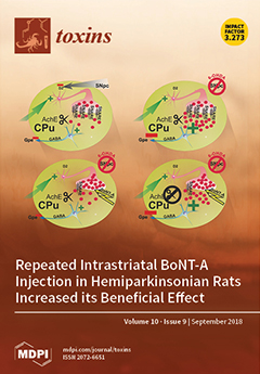
Using systemic donepezil application, we studied if the reduction of apomorphine-induced rotations caused by intrastriatal BoNT-A in hemiparkinsonian rats is dependent on acetylcholine (ACh). Schemes of the indirect pathway of the basal ganglia circuitry with main neurons and connections of the striatum are depicted under normal and experimental conditions. An exemplary presynapse of a cholinergic interneuron contains ACh (dots). Rectangles symbolize SNARE complexes which convey the fusion of ACh-containing vesicles with the presynaptic membrane. Scissors represent acetylcholinesterase (AChE) which cleaves ACh, and the medium spiny neuron (colored blue) inhibits the globus pallidus externus (Gpe) by GABA. View this paper.
  • Issues are regarded as officially published after their release is announced to the table of contents alert mailing list.
  • You may sign up for e-mail alerts to receive table of contents of newly released issues.
  • PDF is the official format for papers published in both, html and pdf forms. To view the papers in pdf format, click on the "PDF Full-text" link, and use the free Adobe Reader to open them.
Order results
Result details
Section
Select all
Export citation of selected articles as:
9 pages, 540 KiB  
Article
Soluble Toll-Like Receptor 4 Impairs the Interaction of Shiga Toxin 2a with Human Serum Amyloid P Component
by Maurizio Brigotti, Valentina Arfilli, Domenica Carnicelli, Francesca Ricci, Pier Luigi Tazzari, Gianluigi Ardissino, Gaia Scavia, Stefano Morabito and Xiaohua He
Toxins 2018, 10(9), 379; https://doi.org/10.3390/toxins10090379 - 18 Sep 2018
Cited by 9 | Viewed by 3053
Abstract
Shiga toxin 2a (Stx2a) is the main virulence factor produced by pathogenic Escherichia coli strains (Stx-producing E. coli, STEC) responsible for hemorrhagic colitis and the life-threatening sequela hemolytic uremic syndrome in children. The toxin released in the intestine by STEC targets the [...] Read more.
Shiga toxin 2a (Stx2a) is the main virulence factor produced by pathogenic Escherichia coli strains (Stx-producing E. coli, STEC) responsible for hemorrhagic colitis and the life-threatening sequela hemolytic uremic syndrome in children. The toxin released in the intestine by STEC targets the globotriaosylceramide receptor (Gb3Cer) present on the endothelial cells of the brain and the kidney after a transient blood phase during which Stx2a interacts with blood components, such as neutrophils, which, conversely, recognize Stx through Toll-like receptor 4 (TLR4). Among non-cellular blood constituents, human amyloid P component (HuSAP) is considered a negative modulating factor that specifically binds Stx2a and impairs its toxic action. Here, we show that the soluble extracellular domain of TLR4 inhibits the binding of Stx2a to neutrophils, assessed by indirect flow cytometric analysis. Moreover, by using human sensitive Gb3Cer-expressing cells (Raji cells) we found that the complex Stx2a/soluble TLR4 escaped from capture by HuSAP allowing the toxin to target and damage human cells, as assayed by measuring translation inhibition, the typical Stx-induced functional impairment. Thus, soluble TLR4 stood out as a positive modulating factor for Stx2a. In the paper, these findings have been discussed in the context of the pathogenesis of hemolytic uremic syndrome. Full article
(This article belongs to the Section Bacterial Toxins)
Show Figures

Figure 1

15 pages, 2001 KiB  
Article
The Streptococcus pneumoniae yefM-yoeB and relBE Toxin-Antitoxin Operons Participate in Oxidative Stress and Biofilm Formation
by Wai Ting Chan, Mirian Domenech, Inmaculada Moreno-Córdoba, Verónica Navarro-Martínez, Concha Nieto, Miriam Moscoso, Ernesto García and Manuel Espinosa
Toxins 2018, 10(9), 378; https://doi.org/10.3390/toxins10090378 - 18 Sep 2018
Cited by 31 | Viewed by 5280
Abstract
Type II (proteic) toxin-antitoxin systems (TAs) are widely distributed among bacteria and archaea. They are generally organized as operons integrated by two genes, the first encoding the antitoxin that binds to its cognate toxin to generate a harmless protein–protein complex. Under stress conditions, [...] Read more.
Type II (proteic) toxin-antitoxin systems (TAs) are widely distributed among bacteria and archaea. They are generally organized as operons integrated by two genes, the first encoding the antitoxin that binds to its cognate toxin to generate a harmless protein–protein complex. Under stress conditions, the unstable antitoxin is degraded by host proteases, releasing the toxin to achieve its toxic effect. In the Gram-positive pathogen Streptococcus pneumoniae we have characterized four TAs: pezAT, relBE, yefM-yoeB, and phD-doc, although the latter is missing in strain R6. We have assessed the role of the two yefM-yoeB and relBE systems encoded by S. pneumoniae R6 by construction of isogenic strains lacking one or two of the operons, and by complementation assays. We have analyzed the phenotypes of the wild type and mutants in terms of cell growth, response to environmental stress, and ability to generate biofilms. Compared to the wild-type, the mutants exhibited lower resistance to oxidative stress. Further, strains deleted in yefM-yoeB and the double mutant lacking yefM-yoeB and relBE exhibited a significant reduction in their ability for biofilm formation. Complementation assays showed that defective phenotypes were restored to wild type levels. We conclude that these two loci may play a relevant role in these aspects of the S. pneumoniae lifestyle and contribute to the bacterial colonization of new niches. Full article
(This article belongs to the Special Issue Toxin-antitoxin (TA) systems)
Show Figures

Figure 1

16 pages, 3018 KiB  
Article
A Critical Role for HlgA in Staphylococcus aureus Pathogenesis Revealed by A Switch in the SaeRS Two-Component Regulatory System
by Arundhathi Venkatasubramaniam, Tulasikumari Kanipakala, Nader Ganjbaksh, Rana Mehr, Ipsita Mukherjee, Subramaniam Krishnan, Taeok Bae, M. Javad Aman and Rajan P. Adhikari
Toxins 2018, 10(9), 377; https://doi.org/10.3390/toxins10090377 - 18 Sep 2018
Cited by 18 | Viewed by 4768
Abstract
Cytolytic pore-forming toxins including alpha hemolysin (Hla) and bicomponent leukotoxins play an important role in the pathogenesis of Staphylococcus aureus. These toxins kill the polymorphonuclear phagocytes (PMNs), disrupt epithelial and endothelial barriers, and lyse erythrocytes to provide iron for bacterial growth. The [...] Read more.
Cytolytic pore-forming toxins including alpha hemolysin (Hla) and bicomponent leukotoxins play an important role in the pathogenesis of Staphylococcus aureus. These toxins kill the polymorphonuclear phagocytes (PMNs), disrupt epithelial and endothelial barriers, and lyse erythrocytes to provide iron for bacterial growth. The expression of these toxins is regulated by the two-component sensing systems Sae and Agr. Here, we report that a point mutation (L18P) in SaeS, the histidine kinase sensor of the Sae system, renders the S. aureus Newman hemolytic activity fully independent of Hla and drastically increases the PMN lytic activity. Furthermore, this Hla-independent activity, unlike Hla itself, can lyse human erythrocytes. The Hla-independent activity towards human erythrocytes was also evident in USA300, however, under strict agr control. Gene knockout studies revealed that this Hla-independent Sae-regulated activity was entirely dependent on gamma hemolysin A subunit (HlgA). In contrast, hemolytic activity of Newman towards human erythrocytes from HlgAB resistant donors was completely dependent on agr. The culture supernatant from Newman S. aureus could be neutralized by antisera against two vaccine candidates based on LukS and LukF subunits of Panton-Valentine leukocidin but not by an anti-Hla neutralizing antibody. These findings display the complex involvement of Sae and Agr systems in regulating the virulence of S. aureus and have important implications for vaccine and immunotherapeutics development for S. aureus disease in humans. Full article
(This article belongs to the Special Issue Leukotoxins)
Show Figures

Figure 1

11 pages, 2207 KiB  
Article
Dietary Deoxynivalenol (DON) May Impair the Epithelial Barrier and Modulate the Cytokine Signaling in the Intestine of Atlantic Salmon (Salmo salar)
by Torfinn Moldal, Aksel Bernhoft, Grethe Rosenlund, Magne Kaldhusdal and Erling Olaf Koppang
Toxins 2018, 10(9), 376; https://doi.org/10.3390/toxins10090376 - 14 Sep 2018
Cited by 15 | Viewed by 4988
Abstract
Impaired growth, immunity, and intestinal barrier in mammals, poultry, and carp have been attributed to the mycotoxin deoxynivalenol (DON). The increased use of plant ingredients in aquaculture feed implies a risk for contamination with mycotoxins. The effects of dietary DON were explored in [...] Read more.
Impaired growth, immunity, and intestinal barrier in mammals, poultry, and carp have been attributed to the mycotoxin deoxynivalenol (DON). The increased use of plant ingredients in aquaculture feed implies a risk for contamination with mycotoxins. The effects of dietary DON were explored in 12-month-old Atlantic salmon (Salmo salar) (start weight of 58 g) that were offered a standard feed with non-detectable levels of mycotoxins (control group) or 5.5 mg DON/kg feed (DON group). Each group comprised two tanks with 25 fish per tank. Five fish from each tank were sampled eight weeks after the start of the feeding trial, when mean weights for the control and DON groups were 123.2 g and 80.2 g, respectively. The relative expression of markers for three tight junction proteins (claudin 25b, occludin, and tricellulin) were lower, whereas the relative expression of a marker for proliferating cell nuclear antigen was higher in both the mid-intestine and the distal intestine in fish fed DON compared with fish from the control group. The relative expression of markers for two suppressors of cytokine signaling (SOCS1 and SOCS2) were higher in the distal intestine in fish fed DON. There was no indication of inflammation attributed to the feed in any intestinal segments. Our findings suggest that dietary DON impaired the intestinal integrity, while an inflammatory response appeared to be mitigated by suppressors of cytokine signaling. A dysfunctional intestinal barrier may have contributed to the impaired production performance observed in the DON group. Full article
(This article belongs to the Special Issue Effects of Mycotoxins on the Intestine)
Show Figures

Figure 1

22 pages, 1981 KiB  
Article
Extended Targeted and Non-Targeted Strategies for the Analysis of Marine Toxins in Mussels and Oysters by (LC-HRMS)
by Inès Dom, Ronel Biré, Vincent Hort, Gwenaëlle Lavison-Bompard, Marina Nicolas and Thierry Guérin
Toxins 2018, 10(9), 375; https://doi.org/10.3390/toxins10090375 - 14 Sep 2018
Cited by 28 | Viewed by 5934
Abstract
When considering the geographical expansion of marine toxins, the emergence of new toxins and the associated risk for human health, there is urgent need for versatile and efficient analytical methods that are able to detect a range, as wide as possible, of known [...] Read more.
When considering the geographical expansion of marine toxins, the emergence of new toxins and the associated risk for human health, there is urgent need for versatile and efficient analytical methods that are able to detect a range, as wide as possible, of known or emerging toxins. Current detection methods for marine toxins rely on a priori defined target lists of toxins and are generally inappropriate for the detection and identification of emerging compounds. The authors describe the implementation of a recent approach for the non-targeted analysis of marine toxins in shellfish with a focus on a comprehensive workflow for the acquisition and treatment of the data generated after liquid chromatography coupled with high resolution mass spectrometry (LC-HRMS) analysis. First, the study was carried out in targeted mode to assess the performance of the method for known toxins with an extended range of polarities, including lipophilic toxins (okadaic acid, dinophysistoxins, azaspiracids, pectenotoxins, yessotoxins, cyclic imines, brevetoxins) and domoic acid. The targeted method, assessed for 14 toxins, shows good performance both in mussel and oyster extracts. The non-target potential of the method was then challenged via suspects and without a priori screening by blind analyzing mussel and oyster samples spiked with marine toxins. The data processing was optimized and successfully identified the toxins that were spiked in the blind samples. Full article
(This article belongs to the Special Issue Emerging Marine Biotoxins)
Show Figures

Graphical abstract

14 pages, 1064 KiB  
Review
Variability of Botulinum Toxins: Challenges and Opportunities for the Future
by Christine Rasetti-Escargueil, Emmanuel Lemichez and Michel R. Popoff
Toxins 2018, 10(9), 374; https://doi.org/10.3390/toxins10090374 - 13 Sep 2018
Cited by 13 | Viewed by 7160
Abstract
Botulinum neurotoxins (BoNTs) are the most potent known toxins, and are therefore classified as extremely harmful biological weapons. However, BoNTs are therapeutic drugs that are widely used and have an increasing number of applications. BoNTs show a high diversity and are divided into [...] Read more.
Botulinum neurotoxins (BoNTs) are the most potent known toxins, and are therefore classified as extremely harmful biological weapons. However, BoNTs are therapeutic drugs that are widely used and have an increasing number of applications. BoNTs show a high diversity and are divided into multiple types and subtypes. Better understanding of the activity at the molecular and clinical levels of the natural BoNT variants as well as the development of BoNT-based chimeric molecules opens the door to novel medical applications such as silencing the sensory neurons at targeted areas and dermal restoration. This short review is focused on BoNTs’ variability and the opportunities or challenges posed for future clinical applications. Full article
(This article belongs to the Collection Botulinum Toxins on Human Pain)
Show Figures

Figure 1

19 pages, 1338 KiB  
Article
Proteomic Deep Mining the Venom of the Red-Headed Krait, Bungarus flaviceps
by Alex Chapeaurouge, Andreza Silva, Paulo Carvalho, Ryan J. R. McCleary, Cassandra Marie Modahl, Jonas Perales, R. Manjunatha Kini and Stephen P. Mackessy
Toxins 2018, 10(9), 373; https://doi.org/10.3390/toxins10090373 - 13 Sep 2018
Cited by 15 | Viewed by 5215
Abstract
The use of -omics technologies allows for the characterization of snake venom composition at a fast rate and at high levels of detail. In the present study, we investigated the protein content of Red-headed Krait (Bungarus flaviceps) venom. This analysis revealed [...] Read more.
The use of -omics technologies allows for the characterization of snake venom composition at a fast rate and at high levels of detail. In the present study, we investigated the protein content of Red-headed Krait (Bungarus flaviceps) venom. This analysis revealed a high diversity of snake venom protein families, as evidenced by high-throughput mass spectrometric analysis. We found all six venom protein families previously reported in a transcriptome study of the venom gland of B. flaviceps, including phospholipases A2 (PLA2s), Kunitz-type serine proteinase inhibitors (KSPIs), three-finger toxins (3FTxs), cysteine-rich secretory proteins (CRISPs), snaclecs, and natriuretic peptides. A combined approach of automated database searches and de novo sequencing of tandem mass spectra, followed by sequence similarity searches, revealed the presence of 12 additional toxin families. De novo sequencing alone was able to identify 58 additional peptides, and this approach contributed significantly to the comprehensive description of the venom. Abundant protein families comprise 3FTxs (22.3%), KSPIs (19%), acetylcholinesterases (12.6%), PLA2s (11.9%), venom endothelial growth factors (VEGFs, 8.4%), nucleotidases (4.3%), and C-type lectin-like proteins (snaclecs, 3.3%); an additional 11 toxin families are present at significantly lower concentrations, including complement depleting factors, a family not previously detected in Bungarus venoms. The utility of a multifaceted approach toward unraveling the proteome of snake venoms, employed here, allowed detection of even minor venom components. This more in-depth knowledge of the composition of B. flaviceps venom facilitates a better understanding of snake venom molecular evolution, in turn contributing to more effective treatment of krait bites. Full article
(This article belongs to the Section Animal Venoms)
Show Figures

Graphical abstract

25 pages, 2117 KiB  
Article
A New Concept to Secure Food Safety Standards against Fusarium Species and Aspergillus Flavus and Their Toxins in Maize
by Balazs Szabo, Beata Toth, Eva Toth Toldine, Monika Varga, Nandor Kovacs, Janos Varga, Sandor Kocsube, Andrea Palagyi, Ferenc Bagi, Dragana Budakov, Vera Stojšin, Sanja Lazić, Marija Bodroža-Solarov, Radmilo Čolović, Goran Bekavac, Božana Purar, Djordje Jocković and Akos Mesterházy
Toxins 2018, 10(9), 372; https://doi.org/10.3390/toxins10090372 - 13 Sep 2018
Cited by 25 | Viewed by 4161
Abstract
Commercial maize hybrids are exposed to different degrees of ear infection by toxigenic fungal species and toxin contamination. Their resistance to different fungi and toxin relationships are largely unknown. Without this knowledge, screening and breeding are not possible for these pathogens. Seven- to [...] Read more.
Commercial maize hybrids are exposed to different degrees of ear infection by toxigenic fungal species and toxin contamination. Their resistance to different fungi and toxin relationships are largely unknown. Without this knowledge, screening and breeding are not possible for these pathogens. Seven- to tenfold differences were found in resistance to Fusarium spp., and there was a five-fold difference in ear coverage (%) in response to A. flavus. Three hybrids of the twenty entries had lower infection severity compared with the general means for toxigenic species. Three were highly susceptible to each, and 14 hybrids reacted differently to the different fungi. Differences were also observed in the toxin content. Again, three hybrids had lower toxin content in response to all toxigenic species, one had higher values for all, and 16 had variable resistance levels. Correlations between infection severity and deoxynivalenol (DON) content were 0.95 and 0.82 (p = 0.001) for F. graminearum and F. culmorum, respectively. For fumonisin and F. verticillioides ear rot, the Pearson correlation coefficient (r) was 0.45 (p = 0.05). Two independent isolates with different aggressiveness were used, and their mean X values better described the resistance levels. This increased the reliability of the data. With the introduction of this methodological concept (testing the resistance levels separately for different fungi and with two isolates independently), highly significant resistance differences were found. The resistance to different fungal species correlated only in certain cases; thus, each should be tested separately. This is very useful in registration tests and post-registration screening and breeding. This would allow a rapid increase in food and feed safety. Full article
(This article belongs to the Section Mycotoxins)
Show Figures

Figure 1

13 pages, 1472 KiB  
Article
Peptide Mimics of the Ribosomal P Stalk Inhibit the Activity of Ricin A Chain by Preventing Ribosome Binding
by Xiao-Ping Li, Jennifer N. Kahn and Nilgun E. Tumer
Toxins 2018, 10(9), 371; https://doi.org/10.3390/toxins10090371 - 13 Sep 2018
Cited by 11 | Viewed by 3338
Abstract
Ricin A chain (RTA) depurinates the sarcin/ricin loop (SRL) by interacting with the C-termini of the ribosomal P stalk. The ribosome interaction site and the active site are located on opposite faces of RTA. The interaction with P proteins allows RTA to depurinate [...] Read more.
Ricin A chain (RTA) depurinates the sarcin/ricin loop (SRL) by interacting with the C-termini of the ribosomal P stalk. The ribosome interaction site and the active site are located on opposite faces of RTA. The interaction with P proteins allows RTA to depurinate the SRL on the ribosome at physiological pH with an extremely high activity by orienting the active site towards the SRL. Therefore, if an inhibitor disrupts RTA–ribosome interaction by binding to the ribosome binding site of RTA, it should inhibit the depurination activity. To test this model, we synthesized peptides mimicking the last 3 to 11 amino acids of P proteins and examined their interaction with wild-type RTA and ribosome binding mutants by Biacore. We measured the inhibitory activity of these peptides on RTA-mediated depurination of yeast and rat liver ribosomes. We found that the peptides interacted with the ribosome binding site of RTA and inhibited depurination activity by disrupting RTA–ribosome interactions. The shortest peptide that could interact with RTA and inhibit its activity was four amino acids in length. RTA activity was inhibited by disrupting its interaction with the P stalk without targeting the active site, establishing the ribosome binding site as a new target for inhibitor discovery. Full article
(This article belongs to the Special Issue Ricin Toxins)
Show Figures

Figure 1

13 pages, 5559 KiB  
Article
Oxidative Damage and Nrf2 Translocation Induced by Toxicities of Deoxynivalenol on the Placental and Embryo on Gestation Day 12.5 d and 18.5 d
by Miao Yu, Zhi-Yuan Wei, Zhou-Heng Xu, Jia-Qi Pan and Jian-Huan Chen
Toxins 2018, 10(9), 370; https://doi.org/10.3390/toxins10090370 - 13 Sep 2018
Cited by 12 | Viewed by 2998
Abstract
Deoxynivalenol (DON) is a kind of natural pollutant belonging to the trichothecenes family. The aim of this study is to use diverse assays to evaluate oxidative damage as well as translocation of nuclear factor erythroid 2-related factor 2 (Nrf2), and to investigate their [...] Read more.
Deoxynivalenol (DON) is a kind of natural pollutant belonging to the trichothecenes family. The aim of this study is to use diverse assays to evaluate oxidative damage as well as translocation of nuclear factor erythroid 2-related factor 2 (Nrf2), and to investigate their mechanisms in DON-induced toxicities on a placenta and embryo. Pregnant C57BL/6 mice were randomly assigned to three groups with different doses of DON: 0, 1.0, 2.5 mg/(kg·day). In gestation day (GD) 12.5 d and 18.5 d, DON induced an elevated resorption rate of the embryos as well as structural and functional damage of the placenta. In the placenta, altered levels of the antioxidant enzymes malondialdehyde, superoxide dismutase and glutathione indicated remarkable oxidative stress. Furthermore, an elevated level of heme oxygenase-1 (HO-1) and the translocation of Nrf2 from nucleus to cytoplasm indicated Nrf2/HO-1 pathway activation in DON-L group (1.0 mg/(kg·day)). It is noteworthy that the results in this experiment in GD 12.5 d were similar to those in GD 18.5 d. In conclusion, DON-induced placental oxidative damage and Nrf2 translocation were similar in GD 12.5 d and GD 18.5 d. Oxidative stress is one of the most important molecular mechanisms for embryotoxicity induced by DON, and Nrf2 translocation may play a substantial role against it. Full article
(This article belongs to the Section Mycotoxins)
Show Figures

Figure 1

15 pages, 654 KiB  
Article
Malting of Fusarium Head Blight-Infected Rye (Secale cereale): Growth of Fusarium graminearum, Trichothecene Production, and the Impact on Malt Quality
by Zhao Jin, James Gillespie, John Barr, Jochum J. Wiersma, Mark E. Sorrells, Steve Zwinger, Thomas Gross, Jaime Cumming, Gary C. Bergstrom, Robert Brueggeman, Richard D. Horsley and Paul B. Schwarz
Toxins 2018, 10(9), 369; https://doi.org/10.3390/toxins10090369 - 11 Sep 2018
Cited by 14 | Viewed by 3532
Abstract
This project was initiated with the goal of investigating the malt quality of winter rye cultivars and hybrids grown in the United States in 2014 and 2015, but high levels of deoxynivalenol (DON) were subsequently found in many of the malt samples. DON [...] Read more.
This project was initiated with the goal of investigating the malt quality of winter rye cultivars and hybrids grown in the United States in 2014 and 2015, but high levels of deoxynivalenol (DON) were subsequently found in many of the malt samples. DON levels in 75% of the investigated rye samples (n = 117) were actually below 1.0 mg/kg, as quantified by a gas chromatography combined with electron capture detector (GC-ECD). However, 83% of the samples had DON in excess of 1.0 mg/kg following malting, and the average DON level in malted rye was 10.6 mg/kg. In addition, relatively high levels of 3-acetate DON (3-ADON), 15-acetate DON (15-ADON), nivalenol (NIV), and DON-3-glucoside (D3G) were observed in some rye malts. Our results show that rye grain DON is likely a poor predicator of type B trichothecenes in malt in practice, because high levels of malt DON, 15-ADONm and D3G were produced, even when the rye samples with DON levels below 0.50 mg/kg were processed. Fusarium Tri5 DNA content in rye was highly associated with malt DON levels (r = 0.83) in a small subset of samples (n = 55). The impact of Fusarium infection on malt quality was demonstrated by the significant correlations between malt DON levels and wort viscosity, β-glucan content, wort color, wort p-coumaric acid content, and total phenolic content. Additional correlations of rye Fusarium Tri5 DNA contents with malt diastatic power (DP), wort free amino nitrogen (FAN) content, and arabinoxylan content were observed. Full article
(This article belongs to the Collection Fusarium Toxins – Relevance for Human and Animal Health)
Show Figures

Figure 1

22 pages, 2918 KiB  
Article
Repeated Intrastriatal Botulinum Neurotoxin-A Injection in Hemiparkinsonian Rats Increased the Beneficial Effect on Rotational Behavior
by Alexander Hawlitschka, Carsten Holzmann, Andreas Wree and Veronica Antipova
Toxins 2018, 10(9), 368; https://doi.org/10.3390/toxins10090368 - 11 Sep 2018
Cited by 9 | Viewed by 4381
Abstract
Injection of botulinum neurotoxin-A (BoNT-A) into the striatum of hemiparkinsonian (hemi-PD) rats reduced apomorphine-induced rotation behavior significantly, for at least 3 months. Thereafter, rotation behavior increased again. We injected hemi-PD rats with 1 ng BoNT-A twice, the second injection following 6 months after [...] Read more.
Injection of botulinum neurotoxin-A (BoNT-A) into the striatum of hemiparkinsonian (hemi-PD) rats reduced apomorphine-induced rotation behavior significantly, for at least 3 months. Thereafter, rotation behavior increased again. We injected hemi-PD rats with 1 ng BoNT-A twice, the second injection following 6 months after the first one and tested the rats for apomorphine-induced rotations and spontaneous motor behaviors, i.e., corridor task and stepping test. To test the hypothesis that BoNT-A reduced striatal hypercholinism in hemi-PD rats, the acetylcholinesterase inhibitor donepezil was injected prior to separate apomorphine-induced rotation tests. In hemi-PD rats, the first BoNT-A injection led to a clear reduction of the apomorphine-induced rotations, and the second BoNT-A injection to a more massive and prolonged reaction. In hemi-PD rats whose apomorphine-induced rotation behavior was strongly reduced by an intrastriatal BoNT-A, subsequent donepezil injections led to significant increases of the rotation rate. Concerning corridor task and stepping test, neither first nor second BoNT-A injections changed hemi-PD rats’ behavior significantly. The data give evidence for the possibility of repeated intrastriatal administrations of BoNT-A, for treatment of motor symptoms in experimental hemi-PD over a longer time. Full article
(This article belongs to the Special Issue Botulinum Toxin Treatment of Movement Disorders)
Show Figures

Figure 1

22 pages, 722 KiB  
Review
Impacts of Indoxyl Sulfate and p-Cresol Sulfate on Chronic Kidney Disease and Mitigating Effects of AST-120
by Wen-Chih Liu, Yasuhiko Tomino and Kuo-Cheng Lu
Toxins 2018, 10(9), 367; https://doi.org/10.3390/toxins10090367 - 11 Sep 2018
Cited by 107 | Viewed by 10265
Abstract
Uremic toxins, such as indoxyl sulfate (IS) and p-cresol, or p-cresyl sulfate (PCS), are markedly accumulated in the organs of chronic kidney disease (CKD) patients. These toxins can induce inflammatory reactions and enhance oxidative stress, prompting glomerular sclerosis and interstitial fibrosis, to aggravate [...] Read more.
Uremic toxins, such as indoxyl sulfate (IS) and p-cresol, or p-cresyl sulfate (PCS), are markedly accumulated in the organs of chronic kidney disease (CKD) patients. These toxins can induce inflammatory reactions and enhance oxidative stress, prompting glomerular sclerosis and interstitial fibrosis, to aggravate the decline of renal function. Consequently, uremic toxins play an important role in the worsening of renal and cardiovascular functions. Furthermore, they destroy the quantity and quality of bone. Oral sorbent AST-120 reduces serum levels of uremic toxins in CKD patients by adsorbing the precursors of IS and PCS generated by amino acid metabolism in the intestine. Accordingly, AST-120 decreases the serum IS levels and reduces the production of reactive oxygen species by endothelial cells, to impede the subsequent oxidative stress. This slows the progression of cardiovascular and renal diseases and improves bone metabolism in CKD patients. Although large-scale studies showed no obvious benefits from adding AST-120 to the standard therapy for CKD patients, subsequent sporadic studies may support its use. This article summarizes the mechanisms of the uremic toxins, IS, and PCS, and discusses the multiple effects of AST-120 in CKD patients. Full article
(This article belongs to the Section Uremic Toxins)
Show Figures

Figure 1

1 pages, 187 KiB  
Correction
Correction: Rajput, S.A., et al. Ameliorative Effects of Grape Seed Proanthocyanidin Extract on Growth Performance, Immune Function, Antioxidant Capacity, Biochemical Constituents, Liver Histopathology and Aflatoxin Residues in Broilers Exposed to Aflatoxin B1. Toxins 2017, 9, 371
by Shahid Ali Rajput, Lvhui Sun, Niya Zhang, Mahmoud Mohamed Khalil, Xin Gao, Zhao Ling, Luoyi Zhu, Farhan Anwar Khan, Jiacai Zhang and Desheng Qi
Toxins 2018, 10(9), 366; https://doi.org/10.3390/toxins10090366 - 10 Sep 2018
Cited by 7 | Viewed by 2765
Abstract
The authors wish to make the following correction to their paper [...] Full article
(This article belongs to the Section Mycotoxins)
26 pages, 1345 KiB  
Review
Venomous Arachnid Diagnostic Assays, Lessons from Past Attempts
by Camila Dias-Lopes, Ana Luiza Paiva, Clara Guerra-Duarte, Franck Molina and Liza Felicori
Toxins 2018, 10(9), 365; https://doi.org/10.3390/toxins10090365 - 10 Sep 2018
Cited by 12 | Viewed by 4756
Abstract
Diagnostic tests for arachnid accidents remain unavailable for patients and clinicians. Together with snakes, these accidents are still a global medical concern, and are recognized as neglected tropical issues. Due to arachnid toxins’ fast mechanism of action, quick detection and quantification of venom [...] Read more.
Diagnostic tests for arachnid accidents remain unavailable for patients and clinicians. Together with snakes, these accidents are still a global medical concern, and are recognized as neglected tropical issues. Due to arachnid toxins’ fast mechanism of action, quick detection and quantification of venom is required to accelerate treatment decisions, rationalize therapy, and reduce costs and patient risks. This review aims to understand the current limitations for arachnid venom identification and quantification in biological samples. We benchmarked the already existing initiatives regarding test requirements (sample or biomarkers of choice), performances (time, detection limit, sensitivity and specificity) and their validation (on animal models or on samples from envenomed humans). Our analysis outlines unmet needs for improving diagnosis and consequently treatment of arachnid accidents. Hence, based on lessons from past attempts, we propose a road map for raising best practice guidelines, leading to recommendations for future progress in the development of arachnid diagnostic assays. Full article
(This article belongs to the Section Animal Venoms)
Show Figures

Figure 1

10 pages, 1329 KiB  
Review
Lipopolysaccharide Structural Differences between Western and Asian Helicobacter pylori Strains
by Hong Li, Hong Tang, Aleksandra W. Debowski, Keith A. Stubbs, Barry J. Marshall and Mohammed Benghezal
Toxins 2018, 10(9), 364; https://doi.org/10.3390/toxins10090364 - 08 Sep 2018
Cited by 10 | Viewed by 4767
Abstract
Recent structural analysis of the lipopolysaccharide (LPS) isolated from Helicobacter pylori G27 wild-type and O-antigen ligase mutant resulted in the redefinition of the core-oligosaccharide and O-antigen domains. The short core-oligosaccharide (Glc–Gal–Hep-III–Hep-II–Hep-I–KDO) and its attached trisaccharide (Trio, GlcNAc–Fuc–Hep) appear to be highly [...] Read more.
Recent structural analysis of the lipopolysaccharide (LPS) isolated from Helicobacter pylori G27 wild-type and O-antigen ligase mutant resulted in the redefinition of the core-oligosaccharide and O-antigen domains. The short core-oligosaccharide (Glc–Gal–Hep-III–Hep-II–Hep-I–KDO) and its attached trisaccharide (Trio, GlcNAc–Fuc–Hep) appear to be highly conserved structures among H. pylori strains. The G27 LPS contains a linear glucan–heptan linker between the core-Trio and distal Lewis antigens. This linker domain was commonly identified in Western strains. In contrast, out of 12 partial LPS structures of Asian strains, none displayed the heptan moiety, despite the presence of Lewis antigens. This raises the question of how Lewis antigens are attached to the Trio, and whether the LPS structure of Asian strains contain another linker. Of note, a riban was identified as a linker in LPS of the mouse-adapted SS1 strain, suggesting that alternative linker structures can occur. In summary, additional full structural analyses of LPS in Asian strains are required to assess the presence or absence of an alternative linker in these strains. It will also be interesting to study the glucan-heptan linker moieties in pathogenesis as H. pylori infections in Asia are usually more symptomatic than the ones presented in the Western world. Full article
(This article belongs to the Special Issue Lipopolysaccharide: Bacterial Endotoxin)
Show Figures

Figure 1

11 pages, 3468 KiB  
Article
Removal of Microcystin-LR by a Novel Native Effective Bacterial Community Designated as YFMCD4 Isolated from Lake Taihu
by Fei Yang, Jian Guo, Feiyu Huang, Isaac Yaw Massey, Ruixue Huang, Yunhui Li, Cong Wen, Ping Ding, Weiming Zeng and Geyu Liang
Toxins 2018, 10(9), 363; https://doi.org/10.3390/toxins10090363 - 08 Sep 2018
Cited by 31 | Viewed by 4102
Abstract
Microcystin-LR (MC-LR) is the most toxic and frequently detected monocyclic heptapeptide hepatotoxin produced by cyanobacteria, which poses a great threat to the natural ecosystem and public health. It is very important to seek environment-friendly and cost-efficient methods to remove MC-LR in water. In [...] Read more.
Microcystin-LR (MC-LR) is the most toxic and frequently detected monocyclic heptapeptide hepatotoxin produced by cyanobacteria, which poses a great threat to the natural ecosystem and public health. It is very important to seek environment-friendly and cost-efficient methods to remove MC-LR in water. In this study, the MC-degrading capacities of a novel indigenous bacterial community designated as YFMCD4 and the influence of environmental factors including various temperatures, MC concentrations and pH on the MC-degrading activities were investigated utilizing high-performance liquid chromatography (HPLC). In addition, the MC-degrading mechanism of YFMCD4 was also studied using HPLC coupled with a mass spectrometry equipped with electrospray ionization interface (HPLC-ESI-MS). The data showed MC-LR was completely removed at the maximum rate of 0.5 µg/(mL·h) under the optimal condition by YFMCD4. Two pure bacterial strains Alcaligenes faecalis and Stenotrophomonas acidaminiohila were isolated from YFMCD4 degraded MC-LR at a slower rate. The MC-degrading rates of YFMCD4 were significantly affected by different temperatures, pH and MC-LR concentrations. Two intermediates of a tetrapeptide and Adda appeared in the degradation process. These results illustrate that the novel YFMCD4 is one of the highest effective MC-degrading bacterial community, which can completely remove MC-LR and possesses a significant potential to treat water bodies contaminated by MC-LR. Full article
(This article belongs to the Collection Freshwater HABs and Health in a Changing World)
Show Figures

Figure 1

13 pages, 1327 KiB  
Article
Paralytic Shellfish Toxins Occurrence in Non-Traditional Invertebrate Vectors from North Atlantic Waters (Azores, Madeira, and Morocco)
by Marisa Silva, Verónica Rey, Aldo Barreiro, Manfred Kaufmann, Ana Isabel Neto, Meryem Hassouani, Brahim Sabour, Ana Botana, Luis M. Botana and Vitor Vasconcelos
Toxins 2018, 10(9), 362; https://doi.org/10.3390/toxins10090362 - 06 Sep 2018
Cited by 16 | Viewed by 5417
Abstract
Paralytic shellfish toxins (PSTs) are potent alkaloids of microalgal and cyanobacterial origin, with worldwide distribution. Over the last 20 years, the number of poisoning incidents has declined as a result of the implementation of legislation and monitoring programs based on bivalves. In the [...] Read more.
Paralytic shellfish toxins (PSTs) are potent alkaloids of microalgal and cyanobacterial origin, with worldwide distribution. Over the last 20 years, the number of poisoning incidents has declined as a result of the implementation of legislation and monitoring programs based on bivalves. In the summer of 2012 and 2013, we collected a total of 98 samples from 23 different species belonging to benthic and subtidal organisms, such as echinoderms, crustaceans, bivalves, and gastropods. The sampling locations were Madeira, São Miguel Island (Azores archipelago), and the northwestern coast of Morocco. The samples were analyzed using post-column oxidation liquid chromatography with a fluorescence detection method. Our main goal was to detect new vectors for these biotoxins. After reporting a total of 59 positive results for PSTs with 14 new vectors identified, we verified that some of the amounts exceeded the limit value established in the EU. These results suggest that routine monitoring of saxitoxin and its analogs should be extended to more potential vectors other than bivalves, including other edible organisms, for a better protection of public health. Full article
(This article belongs to the Special Issue Paralytic Shellfish Toxins)
Show Figures

Figure 1

13 pages, 1888 KiB  
Article
A Novel Adsorbent Magnetic Graphene Oxide Modified with Chitosan for the Simultaneous Reduction of Mycotoxins
by Atena Abbasi Pirouz, Roghayeh Abedi Karjiban, Fatimah Abu Bakar and Jinap Selamat
Toxins 2018, 10(9), 361; https://doi.org/10.3390/toxins10090361 - 06 Sep 2018
Cited by 24 | Viewed by 3632
Abstract
A novel magnetic graphene oxide modified with chitosan (MGO-CTS) was synthesised as an adsorbent aimed to examine the simultaneous removal of mycotoxins. The composite was characterised by various procedures, namely Fourier-transform infrared spectroscopy (FTIR), X-ray diffraction (XRD) and a scanning electron microscope (SEM). [...] Read more.
A novel magnetic graphene oxide modified with chitosan (MGO-CTS) was synthesised as an adsorbent aimed to examine the simultaneous removal of mycotoxins. The composite was characterised by various procedures, namely Fourier-transform infrared spectroscopy (FTIR), X-ray diffraction (XRD) and a scanning electron microscope (SEM). The adsorption evaluation was considered via pH effects, initial mycotoxin concentration, adsorption time and temperature. Adsorption isotherm data and kinetics experiments were acquired at the optimum pH 5 fit Freundlich isotherm as well as pseudo-second-order kinetic models. The thermodynamic results indicated that the adsorption of the mycotoxins was spontaneous, endothermic and favourable. Full article
Show Figures

Figure 1

10 pages, 1065 KiB  
Article
Cell-Based Reporter Release Assay to Determine the Potency of Proteolytic Bacterial Neurotoxins
by Andrea Pathe-Neuschäfer-Rube, Frank Neuschäfer-Rube, Gerald Haas, Nina Langoth-Fehringer and Gerhard Paul Püschel
Toxins 2018, 10(9), 360; https://doi.org/10.3390/toxins10090360 - 05 Sep 2018
Cited by 12 | Viewed by 3437
Abstract
Despite the implementation of cell-based replacement methods, the mouse lethality assay is still frequently used to determine the activity of botulinum toxin (BoNT) for medical use. One explanation is that due to the use of neoepitope-specific antibodies to detect the cleaved BoNT substrate, [...] Read more.
Despite the implementation of cell-based replacement methods, the mouse lethality assay is still frequently used to determine the activity of botulinum toxin (BoNT) for medical use. One explanation is that due to the use of neoepitope-specific antibodies to detect the cleaved BoNT substrate, the currently devised assays can detect only one specific serotype of the toxin. Recently, we developed a cell-based functional assay, in which BoNT activity is determined by inhibiting the release of a reporter enzyme that is liberated concomitantly with the neurotransmitter from neurosecretory vesicles. In theory, this assay should be suitable to detect the activity of any BoNT serotype. Consistent with this assumption, the current study shows that the stimulus-dependent release of a luciferase from a differentiated human neuroblastoma-based reporter cell line (SIMA-hPOMC1-26-GLuc cells) was inhibited by BoNT-A and-C. Furthermore, this was also inhibited by BoNT-B and tetanus toxin to a lesser extent and at higher concentrations. In order to provide support for the suitability of this technique in practical applications, a dose–response curve obtained with a pharmaceutical preparation of BoNT-A closely mirrored the activity determined in the mouse lethality assay. In summary, the newly established cell-based assay may represent a versatile and specific alternative to the mouse lethality assay and other currently established cell-based assays. Full article
(This article belongs to the Section Bacterial Toxins)
Show Figures

Figure 1

25 pages, 4127 KiB  
Article
Transcriptomic and Proteomic Analyses Reveal the Diversity of Venom Components from the Vaejovid Scorpion Serradigitus gertschi
by Maria Teresa Romero-Gutiérrez, Carlos Eduardo Santibáñez-López, Juana María Jiménez-Vargas, Cesar Vicente Ferreira Batista, Ernesto Ortiz and Lourival Domingos Possani
Toxins 2018, 10(9), 359; https://doi.org/10.3390/toxins10090359 - 05 Sep 2018
Cited by 27 | Viewed by 5404
Abstract
To understand the diversity of scorpion venom, RNA from venomous glands from a sawfinger scorpion, Serradigitus gertschi, of the family Vaejovidae, was extracted and used for transcriptomic analysis. A total of 84,835 transcripts were assembled after Illumina sequencing. From those, 119 transcripts [...] Read more.
To understand the diversity of scorpion venom, RNA from venomous glands from a sawfinger scorpion, Serradigitus gertschi, of the family Vaejovidae, was extracted and used for transcriptomic analysis. A total of 84,835 transcripts were assembled after Illumina sequencing. From those, 119 transcripts were annotated and found to putatively code for peptides or proteins that share sequence similarities with the previously reported venom components of other species. In accordance with sequence similarity, the transcripts were classified as potentially coding for 37 ion channel toxins; 17 host defense peptides; 28 enzymes, including phospholipases, hyaluronidases, metalloproteases, and serine proteases; nine protease inhibitor-like peptides; 10 peptides of the cysteine-rich secretory proteins, antigen 5, and pathogenesis-related 1 protein superfamily; seven La1-like peptides; and 11 sequences classified as “other venom components”. A mass fingerprint performed by mass spectrometry identified 204 components with molecular masses varying from 444.26 Da to 12,432.80 Da, plus several higher molecular weight proteins whose precise masses were not determined. The LC-MS/MS analysis of a tryptic digestion of the soluble venom resulted in the de novo determination of 16,840 peptide sequences, 24 of which matched sequences predicted from the translated transcriptome. The database presented here increases our general knowledge of the biodiversity of venom components from neglected non-buthid scorpions. Full article
(This article belongs to the Special Issue Scorpion Toxins)
Show Figures

Graphical abstract

9 pages, 1689 KiB  
Article
Engineering Gain-of-Function Analogues of the Spider Venom Peptide HNTX-I, A Potent Blocker of the hNaV1.7 Sodium Channel
by Yunxiao Zhang, Qiuchu Yang, Qingfeng Zhang, Dezheng Peng, Minzhi Chen, Songping Liang, Xi Zhou and Zhonghua Liu
Toxins 2018, 10(9), 358; https://doi.org/10.3390/toxins10090358 - 04 Sep 2018
Cited by 9 | Viewed by 3420
Abstract
Pain is a medical condition that interferes with normal human life and work and reduces human well-being worldwide. Human voltage-gated sodium channel NaV1.7 (hNaV1.7) is a compelling target that plays a key role in human pain signaling. The 33-residue peptide µ-TRTX-Hhn2b (HNTX-I), a [...] Read more.
Pain is a medical condition that interferes with normal human life and work and reduces human well-being worldwide. Human voltage-gated sodium channel NaV1.7 (hNaV1.7) is a compelling target that plays a key role in human pain signaling. The 33-residue peptide µ-TRTX-Hhn2b (HNTX-I), a member of NaV-targeting spider toxin (NaSpTx) family 1, has shown negligible activity on mammalian voltage-gated sodium channels (VGSCs), including the hNaV1.7 channel. We engineered analogues of HNTX-I based on sequence conservation in NaSpTx family 1. Substitution of Asn for Ser at position 23 or Asp for His at position 26 conferred potent activity against hNaV1.7. Moreover, multiple site mutations combined together afforded improvements in potency. Ultimately, we generated an analogue E1G–N23S–D26H–L32W with >300-fold improved potency compared with wild-type HNTX-I on hNaV1.7 (IC50 0.036 ± 0.007 µM). Structural simulation suggested that the charged surface and the hydrophobic surface of the modified peptide are responsible for binding affinity to the hNaV1.7 channel, while variable residues may determine pharmacological specificity. Therefore, this study provides a profile for drug design targeting the hNaV1.7 channel. Full article
Show Figures

Figure 1

15 pages, 771 KiB  
Article
Fumonisin Distribution in Maize Dry-Milling Products and By-Products: Impact of Two Industrial Degermination Systems
by Francesca Vanara, Valentina Scarpino and Massimo Blandino
Toxins 2018, 10(9), 357; https://doi.org/10.3390/toxins10090357 - 04 Sep 2018
Cited by 25 | Viewed by 8311
Abstract
In temperate areas, the main limitation to the use of maize in the food chain is its contamination by B-series fumonisins (FBs) during cultivation. Since the content of this group of mycotoxins may be distributed unevenly after milling, the aim of this study [...] Read more.
In temperate areas, the main limitation to the use of maize in the food chain is its contamination by B-series fumonisins (FBs) during cultivation. Since the content of this group of mycotoxins may be distributed unevenly after milling, the aim of this study was to compare the distribution of FBs in maize fractions derived from two industrial dry-milling processes, that is, a dry-degermination (DD) system and a tempering-degermination (TD) system. Grain cleaning reduces FBs by about 42%. The germ of the two degermination processes showed a similar FB content of kernel after cleaning. Conversely, an animal feed flour resulted in a FB content that was two times higher than whole grain before cleaning. A significant FB reduction was observed in the milling fractions in both processes, with a higher reduction in the TD system than in the DD one. The average decontamination respect to uncleaned kernels in the DD process was 50%, 83% and 87%, for maize flour, break meal and pearl meal, respectively, while it was 78%, 88% and 94% in the TD process for small, medium and flaking grits, respectively. Among the milling fractions, the flaking grits with the highest particle size resulted in the highest FB reduction. Full article
(This article belongs to the Collection Understanding Mycotoxin Occurrence in Food and Feed Chains)
Show Figures

Graphical abstract

12 pages, 1604 KiB  
Article
Release of Indospicine from Contaminated Camel Meat following Cooking and Simulated Gastrointestinal Digestion: Implications for Human Consumption
by Saira Sultan, Cindy Giles, Gabriele Netzel, Simone A. Osborne, Michael E. Netzel and Mary T. Fletcher
Toxins 2018, 10(9), 356; https://doi.org/10.3390/toxins10090356 - 03 Sep 2018
Cited by 5 | Viewed by 4306
Abstract
Indospicine, a hepatotoxic arginine analog, occurs in leguminous plants of the Indigofera genus and accumulates in the tissues of grazing animals that consume these plants. Furthermore, indospicine has caused toxicity in dogs following consumption of indospicine-contaminated meat; however, the potential impact on human [...] Read more.
Indospicine, a hepatotoxic arginine analog, occurs in leguminous plants of the Indigofera genus and accumulates in the tissues of grazing animals that consume these plants. Furthermore, indospicine has caused toxicity in dogs following consumption of indospicine-contaminated meat; however, the potential impact on human health is unknown. The present study was designed to determine the effect of simulated human gastrointestinal digestion on the release and degradation of indospicine from contaminated camel meat following microwave cooking. Results showed no significant (p > 0.05) indospicine degradation during cooking or in vitro digestion. However, approximately 70% indospicine was released from the meat matrix into the liquid digesta during the gastric phase (in the presence of pepsin) and increased to >90% in the intestinal phase (with pancreatic enzymes). Following human consumption of contaminated meat, this soluble and more bioaccessible fraction of intact indospicine could be readily available for absorption by the small intestine, potentially circulating indospicine throughout the human body to tissues where it could accumulate and cause detrimental toxic effects. Full article
(This article belongs to the Special Issue Food Safety and Natural Toxins)
Show Figures

Graphical abstract

7 pages, 578 KiB  
Article
Safety and Efficacy of PrabotulinumtoxinA (Nabota®) Injection for Cervical and Shoulder Girdle Myofascial Pain Syndrome: A Pilot Study
by Da-ye Kim and Jae Min Kim
Toxins 2018, 10(9), 355; https://doi.org/10.3390/toxins10090355 - 03 Sep 2018
Cited by 11 | Viewed by 5200
Abstract
Myofascial pain syndrome is a common painful condition encountered in the general population. Previous studies evaluating the efficacy of botulinum toxin for the treatment of myofascial pain syndrome are limited, with variable results. This prospective study investigated the efficacy and safety of direct [...] Read more.
Myofascial pain syndrome is a common painful condition encountered in the general population. Previous studies evaluating the efficacy of botulinum toxin for the treatment of myofascial pain syndrome are limited, with variable results. This prospective study investigated the efficacy and safety of direct injection of Prabotulinumtoxin A (Nabota®) into painful muscle groups for cervical and shoulder girdle myofascial pain. Twelve patients with chronic myofascial pain syndrome of the neck and shoulder underwent an injection of Prabotulinumtoxin A. Painful muscles containing trigger points were injected in the mid-belly. Pain scores and quality of life measurements were assessed at baseline, as well as 6 weeks and 12 weeks post-injection. Safety and tolerability were also assessed. This trial is registered under clinical research information service (CRIS) number KCT0001634. Patients injected with Prabotulinumtoxin A showed a significant improvement in pain at 12 weeks (p < 0.001). At 6 weeks, the pain had not significantly improved compared with baseline (p = 0.063). However, at that time, 41.7% of patients were characterized as Prabotulinumtoxin A responders, with a 30% reduction in pain rating score compared to baseline. In the Neck Disability Index scores, the patients demonstrated significant improvement at both 6 weeks and 12 weeks. No serious adverse effects occurred during the study. Prabotulinumtoxin A injection into chronically painful muscles associated with cervical and shoulder girdle myofascial pain syndrome resulted in an improvement in pain scores and quality of life lasting at least 12 weeks. Additionally, the injections were well tolerated. As these are preliminary findings in a pilot study, future studies should carefully consider using randomized, controlled, prospective trials. Full article
(This article belongs to the Collection Botulinum Toxins on Human Pain)
Show Figures

Figure 1

14 pages, 3442 KiB  
Article
Development of a Sensitive Enzyme-Linked Immunosorbent Assay and Rapid Gold Nanoparticle Immunochromatographic Strip for Detecting Citrinin in Monascus Fermented Food
by Shih-Wei Wu, Yao-An Yu, Biing-Hui Liu and Feng-Yih Yu
Toxins 2018, 10(9), 354; https://doi.org/10.3390/toxins10090354 - 02 Sep 2018
Cited by 16 | Viewed by 4582
Abstract
Antibodies against citrinin (CTN) were generated from rabbits, which were injected with CTN-keyhole limpet hemocyanin (KLH). This work involved the development of a sensitive competitive direct enzyme-linked immunosorbent assay (cdELISA) and a rapid gold nanoparticle immunochromatographic strip (immunostrip) method for analyzing CTN in [...] Read more.
Antibodies against citrinin (CTN) were generated from rabbits, which were injected with CTN-keyhole limpet hemocyanin (KLH). This work involved the development of a sensitive competitive direct enzyme-linked immunosorbent assay (cdELISA) and a rapid gold nanoparticle immunochromatographic strip (immunostrip) method for analyzing CTN in Monascus-fermented food. CTN at a concentration of 5.0 ng/mL caused 50% inhibition (IC50) of CTN-horseradish peroxidase (CTN-HRP) binding to the antibodies in the cdELISA. The capable on-site detection of CTN was accomplished by a rapid antibody-gold nanoparticle immunostrip with a detection limit of 20 ng/mL and that was completed within 15 min. A close inspection of 19 Monascus-fermented foods by cdELISA confirmed that 14 were contaminated with citrinin at levels from 28.6–9454 ng/g. Further analysis with the immunostrip is consistent with those results obtained using cdELISA. Both means are sensitive enough for the rapid examination of CTN in Monascus-fermented food products. Full article
(This article belongs to the Special Issue Advanced Methods for Mycotoxins Detection)
Show Figures

Graphical abstract

16 pages, 5030 KiB  
Article
Interaction of 2′R-ochratoxin A with Serum Albumins: Binding Site, Effects of Site Markers, Thermodynamics, Species Differences of Albumin-binding, and Influence of Albumin on Its Toxicity in MDCK Cells
by Zelma Faisal, Diána Derdák, Beáta Lemli, Sándor Kunsági-Máté, Mónika Bálint, Csaba Hetényi, Rita Csepregi, Tamás Kőszegi, Franziska Sueck, Benedikt Cramer, Hans-Ulrich Humpf and Miklós Poór
Toxins 2018, 10(9), 353; https://doi.org/10.3390/toxins10090353 - 01 Sep 2018
Cited by 10 | Viewed by 3977
Abstract
Ochratoxin A (OTA) is a nephrotoxic mycotoxin. Roasting of OTA-contaminated coffee results in the formation of 2′R-ochratoxin A (2′R-OTA), which appears in the blood of coffee drinkers. Human serum albumin (HSA) binds 2′R-OTA (and OTA) with high affinity; therefore, albumin may influence the [...] Read more.
Ochratoxin A (OTA) is a nephrotoxic mycotoxin. Roasting of OTA-contaminated coffee results in the formation of 2′R-ochratoxin A (2′R-OTA), which appears in the blood of coffee drinkers. Human serum albumin (HSA) binds 2′R-OTA (and OTA) with high affinity; therefore, albumin may influence the tissue uptake and elimination of ochratoxins. We aimed to investigate the binding site of 2′R-OTA (verses OTA) in HSA and the displacing effects of site markers to explore which molecules can interfere with its albumin-binding. Affinity of 2′R-OTA toward albumins from various species (human, bovine, porcine and rat) was tested to evaluate the interspecies differences regarding 2′R-OTA-albumin interaction. Thermodynamic studies were performed to give a deeper insight into the molecular background of the complex formation. Besides fluorescence spectroscopic and modeling studies, effects of HSA, and fetal bovine serum on the cytotoxicity of 2′R-OTA and OTA were tested in MDCK kidney cell line in order to demonstrate the influence of albumin-binding on the cellular uptake of ochratoxins. Site markers displaced more effectively 2′R-OTA than OTA from HSA. Fluorescence and binding constants of 2′R-OTA-albumin and OTA-albumin complexes showed different tendencies. Albumin significantly decreased the cytotoxicity of ochratoxins. 2′R-OTA, even at sub-toxic concentrations, increased the toxic action of OTA. Full article
(This article belongs to the Collection Ochratoxins-Collection)
Show Figures

Figure 1

29 pages, 352 KiB  
Review
Cardiotoxicity of Uremic Toxins: A Driver of Cardiorenal Syndrome
by Suree Lekawanvijit
Toxins 2018, 10(9), 352; https://doi.org/10.3390/toxins10090352 - 01 Sep 2018
Cited by 79 | Viewed by 6123
Abstract
Cardiovascular disease (CVD) is highly prevalent in the setting of chronic kidney disease (CKD). Such coexistence of CVD and CKD—the so-called “cardiorenal or renocardiac syndrome”—contributes to exponentially increased risk of cardiovascular (CV) mortality. Uremic cardiomyopathy is a characteristic cardiac pathology commonly found in [...] Read more.
Cardiovascular disease (CVD) is highly prevalent in the setting of chronic kidney disease (CKD). Such coexistence of CVD and CKD—the so-called “cardiorenal or renocardiac syndrome”—contributes to exponentially increased risk of cardiovascular (CV) mortality. Uremic cardiomyopathy is a characteristic cardiac pathology commonly found in CKD. CKD patients are also predisposed to heart rhythm disorders especially atrial fibrillation. Traditional CV risk factors as well as known CKD-associated CV risk factors such as anemia are insufficient to explain CV complications in the CKD population. Accumulation of uremic retention solutes is a hallmark of impaired renal excretory function. Many of them have been considered inert solutes until their biological toxicity is unraveled and they become accepted as “uremic toxins”. Direct cardiotoxicity of uremic toxins has been increasingly demonstrated in recent years. This review offers a mechanistic insight into the pathological cardiac remodeling and dysfunction contributed by uremic toxins with a main focus on fibroblastic growth factor-23, an emerging toxin playing a central role in the chronic kidney disease–mineral bone disorder, and the two most investigated non-dialyzable protein-bound uremic toxins, indoxyl sulfate and p-cresyl sulfate. Potential therapeutic strategies that could address these toxins and their relevant mediated pathways since pre-dialysis stages are also discussed. Full article
(This article belongs to the Special Issue Uremia and Cardiovascular Disease)
13 pages, 2057 KiB  
Article
Enterohemorrhagic E. coli (EHEC)—Secreted Serine Protease EspP Stimulates Electrogenic Ion Transport in Human Colonoid Monolayers
by C. Ming Tse, Julie G. In, Jianyi Yin, Mark Donowitz, Michele Doucet, Jennifer Foulke-Abel, Fernando Ruiz-Perez, James P. Nataro, Nicholas C. Zachos, James B. Kaper and Olga Kovbasnjuk
Toxins 2018, 10(9), 351; https://doi.org/10.3390/toxins10090351 - 01 Sep 2018
Cited by 16 | Viewed by 4397
Abstract
One of the characteristic manifestations of Shiga-toxin-producing Escherichia coli (E. coli) infection in humans, including EHEC and Enteroaggregative E. coli O104:H4, is watery diarrhea. However, neither Shiga toxin nor numerous components of the type-3 secretion system have been found to independently [...] Read more.
One of the characteristic manifestations of Shiga-toxin-producing Escherichia coli (E. coli) infection in humans, including EHEC and Enteroaggregative E. coli O104:H4, is watery diarrhea. However, neither Shiga toxin nor numerous components of the type-3 secretion system have been found to independently elicit fluid secretion. We used the adult stem-cell-derived human colonoid monolayers (HCM) to test whether EHEC-secreted extracellular serine protease P (EspP), a member of the serine protease family broadly expressed by diarrheagenic E. coli can act as an enterotoxin. We applied the Ussing chamber/voltage clamp technique to determine whether EspP stimulates electrogenic ion transport indicated by a change in short-circuit current (Isc). EspP stimulates Isc in HCM. The EspP-stimulated Isc does not require protease activity, is not cystic fibrosis transmembrane conductance regulator (CFTR)-mediated, but is partially Ca2+-dependent. EspP neutralization with a specific antibody reduces its potency in stimulating Isc. Serine Protease A, secreted by Enteroaggregative E. coli, also stimulates Isc in HCM, but this current is CFTR-dependent. In conclusion, EspP stimulates colonic CFTR-independent active ion transport and may be involved in the pathophysiology of EHEC diarrhea. Serine protease toxins from E. coli pathogens appear to serve as enterotoxins, potentially significantly contributing to watery diarrhea. Full article
(This article belongs to the Section Bacterial Toxins)
Show Figures

Figure 1

13 pages, 1326 KiB  
Article
The Genotoxicity of Caecal Water in Gilts Exposed to Low Doses of Zearalenone
by Katarzyna Cieplińska, Magdalena Gajęcka, Adriana Nowak, Michał Dąbrowski, Łukasz Zielonka and Maciej T. Gajęcki
Toxins 2018, 10(9), 350; https://doi.org/10.3390/toxins10090350 - 01 Sep 2018
Cited by 10 | Viewed by 3088
Abstract
Zearalenone is a toxic low-molecular-weight molecule that is naturally produced by moulds on crops as a secondary metabolite. The aim of this study was to determine the genotoxicity of caecal water collected successively from the caecal contents of gilts exposed to low doses [...] Read more.
Zearalenone is a toxic low-molecular-weight molecule that is naturally produced by moulds on crops as a secondary metabolite. The aim of this study was to determine the genotoxicity of caecal water collected successively from the caecal contents of gilts exposed to low doses (LOAEL, NOAEL, and MABEL) of zearalenone. The experiment was performed on 60 clinically healthy gilts with average BW of 14.5 ± 2 kg, divided into three experimental groups and a control group. Group ZEN5 were orally administered ZEN at 5 μg/kg BW, group ZEN10—10 μg ZEN/kg BW and group ZEN15—15 µg ZEN/kg BW. Five gilts from every group were euthanized on analytical dates 1, 2, and 3. Caecal water samples for in vitro analysis were collected from the ileocaecal region. The genotoxicity of caecal water was noted, particularly after date 1 in groups ZEN10 and ZEN15 with a decreasing trend. Electrophoresis revealed the presence of numerous comets without tails in groups C and ZEN5 and fewer comets with clearly expressed tails in groups ZEN10 and ZEN15. The distribution of LLC-PK1 cells ranged from 15% to 20% in groups C and ZEN5, and from 30% to 60% in groups ZEN10 and ZEN15. The analysis of caecal water genotoxicity during exposure to very low doses of ZEN revealed the presence of a counter response and a compensatory effect in gilts. Full article
(This article belongs to the Special Issue Effects of Mycotoxins on the Intestine)
Show Figures

Figure 1

Previous Issue
Next Issue
Back to TopTop