Towards New Uses of Botulinum Toxin as a Novel Therapeutic Tool

Biologicals Science and Technology, Ipsen Biopharm Limited, Ash Road North, Wrexham, LL13 9UF, UK
*
Author to whom correspondence should be addressed.
Toxins 2011, 3(1), 63-81; https://doi.org/10.3390/toxins3010063
Submission received: 7 December 2010
/
Revised: 3 January 2011
/
Accepted: 4 January 2011
/
Published: 12 January 2011
(This article belongs to the Special Issue Toxins as Therapeutics)
Abstract
:The uses of botulinum toxin in the fields of neurology, ophthalmology, urology, rehabilitation medicine and aesthetic applications have been revolutionary for the treatment of patients. This non-invasive therapeutic has continually been developed since first discovered in the 1970s as a new approach to what were previously surgical treatments. As these applications develop, so also the molecules are developing into tools with new therapeutic properties in specific clinical areas. This review examines how the botulinum toxin molecule is being adapted to new therapeutic uses and also how new areas of use for the existing molecules are being identified. Prospects for future developments are also considered.
1. Introduction
Botulinum neurotoxins (BoNTs) have shown considerable clinical efficacy in treating a large range of disorders. Historically, the therapeutic utilization of BoNTs has progressed by administration for novel therapeutic interventions and future progress is likely to follow a similar path. Most recently, BoNT has been increasingly employed for example in the field of urology [1] and also BoNTs have been evaluated for the treatment of other new indications [2], for example painful keloid [3], diabetic neuropathic pain [4], refractory knee pain [5], trigeminal neuralgia trigger-zone application to control pain [6], scarring after cleft-lip surgery [7], cancer [8] and depression [9]. These all seek to use the toxin in the unmodified, i.e., native form, to treat new indications. This review will, however concentrate on the adaptation of the BoNT molecule itself, engineered from the wild-type form to either alter the mode of action or to expand the range of therapeutic targets.
The clostridial neurotoxin family comprises seven BoNT serotypes (A-G), produced mainly by Clostridium botulinum and the tetanus neurotoxin (TeNT), produced by Clostridium tetani [10]. Although the BoNTs and TeNT function via a similar initial physiological mechanism of action, producing paralysis by inhibition of neurotransmission, they differ in their clinical response (flaccid paralysis for BoNT, rigid paralysis for TeNT), cellular targeting, substrate and duration of action. There are currently three major commercially available preparations of BoNT Type A (BoNT-A) toxins: Dysport®, Botox® and Xeomin®, although several others are currently available in a few countries (e.g., Neuronox®, BTXA) and others are being developed (e.g., PurTox®), and one Type B toxin exists: Myobloc®[11]. In clinical treatment, BoNTs are traditionally administered into peripheral tissue, resulting in reversible blockade of the neuromuscular junction.
Here, we review the potential of modifying BoNTs to extend their range and number of therapeutic applications and to provide novel therapeutic tools for the future. We will summarize current knowledge of BoNT genetics and discuss the design of novel toxins for application in new therapeutic interventions.
2. Genetic Organization of BoNT
BoNTs are synthesized as a single polypeptide chain comprising several domains with distinct functions that contribute to the mechanism of toxicity (discussed in further detail below). Other proteins produced from Clostridium botulinum form a complex with BoNT that may contribute to toxicity and the stability of the BoNT in the natural environment of food poisoning [12,13,14]. The potential of these accessory proteins to facilitate function within a therapeutic setting is not currently known.
Genes encoding the BoNTs, other members of the protein complex and genes that regulate expression of the toxin, are grouped in clusters. Great variation exists between BoNT gene clusters (Figure 1). Unique clusters containing the BoNT-A gene are designated as subtypes A1-A5 [15], with new subtypes of the BoNTs regularly being identified [16,17,18].
Both the BoNT complex and the functional domains of the toxic BoNT peptide are modular in nature (Figure 2). This is a reflection of the arrangement of the genes in the cluster, making the toxin amenable to genetic engineering. The prospect of genetic engineering is further enriched by the functional domains of the BoNTs themselves being arranged in a linear fashion, so that domains at either end of the molecule can be manipulated with minimal impact on the central domain [19].
Genomes from several C. botulinum strains have now been sequenced [20], revealing the genetic diversity of BoNT [21]. Comparison of BoNT nucleotide and amino acid sequences, which differ by up to 8% and 16%, respectively [21], has been used to predict differences in substrate binding and catalysis [22]. In contrast, the number and composition of reading frames within gene clusters (Figure 1) [15,16,23], and their location, on chromosomes, plasmids or bacteriophages, all vary in a serotype-specific manner [23,24,25,26,27].
Figure 1.
Arrangements of 18 BoNT gene clusters. Arrows indicate the respective positions and direction of genes identified in BoNT gene clusters. Gene nomenclature is provided beneath and strain identifications on the right. BoNT serotypes are indicated with a capital letter and subtypes by number. Silent genes are indicated in lower case. Partial genes are indicated with an apostrophe. Brackets indicate a second, partial sequence is expressed. HA, haemagglutinin; ORF, open reading frame; IS, insertion sequence; NTNH, non-toxic non-haemagglutinin. BOTR is a regulatory gene identified in the HA cluster [15,17,20,23,27,111,112].

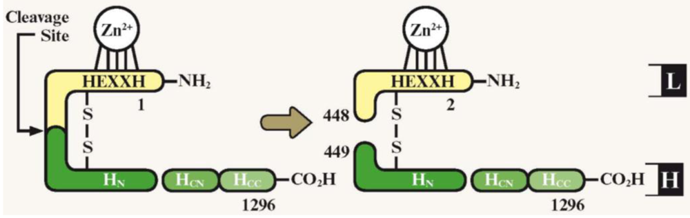
Figure 2.
Schematic representation of BoNT-A domain structure. Proteolytic cleavage activates BoNT, yielding a di-chain protein joined by a disulfide bond. The heavy chain (green) is composed of domains: HN and the HC, which is involved in translocation of the light chain (L, yellow). The HC is further divided into two subdomains: HCN and the HCC, which is involved in neurospecific binding. The light chain possesses endopeptidase activity, with a zinc-binding motif (HEXXH). Numbers indicate amino acid residues within the complete neurotoxin gene [8]. H: Heavy chain; HN: Heavy chain N-terminal fragment; HCN: Heavy chain C-terminal fragment, N-terminal subdomain; HCC: Heavy chain C-terminal fragment, C-terminal subdomain; L: Light chain. Figure adapted from [8], permission obtained.

3. Membrane Binding Initiates BoNT Mechanism of Action
All BoNT neurotoxins are synthesized initially as a ~150 kDa single chain, which is cleaved by an (unidentified) clostridial enzyme to form the active BoNT complex. This comprises a ~50 kDa light chain, which is a zinc-dependent endopeptidase, and a ~100 kDa heavy chain; the two are linked by a single disulfide bond [10]. The heavy chain, particularly the C-terminal domain (HC), mediates uptake of the toxin into the neuron, by binding to recycling synaptic vesicles in a stimulation-dependent manner [28,29]. Uptake is selectively directed to neuronal targets by specific high-affinity binding domains on the heavy chain, which interact with protein and ganglioside components of the cell membrane in a serotype-dependent manner (Table 1) [30,31].
The highly-expressed gangliosides [32] represent the lower-affinity receptors [33] responsible for accumulating BoNT on the neuronal membrane. Upon stimulation of the neuromuscular junction and subsequent recycling of synaptic vesicles, the amino-terminal intra-vesicular domain of the protein receptor is exposed, allowing toxin binding and endocytosis [34,35].
Comparison of the crystal structures of BoNT-A and BoNT-F receptor-binding domains reveals the heavy chain folds of BoNT-A and BoNT-F are similar, except for the region implicated in neuron binding [36]. A similar trend has also been reported for BoNT-B and BoNT-G, with differences in crystal structure explaining both ganglioside- and protein-receptor specificities [37]. Understanding such interactions in the future may allow BoNT engineering to bind non-neuronal cells.
Despite the variation exhibited by this region, a single, highly conserved ganglioside-binding motif E(D)…H…S(G)XWY…G(S) has been identified in the HC of BoNT-A, BoNT-B, BoNT-E, BoNT-F and BoNT-G [38,39,40]. An additional carbohydrate-binding domain has also been identified in the HC of BoNT-D [41]. These motifs determine the serotype specificity of the BoNT/carbohydrate interaction (Table 1) [37,39,42], as modification of residues within these sites is reported to alter the binding, uptake and toxicity of BoNT [38,39,40,41,42,43,44,45,46,47,48,49]. In particular, substitutions have been identified that enhance BoNT binding and toxicity [43], an effect that has been proposed to increase potency for use in the clinic [44].

Table 1.
Binding and catalytic targets of BoNT serotypes serotypes (A-G). TeNT, tetanus toxin; SV2A, B and C, synaptic vesicle glycoprotein 2A/B/C, Syn I and II, synaptotagmins I/II, SNAP-25, synaptosomal-associated protein 25; VAMP, vesicle-associated membrane protein; ThyI, Thy-1 cell surface antigen [33,37,41,42,44].
| Serotype | Cellular binding receptors | Catalytic target | |
|---|---|---|---|
| Carbohydrate | Protein | ||
| A | GD1a, GD1b, GT1b, GQ1b | SV2A, B and C | SNAP-25 |
| B | GD1a, GD1b, GT1b | Syn I and II | VAMP |
| C1 | GD1a, GD1b ,GT1b | SNAP-25 and syntaxin | |
| D | GT1b, GD2 | VAMP | |
| E | GD1a, GT1b, GQ1b | Glycosylated SV2A and B | SNAP-25 |
| F | GD1a, GD1b, GT1b | SV2 | VAMP |
| G | GT1b | Syn I and II | VAMP |
| TeNT | GT1b, GD1b GM1a GD3 | ThyI | VAMP |
4. Modifying the Binding Domain of BoNT to Retarget the Native Catalytic Domain
The light chain is catalytically active if introduced to non-neuronal cells via permeabilization [45,46], microinjection [47], transfection with the gene encoding the light chain [48] and modification of the BoNT-binding domain [49,50,51]. Retargeting the neuronal specificity of binding is a key aim of BoNT engineering, as delivery of the light chain, or non-native proteins, to the cytosol of non-neuronal cells may allow, for example, treatment of non-neuronal secretory diseases.
Several approaches have been used to target non-neuronal cells with BoNT. Co-application with lipid-based DNA transfection reagents resulted in BoNT-A activity in non-neuronal cell lines that are resistant to the toxin when applied alone [52]. Other approaches have exploited elements of the actual BoNT, substituting single amino acids [53] and whole domains of the toxin, summarized in Table 2 and discussed in detail here.
Generating chimeric proteins from BoNT has combined desired BoNT characteristics within one toxin. A chimera composed of the light chain and N-terminal heavy chain (HN) of BoNT-E with the heavy chain of BoNT-A (chimera E/A) displayed the rapid uptake and block of neuromuscular transmission exhibited by BoNT-E [54]. This chimera has been used to target a sensory relay centre implicated in pain mediation that is resistant to both parent toxins [55].
Conjugating light chain catalytic domains of BoNT to cell-binding domains of non-toxic proteins has rendered refractory cells sensitive to the toxin. A heterodimer (termed LHN) consisting of the light chain and heavy chain N-terminus (HN) is of particular interest for the design of novel therapeutic conjugates as this still contains both cell-membrane transportation and catalytic actions of the original BoNT molecule [19,51,56]. Recombinant studies have produced conjugates of the LHN heterodimer containing fragments of BoNT-A, BoNT-B and BoNT-C [57]. The LHN of BoNT-A has also been conjugated to lectin from Erythrina cristagalli, which binds to galactose-containing carbohydrates found on the surface of nociceptive afferents, to target pain signalling in vivo [49]. Chaddock et al. showed that the same BoNT-A LHN conjugated to wheat germ agglutinin inhibited neurotransmitter release from neuronal cell lines normally resistant to the neurotoxin [50]. Using a similar approach, this BoNT-A LHN was targeted to neuroblastoma cells that are normally refractory to the fragment by conjugation to nerve growth factor, inhibiting neurotransmitter release [51].
Table 2.
Summary of modifications to the BoNT-binding domain to generate molecules with new therapeutic potentials as indicated. SNAP-25, synaptosomal-associated protein 25; C2IN, enzymatically-inactive binding domain C2IN of BoNT-C2; GFP, green fluorescent protein; LHN, heterodimer consisting of the light chain and amino-terminal domain of the heavy chain; PEP-1, carrier protein.
| Modification to Binding Domain | Effect | Therapeutic Potential | Reference |
|---|---|---|---|
| BoNT-A/E chimera | SNAP-25 cleavage similar to BoNT-A | Similar to that of BoNT-A | [54,55] |
| BoNT-E/A chimera | Rapid uptake similar to BoNT-E | More persistent muscle weakening, targeted pain mediation | [54,55] |
| C2IN-streptavidin | Delivery of biotinylated molecules | Drug delivery | [61] |
| S6 peptide | Delivery of small molecules | Drug delivery | [60] |
| Fluorescent proteins e.g., GFP | Tracer molecules | Analysing neuronal circuit plasticity | [64,65] |
| Drug activating enzyme | Drug activation | Chemotherapy | [67] |
| Poly-lysine | DNA delivery | Gene therapy | [68,70] |
| Lectin | Binds nociceptive afferents | Targeted pain medication | [49] |
| Wheatgerm agglutinin | Targeted light chain to neuronal cells | Inhibited refractory neurotransmitter release | [50] |
| Nerve growth factor | Targeted LHN neuronal cells | Inhibited refractory neurotransmitter release | [51] |
| Epidermal growth factor | Targeted epithelial cells | Inhibited mucus secretion | [58] |
| Addition of PEP-1 peptide | Penetrated skin | Novel administration technique | [88] |
Providing proof-of-principle that a retargeted BoNT derivative can prevent secretion in non-neuronal cells, Foster et al. conjugated BoNT-C LHN and epidermal growth factor (EGF) to inhibit the secretion of mucus from epithelial cells that are refractory to the LHN alone [58]. This BoNT-EGF derivative has the potential to treat the hyperactive mucus secretion associated with asthma and chronic obstructive pulmonary disease and the approach could be used, in concert with a BoNT, to relieve overactive smooth muscle contraction that contributes to such respiratory diseases [59].
5. Employing the BoNT-Binding Domain to Deliver Non-Native Proteins
Chimeric BoNTs have also been developed that employ the cellular binding activity of BoNT as a targeting moiety to deliver the activity of a conjugated, non-native protein. Removing or inactivating the catalytic light chain domain of BoNT is required in order to prevent the blockade of neurotransmission. Catalytically-inactive BoNTs are therefore proposed to have prolonged cellular uptake, in contrast to active BoNTs that disable their own uptake [60].
Genetic fusion of the enzymatically-inactive binding domain C2IN of BoNT-C2 to streptavidin allowed delivery of biotinylated molecules into the cytosol of mammalian cells [61]. Full-length BoNTs containing activating mutations have been fused to an S6 peptide sequence, allowing the attachment and delivery of a fluorescent small-molecule to the target cell cytoplasm [60]. Conjugating BoNT to such carrier proteins exponentially increases the number of potential targeted cargos, providing vehicles for future targeting of small molecule drugs.
Various tracer proteins have also been conjugated to BoNT peptides, including those containing inactivating mutations [62]. Conjugates with β-galactosidase [63] and fluorescent proteins [62,64,65] have revealed BoNT and TeNT distribution profiles, allowing mapping of neuronal circuits [65] and analysis of neuronal circuit plasticity after traumatic injury or neurodegenerative diseases [66].
The BoNT cellular-binding activity has also been proposed to deliver an enzyme to cancer cells that activates a prodrug, administered systemically [67]. However, catalytically active BoNTs, which are proposed to enhance tumor perfusion and subsequent access of cytotoxic agents and oxygen in order to potentiate radiotherapy, may be more appropriate for cancer therapy [8].
As TeNT has a similar modular arrangement of functional domains as BoNT, advances in TeNT engineering may also be applied to BoNT. For example, the binding domains of both BoNT and TeNT have been utilized to deliver DNA to target cells and enhance targeting of other transfection methods. The TeNT heavy chain fragment has been conjugated to poly-lysine, which has a high capacity to bind DNA, allowing transfection of a range of neuronal cell lines [68,69]. This TeNT-poly-lysine derivative has also been shown to enhance adenoviral infection of primary neuronal cultures while increasing neuronal specificity of transfection [70]. TeNT and BoNT heavy chain have also been added to liposomes, targeting gene delivery in vitro [71].
Recent studies with recombinantly produced BoNT domains show that proteins can be assembled by non-chemical linking, using tagging with helical motifs from the family of soluble N-ethylamide-sensitive factor attachment protein receptor (SNARE) proteins. This may potentially be exploited to use the BoNT-binding domain to deliver future therapeutics or other cargo into neurons, or to facilitate re-targeting of the light chain [72].
6. Modifying the BoNT Active Site to Target Non-Native Substrates
BoNT-A has the potential to inhibit the release of multiple, but not all, neurotransmitters [73,74]. Thus, in addition to neuronal specificity being conferred by the binding domain, a degree of specificity is also bestowed by the catalytic domain. Manipulation of the BoNT light chain catalytic domain may therefore also be important when designing BoNT to target non-neuronal cells or in modifying the targets within neuronal systems.
Once taken up into the neuron, BoNT light chain cleaves members of the SNARE family of proteins which mediate docking of synaptic vesicles with the neural cell membrane [75]. The SNARE proteins are a large family: 36 members have so far been identified in mammalian cells [76], including a 25 kDa synaptosomal-associated protein (SNAP-25), synaptobrevin (also called vesicle-associated membrane protein (VAMP) and syntaxin. Cleavage and inactivation of SNAREs by the BoNTs results in inhibition of neurotransmission and concomitant prevention of further toxin uptake. BoNT-A, BoNT-C and BoNT-E cleave SNAP-25 while BoNT-B, BoNT-D, BoNT-F and BoNT-G cleave proteins of the VAMP family (Table 1); [44]. BoNT-C also cleaves syntaxin [44].
In addition to neurotransmission, SNAREs have been implicated in non-neuronal secretory processes. The effectiveness of BoNT on these processes is determined by both the SNARE member mediating secretion and the BoNT serotype. For example, BoNT-A is reported to inhibit insulin secretion from permeabilized β-cell lines, as this process is mediated by SNAP-25 [45]. In contrast, insulin-stimulated uptake of glucose to permeabilized adipocytes, which is mediated by SNAP-23, is resistant to BoNT-A [77]. Engineering BoNT to modulate insulin signalling may allow application of BoNT in diabetes therapy. Moreover, this approach may target non-neuronal SNAREs in a variety of non-neuronal secretory disorders.
Regions of BoNT involved in SNARE recognition and cleavage have been identified [78,79], including the residues directly involved in catalysis [80], although residues both near and distal to the active site are reported to be important in their proteolytic action [81,82]. Primary sequences are also now available for 36 human SNAREs [76] and mapping the sites cleaved by members of the clostridial neurotoxins family has been completed (Table 3) [83,84]. This indicates that specific residues on both the BoNT and the substrate are involved in catalysis.
The BoNT regions implicated in catalysis are supported by crystal structures of inactive BoNT-A light chain bound to SNAP-25, confirming exosite and active site interactions [85]. Correlating structural data with sequence identity has explained serotype-specificity of substrate binding and catalysis, allowing substrate prediction in newly discovered BoNT species [22]. Structure-function relationships may allow directed engineering of BoNT to provide alternatively targeted molecules.
Although the minimal substrate size of BoNT-A required for catalysis is relatively large, only a few non-conserved residues proximal to the active site have been shown to influence catalysis of the substrate [82]. Such residues are sensitive to subtle changes in amino acid composition; substituting one positively-charged residue (arginine) for another (lysine) in BoNT-A(R230K), for example, abolishes activity [82]. Taken together, these data have allowed rational, directed approaches to retarget the catalytic activity of BoNT and cleave non-neuronal SNARE proteins.
In order to target non-neuronal SNARE proteins, Chen et al. [53] targeted position 224 within the catalytic site of BoNT-E, which specifically cleaves the neuronal SNARE, SNAP-25. In addition to cleaving SNAP-25, the engineered BoNT-E (K224D) cleaved non-neuronal SNAP-23, at a similar rate to that at which the wild-type toxin cleaves its native target [53].
Table 3.
SNARE isoform sequences indicating BoNT cleavage sites. Amino acid residues are indicated in lower case. Cleavage sites are indicated by a dashed line between specific amino acids of the toxin sequence. Highlighted residues indicate non-conserved mutations at or around cleavage sites. SNARE, soluble N-ethylamide-sensitive factor attachment protein receptor; VAMP, vesicle-associated membrane protein; SNAP, synaptosomal-associated protein. Table adapted from [94] copyright retained by Inderscience.
|
Catalytically-retargeted BoNT may be applied clinically in the future to target non-neuronal cells in concert with novel administration technologies, such as iontophoresis [86], nasal inhalation [87], and fusion proteins capable of penetrating the skin [88]. A recent report also demonstrated intranasal administration of BoNT-A HC in a ‘nanometer-sized hydrogel’ (nanogel) [89], allowing binding and penetration of nasal epithelial cells. Once the HC was released from the nanogel, the peptide was taken up by nasal dendritic cells, without accumulation in the brain [89]. Several patents for novel BoNT administration have also been filed (Table 4), including skin disruption with transdermal patches [90], release of polymeric microspheres from implants [91] and toxin application in phospholipid micelles [92] and solvents [93].
Table 4.
Recent patents involving modifications to botulinum toxin molecules intended to generate new molecules with additional therapeutic potentials and targets.
| Invention | Author(s) | Patent No. | Reference |
|---|---|---|---|
| Application via transdermal patches | Donovan | US20017758871 | [90] |
| Application via skin disruption | Donovan | US20017758871 | [90] |
| Application in polymeric microsphere-containing implant | Donovan | US20080028216 | [91] |
| Application in phospholipid micelles | Modi | US20080220021 | [92] |
| Application in non-polar solvent | Petrou and Vedra | US20090304747 | [93] |
| PEGylated mutated BoNT | Frevert and Specht | EP1834962 | [107] |
| Formulations for oral administration | Donovan | US20040086532 | [108] |
| Biodegradable neurotoxin implants | Hughes and Orest | US20050232966 | [109] |
| Leucine-based motif and Clostridia neurotoxins | Steward et al. | US20080177041 | [110] |
7. Modifications to BoNT Duration of Action
In most clinical applications, the actions of BoNT are temporary, lasting from several days (BoNT-E) to months (BoNT-A) [19]. The duration of action is also highly species-dependant: BoNTs display the shortest action at the mouse neuromuscular junction, followed by rat, then human [94]. Adults are also reported to display greater sensitivity than juveniles [95], which may arise from differences in the abundance of motor endplates. Although the reversibility of BoNT activity can be considered desirable, for altering muscle involvement or fine-tuning aesthetic effects, a long duration of action is also advantageous, to reduce administration frequency.
Differences in duration of the neuroparalytic effects between BoNT-A and BoNT-E may be due to the nature of the cleavage of the target protein [96]. Both cleave the SNARE complex component SNAP-25; BoNT-A truncates this protein by removal of 9 amino acids from the C-terminal end and BoNT-E cleaves 26 residues from the C-terminal end. Potentially the BoNT-A truncated SNAP-25 can still interact with the other SNARE proteins to form non-functioning SNARE complexes and so prevent exocytosis. Newly synthesised SNAP-25 cannot then interact with these blocked complexes, so causing paralysis to persist. Conversely, BoNT-E cleaves the SNAP-25 in the cell, causing paralysis, but the cleavage product does not associate with the other SNARE proteins in the same way, and newly synthesised intact SNAP-25 can then interact as normal to form the SNARE complex and the neuromuscular function is therefore resumed earlier than when BoNT-A is present.
The duration of action is also proposed to be determined by the intracellular persistence of the light chain [97], which in turn results from the cellular distribution of the peptide. Fernández-Salas et al. [98,99] compared the distribution of green fluorescent protein (GFP) and GFP conjugated to BoNT-A light chain when expressed in neuroblastoma cell lines. Cells expressing unconjugated GFP exhibited diffuse fluorescence throughout the cell. In contrast, cells expressing the GFP- light chain conjugate displayed a discrete pattern of fluorescence distributed in a manner resembling a cell outline, which co-distributed with SNAP-25 [98], indicating BoNT-A light chain directed distribution away from the cytosol. They showed that the duration of action of light chain from BoNT-A, BoNT-B and BoNT-E corresponded with cellular distribution. In particular, a cytosolic distribution was shown to correlate with a short duration of action [99]. Building on these data, Fernández-Salas et al. reported the catalytic light chain domain of BoNT-A, which is not involved in the neuron-specific endocytotic cellular binding, contains signals in the N- and C-termini that are required for plasma membrane binding [98]. Consistent with this, a chimeric BoNT composed of the light chain N-terminal (LN) of BoNT-A and HC of BoNT-E (chimera AE) possessed similar persistence of SNAP-25 cleavage in vitro and neuromuscular block in vivo to BoNT-A, suggesting regions involved in duration of action are located in the LN. Such regions may be amenable to direct engineering in the future to extend the duration of toxin action in the clinic. In the past, many genetic approaches have been hindered by the number of clostridial species that are amenable to genetic manipulation, although novel techniques have recently been developed.
Studying and potentially altering potentially altering the duration of action of the BoNTs is particularly relevant, as there may be a limited amount of toxin that can enter the cell. The toxins mode of entry into the cell is by exploiting the synaptic vesicle pathway that couples exocytosis with endocytosis of synaptic vesicles. Binz and Rummel [28] claim that, as toxins interfere with this machinery and disrupt the cycle by cleavage of the proteins involved in this process, they will prevent the cycle from functioning and therefore will prevent their own further uptake into the cell. On this premise, there is a limited amount of toxin that can get into the cell and its longevity inside the cell therefore governs the length of the effect of the toxin. However, recent studies have indicated that the endocytosis of synaptic vesicles is still occurring in toxin poisoned cells [100,101]. These papers describe the potential for a modified BoNT (encompassing a cargo of 10 kDa amino dextran molecule coupled to a non-toxic recombinant heavy chain) to be transported into the cell as a drug delivery vehicle for rescue of botulinum toxin poisoned synapses as a countermeasure for botulism victims. Overall therefore, the exact mechanism of persistence of BoNT activity remains to be elucidated.
8. Alternative Methods of Modifying BoNT
Exploitation of C. botulinum sequence data has been hindered by the number of mutations generated for functional genomic studies, owing to a lack of basic molecular biology tools required for directed mutation. Targeted inactivation of clostridial genes has been almost exclusively limited to single crossover knockouts via integration through homologous recombination of a replication-defective plasmid [102]. However, capitalizing on these mutants has been restricted by the unstable integration of a plasmid with the chromosome. To address these issues, recombination-independent strategies have been devised that utilize a retargeted group II intron. One of these, “ClosTron”, allows systematic inactivation of genes to evaluate their function [103,104].
ClosTron provides the facility for positive selection of desired mutants, which are highly stable and reproducible, expanding the current options for functional genomic studies in clostridia. Although use in BoNT engineering is still in its infancy, ClosTron has huge potential. To date, ClosTron has been utilized to generate a non-toxigenic mutant strain of C. botulinum [105] and inactivate restriction endonuclease activity to allow transformation with unmethylated DNA as efficiently as with methylated DNA in C. acetobutylicum [106].
9. Summary and Conclusion
BoNTs have unique and well-characterized structural properties that make them particularly well suited to engineering for therapeutic use. The potential therapeutic capacity of BoNT is being realized through characterization and genetic engineering to alter the binding, catalysis and duration of the toxin, allowing specific targeting and therapeutic tailoring. Structure-function relationships allow rational design of catalytic specificity, allowing application of BoNT to numerous SNARE-mediated secretory processes, including those involved in diabetes [45,77], respiratory disorders [53] and processes mediating immune and inflammatory disorders [58]. Genetic engineering and functional studies are revealing the true potential of BoNT and have already expanded and diversified the potential therapeutic applications of BoNT, with each development yielding more ways to exploit and capitalize on the actions of this remarkable toxin.
Acknowledgements
Editorial support for the preparation of this manuscript was provided by Ogilvy Healthworld Medical Education.
References
- Apostolidis, A.; Fowler, C.J. The use of botulinum neurotoxin type A (BoNTA) in urology. J. Neural Transm. 2008, 115, 593–605. [Google Scholar] [CrossRef] [PubMed]
- Cordivari, C.; Misra, V.P.; Catania, S.; Lees, A.J. New therapeutic indications for botulinum toxins. Mov. Disord. 2004, 19, S157–S161. [Google Scholar] [CrossRef] [PubMed]
- Uyesugi, B.; Lippincott, B.; Dave, S. Treatment of a painful keloid with botulinum toxin type A. Am. J. Phys. Med. Rehabil. 2010, 89, 153–155. [Google Scholar] [CrossRef] [PubMed]
- Torgovnick, J.; Arsura, E.; Sethi, N.K.; Hu, C.J.; Yuan, R.Y.; Sheu, J.J.; Apfel, S.C. Botulinum toxin for diabetic neuropathic pain: A randomized double-blind crossover trial: Botulinum toxin for neuropathic pain? Neurology 2010, 74, 92–93. [Google Scholar] [PubMed]
- Singer, B.J.; Silbert, P.L.; Song, S.; Dunne, J.W.; Singer, K.P. Treatment of refractory anterior knee pain using botulinum toxin type A (Dysport) injection to the distal vastus lateralis muscle: A randomised placebo controlled crossover trial. Br. J. Sports Med. 2010. [Google Scholar] [CrossRef]
- Ngeow, W.C.; Nair, R. Injection of botulinum toxin type A (BOTOX) into trigger zone of trigeminal neuralgia as a means to control pain. Oral Surg. Oral Med. Oral Pathol. Oral Radiol. Endod. 2010, 109, e47–e50. [Google Scholar] [CrossRef]
- Liu, R.K.; Li, C.H.; Zou, S.J. Reducing scar formation after lip repair by injecting botulinum toxin. Plast. Reconstr. Surg. 2010, 125, 1573–1574. [Google Scholar] [PubMed]
- Ansiaux, R.; Gallez, B. Use of botulinum toxins in cancer therapy. ExpertOpin. Investig. Drugs 2007, 16, 209–218. [Google Scholar] [CrossRef]
- Beer, K. Cost effectiveness of botulinum toxins for the treatment of depression: Preliminary observations. J. Drugs Dermatol. 2010, 9, 27–30. [Google Scholar] [PubMed]
- Montal, M. Botulinum neurotoxin: A marvel of protein design. Annu. Rev. Biochem. 2010, 79, 591–617. [Google Scholar] [CrossRef] [PubMed]
- Pickett, A.; Perrow, K. Formulation composition of botulinum toxins in clinical use. J. Drugs Dermatol. 2010, 9, 1085–1091. [Google Scholar] [PubMed]
- Fujinaga, Y.; Inoue, K.; Nomura, T.; Sasaki, J.; Marvaud, J.C.; Popoff, M.R.; Kozaki, S.; Oguma, K. Identification and characterization of functional subunits of Clostridium botulinum type A progenitor toxin involved in binding to intestinal microvilli and erythrocytes. FEBS Lett. 2000, 467, 179–183. [Google Scholar] [CrossRef] [PubMed]
- Fu, F.N.; Sharma, S.K.; Singh, B.R. A protease-resistant novel hemagglutinin purified from type A Clostridium botulinum. J. Protein Chem. 1998, 17, 53–60. [Google Scholar] [CrossRef] [PubMed]
- Fujinaga, Y.; Inoue, K.; Watanabe, S.; Yokota, K.; Hirai, Y.; Nagamachi, E.; Oguma, K. The haemagglutinin of Clostridium botulinum type C progenitor toxin plays an essential role in binding of toxin to the epithelial cells of guinea pig small intestine, leading to the efficient absorption of the toxin. Microbiology 1997, 143, 3841–3847. [Google Scholar] [CrossRef] [PubMed]
- Jacobson, M.J.; Lin, G.; Raphael, B.; Andreadis, J.; Johnson, E.A. Analysis of neurotoxin cluster genes in Clostridium botulinum strains producing botulinum neurotoxin serotype A subtypes. Appl. Environ. Microbiol. 2008, 74, 2778–2786. [Google Scholar] [CrossRef] [PubMed]
- Sakaguchi, Y.; Hayashi, T.; Yamamoto, Y.; Nakayama, K.; Zhang, K.; Ma, S.; Arimitsu, H.; Oguma, K. Molecular analysis of an extrachromosomal element containing the C2 toxin gene discovered in Clostridium botulinum type C. J. Bacteriol. 2009, 191, 3282–3291. [Google Scholar] [CrossRef] [PubMed]
- Dover, N.; Barash, J.R.; Arnon, S.S. Novel Clostridium botulinum Toxin Gene Arrangement with Subtype A5 and Partial Subtype B3 Botulinum Neurotoxin Genes. J. Clin. Microbiol. 2009, 47, 2349–2350. [Google Scholar] [CrossRef] [PubMed]
- Luquez, C.; Raphael, B.H.; Maslanka, S.E. Neurotoxin gene clusters in Clostridium botulinum type Ab strains. Appl. Environ. Microbiol. 2009, 75, 6094–6101. [Google Scholar] [PubMed]
- Chaddock, J.A.; Marks, P.M. Clostridial neurotoxins: Structure-function led design of new therapeutics. Cell Mol. Life Sci. 2006, 63, 540–551. [Google Scholar] [CrossRef] [PubMed]
- Sebaihia, M.; Peck, M.W.; Minton, N.P.; Thomson, N.R.; Holden, M.T.; Mitchell, W.J.; Carter, A.T.; Bentley, S.D.; Mason, D.R.; Crossman, L.; Paul, C.J.; Ivens, A.; Wells-Bennik, M.H.; Davis, I.J.; Cerdeno-Tarraga, A.M.; Churcher, C.; Quail, M.A.; Chillingworth, T.; Feltwell, T.; Fraser, A.; Goodhead, I.; Hance, Z.; Jagels, K.; Larke, N.; Maddison, M.; Moule, S.; Mungall, K.; Norbertczak, H.; Rabbinowitsch, E.; Sanders, M.; Simmonds, M.; White, B.; Whithead, S.; Parkhill, J. Genome sequence of a proteolytic (Group I) Clostridium botulinum strain Hall A and comparative analysis of the clostridial genomes. Genome Res. 2007, 17, 1082–1092. [Google Scholar] [PubMed]
- Hill, K.K.; Smith, T.J.; Helma, C.H.; Ticknor, L.O.; Foley, B.T.; Svensson, R.T.; Brown, J.L.; Johnson, E.A.; Smith, L.A.; Okinaka, R.T.; Jackson, P.J.; Marks, J.D. Genetic diversity among Botulinum Neurotoxin-producing clostridial strains. J. Bacteriol. 2007, 189, 818–832. [Google Scholar] [PubMed]
- Henkel, J.S.; Jacobson, M.; Tepp, W.; Pier, C.; Johnson, E.A.; Barbieri, J.T. Catalytic Properties of Botulinum Neurotoxin Subtypes A3 and A4 (dagger). Biochemistry 2009, 48, 2522–2528. [Google Scholar] [PubMed]
- Smith, T.J.; Hill, K.K.; Foley, B.T.; Detter, J.C.; Munk, A.C.; Bruce, D.C.; Doggett, N.A.; Smith, L.A.; Marks, J.D.; Xie, G.; Brettin, T.S. Analysis of the neurotoxin complex genes in Clostridium botulinum A1-A4 and B1 strains: BoNT/A3, /Ba4 and /B1 clusters are located within plasmids. PLoS One 2007, 2, e1271. [Google Scholar] [PubMed]
- Marshall, K.M.; Bradshaw, M.; Pellett, S.; Johnson, E.A. Plasmid encoded neurotoxin genes in Clostridium botulinum serotype A subtypes. Biochem. Biophys. Res. Commun. 2007, 361, 49–54. [Google Scholar] [CrossRef] [PubMed]
- Franciosa, G.; Maugliani, A.; Scalfaro, C.; Aureli, P. Evidence that plasmid-borne botulinum neurotoxin type B genes are widespread among Clostridium botulinum serotype B strains. PLoS One 2009, 4, e4829. [Google Scholar] [PubMed]
- Hill, K.K.; Xie, G.; Foley, B.T.; Smith, T.J.; Munk, A.C.; Bruce, D.; Smith, L.A.; Brettin, T.S.; Detter, J.C. Recombination and insertion events involving the botulinum neurotoxin complex genes in Clostridium botulinum types A, B, E and F and Clostridium butyricum type E strain. BMC Biol. 2009, 7, 66. [Google Scholar] [CrossRef] [PubMed]
- Brussow, H.; Canchaya, C.; Hardt, W.D. Phages and the evolution of bacterial pathogens: From genomic rearrangements to lysogenic conversion. Microbiol. Mol. Biol. Rev. 2004, 68, 560–602. [Google Scholar] [CrossRef] [PubMed]
- Binz, T.; Rummel, A. Cell entry strategy of clostridial neurotoxins. J. Neurochem. 2009, 109, 1584–1595. [Google Scholar] [CrossRef] [PubMed]
- Keller, J.E.; Cai, F.; Neale, E.A. Uptake of botulinum neurotoxin into cultured neurons. Biochemistry 2004, 43, 526–532. [Google Scholar] [CrossRef] [PubMed]
- Baldwin, M.R.; Barbieri, J.T. Association of botulinum neurotoxins with synaptic vesicle protein complexes. Toxicon 2009, 54, 570–574. [Google Scholar] [CrossRef] [PubMed]
- Baldwin, M.R.; Kim, J.J.; Barbieri, J.T. Botulinum neurotoxin B-host receptor recognition: It takes two receptors to tango. Nat. Struct. Mol. Biol. 2007, 14, 9–10. [Google Scholar] [CrossRef] [PubMed]
- van Heyningen, W.E.; Miller, P.A. The fixation of tetanus toxin by ganglioside. J. Gen. Microbiol. 1961, 24, 107–119. [Google Scholar] [PubMed]
- Brunger, A.T.; Rummel, A. Receptor and substrate interactions of clostridial neurotoxins. Toxicon 2009, 54, 550–560. [Google Scholar] [CrossRef] [PubMed]
- Kozaki, S.; Miyazaki, S.; Sakaguchi, G. Development of antitoxin with each of two complementary fragments of Clostridium botulinum type B derivative toxin. Infect. Immun. 1977, 18, 761–766. [Google Scholar] [PubMed]
- Verderio, C.; Rossetto, O.; Grumelli, C.; Frassoni, C.; Montecucco, C.; Matteoli, M. Entering neurons: Botulinum toxins and synaptic vesicle recycling. EMBO Rep. 2006, 7, 995–999. [Google Scholar] [CrossRef] [PubMed]
- Fu, Z.; Chen, C.; Barbieri, J.T.; Kim, J.J.; Baldwin, M.R. Glycosylated SV2 and gangliosides as dual receptors for botulinum neurotoxin serotype F. Biochemistry 2009, 48, 5631–5641. [Google Scholar] [CrossRef] [PubMed]
- Schmitt, J.; Karalewitz, A.; Benefield, D.A.; Mushrush, D.J.; Pruitt, R.N.; Spiller, B.W.; Barbieri, J.T.; Lacy, D.B. Structural analysis of botulinum neurotoxin type G receptor binding. Biochemistry 2010, 49, 5200–5205. [Google Scholar] [PubMed]
- Rummel, A.; Hafner, K.; Mahrhold, S.; Darashchonak, N.; Holt, M.; Jahn, R.; Beermann, S.; Karnath, T.; Bigalke, H.; Binz, T. Botulinum neurotoxins C, E and F bind gangliosides via a conserved binding site prior to stimulation-dependent uptake with botulinum neurotoxin F utilising the three isoforms of SV2 as second receptor. J. Neurochem. 2009, 110, 1942–1954. [Google Scholar] [CrossRef] [PubMed]
- Rummel, A.; Mahrhold, S.; Bigalke, H.; Binz, T. The HCC-domain of botulinum neurotoxins A and B exhibits a singular ganglioside binding site displaying serotype specific carbohydrate interaction. Mol. Microbiol. 2004, 51, 631–643. [Google Scholar] [PubMed]
- Rummel, A.; Eichner, T.; Weil, T.; Karnath, T.; Gutcaits, A.; Mahrhold, S.; Sandhoff, K.; Proia, R.L.; Acharya, K.R.; Bigalke, H.; Binz, T. Identification of the protein receptor binding site of botulinum neurotoxins B and G proves the double-receptor concept. Proc. Natl. Acad. Sci. USA 2007, 104, 359–364. [Google Scholar]
- Strotmeier, J.; Lee, K.; Volker, A.K.; Mahrhold, S.; Zong, Y.; Zeiser, J.; Zhou, J.; Pich, A.; Bigalke, H.; Binz, T.; Rummel, A.; Jin, R. Botulinum neurotoxin serotype D attacks neurons via two carbohydrate binding sites in a ganglioside dependent manner. Biochem. J. 2010, 431, 207–216. [Google Scholar] [CrossRef] [PubMed]
- Yowler, B.C.; Schengrund, C.L. Glycosphingolipids-sweets for botulinum neurotoxin. Glycoconj. J. 2004, 21, 287–293. [Google Scholar] [CrossRef] [PubMed]
- Rummel, A. Transport Protein Which Is Used To Introduce Chemical Compounds into Nerve Cells. US Patent 0299008, 27 12 2007. [Google Scholar]
- Foster, K.A. Engineered toxins: New therapeutics. Toxicon 2009, 54, 587–592. [Google Scholar] [CrossRef] [PubMed]
- Boyd, R.S.; Duggan, M.J.; Shone, C.C.; Foster, K.A. The effect of botulinum neurotoxins on the release of insulin from the insulinoma cell lines HIT-15 and RINm5F. J. Biol. Chem. 1995, 270, 18216–18218. [Google Scholar]
- Lawrence, G.W.; Foran, P.; Mohammed, N.; DasGupta, B.R.; Dolly, J.O. Importance of two adjacent C-terminal sequences of SNAP-25 in exocytosis from intact and permeabilized chromaffin cells revealed by inhibition with botulinum neurotoxins A and E. Biochemistry 1997, 36, 3061–3067. [Google Scholar] [CrossRef] [PubMed]
- Penner, R.; Neher, E.; Dreyer, F. Intracellularly injected tetanus toxin inhibits exocytosis in bovine adrenal chromaffin cells. Nature 1986, 324, 76–78. [Google Scholar] [CrossRef] [PubMed]
- Aguado, F.; Gombau, L.; Majo, G.; Marsal, J.; Blanco, J.; Blasi, J. Regulated secretion is impaired in AtT-20 endocrine cells stably transfected with botulinum neurotoxin type A light chain. J. Biol. Chem. 1997, 272, 26005–26008. [Google Scholar] [PubMed]
- Duggan, M.J.; Quinn, C.P.; Chaddock, J.A.; Purkiss, J.R.; Alexander, F.C.; Doward, S.; Fooks, S.J.; Friis, L.M.; Hall, Y.H.; Kirby, E.R.; Leeds, N.; Moulsdale, H.J.; Dickenson, A.; Green, G.M.; Rahman, W.; Suzuki, R.; Shone, C.C.; Foster, K.A. Inhibition of release of neurotransmitters from rat dorsal root ganglia by a novel conjugate of a Clostridium botulinum toxin A endopeptidase fragment and Erythrina cristagalli lectin. J. Biol. Chem. 2002, 277, 34846–34852. [Google Scholar] [PubMed]
- Chaddock, J.A.; Purkiss, J.R.; Friis, L.M.; Broadbridge, J.D.; Duggan, M.J.; Fooks, S.J.; Shone, C.C.; Quinn, C.P.; Foster, K.A. Inhibition of vesicular secretion in both neuronal and nonneuronal cells by a retargeted endopeptidase derivative of Clostridium botulinum neurotoxin type A. Infect. Immun. 2000, 68, 2587–2593. [Google Scholar] [PubMed]
- Chaddock, J.A.; Purkiss, J.R.; Duggan, M.J.; Quinn, C.P.; Shone, C.C.; Foster, K.A. A conjugate composed of nerve growth factor coupled to a non-toxic derivative of Clostridium botulinum neurotoxin type A can inhibit neurotransmitter release in vitro. Growth Factors 2000, 18, 147–155. [Google Scholar] [CrossRef] [PubMed]
- Kuo, C.L.; Oyler, G.; Shoemaker, C.B. Lipid and cationic polymer based transduction of botulinum holotoxin, or toxin protease alone, extends the target cell range and improves the efficiency of intoxication. Toxicon 2010, 55, 619–629. [Google Scholar] [CrossRef] [PubMed]
- Chen, S.; Barbieri, J.T. Engineering botulinum neurotoxin to extend therapeutic intervention. Proc. Natl. Acad. Sci. USA 2009, 106, 9180–9184. [Google Scholar]
- Wang, J.; Meng, J.; Lawrence, G.W.; Zurawski, T.H.; Sasse, A.; Bodeker, M.O.; Gilmore, M.A.; Fernandez-Salas, E.; Francis, J.; Steward, L.E.; Aoki, K.R.; Dolly, J.O. Novel chimeras of botulinum neurotoxins A and E unveil contributions from the binding, translocation, and protease domains to their functional characteristic. J. Biol. Chem. 2008, 283, 16993–17002. [Google Scholar]
- Meng, J.; Ovsepian, S.V.; Wang, J.; Pickering, M.; Sasse, A.; Aoki, K.R.; Lawrence, G.W.; Dolly, J.O. Activation of TRPV1 mediates calcitonin gene-related peptide release, which excites trigeminal sensory neurons and is attenuated by a retargeted botulinum toxin with anti-nociceptive potential. J. Neurosci. 2009, 29, 4981–4992. [Google Scholar]
- Shone, C.C.; Hambleton, P.; Melling, J. Inactivation of Clostridium botulinum type A neurotoxin by trypsin and purification of two tryptic fragments. Proteolytic action near the COOH-terminus of the heavy subunit destroys toxin-binding activity. Eur. J. Biochem. 1985, 151, 75–82. [Google Scholar] [CrossRef] [PubMed]
- Sutton, J.M.; Wayne, J.; Scott-Tucker, A.; O'Brien, S.M.; Marks, P.M.; Alexander, F.C.; Shone, C.C.; Chaddock, J.A. Preparation of specifically activatable endopeptidase derivatives of Clostridium botulinum toxins type A, B, and C and their application. Protein Expr. Purif. 2005, 40, 31–41. [Google Scholar] [CrossRef] [PubMed]
- Foster, K.A.; Adams, E.J.; Durose, L.; Cruttwell, C.J.; Marks, E.; Shone, C.C.; Chaddock, J.A.; Cox, C.L.; Heaton, C.; Sutton, J.M.; Wayne, J.; Alexander, F.C.; Rogers, D.F. Re-engineering the target specificity of Clostridial neurotoxins-A route to novel therapeutics. Neurotox. Res. 2006, 9, 101–107. [Google Scholar] [PubMed]
- Lim, E.C.; Ong, B.K.; Oh, V.M.; Seet, R.C. Botulinum toxin: A novel therapeutic option for bronchial asthma? Med. Hypotheses 2006, 66, 915–919. [Google Scholar] [CrossRef] [PubMed]
- Band, P.A.; Blais, S.; Neubert, T.A.; Cardozo, T.J.; Ichtchenko, K. Recombinant derivatives of botulinum neurotoxin A engineered for trafficking studies and neuronal delivery. Protein Expr. Purif. 2010, 71, 62–73. [Google Scholar] [CrossRef] [PubMed]
- Fahrer, J.; Plunien, R.; Binder, U.; Langer, T.; Seliger, H.; Barth, H. Genetically engineered clostridial C2 toxin as a novel delivery system for living mammalian cells. Bioconjug. Chem. 2010, 21, 130–139. [Google Scholar] [CrossRef] [PubMed]
- Bade, S.; Rummel, A.; Reisinger, C.; Karnath, T.; Ahnert-Hilger, G.; Bigalke, H.; Binz, T. Botulinum neurotoxin type D enables cytosolic delivery of enzymatically active cargo proteins to neurones via unfolded translocation intermediates. J. Neurochem. 2004, 91, 1461–1472. [Google Scholar] [CrossRef] [PubMed]
- Coen, L.; Osta, R.; Maury, M.; Brulet, P. Construction of hybrid proteins that migrate retrogradely and transynaptically into the central nervous system. Proc. Natl. Acad. Sci. USA 1997, 94, 9400–9405. [Google Scholar]
- Kissa, K.; Mordelet, E.; Soudais, C.; Kremer, E.J.; Demeneix, B.A.; Brulet, P.; Coen, L. In vivo neuronal tracing with GFP-TTC gene delivery. Mol. Cell Neurosci. 2002, 20, 627–637. [Google Scholar] [CrossRef] [PubMed]
- Maskos, U.; Kissa, K.; St. Cloment, C.; Brulet, P. Retrograde trans-synaptic transfer of green fluorescent protein allows the genetic mapping of neuronal circuits in transgenic mice. Proc. Natl. Acad. Sci. USA 2002, 99, 10120–10125. [Google Scholar]
- Huh, Y.; Oh, M.S.; Leblanc, P.; Kim, K.S. Gene transfer in the nervous system and implications for transsynaptic neuronal tracing. Expert Opin. Biol. Ther. 2010, 10, 763–772. [Google Scholar] [PubMed]
- van Mellaert, L.; Barbe, S.; Anne, J. Clostridium spores as anti-tumour agents. Trends Microbiol. 2006, 14, 190–196. [Google Scholar] [CrossRef] [PubMed]
- Knight, A.; Carvajal, J.; Schneider, H.; Coutelle, C.; Chamberlain, S.; Fairweather, N. Non-viral neuronal gene delivery mediated by the HC fragment of tetanus toxin. Eur. J. Biochem. 1999, 259, 762–769. [Google Scholar] [PubMed]
- Box, M.; Parks, D.A.; Knight, A.; Hale, C.; Fishman, P.S.; Fairweather, N.F. A multi-domain protein system based on the HC fragment of tetanus toxin for targeting DNA to neuronal cells. J. Drug Target 2003, 11, 333–343. [Google Scholar] [CrossRef] [PubMed]
- Schneider, H.; Groves, M.; Muhle, C.; Reynolds, P.N.; Knight, A.; Themis, M.; Carvajal, J.; Scaravilli, F.; Curiel, D.T.; Fairweather, N.F.; Coutelle, C. Retargeting of adenoviral vectors to neurons using the Hc fragment of tetanus toxin. Gene Ther. 2000, 7, 1584–1592. [Google Scholar] [CrossRef] [PubMed]
- Andreu, A.; Fairweather, N.; Miller, A.D. Clostridium neurotoxin fragments as potential targeting moieties for liposomal gene delivery to the CNS. Chem. Biochem. 2008, 9, 219–231. [Google Scholar]
- Darios, F.; Niranjan, D.; Ferrari, E.; Zhang, F.; Soloviev, M.; Rummel, A.; Bigalke, H.; Suckling, J.; Ushkaryov, Y.; Naumenko, N.; Shakirzyanova, A.; Giniatullin, R.; Maywood, E.; Hastings, M.; Binz, T.; Davletov, B. SNARE tagging allows stepwise assembly of a multimodular medicinal toxin. Proc. Natl. Acad. Sci. USA 2010, 107, 18197–18201. [Google Scholar]
- Ambache, N. A further survey of the action of Clostridium botulinum toxin upon different types of autonomic nerve fibre. J. Physiol. 1951, 113, 1–17. [Google Scholar] [PubMed]
- Chaddock, J.A.; Purkiss, J.R.; Alexander, F.C.; Doward, S.; Fooks, S.J.; Friis, L.M.; Hall, Y.H.; Kirby, E.R.; Leeds, N.; Moulsdale, H.J.; Dickenson, A.; Green, G.M.; Rahman, W.; Suzuki, R.; Duggan, M.J.; Quinn, C.P.; Shone, C.C.; Foster, K.A. Retargeted clostridial endopeptidases: Inhibition of nociceptive neurotransmitter release in vitro, and antinociceptive activity in in vivo models of pain. Mov. Disord. 2004, 19, S42–S47. [Google Scholar] [PubMed]
- Sudhof, T.C.; Rothman, J.E. Membrane fusion: Grappling with SNARE and SM proteins. Science 2009, 323, 474–477. [Google Scholar] [CrossRef] [PubMed]
- Hong, W. SNAREs and traffic. Biochim. Biophys. Acta 1744, 120–144. [Google Scholar]
- Chen, F.; Foran, P.; Shone, C.C.; Foster, K.A.; Melling, J.; Dolly, J.O. Botulinum neurotoxin B inhibits insulin-stimulated glucose uptake into 3T3-L1 adipocytes and cleaves cellubrevin unlike type A toxin which failed to proteolyze the SNAP-23 present. Biochemistry 1997, 36, 5719–5728. [Google Scholar] [CrossRef] [PubMed]
- Baldwin, M.R.; Bradshaw, M.; Johnson, E.A.; Barbieri, J.T. The C-terminus of botulinum neurotoxin type A light chain contributes to solubility, catalysis, and stability. Protein Expr. Purif. 2004, 37, 187–195. [Google Scholar] [CrossRef] [PubMed]
- Chen, S.; Kim, J.J.; Barbieri, J.T. Mechanism of substrate recognition by botulinum neurotoxin serotype A. J. Biol. Chem. 2007, 282, 9621–9627. [Google Scholar] [CrossRef] [PubMed]
- Fu, Z.; Chen, S.; Baldwin, M.R.; Boldt, G.E.; Crawford, A.; Janda, K.D.; Barbieri, J.T.; Kim, J.J. Light chain of botulinum neurotoxin serotype A: Structural resolution of a catalytic intermediate. Biochemistry 2006, 45, 8903–8911. [Google Scholar] [PubMed]
- Fang, H.; Luo, W.; Henkel, J.; Barbieri, J.; Green, N. A yeast assay probes the interaction between botulinum neurotoxin serotype B and its SNARE substrate. Proc. Natl. Acad. Sci. USA 2006, 103, 6958–6963. [Google Scholar]
- Ahmed, S.A.; Olson, M.A.; Ludivico, M.L.; Gilsdorf, J.; Smith, L.A. Identification of residues surrounding the active site of type A botulinum neurotoxin important for substrate recognition and catalytic activity. Protein J. 2008, 27, 151–162. [Google Scholar] [CrossRef] [PubMed]
- Chen, S.; Barbieri, J.T. Unique substrate recognition by botulinum neurotoxins serotypes A and E. J. Biol. Chem. 2006, 281, 10906–10911. [Google Scholar] [CrossRef] [PubMed]
- Vaidyanathan, V.V.; Yoshino, K.; Jahnz, M.; Dorries, C.; Bade, S.; Nauenburg, S.; Niemann, H.; Binz, T. Proteolysis of SNAP-25 isoforms by botulinum neurotoxin types A, C, and E: Domains and amino acid residues controlling the formation of enzyme-substrate complexes and cleavage. J. Neurochem. 1999, 72, 327–337. [Google Scholar] [PubMed]
- Breidenbach, M.A.; Brunger, A.T. Substrate recognition strategy for botulinum neurotoxin serotype A. Nature 2004, 432, 925–929. [Google Scholar] [CrossRef] [PubMed]
- Kavanagh, G.M.; Oh, C.; Shams, K. BOTOX delivery by iontophoresis. Br. J. Dermatol. 2004, 151, 1093–1095. [Google Scholar] [CrossRef] [PubMed]
- Taysse, L.; Daulon, S.; Calvet, J.; Delamanche, S.; Hilaire, D.; Bellier, B.; Breton, P. Induction of acute lung injury after intranasal administration of toxin botulinum a complex. Toxicol. Pathol. 2005, 33, 336–342. [Google Scholar] [CrossRef] [PubMed]
- Kim, D.W.; Kim, S.Y.; An, J.J.; Lee, S.H.; Jang, S.H.; Won, M.H.; Kang, T.C.; Chung, K.H.; Jung, H.H.; Cho, S.W.; Choi, J.H.; Park, J.; Eum, W.S.; Choi, S.Y. Expression, purification and transduction of PEP-1-botulinum neurotoxin type A (PEP-1-BoNT/A) into skin. J. Biochem. Mol. Biol. 2006, 39, 642–647. [Google Scholar] [CrossRef] [PubMed]
- Nochi, T.; Yuki, Y.; Takahashi, H.; Sawada, S.; Mejima, M.; Kohda, T.; Harada, N.; Kong, I.G.; Sato, A.; Kataoka, N.; Tokuhara, D.; Kurokawa, S.; Takahashi, Y.; Tsukada, H.; Kozaki, S.; Akiyoshi, K.; Kiyono, H. Nanogel antigenic protein-delivery system for adjuvant-free intranasal vaccines. Nat. Mater. 2010, 9, 572–578. [Google Scholar] [CrossRef] [PubMed]
- Donovan, S. Transdermal botulinum toxin administration. US Patent 20017758871, 20 07 2010. [Google Scholar]
- Donovan, S. Botulinum Toxin Implant. US Patent 20,080,028,216, 6 11 2001. [Google Scholar]
- Modi, P. Topical botulinum toxin compositions for the treatment of hyperhidrosis. US Patent 20080220021, 23 05 2008. [Google Scholar]
- Petrou, S. Use of DMSO and botulinum toxin therapy for urinary incontinence and related disorders. US Patent 20090304747, 10 12 2009. [Google Scholar]
- Poulain, B.; Popoff, M.; Molgo, J. How do the botulinum neurotoxins block neurotransmitter release: From botulism to the molecular mechanism of action. Botulinum J. 2008, 1, 14–87. [Google Scholar] [CrossRef]
- Bambrick, L.L.; Gordon, T. Comparison of the effects of botulinum toxin in adult and neonatal rats: Neuromuscular blockade and toxicity. Can. J. Physiol. Pharmacol. 1989, 67, 879–882. [Google Scholar] [CrossRef] [PubMed]
- Meunier, F.A.; Lisk, G.; Sesardic, D.; Dolly, J.O. Dynamics of motor nerve terminal remodeling unveiled using SNARE-cleaving botulinum toxins: The extent and duration are dictated by the sites of SNAP-25 truncation. Mol. Cell Neurosci. 2003, 22, 454–466. [Google Scholar] [CrossRef] [PubMed]
- Keller, J.E.; Neale, E.A.; Oyler, G.; Adler, M. Persistence of botulinum neurotoxin action in cultured spinal cord cells. FEBS Lett. 1999, 456, 137–142. [Google Scholar] [CrossRef] [PubMed]
- Fernandez-Salas, E.; Steward, L.E.; Ho, H.; Garay, P.E.; Sun, S.W.; Gilmore, M.A.; Ordas, J.V.; Wang, J.; Francis, J.; Aoki, K.R. Plasma membrane localization signals in the light chain of botulinum neurotoxin. Proc. Natl. Acad. Sci. USA 2004, 101, 3208–3213. [Google Scholar]
- Fernandez-Salas, E.; Ho, H.; Garay, P.; Steward, L.E.; Aoki, K.R. Is the light chain subcellular localization an important factor in botulinum toxin duration of action? Mov. Disord. 2004, 19, S23–S34. [Google Scholar] [CrossRef]
- Zhang, P.; Ray, R.; Singh, B.R.; Li, D.; Adler, M.; Ray, P. An efficient drug delivery vehicle for botulism countermeasure. BMC Pharmacol. 2009, 9, 12. [Google Scholar] [PubMed]
- Singh, B.R.; Thirunavukkarasu, N.; Ghosal, K.; Ravichandran, E.; Kukreja, R.; Cai, S.; Zhang, P.; Ray, R.; Ray, P. Clostridial neurotoxins as a drug delivery vehicle targeting nervous system. Biochimie 2010, 92, 1252–1259. [Google Scholar] [CrossRef] [PubMed]
- Shimizu, T.; Ba-Thein, W.; Tamaki, M.; Hayashi, H. The virR gene, a member of a class of two-component response regulators, regulates the production of perfringolysin O, collagenase, and hemagglutinin in Clostridium perfring. J. Bacteriol. 1994, 176, 1616–1623. [Google Scholar] [PubMed]
- Heap, J.T.; Pennington, O.J.; Cartman, S.T.; Carter, G.P.; Minton, N.P. The ClosTron: A universal gene knock-out system for the genus Clostridium. J. Microbiol. Methods 2007, 70, 452–464. [Google Scholar] [CrossRef] [PubMed]
- Heap, J.T.; Kuehne, S.A.; Ehsaan, M.; Cartman, S.T.; Cooksley, C.M.; Scott, J.C.; Minton, N.P. The ClosTron: Mutagenesis in Clostridium refined and streamlined. J. Microbiol. Methods 2010, 80, 49–55. [Google Scholar] [CrossRef] [PubMed]
- Bradshaw, M.; Marshall, K.M.; Heap, J.T.; Tepp, W.H.; Minton, N.P.; Johnson, E.A. Construction of a nontoxigenic Clostridium botulinum strain for food challenge studies. Appl. Environ. Microbiol. 2010, 76, 387–393. [Google Scholar] [PubMed]
- Dong, H.; Zhang, Y.; Dai, Z.; Li, Y. Engineering clostridium strain to accept unmethylated DNA. PLoS One 2010, 5, e9038. [Google Scholar] [PubMed]
- Frevert, J.; Specht, V. PEGylated mutated BoNT. US Patent 1834962, 15 05 2006. [Google Scholar]
- Donovan, S. Botulinum toxin formulations for oral administration. US Patent 20040086532, 22 11 2007. [Google Scholar]
- Hughes, P.; Orest, O. Stabilzed biodegradeable neurotoxin implants. US Patent 20050232966, 20 10 2005. [Google Scholar]
- Steward, L. Leucine-based motif and Closridia neurotoxins. US Patent 20080177041, 24 07 2008. [Google Scholar]
- Chen, Y.; Korkeala, H.; Aarnikunnas, J.; Lindstrom, M. Sequencing the botulinum neurotoxin gene and related genes in Clostridium botulinum type E strains reveals orfx3 and a novel type E neurotoxin subtype. J. Bacteriol. 2007, 189, 8643–8650. [Google Scholar] [CrossRef] [PubMed]
- Franciosa, G.; Maugliani, A.; Floridi, F.; Aureli, P. A novel type A2 neurotoxin gene cluster in Clostridium botulinum strain Mascarpone. FEMS Microbiol. Lett. 2006, 261, 88–94. [Google Scholar] [CrossRef] [PubMed]
© 2011 by the authors; licensee MDPI, Basel, Switzerland This article is an open-access article distributed under the terms and conditions of the Creative Commons Attribution license (http://creativecommons.org/licenses/by/3.0/).
Share and Cite
MDPI and ACS Style
Pickett, A.; Perrow, K. Towards New Uses of Botulinum Toxin as a Novel Therapeutic Tool. Toxins 2011, 3, 63-81. https://doi.org/10.3390/toxins3010063
AMA Style
Pickett A, Perrow K. Towards New Uses of Botulinum Toxin as a Novel Therapeutic Tool. Toxins. 2011; 3(1):63-81. https://doi.org/10.3390/toxins3010063
Chicago/Turabian StylePickett, Andy, and Karen Perrow. 2011. "Towards New Uses of Botulinum Toxin as a Novel Therapeutic Tool" Toxins 3, no. 1: 63-81. https://doi.org/10.3390/toxins3010063