Article

In Vitro Induction of Erythrocyte Phosphatidylserine Translocation by the Natural Naphthoquinone Shikonin

Department of Physiology, University of Tuebingen, Gmelinstreet 5, Tuebingen 72076, Germany
*
Author to whom correspondence should be addressed.
Toxins 2014, 6(5), 1559-1574; https://doi.org/10.3390/toxins6051559
Submission received: 4 March 2014 / Revised: 5 May 2014 / Accepted: 5 May 2014 / Published: 13 May 2014

Abstract

:
Shikonin, the most important component of Lithospermum erythrorhizon, has previously been shown to exert antioxidant, anti-inflammatory, antithrombotic, antiviral, antimicrobial and anticancer effects. The anticancer effect has been attributed to the stimulation of suicidal cell death or apoptosis. Similar to the apoptosis of nucleated cells, erythrocytes may experience eryptosis, the suicidal erythrocyte death characterized by cell shrinkage and by phosphatidylserine translocation to the erythrocyte surface. Triggers of eryptosis include the increase of cytosolic Ca2+-activity ([Ca2+]i) and ceramide formation. The present study explored whether Shikonin stimulates eryptosis. To this end, Fluo 3 fluorescence was measured to quantify [Ca2+]i, forward scatter to estimate cell volume, annexin V binding to identify phosphatidylserine-exposing erythrocytes, hemoglobin release to determine hemolysis and antibodies to quantify ceramide abundance. As a result, a 48 h exposure of human erythrocytes to Shikonin (1 µM) significantly increased [Ca2+]i, increased ceramide abundance, decreased forward scatter and increased annexin V binding. The effect of Shikonin (1 µM) on annexin V binding was significantly blunted, but not abolished by the removal of extracellular Ca2+. In conclusion, Shikonin stimulates suicidal erythrocyte death or eryptosis, an effect at least partially due to the stimulation of Ca2+ entry and ceramide formation.

1. Introduction

Shikonin, a naphthoquinone, is the most important component of Lithospermum erythrorhizon, a traditional Chinese herbal medicine [1]. Shikonin has antioxidant [1], anti-inflammatory [1,2,3], antithrombotic [1,2], antiviral [2,4], antimicrobial [1,2], as well as anticancer [2,5,6,7,8] potency and fosters wound healing [1,2,5]. The anticancer effect of Shikonin has been attributed at least in part to the stimulation of suicidal cell death or apoptosis [9,10,11,12,13,14,15,16,17]. Mechanisms involved in Shikonin-induced apoptosis include reactive oxidant species [18,19,20,21,22,23], altered gene expression [15], protein phosphorylation [18] and caspase activation [24].
Similar to the apoptosis of nucleated cells, erythrocytes may undergo eryptosis, a suicidal erythrocyte death characterized by phosphatidylserine translocation and cell shrinkage [25]. Eryptosis is stimulated by increase of cytosolic Ca2+ concentration ([Ca2+]i), e.g., due to stimulation of Ca2+ entry [25]. Increased [Ca2+]i leads to the activation of Ca2+-sensitive K+ channels with subsequent cell shrinkage due to K+ exit, hyperpolarization, Cl exit and thus to cellular loss of KCl and osmotically obliged water [26]. Increased [Ca2+]i is further followed by phospholipid scrambling of the cell membrane with phosphatidylserine exposure at the erythrocyte surface [25]. Eryptosis may further be triggered by ceramide formation [25], caspase activation [27,28,29,30,31] and deranged activities of AMP activated kinase (AMPK) [32], casein kinase 1α [33,34], cGMP-dependent protein kinase [35], Janus-activated kinase (JAK3) [36], protein kinase C [37], p38 kinase [38], PAK2 kinase [39], and/or sorafenib- [40] and sunitinib- [41] sensitive kinases.
Eryptosis is triggered by a myriad of xenobiotics [25,41,42,43,44,45,46,47,48,49,50,51,52,53,54,55,56,57,58,59,60,61,62,63,64,65,66,67,68,69,70,71], and excessive eryptosis is observed in a wide variety of clinical conditions, such as diabetes, renal insufficiency, hemolytic uremic syndrome, sepsis, malaria, sickle cell disease, Wilson’s disease, iron deficiency, malignancy, phosphate depletion and metabolic syndrome [25].
The present study explored, whether and, if so, how Shikonin stimulates eryptosis. To this end, [Ca2+]i, cell volume and phosphatidylserine exposure were determined in the absence and presence of Shikonin.

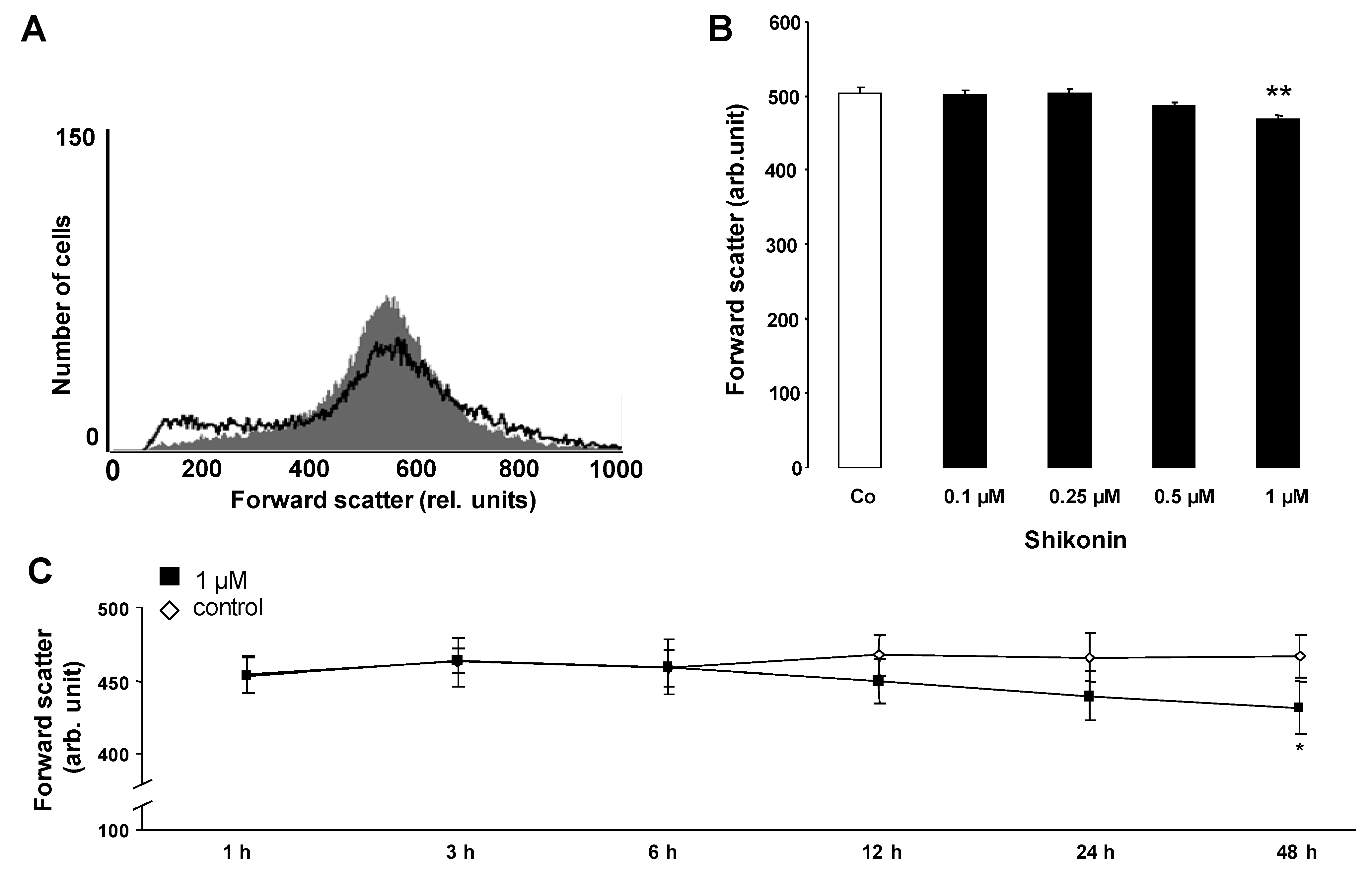
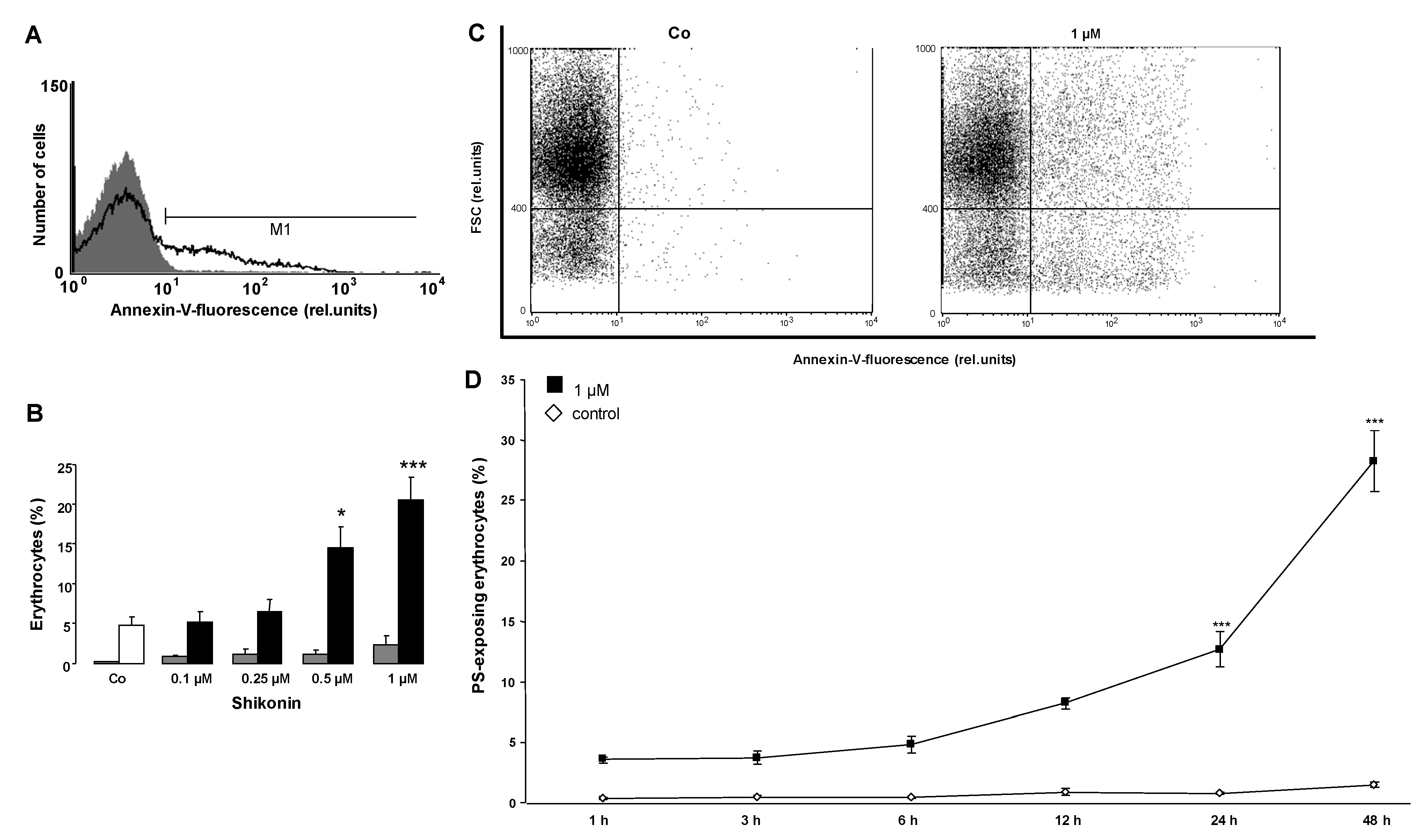
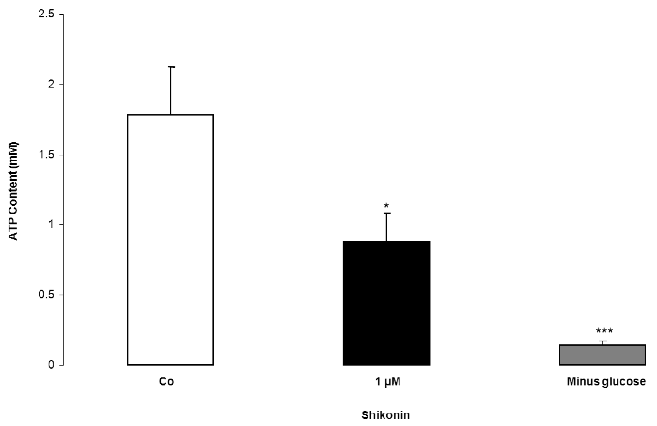
2. Results and Discussion

The present study explored whether the naphthoquinone Shikonin triggers eryptosis, the suicidal erythrocyte death characterized by cell shrinkage and phosphatidylserine translocation. As both hallmarks of eryptosis are stimulated by the increase of cytosolic Ca2+ activity ([Ca2+]i), the effect of Shikonin on [Ca2+]i was tested in a first series of experiments. To this end, human erythrocytes were loaded with Fluo3-AM and the Fluo 3 fluorescence determined by flow cytometry. Prior to the determination of Fluo 3 fluorescence, the erythrocytes were incubated for 48 h in Ringer solution without or with Shikonin (0.1–1 µM). As illustrated in Figure 1, the exposure to Shikonin was followed by an increase of Fluo 3 fluorescence, an effect reaching statistical significance at a 1-µM Shikonin concentration. Accordingly, Shikonin increased cytosolic Ca2+ concentration.
Figure 1. The effect of Shikonin on erythrocyte cytosolic Ca2+ concentration. (A) Original histogram of the Fluo 3 fluorescence in erythrocytes following exposure for 48 h to Ringer solution without (grey area) and with (black line) the presence of 1 µM Shikonin; (B) Arithmetic means ± SEM (n = 8) of the Fluo 3 fluorescence (arbitrary units) in erythrocytes exposed for 48 h to Ringer solution without (white bar) or with (black bars) Shikonin (0.1–1 µM). *** (p < 0.001) indicates significant difference from the absence of Shikonin (ANOVA); (C) Original dot blots of the Fluo 3 fluorescence as a function of forward scatter following exposure for 48 h to Ringer solution without and with the presence of 1 µM Shikonin; (D) Arithmetic means ± SEM (n = 4) of the Fluo 3 fluorescence (arbitrary units) in erythrocytes exposed for 1–48 h to Ringer solution without (white squares) or with 1 µM Shikonin (black squares). * (p < 0.05) indicates significant difference from the absence of Shikonin (ANOVA).
Toxins 06 01559 g001
An increase of [Ca2+]i may trigger cell shrinkage due to the activation of Ca2+-sensitive K+ channels and the subsequent exit of KCl and osmotically obliged water. Thus, cell volume was estimated from forward scatter in flow cytometry. As illustrated in Figure 2, a 48 h exposure to Shikonin was followed by a decrease of forward scatter, an effect reaching statistical significance at 1 µM Shikonin concentration. Accordingly, Shikonin treatment was followed by erythrocyte shrinkage.
An increased [Ca2+]i may further trigger cell membrane phospholipid scrambling with phosphatidylserine translocation to the erythrocyte surface. Phosphatidylserine exposing erythrocytes were identified utilizing annexin V binding as determined in flow cytometry. As illustrated in Figure 3, a 48 h exposure to Shikonin was followed by an increase of the percentage of annexin V binding erythrocytes, an effect reaching statistical significance at a 0.5 µM Shikonin concentration. Accordingly, Shikonin triggered erythrocyte phosphatidylserine translocation with the subsequent translocation of phosphatidylserine to the cell surface.
Figure 2. The effect of Shikonin on erythrocyte forward scatter. (A) Original histogram of the forward scatter of erythrocytes following exposure for 48 h to Ringer solution without (grey area) and with (black line) the presence of 1 µM Shikonin; (B) Arithmetic means ± SEM (n = 8) of the normalized erythrocyte forward scatter (FSC) following incubation for 48 h to Ringer solution without (white bar) or with (black bars) Shikonin (0.1–1 µM). ** (p < 0.01) indicates significant difference from the absence of Shikonin (ANOVA); (C) Arithmetic means ± SEM (n = 4) of the forward scatter (arbitrary units) in erythrocytes exposed for 1–48 h to Ringer solution without (white squares) or with 1 µM Shikonin (black squares). * (p < 0.05) indicates significant difference from the absence of Shikonin (ANOVA).
Toxins 06 01559 g002
Further experiments were performed to quantify the effect of Shikonin on hemolysis. The percentage of hemolyzed erythrocytes was calculated from the hemoglobin concentration in the supernatant. As illustrated in Figure 3, the percentage of hemolyzed erythrocytes did not significantly increase following Shikonin exposure.
In order to test whether the Shikonin induced phosphatidylserine translocation required the entry of extracellular Ca2+, erythrocytes were treated for 48 h with 1 µM Shikonin in either the presence or nominal absence of extracellular Ca2+. As illustrated in Figure 4, the effect of Shikonin on annexin V binding was significantly blunted in the nominal absence of Ca2+. However, even in the nominal absence of Shikonin, the percentage of annexin V binding erythrocytes increased significantly. Thus, the effect of Shikonin was apparently not fully dependent on Ca2+ entry.
Figure 3. The effect of Shikonin on phosphatidylserine exposure. (A) Original histogram of the annexin V binding of erythrocytes following exposure for 48 h to Ringer solution without (grey area) and with (black line) the presence of 1 µM Shikonin; (B) Arithmetic means ± SEM of the erythrocyte annexin V binding (n = 8) following incubation for 48 h to Ringer solution without (white bar) or with (black bars) the presence of Shikonin (0.1–1 µM). For comparison, the arithmetic means ± SEM of hemolysis (n = 8) following incubation for 48 h to Ringer solution without or with the presence of Shikonin (grey bars) is shown. * (p < 0.05), *** (p < 0.001) indicate significant difference from the absence of Shikonin (ANOVA); (C) Original dot blots of the annexin V binding as a function of forward scatter following exposure for 48 h to Ringer solution without and with the presence of 1 µM Shikonin; (D) Arithmetic means ± SEM (n = 4) of the annexin V binding erythrocytes (arbitrary units) following exposure for 1–48 h to Ringer solution without (white squares) or with 1 µM Shikonin (black squares). *** (p < 0.001) indicates significant difference from the absence of Shikonin (ANOVA).
Toxins 06 01559 g003
In the search for an additional mechanism, further experiments were performed in order to test whether Shikonin increased the formation of ceramide. Ceramide abundance at the erythrocyte surface was determined utilizing an anti-ceramide antibody. As illustrated in Figure 5, the exposure of erythrocytes to 1 µM Shikonin significantly increased the ceramide abundance at the erythrocyte surface.
Additional experiments tested the effect of Shikonin on erythrocytic ATP concentration. As illustrated in Figure 6, a 48 h incubation of erythrocytes with 1 µM Shikonin was followed by a significant decrease of erythrocytic ATP concentration. For a comparison, the effect of glucose removal on erythrocytic ATP concentration is shown in Figure 6.
Figure 4. The effect of Ca2+ withdrawal on Shikonin-induced annexin V binding. (A) Original histogram of the annexin V binding of erythrocytes following exposure for 48 h to Ringer solution with (grey area) and without (black line) calcium in the presence of 1 µM Shikonin; (B) Arithmetic means ± SEM (n = 4) of the percentage of annexin V binding of erythrocytes after a 48 h treatment with Ringer solution without (white bar) or with (black bars) 1 µM Shikonin in the presence (left bars, plus calcium) and absence (right bars, minus calcium) of calcium. * (p < 0.05), ** (p < 0.01) indicate significant difference from the respective values in the absence of Shikonin; # (p < 0.05) indicates significant difference from the respective value in the presence of Ca2+ (ANOVA).
Toxins 06 01559 g004
Figure 5. The effect of Shikonin on ceramide formation. (A) Original histogram of the ceramide surface abundance of erythrocytes following exposure for 48 h to Ringer solution without (grey shadow) and with (black line) the presence of 1 µM Shikonin. (B) Arithmetic means ± SEM (n = 4) of the ceramide abundance after a 48 h incubation in Ringer solution without (white bars) or with 1 µM Shikonin (black bars). * (p < 0.05) indicates significant difference from the absence of Shikonin (t-test).
Toxins 06 01559 g005
Figure 6. The effect of Shikonin on erythrocyte cytosolic ATP content. Arithmetic means ± SEM (n = 4) of the ATP concentration after a 48 h incubation in Ringer solution without (white bar) or with 1 µM Shikonin (black bar) or in glucose-depleted Ringer solution (grey bar, minus glucose) * (p < 0.05) and *** (p < 0.001) indicate significant difference from the control (absence of Shikonin and presence of glucose) (ANOVA).
Toxins 06 01559 g006
The present study reveals that Shikonin stimulates eryptosis, the suicidal death of erythrocytes [25]. The hallmark of eryptosis is phosphatidylserine translocation to the erythrocyte surface [25]. Eryptosis is further characterized by cell shrinkage [25]. Shikonin exposure of human erythrocytes leads to phosphatidylserine translocation and to cell shrinkage.
The Shikonin-induced cell shrinkage is presumably due to the increase of [Ca2+]i, which activates Ca2+-sensitive K+ channels [25], leading to K+ exit, cell membrane hyperpolarization, Cl exit and, thus, cellular loss of KCl with osmotically obliged water [26]. The cellular loss of KCl counteracts erythrocyte swelling, which may jeopardize the integrity of the erythrocyte membrane. Excessive erythrocyte swelling leads to hemolysis with the subsequent release of hemoglobin, which subsequently undergoes glomerular filtration and precipitation in the acidic tubular lumina [72].
Stimulation of phosphatidylserine translocation following Shikonin treatment is similarly a result of Ca2+ entry. Accordingly, the effect of Shikonin was significantly blunted in the nominal absence of Ca2+. Removal of Ca2+ decreased phosphatidylserine translocation even in the absence of Shikonin, indicating that spontaneous eryptosis following the exposure of erythrocytes to Ringer without Shikonin was similarly, in part, caused by cytosolic Ca2+. However, significant phosphatidylserine exposure was observed in the presence of Shikonin even in the nominal absence of Ca2+, indicating that Shikonin was effective by an additional mechanism. As a matter of fact, Shikonin triggered the formation of ceramide, which, in turn, fosters phosphatidylserine translocation [25]. Ceramide is formed by a sphingomyelinase, which is, in turn, activated by a platelet activating factor [25]. Ceramide fosters suicidal death similarly in nucleated cells [73]. It is effective by potentiating proapoptotic signaling [73]. Shikonin further decreases erythrocytic ATP concentration, another known trigger of eryptosis [25].
The molecular mechanism underlying phosphatidylserine translocation has not yet been identified [25]. Recently, Anoctamin 6 (Ano6; TMEM16F gene) has been suggested to mediate cell membrane scrambling [74]. The molecule may function as a Cl channel, a Ca2+-regulated nonselective Ca2+ permeable cation channel, a scramblase mediating phosphatidylserine translocation upon the increase of cytosolic Ca2+ and a regulator of cell membrane blebbing and microparticle shedding [74]. However, Ano6 is inhibited by tannic acid [75], which itself triggers phosphatidylserine translocation in erythrocytes [60]. Thus, the role of Ano6 in the regulation of phosphatidylserine translocation in the erythrocyte membrane remains elusive.
Stimulation of eryptosis may be favorable, as it allows the elimination of defective erythrocytes prior to hemolysis [25]. The phosphatidylserine exposure at the cell surface may be particularly important during the course of malaria, as infected phosphatidylserine exposing cells may be recognized and, thus, rapidly removed from circulating blood. [76]. Infected erythrocytes undergo eryptosis, as the intraerythrocytic parasite activates several ion channels, including the Ca2+-permeable erythrocyte cation channels [77,78]. Activation of the channels in the host cell membrane is required for the intraerythrocytic survival of the pathogen [77,78], as the channels provide the pathogen with nutrients, Na+ and Ca2+, and they mediate the disposal of intracellular waste products [78]. By the same token, the Ca2+ entry through the Ca2+-permeable cation channels triggers eryptosis [76], which is, in turn, followed by the rapid clearance of the infected erythrocytes from circulating blood [25]. Ca2+ entry and Ca2+-induced eryptosis thus lead to elimination, not only of the infected erythrocyte, but also of the pathogen [76].
Accordingly, genetic disorders predisposing to accelerated eryptosis, such as the sickle-cell trait, the beta-thalassemia trait, homozygous Hb-C and G6PD deficiency [25], lead to a relatively mild clinical course of malaria following infection with Plasmodia [79,80,81]. Moreover, several clinical conditions, such as iron deficiency [82], and eryptosis stimulating drugs, such as lead [83], chlorpromazine [84] or NO synthase inhibitors [85], have been shown to favorably influence the clinical course of malaria. It may be worth considering whether Shikonin similarly decreases parasitemia in malaria. At least in theory, Shikonin and further proeryptotic substances could be employed for the treatment of malaria. However, the applicability of Shikonin in the treatment of malaria has not been tested, and its clinical use may depend on further properties and side effects not studied here.
Excessive stimulation of eryptosis may lead to anemia. Phosphatidylserine at the surface of eryptotic cells triggers the phagocytosis of the cells and, thus, leads to the rapid removal of the suicidal erythrocytes from circulating blood [25]. Anemia develops, if the accelerated clearance of erythrocytes during stimulated eryptosis outcasts the formation of new erythrocytes [25]. Phosphatidylserine exposing erythrocytes may further interfere with microcirculation [86,87,88,89,90,91]. The phosphatidylserine exposing cells adhere to endothelial CXCL16/SR-PSO [87] and may stimulate blood clotting and thrombosis [86,92,93].
Anemia and compromised microcirculation may, at least in theory, limit the use of Shikonin. The substance has been proposed to counteract oxidative stress [1], inflammation [1,2,3], thrombosis [1,2], infections [1,2] and cancer [2,5,6,7,8]. Moreover, Shikonin has been used to support wound healing [1,2,5]. However, the toxicity of Shikonin is still ill-defined [1]. The present observations point to a novel potentially toxic effect of Shikonin. The local application of the substance, such as in creams and ointments [1], is not expected to trigger eryptosis. Systemic application of the substance may, however, jeopardize erythrocyte survival and microcirculation, thus potentially limiting the use of the substance. This may particularly be true in patients suffering from diseases with enhanced eryptosis [25] or receiving other eryptosis-inducing xenobiotics [25,41,42,43,44,45,46,47,48,49,50,51,52,53,54,55,56,57,58,59,60,61,62,63,64,65,66,67,68,69,70,71]. After oral and intramuscular administration, Shikonin is rapidly absorbed and has a half-life in plasma of 8.8 h and a distribution volume of 8.91 L/kg [1]. Several approaches have improved the stability and water solubility of Shikonin, thus providing the basis for the systemic application of the substance [1]. Future studies will be required to define the impact of Shikonin on erythrocyte survival and microcirculation following systemic application of Shikonin.

3. Experimental Section

3.1. Erythrocytes, Solutions and Chemicals

Leukocyte-depleted erythrocytes were kindly provided by the blood bank of the University of Tübingen. The study is approved by the ethics committee of the University of Tübingen (184/2003V). Erythrocytes were incubated in vitro at a hematocrit of 0.4% in Ringer solution containing (in mM) 125 NaCl, 5 KCl, 1 MgSO4, 32 N-2-hydroxyethylpiperazine-N-2-ethanesulfonic acid (HEPES), 5 glucose, 1 CaCl2; pH 7.4 at 37°C for 48 h. Where indicated, erythrocytes were exposed to Shikonin (Enzo, Lörrach, Germany) at the indicated concentrations. In Ca2+-free Ringer solution, 1-mM CaCl2 was substituted by 1-mM glycol-bis(2-aminoethylether)-N,N,N',N'-tetraacetic acid (EGTA).

3.2. Analysis of Annexin V Binding and Forward Scatter

After incubation under the respective experimental condition, a 50 µL cell suspension was washed in Ringer solution containing 5-mM CaCl2 and then stained with annexin V FITC (1:200 dilution; ImmunoTools, Friesoythe, Germany) in this solution at 37 °C for 20 min under protection from light. In the following, the forward scatter (FSC) of the cells was determined, and the annexin V fluorescence intensity was measured with an excitation wavelength of 488 nm and an emission wavelength of 530 nm on a FACS Calibur (BD, Heidelberg, Germany).

3.3. Measurement of Intracellular Ca2+

After incubation, erythrocytes were washed in Ringer solution and then loaded with Fluo-3/AM (Biotium, Hayward, USA) in Ringer solution containing 5-mM CaCl2 and 5-µM Fluo-3/AM. The cells were incubated at 37 °C for 30 min and washed twice in Ringer solution containing 5-mM CaCl2. The Fluo-3/AM-loaded erythrocytes were resuspended in 200 µL of Ringer. Then, Ca2+-dependent fluorescence intensity was measured with an excitation wavelength of 488 nm and an emission wavelength of 530 nm on a FACS Calibur.

3.4. Determination of Ceramide Formation

For the determination of ceramide, a monoclonal antibody-based assay was used. After incubation, cells were stained for 1 hour at 37 °C with 1 µg/mL of anti-ceramide antibody (clone MID 15B4, Alexis, Grünberg, Germany) in PBS containing 0.1% bovine serum albumin (BSA) at a dilution of 1:5. The samples were washed twice with PBS-BSA. Subsequently, the cells were stained for 30 minutes with polyclonal fluorescein isothiocyanate (FITC) conjugated goat anti-mouse IgG and IgM-specific antibody (Pharmingen, Hamburg, Germany) diluted 1:50 in PBS-BSA. Unbound secondary antibody was removed by repeated washing with PBS-BSA. The samples were then analyzed by flow cytometric analysis with an excitation wavelength of 488 nm and an emission wavelength of 530 nm.

3.5. ATP Content

For the determination of the intracellular ATP concentration, erythrocytes were lysed in distilled water and proteins were precipitated by HClO4 (5%). After centrifugation, an aliquot of the supernatant (400 µL) was adjusted to pH 7.7 by the addition of saturated KHCO3 solution. All manipulations were performed at 4 °C to avoid ATP degradation. After dilution of the supernatant, the ATP concentration of the aliquots was determined utilizing the luciferin-luciferase assay kit (Roche Diagnostics, Mannheim, Germany) and a luminometer (Berthold Biolumat LB9500, Bad Wildbad, Germany) according to the manufacturer´s protocol.

3.6. Statistics

Data are expressed as arithmetic means ± SEM. As indicated in the figure legends, statistical analysis was made using ANOVA with Tukey’s test as the post-test and the t-test as appropriate. n denotes the number of different erythrocyte specimens studied. Since different erythrocyte specimens used in distinct experiments are differently susceptible to the triggers of eryptosis, the same erythrocyte specimens have been used for control and experimental conditions.

4. Conclusions

Shikonin stimulates Ca2+ entry and triggers ceramide formation, which, in turn, leads to shrinkage and phosphatidylserine translocation of erythrocytes. Accordingly, Shikonin triggers eryptosis, the suicidal erythrocyte death.

Acknowledgments

The authors acknowledge the meticulous preparation of the manuscript by Tanja Loch. The study was supported by the Deutsche Forschungsgemeinschaft and the Open Access Publishing Fund of Tuebingen University.

Authors Contributions

Adrian Lupescu organized and supervised the study. He executed the statistical analysis and prepared the figures. Rosi Bissinger and Kashif Jilani performed the experiments. Florian Lang designed the study and drafted the manuscript. All authors carefully read and approved the manuscript.

Conflicts of Interest

The authors declare no conflict of interest.

References

  1. Andujar, I.; Rios, J.L.; Giner, R.M.; Recio, M.C. Pharmacological properties of shikonin—A review of literature since 2002. Planta Med. 2013, 79, 1685–1697. [Google Scholar] [CrossRef]
  2. Chen, X.; Yang, L.; Oppenheim, J.J.; Howard, O.M.Z. Cellular pharmacology studies of shikonin derivatives. Phytother. Res. 2002, 16, 199–209. [Google Scholar] [CrossRef]
  3. Lu, L.; Qin, A.; Huang, H.; Zhou, P.; Zhang, C.; Liu, N.; Li, S.; Wen, G.; Zhang, C.; Dong, W.; et al. Shikonin extracted from medicinal chinese herbs exerts anti-inflammatory effect via proteasome inhibition. Eur. J. Pharmacol. 2011, 658, 242–247. [Google Scholar] [CrossRef]
  4. Gao, H.; Liu, L.; Qu, Z.Y.; Wei, F.X.; Wang, S.Q.; Chen, G.A.; Qin, L.; Jiang, F.Y.; Wang, Y.C.; Shang, L.; et al. Anti-adenovirus activities of shikonin, a component of chinese herbal medicine in vitro. Biol. Pharm. Bull. 2011, 34, 197–202. [Google Scholar] [CrossRef]
  5. Andujar, I.; Rios, J.L.; Giner, R.M.; Recio, M.C. Shikonin promotes intestinal wound healing in vitro via induction of tgf-beta release in iec-18 cells. Eur. J. Pharm. Sci. 2013, 49, 637–641. [Google Scholar] [CrossRef]
  6. Chen, H.M.; Wang, P.H.; Chen, S.S.; Wen, C.C.; Chen, Y.H.; Yang, W.C.; Yang, N.S. Shikonin induces immunogenic cell death in tumor cells and enhances dendritic cell-based cancer vaccine. Cancer Immunol. Immunother. 2012, 61, 1989–2002. [Google Scholar] [CrossRef]
  7. Wang, Y.; Zhou, Y.; Jia, G.; Han, B.; Liu, J.; Teng, Y.; Lv, J.; Song, Z.; Li, Y.; Ji, L.; et al. Shikonin suppresses tumor growth and synergizes with gemcitabine in a pancreatic cancer xenograft model: Involvement of nf-kappab signaling pathway. Biochem. Pharmacol. 2014, 88, 322–333. [Google Scholar] [CrossRef]
  8. Wang, R.; Yin, R.; Zhou, W.; Xu, D.; Li, S. Shikonin and its derivatives: A patent review. Expert Opin. Ther. Pat. 2012, 22, 977–997. [Google Scholar] [CrossRef]
  9. Chen, C.H.; Lin, M.L.; Ong, P.L.; Yang, J.T. Novel multiple apoptotic mechanism of shikonin in human glioma cells. Ann. Surg. Oncol. 2012, 19, 3097–3106. [Google Scholar] [CrossRef]
  10. Han, W.; Li, L.; Qiu, S.; Lu, Q.; Pan, Q.; Gu, Y.; Luo, J.; Hu, X. Shikonin circumvents cancer drug resistance by induction of a necroptotic death. Mol. Cancer Ther. 2007, 6, 1641–1649. [Google Scholar] [CrossRef]
  11. Hou, Y.; Guo, T.; Wu, C.; He, X.; Zhao, M. Effect of shikonin on human breast cancer cells proliferation and apoptosis in vitro. J. Pharm. Soc. Jap. 2006, 126, 1383–1386. [Google Scholar]
  12. Huang, C.J.; Luo, Y.A.; Zhao, J.W.; Yang, F.W.; Zhao, H.W.; Fan, W.H.; Ge, P.F. Shikonin kills glioma cells through necroptosis mediated by rip-1. PLoS ONE 2013, 8, e66326. [Google Scholar]
  13. Liu, C.; Yin, L.; Chen, J.; Chen, J. The apoptotic effect of shikonin on human papillary thyroid carcinoma cells through mitochondrial pathway. Tumour. Biol. 2013, 35, 1791–1798. [Google Scholar]
  14. Park, S.; Shin, H.; Cho, Y. Shikonin induces programmed necrosis-like cell death through the formation of receptor interacting protein 1 and 3 complex. Food Chem. Toxicol. 2013, 55, 36–41. [Google Scholar] [CrossRef]
  15. Piao, J.L.; Cui, Z.G.; Furusawa, Y.; Ahmed, K.; Rehman, M.U.; Tabuchi, Y.; Kadowaki, M.; Kondo, T. The molecular mechanisms and gene expression profiling for shikonin-induced apoptotic and necroptotic cell death in u937 cells. Chem. Biol. Interact. 2013, 205, 119–127. [Google Scholar] [CrossRef]
  16. Yingkun, N.; Lvsong, Z.; Huimin, Y. Shikonin inhibits the proliferation and induces the apoptosis of human hepg2 cells. Can. J. Physiol. Pharmacol. 2010, 88, 1138–1146. [Google Scholar] [CrossRef]
  17. Zhang, F.L.; Wang, P.; Liu, Y.H.; Liu, L.B.; Liu, X.B.; Li, Z.; Xue, Y.X. Topoisomerase inhibitors, shikonin and topotecan, inhibit growth and induce apoptosis of glioma cells and glioma stem cells. PLoS ONE 2013, 8, e81815. [Google Scholar]
  18. Ahn, J.; Won, M.; Choi, J.H.; Kim, Y.S.; Jung, C.R.; Im, D.S.; Kyun, M.L.; Lee, K.; Song, K.B.; Chung, K.S. Reactive oxygen species-mediated activation of the akt/ask1/p38 signaling cascade and p21(cip1) downregulation are required for shikonin-induced apoptosis. Apoptosis 2013, 18, 870–881. [Google Scholar]
  19. Chang, I.C.; Huang, Y.J.; Chiang, T.I.; Yeh, C.W.; Hsu, L.S. Shikonin induces apoptosis through reactive oxygen species/extracellular signal-regulated kinase pathway in osteosarcoma cells. Biol. Pharm. Bull. 2010, 33, 816–824. [Google Scholar] [CrossRef]
  20. Chen, C.H.; Chern, C.L.; Lin, C.C.; Lu, F.J.; Shih, M.K.; Hsieh, P.Y.; Liu, T.Z. Involvement of reactive oxygen species, but not mitochondrial permeability transition in the apoptotic induction of human sk-hep-1 hepatoma cells by shikonin. Planta Med. 2003, 69, 1119–1124. [Google Scholar] [CrossRef]
  21. Gong, K.; Li, W. Shikonin, a chinese plant-derived naphthoquinone, induces apoptosis in hepatocellular carcinoma cells through reactive oxygen species: A potential new treatment for hepatocellular carcinoma. Free Radic. Biol. Med. 2011, 51, 2259–2271. [Google Scholar] [CrossRef]
  22. Lee, M.J.; Kao, S.H.; Hunag, J.E.; Sheu, G.T.; Yeh, C.W.; Hseu, Y.C.; Wang, C.J.; Hsu, L.S. Shikonin time-dependently induced necrosis or apoptosis in gastric cancer cells via generation of reactive oxygen species. Chem. Biol. Interact. 2014, 211C, 44–53. [Google Scholar]
  23. Mao, X.; Yu, C.R.; Li, W.H.; Li, W.X. Induction of apoptosis by shikonin through a ros/jnk-mediated process in bcr/abl-positive chronic myelogenous leukemia (cml) cells. Cell. Res. 2008, 18, 879–888. [Google Scholar] [CrossRef]
  24. Yeh, C.C.; Kuo, H.M.; Li, T.M.; Lin, J.P.; Yu, F.S.; Lu, H.F.; Chung, J.G.; Yang, J.S. Shikonin-induced apoptosis involves caspase-3 activity in a human bladder cancer cell line (t24). In Vivo 2007, 21, 1011–1019. [Google Scholar]
  25. Lang, E.; Qadri, S.M.; Lang, F. Killing me softly—Suicidal erythrocyte death. Int. J. Biochem. Cell. Biol. 2012, 44, 1236–1243. [Google Scholar] [CrossRef]
  26. Lang, P.A.; Kaiser, S.; Myssina, S.; Wieder, T.; Lang, F.; Huber, S.M. Role of Ca2+-activated K+ channels in human erythrocyte apoptosis. Am. J. Physiol. Cell. Physiol. 2003, 285, C1553–C1560. [Google Scholar] [CrossRef]
  27. Bhavsar, S.K.; Bobbala, D.; Xuan, N.T.; Foller, M.; Lang, F. Stimulation of suicidal erythrocyte death by alpha-lipoic acid. Cell. Physiol. Biochem. 2010, 26, 859–868. [Google Scholar] [CrossRef]
  28. Foller, M.; Huber, S.M.; Lang, F. Erythrocyte programmed cell death. IUBMB Life 2008, 60, 661–668. [Google Scholar] [CrossRef]
  29. Foller, M.; Mahmud, H.; Gu, S.; Wang, K.; Floride, E.; Kucherenko, Y.; Luik, S.; Laufer, S.; Lang, F. Participation of leukotriene c(4) in the regulation of suicidal erythrocyte death. J. Physiol. Pharmacol. 2009, 60, 135–143. [Google Scholar]
  30. Lau, I.P.; Chen, H.; Wang, J.; Ong, H.C.; Leung, K.C.; Ho, H.P.; Kong, S.K. In vitro effect of ctab- and peg-coated gold nanorods on the induction of eryptosis/erythroptosis in human erythrocytes. Nanotoxicology 2012, 6, 847–856. [Google Scholar]
  31. Maellaro, E.; Leoncini, S.; Moretti, D.; Del Bello, B.; Tanganelli, I.; De Felice, C.; Ciccoli, L. Erythrocyte caspase-3 activation and oxidative imbalance in erythrocytes and in plasma of type 2 diabetic patients. Acta Diabetol. 2013, 50, 489–495. [Google Scholar]
  32. Foller, M.; Sopjani, M.; Koka, S.; Gu, S.; Mahmud, H.; Wang, K.; Floride, E.; Schleicher, E.; Schulz, E.; Munzel, T.; et al. Regulation of erythrocyte survival by amp-activated protein kinase. FASEB J. 2009, 23, 1072–1080. [Google Scholar]
  33. Kucherenko, Y.; Zelenak, C.; Eberhard, M.; Qadri, S.M.; Lang, F. Effect of casein kinase 1alpha activator pyrvinium pamoate on erythrocyte ion channels. Cell. Physiol. Biochem. 2012, 30, 407–417. [Google Scholar]
  34. Zelenak, C.; Eberhard, M.; Jilani, K.; Qadri, S.M.; Macek, B.; Lang, F. Protein kinase ck1alpha regulates erythrocyte survival. Cell. Physiol. Biochem. 2012, 29, 171–180. [Google Scholar]
  35. Foller, M.; Feil, S.; Ghoreschi, K.; Koka, S.; Gerling, A.; Thunemann, M.; Hofmann, F.; Schuler, B.; Vogel, J.; Pichler, B.; et al. Anemia and splenomegaly in cgki-deficient mice. Proc. Natl. Acad. Sci. USA 2008, 105, 6771–6776. [Google Scholar] [CrossRef] [Green Version]
  36. Bhavsar, S.K.; Gu, S.; Bobbala, D.; Lang, F. Janus kinase 3 is expressed in erythrocytes, phosphorylated upon energy depletion and involved in the regulation of suicidal erythrocyte death. Cell. Physiol. Biochem. 2011, 27, 547–556. [Google Scholar]
  37. Klarl, B.A.; Lang, P.A.; Kempe, D.S.; Niemoeller, O.M.; Akel, A.; Sobiesiak, M.; Eisele, K.; Podolski, M.; Huber, S.M.; Wieder, T.; et al. Protein kinase c mediates erythrocyte “programmed cell death” following glucose depletion. Am. J. Physiol. Cell. Physiol. 2006, 290, C244–C253. [Google Scholar]
  38. Gatidis, S.; Zelenak, C.; Fajol, A.; Lang, E.; Jilani, K.; Michael, D.; Qadri, S.M.; Lang, F. P38 mapk activation and function following osmotic shock of erythrocytes. Cell. Physiol. Biochem. 2011, 28, 1279–1286. [Google Scholar]
  39. Zelenak, C.; Foller, M.; Velic, A.; Krug, K.; Qadri, S.M.; Viollet, B.; Lang, F.; Macek, B. Proteome analysis of erythrocytes lacking amp-activated protein kinase reveals a role of pak2 kinase in eryptosis. J. Proteome Res. 2011, 10, 1690–1697. [Google Scholar]
  40. Lupescu, A.; Shaik, N.; Jilani, K.; Zelenak, C.; Lang, E.; Pasham, V.; Zbidah, M.; Plate, A.; Bitzer, M.; Foller, M.; et al. Enhanced erythrocyte membrane exposure of phosphatidylserine following sorafenib treatment: An in vivo and in vitro study. Cell. Physiol. Biochem. 2012, 30, 876–888. [Google Scholar]
  41. Shaik, N.; Lupescu, A.; Lang, F. Sunitinib-sensitive suicidal erythrocyte death. Cell. Physiol. Biochem. 2012, 30, 512–522. [Google Scholar] [CrossRef]
  42. Abed, M.; Towhid, S.T.; Mia, S.; Pakladok, T.; Alesutan, I.; Borst, O.; Gawaz, M.; Gulbins, E.; Lang, F. Sphingomyelinase-induced adhesion of eryptotic erythrocytes to endothelial cells. Am. J. Physiol. Cell. Physiol. 2012, 303, C991–C999. [Google Scholar] [CrossRef]
  43. Bottger, E.; Multhoff, G.; Kun, J.F.; Esen, M. Plasmodium falciparum-infected erythrocytes induce granzyme b by nk cells through expression of host-hsp70. PLoS ONE 2012, 7, e33774. [Google Scholar] [CrossRef]
  44. Firat, U.; Kaya, S.; Cim, A.; Buyukbayram, H.; Gokalp, O.; Dal, M.S.; Tamer, M.N. Increased caspase-3 immunoreactivity of erythrocytes in stz diabetic rats. Exp. Diabetes Res. 2012, 2012. [Google Scholar] [CrossRef]
  45. Ganesan, S.; Chaurasiya, N.D.; Sahu, R.; Walker, L.A.; Tekwani, B.L. Understanding the mechanisms for metabolism-linked hemolytic toxicity of primaquine against glucose 6-phosphate dehydrogenase deficient human erythrocytes: Evaluation of eryptotic pathway. Toxicology 2012, 294, 54–60. [Google Scholar] [CrossRef]
  46. Gao, M.; Cheung, K.L.; Lau, I.P.; Yu, W.S.; Fung, K.P.; Yu, B.; Loo, J.F.; Kong, S.K. Polyphyllin d induces apoptosis in human erythrocytes through Ca(2)(+) rise and membrane permeabilization. Arch. Toxicol. 2012, 86, 741–752. [Google Scholar]
  47. Ghashghaeinia, M.; Cluitmans, J.C.; Akel, A.; Dreischer, P.; Toulany, M.; Koberle, M.; Skabytska, Y.; Saki, M.; Biedermann, T.; Duszenko, M.; et al. The impact of erythrocyte age on eryptosis. Br. J. Haematol. 2012, 157, 606–614. [Google Scholar] [CrossRef]
  48. Jilani, K.; Lupescu, A.; Zbidah, M.; Abed, M.; Shaik, N.; Lang, F. Enhanced apoptotic death of erythrocytes induced by the mycotoxin ochratoxin A. Kidney Blood Press Res. 2012, 36, 107–118. [Google Scholar]
  49. Jilani, K.; Lupescu, A.; Zbidah, M.; Shaik, N.; Lang, F. Withaferin a-stimulated Ca2+ entry, ceramide formation and suicidal death of erythrocytes. Toxicol. In Vitro 2013, 27, 52–58. [Google Scholar]
  50. Kucherenko, Y.V.; Lang, F. Inhibitory effect of furosemide on non-selective voltage-independent cation channels in human erythrocytes. Cell. Physiol. Biochem. 2012, 30, 863–875. [Google Scholar] [CrossRef]
  51. Lang, E.; Qadri, S.M.; Jilani, K.; Zelenak, C.; Lupescu, A.; Schleicher, E.; Lang, F. Carbon monoxide-sensitive apoptotic death of erythrocytes. Basic Clin. Pharmacol. Toxicol. 2012, 111, 348–355. [Google Scholar]
  52. Polak-Jonkisz, D.; Purzyc, L. Ca influx versus efflux during eryptosis in uremic erythrocytes. Blood Purif. 2012, 34, 209–210. [Google Scholar] [CrossRef]
  53. Qian, E.W.; Ge, D.T.; Kong, S.K. Salidroside protects human erythrocytes against hydrogen peroxide-induced apoptosis. J. Nat. Prod. 2012, 75, 531–537. [Google Scholar] [CrossRef]
  54. Shaik, N.; Zbidah, M.; Lang, F. Inhibition of Ca(2+) entry and suicidal erythrocyte death by naringin. Cell. Physiol. Biochem. 2012, 30, 678–686. [Google Scholar] [CrossRef]
  55. Vota, D.M.; Maltaneri, R.E.; Wenker, S.D.; Nesse, A.B.; Vittori, D.C. Differential erythropoietin action upon cells induced to eryptosis by different agents. Cell. Biochem. Biophys. 2013, 65, 145–157. [Google Scholar] [CrossRef]
  56. Weiss, E.; Cytlak, U.M.; Rees, D.C.; Osei, A.; Gibson, J.S. Deoxygenation-induced and Ca(2+) dependent phosphatidylserine externalisation in red blood cells from normal individuals and sickle cell patients. Cell. Calcium 2012, 51, 51–56. [Google Scholar]
  57. Zappulla, D. Environmental stress, erythrocyte dysfunctions, inflammation, and the metabolic syndrome: Adaptations to CO2 increases? J. Cardiometab. Syndr. 2008, 3, 30–34. [Google Scholar] [CrossRef]
  58. Zbidah, M.; Lupescu, A.; Jilani, K.; Lang, F. Stimulation of suicidal erythrocyte death by fumagillin. Basic Clin. Pharmacol. Toxicol. 2013, 112, 346–351. [Google Scholar]
  59. Zelenak, C.; Pasham, V.; Jilani, K.; Tripodi, P.M.; Rosaclerio, L.; Pathare, G.; Lupescu, A.; Faggio, C.; Qadri, S.M.; Lang, F. Tanshinone IIA stimulates erythrocyte phosphatidylserine exposure. Cell. Physiol. Biochem. 2012, 30, 282–294. [Google Scholar] [CrossRef]
  60. Abed, M.; Herrmann, T.; Alzoubi, K.; Pakladok, T.; Lang, F. Tannic acid induced suicidal erythrocyte death. Cell. Physiol. Biochem. 2013, 32, 1106–1116. [Google Scholar] [CrossRef]
  61. Ahmed, M.S.; Langer, H.; Abed, M.; Voelkl, J.; Lang, F. The uremic toxin acrolein promotes suicidal erythrocyte death. Kidney Blood Press Res. 2013, 37, 158–167. [Google Scholar] [CrossRef]
  62. Ghashghaeinia, M.; Cluitmans, J.C.; Toulany, M.; Saki, M.; Koberle, M.; Lang, E.; Dreischer, P.; Biedermann, T.; Duszenko, M.; Lang, F.; et al. Age sensitivity of nfkappab abundance and programmed cell death in erythrocytes induced by nfkappab inhibitors. Cell. Physiol. Biochem. 2013, 32, 801–813. [Google Scholar] [CrossRef]
  63. Abed, M.; Feger, M.; Alzoubi, K.; Pakladok, T.; Frauenfeld, L.; Geiger, C.; Towhid, S.T.; Lang, F. Sensitization of erythrocytes to suicidal erythrocyte death following water deprivation. Kidney Blood Press Res. 2013, 37, 567–578. [Google Scholar]
  64. Alzoubi, K.; Honisch, S.; Abed, M.; Lang, F. Triggering of suicidal erythrocyte death by penta-o-galloyl-beta-d-glucose. Toxins 2014, 6, 54–65. [Google Scholar] [CrossRef]
  65. Jilani, K.; Qadri, S.M.; Lang, F. Geldanamycin-induced phosphatidylserine translocation in the erythrocyte membrane. Cell. Physiol. Biochem. 2013, 32, 1600–1609. [Google Scholar]
  66. Jilani, K.; Lang, F. Carmustine-induced phosphatidylserine translocation in the erythrocyte membrane. Toxins 2013, 5, 703–716. [Google Scholar] [CrossRef]
  67. Jilani, K.; Enkel, S.; Bissinger, R.; Almilaji, A.; Abed, M.; Lang, F. Fluoxetine induced suicidal erythrocyte death. Toxins 2013, 5, 1230–1243. [Google Scholar] [CrossRef]
  68. Bissinger, R.; Modicano, P.; Frauenfeld, L.; Lang, E.; Jacobi, J.; Faggio, C.; Lang, F. Estramustine-induced suicidal erythrocyte death. Cell. Physiol. Biochem. 2013, 32, 1426–1436. [Google Scholar] [CrossRef]
  69. Lupescu, A.; Jilani, K.; Zbidah, M.; Lang, F. Patulin-induced suicidal erythrocyte death. Cell. Physiol. Biochem. 2013, 32, 291–299. [Google Scholar] [CrossRef]
  70. Lupescu, A.; Bissinger, R.; Jilani, K.; Lang, F. Triggering of suicidal erythrocyte death by celecoxib. Toxins 2013, 5, 1543–1554. [Google Scholar] [CrossRef]
  71. Lang, E.; Modicano, P.; Arnold, M.; Bissinger, R.; Faggio, C.; Abed, M.; Lang, F. Effect of thioridazine on erythrocytes. Toxins 2013, 5, 1918–1931. [Google Scholar] [CrossRef]
  72. Harrison, H.E.; Bunting, H.; Ordway, N.K.; Albrink, W.S. The pathogenesis of the renal injury produced in the dog by hemoglobin or methemoglobin. J. Exp. Med. 1947, 86, 339–356. [Google Scholar] [CrossRef]
  73. Morad, S.A.; Cabot, M.C. Ceramide-orchestrated signalling in cancer cells. Nat. Rev. Cancer 2013, 13, 51–65. [Google Scholar] [CrossRef]
  74. Kunzelmann, K.; Nilius, B.; Owsianik, G.; Schreiber, R.; Ousingsawat, J.; Sirianant, L.; Wanitchakool, P.; Bevers, E.M.; Heemskerk, J.W. Molecular functions of anoctamin 6 (tmem16f): A chloride channel, cation channel, or phospholipid scramblase? Pflugers Arch. 2014, 466, 407–414. [Google Scholar]
  75. Szteyn, K.; Schmid, E.; Nurbaeva, M.K.; Yang, W.; Munzer, P.; Kunzelmann, K.; Lang, F.; Shumilina, E. Expression and functional significance of the Ca(2+)-activated Cl(−) channel ano6 in dendritic cells. Cell. Physiol. Biochem. 2012, 30, 1319–1332. [Google Scholar] [CrossRef]
  76. Foller, M.; Bobbala, D.; Koka, S.; Huber, S.M.; Gulbins, E.; Lang, F. Suicide for survival—Death of infected erythrocytes as a host mechanism to survive malaria. Cell. Physiol. Biochem. 2009, 24, 133–140. [Google Scholar] [CrossRef]
  77. Duranton, C.; Huber, S.; Tanneur, V.; Lang, K.; Brand, V.; Sandu, C.; Lang, F. Electrophysiological properties of the plasmodium falciparum-induced cation conductance of human erythrocytes. Cell. Physiol. Biochem. 2003, 13, 189–198. [Google Scholar] [CrossRef]
  78. Kirk, K. Membrane transport in the malaria-infected erythrocyte. Physiol. Rev. 2001, 81, 495–537. [Google Scholar]
  79. Ayi, K.; Giribaldi, G.; Skorokhod, A.; Schwarzer, E.; Prendergast, P.T.; Arese, P. 16alpha-bromoepiandrosterone, an antimalarial analogue of the hormone dehydroepiandrosterone, enhances phagocytosis of ring stage parasitized erythrocytes: A novel mechanism for antimalarial activity. Antimicrob. Agents Chemother. 2002, 46, 3180–3184. [Google Scholar] [CrossRef]
  80. Ayi, K.; Turrini, F.; Piga, A.; Arese, P. Enhanced phagocytosis of ring-parasitized mutant erythrocytes: A common mechanism that may explain protection against falciparum malaria in sickle trait and beta-thalassemia trait. Blood 2004, 104, 3364–3371. [Google Scholar] [CrossRef]
  81. Cappadoro, M.; Giribaldi, G.; O’Brien, E.; Turrini, F.; Mannu, F.; Ulliers, D.; Simula, G.; Luzzatto, L.; Arese, P. Early phagocytosis of glucose-6-phosphate dehydrogenase (g6pd)-deficient erythrocytes parasitized by plasmodium falciparum may explain malaria protection in g6pd deficiency. Blood 1998, 92, 2527–2534. [Google Scholar]
  82. Koka, S.; Foller, M.; Lamprecht, G.; Boini, K.M.; Lang, C.; Huber, S.M.; Lang, F. Iron deficiency influences the course of malaria in plasmodium berghei infected mice. Biochem. Biophys. Res. Commun. 2007, 357, 608–614. [Google Scholar]
  83. Koka, S.; Huber, S.M.; Boini, K.M.; Lang, C.; Foller, M.; Lang, F. Lead decreases parasitemia and enhances survival of plasmodium berghei-infected mice. Biochem. Biophys. Res. Commun. 2007, 363, 484–489. [Google Scholar] [CrossRef]
  84. Koka, S.; Lang, C.; Boini, K.M.; Bobbala, D.; Huber, S.M.; Lang, F. Influence of chlorpromazine on eryptosis, parasitemia and survival of plasmodium berghe infected mice. Cell. Physiol. Biochem. 2008, 22, 261–268. [Google Scholar] [CrossRef]
  85. Koka, S.; Lang, C.; Niemoeller, O.M.; Boini, K.M.; Nicolay, J.P.; Huber, S.M.; Lang, F. Influence of no synthase inhibitor l-name on parasitemia and survival of plasmodium berghei infected mice. Cell. Physiol. Biochem. 2008, 21, 481–488. [Google Scholar] [CrossRef]
  86. Andrews, D.A.; Low, P.S. Role of red blood cells in thrombosis. Curr. Opin. Hematol. 1999, 6, 76–82. [Google Scholar] [CrossRef]
  87. Borst, O.; Abed, M.; Alesutan, I.; Towhid, S.T.; Qadri, S.M.; Foller, M.; Gawaz, M.; Lang, F. Dynamic adhesion of eryptotic erythrocytes to endothelial cells via cxcl16/sr-psox. Am. J. Physiol. Cell. Physiol. 2012, 302, C644–C651. [Google Scholar] [CrossRef]
  88. Closse, C.; Dachary-Prigent, J.; Boisseau, M.R. Phosphatidylserine-related adhesion of human erythrocytes to vascular endothelium. Br. J. Haematol. 1999, 107, 300–302. [Google Scholar] [CrossRef]
  89. Gallagher, P.G.; Chang, S.H.; Rettig, M.P.; Neely, J.E.; Hillery, C.A.; Smith, B.D.; Low, P.S. Altered erythrocyte endothelial adherence and membrane phospholipid asymmetry in hereditary hydrocytosis. Blood 2003, 101, 4625–4627. [Google Scholar]
  90. Pandolfi, A.; Di Pietro, N.; Sirolli, V.; Giardinelli, A.; Di Silvestre, S.; Amoroso, L.; Di Tomo, P.; Capani, F.; Consoli, A.; Bonomini, M. Mechanisms of uremic erythrocyte-induced adhesion of human monocytes to cultured endothelial cells. J. Cell. Physiol. 2007, 213, 699–709. [Google Scholar]
  91. Wood, B.L.; Gibson, D.F.; Tait, J.F. Increased erythrocyte phosphatidylserine exposure in sickle cell disease: Flow-cytometric measurement and clinical associations. Blood 1996, 88, 1873–1880. [Google Scholar]
  92. Chung, S.M.; Bae, O.N.; Lim, K.M.; Noh, J.Y.; Lee, M.Y.; Jung, Y.S.; Chung, J.H. Lysophosphatidic acid induces thrombogenic activity through phosphatidylserine exposure and procoagulant microvesicle generation in human erythrocytes. Arterioscler. Thromb. Vasc. Biol. 2007, 27, 414–421. [Google Scholar]
  93. Zwaal, R.F.; Comfurius, P.; Bevers, E.M. Surface exposure of phosphatidylserine in pathological cells. Cell. Mol. Life Sci. 2005, 62, 971–988. [Google Scholar] [CrossRef]

