Review

Pokeweed Antiviral Protein: Its Cytotoxicity Mechanism and Applications in Plant Disease Resistance

Department of Plant Biology and Pathology, Rutgers, the State University of New Jersey, 59 Dudley Road, New Brunswick, NJ 08901, USA
*
Author to whom correspondence should be addressed.
Toxins 2015, 7(3), 755-772; https://doi.org/10.3390/toxins7030755
Submission received: 30 November 2014 / Revised: 11 February 2015 / Accepted: 2 March 2015 / Published: 6 March 2015
(This article belongs to the Special Issue Plant Toxins)

Abstract

:
Pokeweed antiviral protein (PAP) is a 29 kDa type I ribosome inactivating protein (RIP) found in pokeweed plants. Pokeweed produces different forms of PAP. This review focuses on the spring form of PAP isolated from Phytolacca americana leaves. PAP exerts its cytotoxicity by removing a specific adenine from the α-sarcin/ricin loop of the large ribosomal RNA. Besides depurination of the rRNA, PAP has additional activities that contribute to its cytotoxicity. The mechanism of PAP cytotoxicity is summarized based on evidence from the analysis of transgenic plants and the yeast model system. PAP was initially found to be anti-viral when it was co-inoculated with plant viruses onto plants. Transgenic plants expressing PAP and non-toxic PAP mutants have displayed broad-spectrum resistance to both viral and fungal infection. The mechanism of PAP-induced disease resistance in transgenic plants is summarized.

1. Introduction

Pokeweed antiviral protein (PAP), purified from the leaves of pokeweed plant (Phytolacca americana), was found to be a potent inhibitor of eukaryotic protein synthesis [1] and several plant and animal viruses [2,3] more than four decades ago [4]. PAP’s inhibitor capacity has been attributed to its enzymatic activity on the ribosomes. PAP removes a specific adenine residue from the highly conserved α-sarcin/ricin loop (SRL) in the 28S rRNA of the eukaryotic ribosome [5] by a process termed depurination, which is characteristic of a group of proteins called ribosome inactivating proteins (RIPs). PAP belongs to type I RIPs, which consist of a single basic polypeptide chain of approximately 30 kDa. Ricin from the castor bean (Ricinus communis) is the type member of the type II RIPs, which consist of a single active A-chain covalently linked to a galactose-binding B-chain [6]. Type II RIPs also include Shiga toxins produced by bacteria Shigella dysenteriae and enterohemorrhagic Escherichia coli (EHEC) [7]. Both type I and type II RIPs are secretory proteins. Other types of RIPs have also been described that are not secretory proteins, including the atypical maize RIP MOD with an internal inactivation loop [8,9,10]. The barley JIP60 (jasmonate-induced protein 60), an inducible protein, has been classified as a type III RIP with two domains: an NH2-terminal domain related to RIPs and an extended COOH-terminal domain similar to the eukaryotic translation initiation factor 4E (eIF4E) [11,12,13].
We have studied PAP since the 1990s, focusing on the mechanism of its cytotoxicity and its applications to agriculture. The general characteristics, enzymatic function, antiviral activity of PAP and its applications in medicine have been reviewed in 1999 [14] and in 2004 [15]. Since then, great advances have been made in understanding the cytotoxicity of PAP and PAP-induced disease resistance in plants. These two aspects of PAP will be the focus of this review.

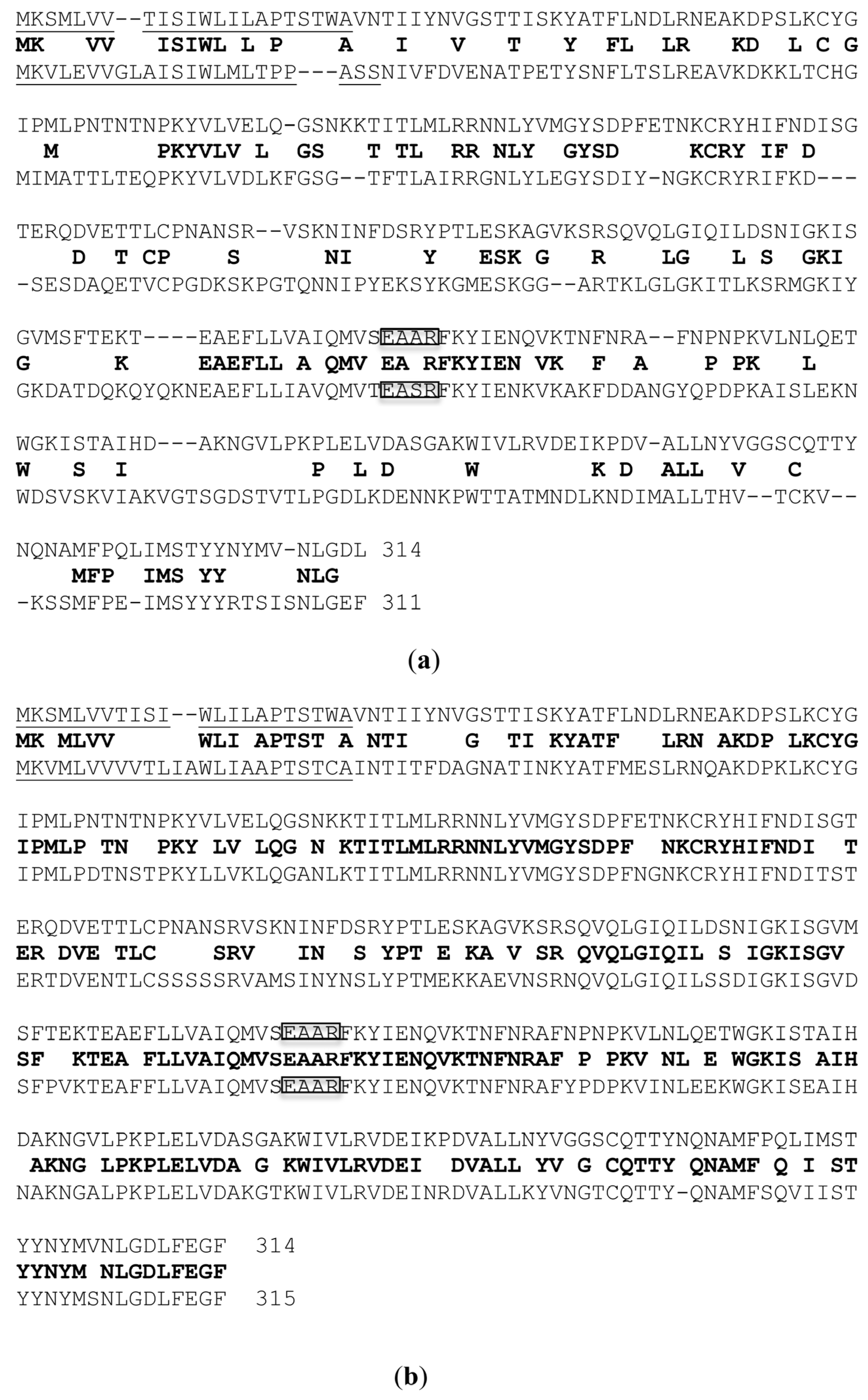
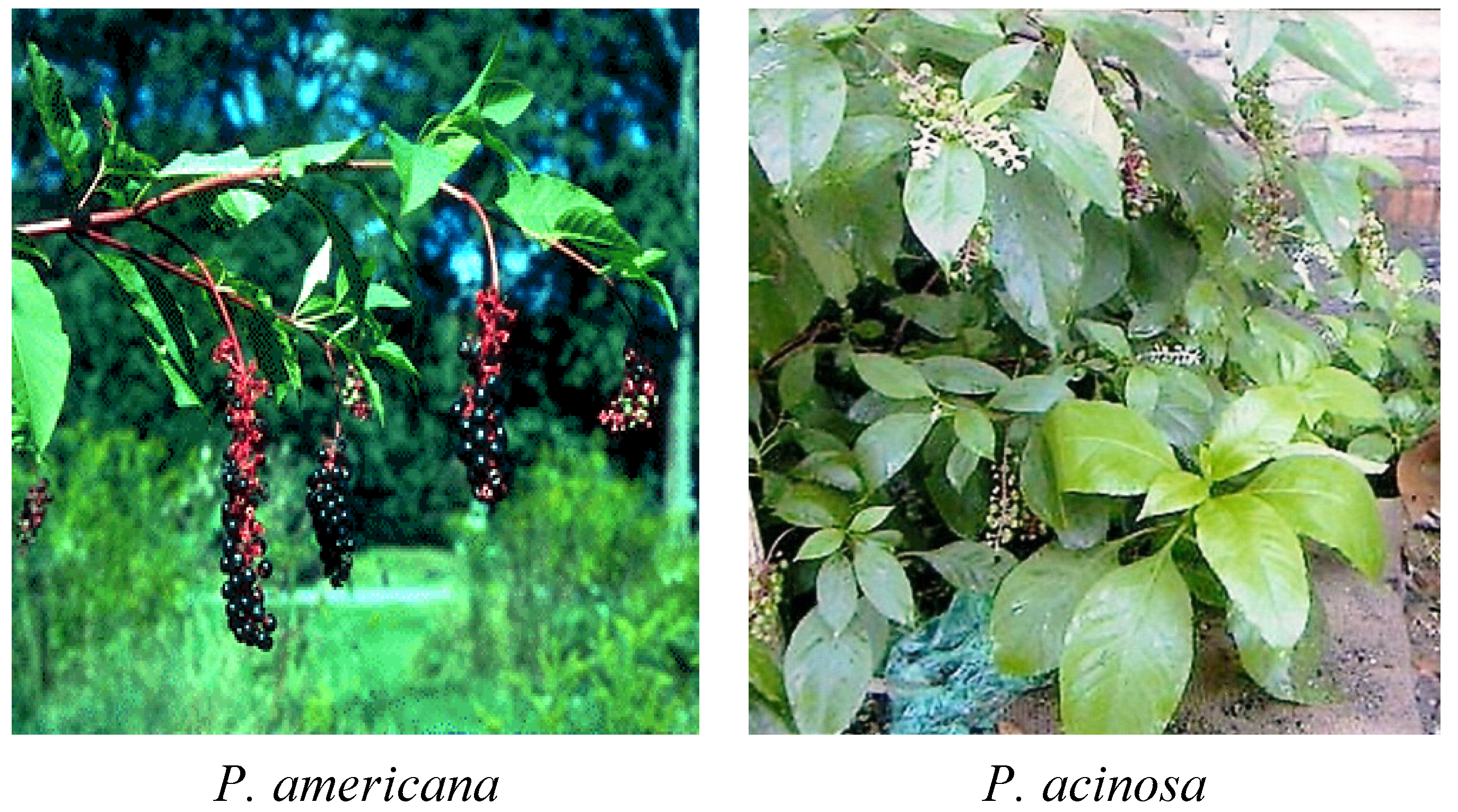
2. Different Forms of PAP

Several different isoforms of PAP have been isolated from Phytolacca americana, namely PAP (sometimes referred as PAP I) from the spring leaves, PAP II and PAP III from the early summer and late summer leaves [16]. Additionally, PAP-S has been purified from pokeweed seeds in 1982 [17]. Later on, two isoforms of PAP-S (PAP-S1 and PAP-S2) from pokeweed seeds were described [18]. Genes encoding PAP [19], PAP II [20] and PAP-S have been cloned and characterized. The complete cDNA sequences for PAP, PAP II and PAP-S are available from GenBank with the following accession numbers: AR009535, X86085 and X98079. PAP, PAP II and PAP-S genes are 942, 933 and 945 nucleotides long and encode 314, 311 and 315 amino acid proteins respectively. The molecular weight of PAP proteins is approximately 29 kDa. The amino acid sequences of PAP, PAP II and PAP-S have been compared in 1997 [21]. The amino acid sequence differences between PAP and the other forms, PAPII and PAP-S are illustrated in Figure 1.
PAP shares 39% and 76% homology with PAP II and PAP-S respectively. It is interesting to note that although both PAP and PAP II are found in pokeweed leaves, PAP is more closely related to PAP-S than to PAP II. It has been shown that both PAP and PAP-S genes do not contain introns while PAP II contains two exons separated by one intron [20,21]. The N-terminal 22, 23 and 24 amino acids have been identified as the putative signal peptides for PAP, PAP II and PAP-S respectively [20,21,22]. PAP, PAP II and PAP-S are considered as secretory RIPs. It was shown earlier by electron microscopy that PAP-specific antibody detected PAP bound within the cell wall matrix of leaf mesophyll cells of P. americana, indicating that PAP is secreted [23]. A recent study showed that PAP formed homodimer complexes in the cytosol of pokeweed cells, and its monomeric form was found in the apoplast [24]. The PAP homodimer was demonstrated to be much less active on rRNA compared to the monomeric PAP. Thus, it was concluded that PAP uses the homodimerization mechanism to avoid depurinating the pokeweed rRNA [24].
Figure 1. Amino acid alignments between pokeweed antiviral protein (PAP) and PAP II (a) PAP and PAP-S (b) using BLAST. The consensus amino acids are listed in the middle in bold between PAP on the top and PAP II or PAP-S at the bottom. The N-terminal signal peptides are underlined. The amino acids in the active sites are boxed.
Toxins 07 00755 g001
PAP has also been isolated and characterized from pokeweed plant found in southwest China, Phytolacca acinosa Roxh. Two accessions are present in the GenBank, PAPa1 (#AY603353) and PAPa2 (#AY603354) with partial sequences starting from the first amino acid to the 238th of the mature protein. Amino acid alignments showed that PAPa1 and PAPa2 are 98.8% and 95% homologous to PAP. Based on the sequence of PAP (accession #AR009535), PCR primers were designed and used to clone the PAP gene from genomic DNA isolated from the leaves of Phytolacca acinosa found in the subtropical Fujian Province in southeast China (Figure 2). The full-length PAP gene was cloned from Phytolacca acinosa and named ChPAP (Chinese Pokeweed Antiviral Protein). Amino acid alignment indicates that ChPAP differs from PAP by only one amino acid.
Figure 2. Pictures of perennial decidious Phytolacca americana from New Jersey, central eastern USA and perennial evergreen Phytolacca acinosa from Fujian Province, southeast China.
Toxins 07 00755 g002

3. Cytotoxicity of PAP

3.1. PAP Cytotoxicity in Plants

PAP removes a specific adenine from the SRL of the large rRNA and inhibits translation, leading to cell death. The cytotoxicity of wild type (wt) PAP made it very difficult to regenerate transgenic tobacco pants (Nicotiana tabacum cv. Samsun) [22]. As reported [22], the transformation efficiency was only 0.7%, compared to 7%–18% for the vector control without the PAP gene. When a variant of PAP (PAPv) containing mutations of L20R and Y49H was used to transform tobacco, 3.7% transformation efficiency was obtained, indicating that PAPv was less toxic than wt PAP. It was shown that high PAP-expressing transgenic tobacco plants showed mottled symptoms on their leaves. They were stunted and their seeds were not viable. However, the low PAP-expressing plants were indistinguishable from non-transformed plants and they produced viable seeds [22]. When the leaves of PAPv expressing transgenic tobacco plants were stained with lactophenol-trypan blue solution (2.5 mg/mL trypan blue, 25% lactic acid, 25% phenol and 25% glycerol) [25] and visualized with a compound microscope, the symptomatic leaves showed the trypan blue staining pattern characteristic of programed cell death. The pattern observed was similar to the necrotic local lesions produced when N. tabacum cv. Samsun plants containing the N gene [26] were infected with the tobacco mosaic virus (TMV) (Figure 3). This pattern of trypan blue staining has also been documented in plants showing the hypersensitive response to bacterial infection [25]. These results showed that PAP expressed in transgenic tobacco plants is toxic and causes cell death.
Figure 3. Trypan blue staining of tobacco leaves. (a) 8 week old wt N. tabacum, 10×; (b) 8 week old symptomless transgenic tobacco, 10×; (c) 8 week old symptomatic transgenic tobacco, 10×; (d) 8 week old symptomatic transgenic tobacco, 20× and (e) N. tabacum NN 7 days post TMV inoculation, 20×. Bars in (a) to (c) = 100 µm. Bars in (d) to (e) = 50 µm.
Toxins 07 00755 g003

3.2. PAP Cytotoxicity in Yeast

A yeast system has been developed as a model to study the cytotoxicity of PAP. The full-length cDNA of PAP (accession #AR009535) was cloned under the control of the galactose inducible GAL1 promoter and transformed into Saccharomyces cerevisiae strain W303 (Mata ade2-1 trp1-1 ura3-1 leu2-3 112 his3-11 15 can1-100) [27]. PAP was properly processed in yeast cells and the induction of PAP expression was lethal to yeast. To understand how PAP interacts with rRNA and causes cell death, the PAP expression plasmid was mutagenized with hydroxylamine and re-transformed into yeast cells. Yeast cells harboring non-toxic PAP mutants were identified as those that could grow after induction of expression on galactose. One of the non-toxic mutants identified was PAPE176V, which abolished PAP cytotoxicity. The Glu-176 in PAP is equivalent to Glu-177 of ricin A chain (RTA) which had been previously implicated as one of the key residues at the active site [28]. PAP and RTA share the same EAAR residues in their active sites (Figure 1) [29]. Glu-177 (E) and Arg-180 (R) residues of RTA were identified as critical for its enzymatic activity [30].
Extensive site-directed mutagenesis of PAP was carried out. Over 30 mutations at the N-terminus, central domain and the C-terminus of PAP were generated, transformed into yeast and characterized [31]. The dual primer extension method was used to measure the depurination of rRNA, and the in vivo (35S)-methionine incorporation assay was used to assess the inhibition of translation in yeast cells transformed with the PAP mutants. Besides PAPE176V, PAPG75D mutation in the central domain led to loss of depurination activity and cytotoxicity. Additionally, both PAPY16M and PAPT18M mutations at the N-terminus were not toxic to yeast cells and these mutants did not depurinate the rRNA. Interestingly, the following PAP mutations in the central domain, PAPN70A, PAPL71R, PAPY72A, PAPV73E, PAPY76A and PAPY123A and PAPY123I, led to loss of cytotoxicity, but not rRNA depurination. These results indicated that cytotoxicity of PAP was lost before ribosome depurination ability, suggesting that ribosome depurination was not the only factor contributing to cytotoxicity [31].
Recent results showed that yeast cells expressing PAP exhibited apoptotic-like features, such as nuclear fragmentation and production of reactive oxygen species (ROS). Arabidopsis thaliana Bax inhibitor-1, a plant antiapoptotic protein, which inhibits Bax induced cell death reduced the cytotoxicity of PAP in yeast, without affecting ribosome depurination and translation inhibition [32]. These results provided further evidence that ribosome depurination and translation inhibition by PAP could be separated from cytotoxicity.

3.3. Mechanism of PAP Cytotoxicity

3.3.1. N-glycosidase Activity

Endo and Tsurugi reported in 1987 that RTA removed a single adenine from position 4324 in the 28S rRNA of rat liver ribosomes [33]. It was demonstrated that RTA cleaved the N-glycosidic bond of A4324 of the 28S rRNA in a hydrolytic fashion, thus defining RIPs as ribosome-specific N-glycosidases (EC 3.2.2.22) [33]. Based on structural analysis, Monzingo and Robertus [34] proposed the following enzymatic action for ricin: Tyr80 (equivalent to PAP Tyr72) and Tyr123 (equivalent to PAP Tyr123) sandwich the target adenine ring, the negative charge of Glu177 (equivalent to PAP Glu176) stabilizes the positive oxycarbonium ion on ribose in the transition state, and Arg180 (equivalent to PAP Arg179) stabilizes the anion on the leaving adenine by protonating it at the N-3 position. Later on, Rajamohan et al. used site-directed mutagenesis, binding assays and surface plasmon resonance (SPR) to provide experimental evidence that Tyr72, Tyr123, Glu176 and Arg179 are catalytic residues that participate in the binding of PAP to the target tetraloop structure of the SRL [35].
Since PAP is a type I RIP, it lacks a sugar-binding moiety, such as the ricin B-chain. Recently, using fluorescence spectroscopy, Nakashima et al. demonstrated that the Trp208 at the C-terminus of PAP cooperated with Tyr72 and bound strongly to N-acetylglucosamine (NAG, a monosaccharide derivative of glucose) [36]. Since Trp208 is considered critical for the enzymatic activity of RIPs, the authors concluded that the sugar-binding may induce a conformational change near the active site of PAP.
RIPs are active on eukaryotic rRNA. PAP, however, was shown to depurinate both the A4324 of the eukaryotic ribosome and the A2660 of the Escherichia coli (E. coli) ribosome [37]. After the crystal structure of PAP was refined at room temperature [38] and lower temperatures [39], Kurinov et al. demonstrated that PAP could release not only adenine but also guanine from Escherichia coli ribosomes at a 20 times slower rate [40]. Although these studies provided information about the N-glycosidase activity of PAP, they did not address its ribosome specificity or its ability to depurinate the eukaryotic and prokaryotic ribosomes.

3.3.2. Interaction with Ribosomal Proteins

To answer the questions above and to elucidate the mechanism of PAP’s interaction with the ribosome, the interaction of PAP with ribosomal proteins has been examined [41]. Ribosomes of yeast cells harboring the mak8-1 allele of the large ribosomal subunit protein L3 (RPL3) were shown to be resistant to PAP depurination [42]. Since the highly conserved RPL3 participates in the formation of the peptidyltransferase center [43,44] and is essential for the catalysis of peptide bond formation [45], RPL3 was shown to be critical for the binding of PAP to ribosomes and the subsequent rRNA depurination. It was shown later by SPR affinity analysis that PAP active site cleft residues Asn69, Asn70 and Asp92 are involved in binding to RPL3 [46].
The interaction between PAP and RPL3 was further examined in transgenic tobacco plants. As mentioned in 3.1, wt PAP and PAPV expressed in transgenic tobacco plants are toxic to tobacco cells [22]. However, when tobacco (N. tabacum cv. Samsun) was transformed with a construct containing both the full-length yeast RPL3 and wt PAP, fertile transgenic plants were produced [47]. The first two leaves of the transgenic tobacco plants showed only mild mottle symptoms and their ribosomes were partially depurinated. However, transgenic tobacco plants expressing the N-terminal 99 amino acids of yeast L3 and wt PAP were phenotypically normal compared to non-transformed tobacco plants and were fertile. Their ribosomes were not depurinated even though PAP was associated with the ribosomes. This work suggested that PAP may interact with the N-terminus of RPL3 in transgenic tobacco plants.
To further study the interaction between PAP and RPL3, the yeast model was used. Yeast cells transformed with both wt PAP and the N-terminal 99 amino acids of RPL3 (L3Δ99) showed markedly reduced growth inhibition and rRNA depurination compared to cells transformed with PAP alone [48]. Additionally, it was shown that the N-terminal 21 amino acids of RPL3 (L3Δ21) were sufficient to reduce the cytotoxicity of PAP in yeast cells [48]. By SPR analysis, PAP interacted with a 27-mer RNA fragment corresponding to the SRL with a binding affinity of 0.365 nM (unpublished data), which was similar to the 0.2 nM affinity published earlier [35]. We also showed that the affinity of PAP for a 40-mer RNA fragment corresponding to L3Δ was 4.91 nM. Furthermore, it was shown that PAP interacted with the yeast L3Δ21 peptide with an affinity of 24.8 nM [48]. These results indicate that PAP interacts with both and RNA fragment and a peptide corresponding to the N-terminus of RPL3. Since RPL3 is highly conserved among different species, the interaction between PAP and RPL3 may partially account for the non-selective cytotoxicity of PAP to rRNA from different organisms. It has been shown that ricin [49], trichosanthin [50,51], Shiga toxin 1 [49,52] and maize RIP MOD [53] interact with the P proteins of the ribosomal stalk. However, this is not the case for PAP [49,51]. Furthermore, it was shown that residues Gly209–Lys225 at the PAP C-terminal domain were critical for its N-glycosidase activity on prokaryotic ribosomes [54]. Since the stalk structure differs between eukaryotic and prokaryotic ribosomes, it has been postulated that the ability of these RIPs to interact with the P protein motif originated during evolution of the eukaryotic elongation factor (EF)-2 [55].

3.3.3. Inhibition of Translation

As early as in 1976 it was shown that protein synthesis was inhibited upon rRNA depurination by RIPs and the effect occurred at the peptide elongation step [56]. Roles of the elongation factors and ribosomes during protein synthesis have been reviewed extensively [57,58,59]. During the elongation step in protein synthesis, EF1 brings aminoacyl-tRNA to the A-site of the ribosome and after codon and anti-codon recognition, EF1 is released. The rRNA catalyzes the peptide formation and transfers the peptidyl-tRNA from the P-site of the ribosome to the A-site. It has been shown that the translocation of peptidyl tRNA from the P-site to A-site by EF2 was inhibited after rRNA was depurinated by RIPs [60]. It was recently demonstrated that PAP depurinated the rRNA prior to the binding of aminoacyl-tRNA to the ribosomal A-site [61].

3.3.4. Other Enzymatic Activity

Different substrates including DNA, RNA and poly (A) RNA have been described for the alternative enzymatic activity of RIPs [62,63]. PAPG75D and PAPW237* (* denotes stop codon) isolated from transgenic tobacco plants did not depurinate the rabbit reticulocyte rRNA; however, they depurinated the capped viral RNAs and inhibited the translation of brome mosaic virus (BMV) and potato virus X (PVX) RNAs [64]. It was shown that PAP’s inhibition of capped BMV RNA translation was overcome in the presence of the cap analog m7GpppG, indicating that PAP recognized the cap structure [64]. Subsequently, PAP was shown to bind to the cap structure of eukaryotic mRNA and depurinate the mRNA downstream of the cap [65]. It was demonstrated that PAP interacted with the eukaryotic initiation factor (eIF)4G and eIFiso4G in the wheat germ lysate, providing a mechanism for PAP to access both uncapped and capped RNAs [66]. Further evidence for PAP binding to the cap analog m7GTP was provided by fluorescence quenching experiments [67].
PAP regulated its own mRNA stability by a mechanism that involved depurination [48,68]. These data suggested that rRNA is not the only substrate for PAP, capped mRNAs and uncapped RNAs are subject to inactivation by PAP. Additionally, double-stranded (ds) supercoiled DNA could be cleaved by PAP [69]. The active site required for rRNA depurination was also required for DNA cleavage, as the non-rRNA depurinating PAPE176V could not cleave the dsDNA [69]. The dsDNA treated with PAP contained apurinic/apyrimidinic (AP) sites due to the removal of adenine [69]. The same phenomenon was observed with different RIPs including gelonin, momordin I, PAP-S and saporin-S6 [70].

3.3.5. C-Terminal Involvement in PAP Processing

Type II RIPs such as ricin and Shiga toxins use their lectin chain to bind to receptors on cells [71,72,73,74]. They are internalized by endocytosis and undergo retrograde transport via the Golgi complex to reach the endoplasmic reticulum (ER) lumen. After processing, they are thought to retro-translocate into the cytosol using the ER-associated degradation (ERAD) pathway. PAP does not contain a lectin chain. It is synthesized as a 314-amino acid precursor containing a 22 residue N-terminal signal sequence and a 29 residue C-terminal extension [75]. The N-terminal 22 amino acids of PAP direct PAP to the apoplast in pokeweed plants [22,23]. When total protein from yeast cells transformed with wt PAP was electrophoresed on the SDS-polyacrylamide gel, two protein forms were observed [76]. It has been shown that both PAP forms associate with the ER in yeast cells, and the smaller PAP form may retrotranslocate into the cytosol [76].
When the C-terminus (249VALLNYVGGSCQTT262) of mature PAP was sequentially truncated, cytotoxicity was lost before rRNA depurination in yeast [31]. Specifically, PAPN253* mutant was toxic, while PAPL252* was not toxic even though it depurinated ribosomes. PAP’s C-terminal motif was similar to those found in ricin and Shiga toxins [75]. Further analysis showed that the C-terminal amino acids were critical for the processing of PAP and the accumulation of mature PAP in the cytosol. Thus, they affected depurination of the rRNA in the cytosol and the cytotoxicity of PAP [75].

4. Applications of PAP in Plant Disease Resistance

4.1. Against Plant Viruses

PAP was first discovered in 1925 for its activity against plant viruses [77]. It was later found that when pokeweed extracts were co-inoculated with a plant virus onto the leaves of susceptible plants, PAP in the crude extract could protect plants from different viruses, such as southern bean mosaic virus [2], cucumber mosaic virus (CMV) [3] and TMV [78]. In 1990s when plant biotechnology became possible, the great potential of PAP as a plant pathogen inhibitor was realized.
Lodge et al. [22] showed for the first time that tobacco and potato plants transformed with wt PAP and PAPL20R/Y49H under the control of CaMV 35S promoter were resistant to potato virus X (PVX), potato virus Y (PVY) and CMV [22]. These results indicated broad spectrum virus resistance, in contrast to viral coat protein-mediated resistance, which was specific to the virus the coat protein gene was derived from. PAP was enriched in the intercellular fluid of transgenic plants and conferred viral resistance by inhibiting an early stage of infection. It was hypothesized that upon infection PAP might enter the host cells along with the virus and depurinate the host rRNA. Since viruses depend on the host machinery for their replication, ribosome-damaged host cells will not be able to support virus replication [22]. This hypothesis was supported by the work of Chen et al. [79], which showed that when PAP and TMV were co-inoculated onto tobacco plants, ribosomes were depurinated as early as 5 min after inoculation and the inhibition of virus infection and ribosome depurination were positively correlated with PAP concentration [79]. Later, Tumer et al. reported that ribosomes of PAPL20R/Y49H expressing transgenic tobacco plants were depurinated [80].
However, when Tumer et al. [80] transformed tobacco with the nontoxic C-terminal deletion mutant PAPW237* and the active site mutant PAPE176V, they found that ribosomes from these transgenic tobacco plants were not depurinated. The extracts from PAPW237* expressing transgenic plants protected tobacco plants from PVX infection, while the extracts from PAPE176V expressing plants did not. Furthermore, they found that transgenic tobacco plants expressing PAPW237* were resistant to PVX infection, while plants expressing PAPE176V were not resistant. These data indicated that the C-terminus of PAP was required for toxicity and depurination of ribosomes, but not for antiviral activity, suggesting that host ribosome depurination was not the only mechanism for PAP-induced virus resistance [80].
It is mentioned earlier that non-depurinating mutants PAPG75D and PAPW237* isolated from transgenic tobacco plants could depurinate the capped BMV and PVX viral RNAs and inhibit their translation in vitro [64]. PAP could inhibit translation of uncapped TBSV (tomato bushy stunt virus) and SPMV (satellite panicum mosaic virus) in vivo without causing detectable depurination in the viral RNAs [81]. The inhibition of PAP on BMV replication was shown later in barley protoplasts [82]. The inhibition was due to the depurination of BMV RNA by PAP and inhibition of RNA replication and subgenomic RNA transcription [82,83]. These data indicate that besides depurinating host ribosomes, PAP can directly depurinate viral RNA and inhibit virus replication.

4.2. Against Plant Fungi

Anti-fungal activities of RIPs have been less frequently described compared to their anti-viral activity. The 30 kDa cytosolic type I RIP from barley was shown to be inactive on plant ribosomes in vitro as other type I RIPs from cereal starchy endosperms [84], but capable of modifying fungal ribosomes [85]. Indeed, when this barley endosperm RIP was transformed into tobacco under the control of an inducible promoter, it conferred resistance to Rhizoctonia solani without affecting the growth of transgenic tobacco plants [86]. Furthermore, co-expression of the barley RIP with a class-II chitinase resulted in synergistically enhanced resistance to R. solani in transgenic tobacco plants [87]. It was proposed that the hydrolytic activity of chitinase could result in an increased uptake of the barley RIP into fungal cells and significantly inhibit the fungal growth [87]. Another cereal endosperm RIP, the maize kernel RIP I, was also shown to be anti-fungal, inhibiting the postdivisional growth of Aspergillus spp. in microculture assays [88]. However, the exact mechanism of how endosperm RIPs affect fungal growth is not well understood.
It was found that PAPL20R/Y49H- and PAPW237*-transgenic plants were resistant to R. solani infection [89]. Additionally, expression of both class I (basic) and class II (acidic) isoforms of pathogenesis-related (PR) (PR1 and PR2) proteins was induced in the transgenic tobacco plants [89,90]. However, the salicylic acid (SA) levels were not elevated [89,90] as in the classical systemic acquired resistance (SAR). Subsequently, it was shown that transgenic tobacco plants expressing the non-toxic mutant PAPG75D were resistant to R. solani infection [91]. PR2 expression was not elevated in PAPG75D-transgenic plants, rather the expression levels of PR1 and wound-inducible protein kinase (WIPK) and protease inhibitor II (PI-II) were up-regulated [91]. The SA level in PAPG75D-plants was also not increased [91]. These results suggested that the PAP expressed in transgenic plants induces expression of several PR proteins, likely through an SA-independent pathway [89,90].

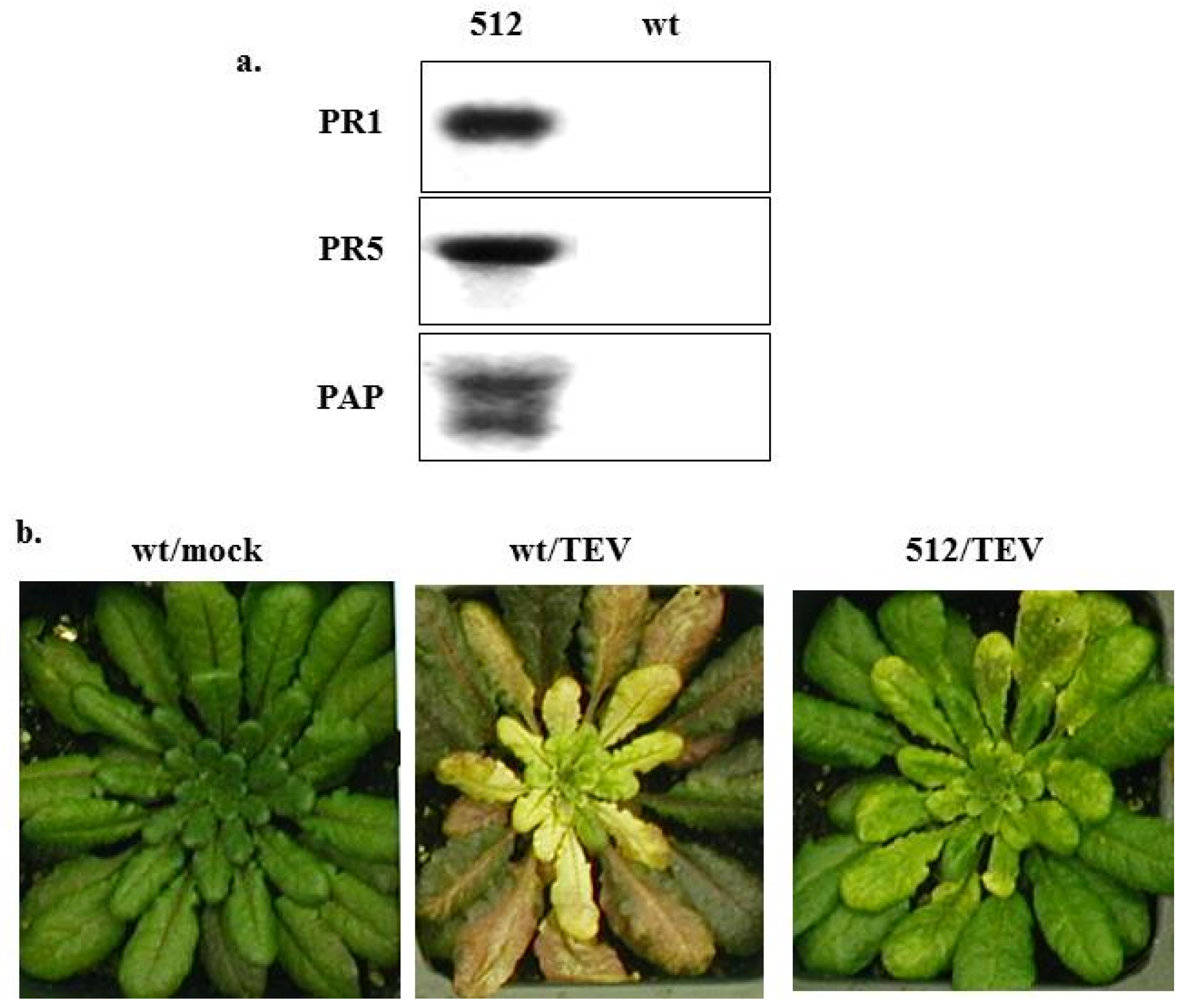
4.3. Mechanisms of PAP-Induced Disease Resistance

To further investigate the disease resistance mechanisms induced by PAP, transgenic Arabidopsis plants expressing the non-toxic C-terminal deletion mutant PAPW237* were produced. Figure 4a shows that PAP was over-expressed in the transgenic line 512 compared to non-transformed wt Arabidopsis thaliana Columbia ecotype by Northern blot analysis using PAP cDNA as a probe. Figure 4a also shows that PR1 [92] and PR5 [93] genes were up-regulated in line 512, supporting the early findings in transgenic tobacco plants [89,91]. Line 512 displayed enhanced resistance to a strain of tobacco etch virus (TEV) that causes systemic infection [94] compared to wt Arabidopsis, as the transgenic plant only showed very mild yellowing symptoms on its leaves (Figure 4b). The transcript profiling analysis using the Affymetrix® Arabidopsis microarrays was conducted. Table 1 lists the genes that were up-regulated in PAPW237* line 512 for more than 3-fold compared to wt Arabidopsis. The expression of several auxin-responsive genes was more than 4-fold higher in line 512 than in wt Arabidopsis. Notably, the lipoxygenase (lox3, AJ249794) gene that is critical for jasmonic acid (JA)-mediated induced systemic resistance (ISR) [92] was up-regulated by 4.6-fold. The other plant innate immunity and disease resistance-related genes that were up-regulated in line 512 include the AtERF6 ethylene responsive element binding factor (AB013301) [95], PR1 (X96600) [92], PR5K (U48698) [93,96], and thaumatin-like protein (U83490) [97]. We have confirmed the expression of these genes by quantitative RT-PCR (qRT-PCR) analysis using gene-specific primers (data not shown).
Figure 4. (a) Northern blot analysis of PAPW237*-transgenic line 512 and wt Arabidopsis plants using PR1, PR5 and PAP specific 32P-labelling cDNA probes; (b) Mock (water) and tobacco etch virus (TEV)-inoculated wt and PAPW237*-transgenic line 512 Arabidopsis plants.
Toxins 07 00755 g004
Table 1. Transcript profiling of PAPW237*-transgenic line 512 vs. wt Arabidopsis using Affymetrix® Arabidopsis microarray: Fold change of up-regulated gene expression.
GeneGene descriptionFold change
U49076early auxin-induced (IAA20) mRNA6.1
AF087819auxin transport protein (PIN6) mRNA5.7
AF082176auxin response factor 9 (ARF9) mRNA5.7
L15448auxin-responsive protein (IAA1) mRNA5.7
AF087819auxin transport protein (PIN6) mRNA5.3
AL035656putative auxin-induced protein4.9
AL035656small auxin up RNA (SAUR-AC1)4.9
AL035656auxin-induced protein-like4.9
S70188small auxin up RNA4.9
AJ249794lipoxygenase (lox3)4.6
AB013301AtERF6 ethylene responsive element binding factor4.3
AJ012745RH27 helicase4.3
Z97341RNA helicase3.7
AJ010475DEAD box RNA helicase, RH283.7
Z97337RNA helicase like protein3.7
X96600pathogenesis-related protein 1 (PR1)3.5
U48698receptor serine/threonine kinase PR5K (PR5K)3.5
U83490 thaumatin-like protein3.2
AL021687cytochrome P4503
AL049659cytochrome P450-like protein3
AL021636cytochrome P450-like protein3
AL049659cytochrome P450-like protein3
These data support the previous findings and indicate that PAP elicits a wide range of defense responses in transgenic plants, which may be responsible for the fungal resistance observed in these plants. The transgene approach has been successfully used to confer plant disease resistance. However, there are few examples of transgenes, which can induce broad-spectrum disease resistance. As plants are often infected with multiple pathogens, engineering them with single genes, such as nontoxic PAP variants that can confer broad-spectrum disease resistance may be more advantageous and may have many applications to agriculture.

Author Contributions

Rong Di wrote the paper and contributed to photo of P. acinosa in Figure 2, data in Figure 3, Figure 4 and Table 1. Nilgun E. Tumer revised the paper.

Conflicts of Interest

The authors declare no conflict of interest.

References

  1. Obrig, T.G.; Irvin, J.D.; Hardesty, B. The effect of an antiviral peptide on the ribosomal reactions of the peptide elongation enzymes, EF-I and EF-II. Arch. Biochem. Biophys. 1973, 155, 278–289. [Google Scholar] [CrossRef] [PubMed]
  2. Wyatt, S.D.; Shepherd, R.J. Isolation and characterization of a virus inhibitor from Phytolacca americana. Phytopathology 1969, 59, 1787–1794. [Google Scholar] [PubMed]
  3. Tomlinson, J.A.; Walker, V.M.; Flewett, T.H.; Barclay, G.R. The inhibition of infection by cucumber mosaic virus and influenza virus by extracts from Phytolacca americana. J. Gen. Virol. 1974, 22, 225–232. [Google Scholar] [CrossRef] [PubMed]
  4. Irvin, J.D. Purification and partial characterization of the antiviral protein from Phytolacca americana which inhibits eukaryotic protein synthesis. Arch. Biochem. Biophys. 1975, 169, 522–528. [Google Scholar] [CrossRef] [PubMed]
  5. Endo, Y.; Tsurugi, K. The RNA N-glycosidase activity of ricin A-chain. The characteristics of the enzymatic activity of ricin A-chain with ribosomes and with rRNA. J. Biol. Chem. 1988, 263, 8735–8739. [Google Scholar] [PubMed]
  6. Olsnes, S.; Kozlov, J.V. Ricin. Toxicon 2001, 39, 1723–1728. [Google Scholar] [CrossRef] [PubMed]
  7. Di, R.; Kyu, E.; Shete, V.; Saidasan, H.; Kahn, P.C.; Tumer, N.E. Identification of amino acids critical for the cytotoxicity of Shiga toxin 1 and 2 in Saccharomyces cerevisiae. Toxicon 2011, 57, 525–539. [Google Scholar] [CrossRef] [PubMed]
  8. Walsh, T.A.; Morgan, A.E.; Hey, T.D. Characterization and molecular cloning of a proenzyme form of a ribosome-inactivating protein from maize. Novel mechanism of proenzyme activation by proteolytic removal of a 2.8-kilodalton internal peptide segment. J. Biol. Chem. 1991, 266, 23422–23427. [Google Scholar] [PubMed]
  9. Bass, H.W.; Webster, C.; GR, O.B.; Roberts, J.K.; Boston, R.S. A maize ribosome-inactivating protein is controlled by the transcriptional activator Opaque-2. Plant Cell 1992, 4, 225–234. [Google Scholar] [CrossRef] [PubMed]
  10. Wong, Y.T.; Ng, Y.M.; Mak, A.N.; Sze, K.H.; Wong, K.B.; Shaw, P.C. Maize ribosome-inactivating protein uses Lys158-lys161 to interact with ribosomal protein P2 and the strength of interaction is correlated to the biological activities. PLoS One 2012, 7, e49608. [Google Scholar] [CrossRef] [PubMed]
  11. Reinbothe, S.; Reinbothe, C.; Lehmann, J.; Becker, W.; Apel, K.; Parthier, B. JIP60, a methyl jasmonate-induced ribosome-inactivating protein involved in plant stress reactions. Proc. Natl. Acad. Sci. USA 1994, 91, 7012–7016. [Google Scholar] [CrossRef] [PubMed]
  12. Chaudhry, B.; Muller-Uri, F.; Cameron-Mills, V.; Gough, S.; Simpson, D.; Skriver, K.; Mundy, J. The barley 60 kDa jasmonate-induced protein (JIP60) is a novel ribosome-inactivating protein. Plant J. 1994, 6, 815–824. [Google Scholar] [CrossRef] [PubMed]
  13. Rustgi, S.; Pollmann, S.; Buhr, F.; Springer, A.; Reinbothe, C.; von Wettstein, D.; Reinbothe, S. JIP60-mediated, jasmonate- and senescence-induced molecular switch in translation toward stress and defense protein synthesis. Proc. Natl. Acad. Sci. USA 2014, 111, 14181–14186. [Google Scholar] [CrossRef] [PubMed]
  14. Tumer, N.E.; Hudak, K.; di, R.; Coetzer, C.; Wang, P.; Zoubenko, O. Pokeweed antiviral protein and its applications. In Plant Biotechnology: New Products and Applications; Hammond, J., McGarvey, P., Yusibov, V., Eds.; Springer: New York, NY, USA, 1999; pp. 139–158. [Google Scholar]
  15. Parikh, B.A.; Tumer, N.E. Antiviral activity of ribosome inactivating proteins in medicine. Mini Rev. Med. Chem. 2004, 4, 523–543. [Google Scholar] [CrossRef] [PubMed]
  16. Irvin, J.D.; Uckun, F.M. Pokeweed antiviral protein: Ribosome inactivation and therapeutic applications. Pharmacol. Ther. 1992, 55, 279–302. [Google Scholar] [CrossRef] [PubMed]
  17. Barbieri, L.; Aron, G.M.; Irvin, J.D.; Stirpe, F. Purification and partial characterization of another form of the antiviral protein from the seeds of Phytolacca americana L. (pokeweed). Biochem. J. 1982, 203, 55–59. [Google Scholar] [PubMed]
  18. Honjo, E.; Dong, D.; Motoshima, H.; Watanabe, K. Genomic clones encoding two isoforms of pokeweed antiviral protein in seeds (PAP-S1 and S2) and the N-glycosidase activities of their recombinant proteins on ribosomes and DNA in comparison with other isoforms. J. Biochem. 2002, 131, 225–231. [Google Scholar] [CrossRef] [PubMed]
  19. Lin, Q.; Chen, Z.C.; Antoniw, J.F.; White, R.F. Isolation and characterization of a cDNA clone encoding the anti-viral protein from Phytolacca americana. Plant Mol. Biol. 1991, 17, 609–614. [Google Scholar] [CrossRef] [PubMed]
  20. Poyet, J.L.; Radom, J.; Hoeveler, A. Isolation and characterization of a cDNA clone encoding the pokeweed antiviral protein II from Phytolacca americana and its expression in E. coli. FEBS Lett. 1994, 347, 268–272. [Google Scholar] [CrossRef] [PubMed]
  21. Poyet, J.L.; Hoeveler, A. cDNA cloning and expression of pokeweed antiviral protein from seeds in Escherichia coli and its inhibition of protein synthesis in vitro. FEBS Lett. 1997, 406, 97–100. [Google Scholar] [CrossRef] [PubMed]
  22. Lodge, J.K.; Kaniewski, W.K.; Tumer, N.E. Broad-spectrum virus resistance in transgenic plants expressing pokeweed antiviral protein. Proc. Natl. Acad. Sci. USA 1993, 90, 7089–7093. [Google Scholar] [CrossRef] [PubMed]
  23. Ready, M.P.; Brown, D.T.; Robertus, J.D. Extracellular localization of pokeweed antiviral protein. Proc. Natl. Acad. Sci. USA 1986, 83, 5053–5056. [Google Scholar] [CrossRef] [PubMed]
  24. Tourlakis, M.E.; Karran, R.A.; Desouza, L.; Siu, K.W.; Hudak, K.A. Homodimerization of pokeweed antiviral protein as a mechanism to limit depurination of pokeweed ribosomes. Mol. Plant Pathol. 2010, 11, 757–767. [Google Scholar] [PubMed]
  25. Choi, D.S.; Kim, N.H.; Hwang, B.K. Pepper mitochondrial FORMATE DEHYDROGENASE1 regulates cell death and defnese responses against bacterial pathogens. Plant Physiol. 2014, 166, 1298–1311. [Google Scholar] [CrossRef] [PubMed]
  26. Marathe, R.; Anandalakshmi, R.; Liu, Y.; Dinesh-Kumar, S.P. The tobacco mosaic virus resistance gene, N. Mol. Plant Pathol. 2002, 3, 167–172. [Google Scholar] [CrossRef] [PubMed]
  27. Hur, Y.; Hwang, D.J.; Zoubenko, O.; Coetzer, C.; Uckun, F.M.; Tumer, N.E. Isolation and characterization of pokeweed antiviral protein mutations in Saccharomyces cerevisiae: Identification of residues important for toxicity. Proc. Natl. Acad. Sci. USA 1995, 92, 8448–8452. [Google Scholar] [CrossRef] [PubMed]
  28. Schlossman, D.; Withers, D.; Welsh, P.; Alexander, A.; Robertus, J.; Frankel, A. Role of glutamic acid 177 of the ricin toxin A chain in enzymatic inactivation of ribosomes. Mol. Cell. Biol. 1989, 9, 5012–5021. [Google Scholar] [PubMed]
  29. Halling, K.C.; Halling, A.C.; Murray, E.E.; Ladin, B.F.; Houston, L.L.; Weaver, R.F. Genomic cloning and characterization of a ricin gene from Ricinus communis. Nucleic Acids Res. 1985, 13, 8019–8033. [Google Scholar] [CrossRef] [PubMed]
  30. Frankel, A.; Welsh, P.; Richardson, J.; Robertus, J.D. Role of arginine 180 and glutamic acid 177 of ricin toxin A chain in enzymatic inactivation of ribosomes. Mol. Cell. Biol. 1990, 10, 6257–6263. [Google Scholar] [PubMed]
  31. Hudak, K.A.; Parikh, B.A.; di, R.; Baricevic, M.; Santana, M.; Seskar, M.; Tumer, N.E. Generation of pokeweed antiviral protein mutations in Saccharomyces cerevisiae: Evidence that ribosome depurination is not sufficient for cytotoxicity. Nucleic Acids Res. 2004, 32, 4244–4256. [Google Scholar] [CrossRef] [PubMed]
  32. Cakir, B.; Tumer, N.E. Arabidopsis Bax inhibitor-1 inhibits cell death induced by pokeweed antiviral protein in Saccharomyces cerevisae. Microl. Cell 2015, 2, 43–56. [Google Scholar] [CrossRef]
  33. Endo, Y.; Tsurugi, K. RNA N-glycosidase activity of ricin A-chain. Mechanism of action of the toxic lectin ricin on eukaryotic ribosomes. J. Biol. Chem. 1987, 262, 8128–8130. [Google Scholar] [PubMed]
  34. Monzingo, A.F.; Robertus, J.D. X-ray analysis of substrate analogs in the ricin A-chain active site. J. Mol. Biol. 1992, 227, 1136–1145. [Google Scholar] [CrossRef] [PubMed]
  35. Rajamohan, F.; Mao, C.; Uckun, F.M. Binding interactions between the active center cleft of recombinant pokeweed antiviral protein and the alpha-sarcin/ricin stem loop of ribosomal RNA. J. Biol. Chem. 2001, 276, 24075–24081. [Google Scholar] [CrossRef] [PubMed]
  36. Nakashima, H.; Fukunaga, Y.; Ueno, R.; Nishimoto, E. Sugar binding effects on the enzymatic reaction and conformation near the active site of pokeweed antiviral protein revealed by fluorescence spectroscopy. J. Fluoresc. 2014, 24, 951–958. [Google Scholar] [CrossRef] [PubMed]
  37. Hartley, M.R.; Legname, G.; Osborn, R.; Chen, Z.; Lord, J.M. Single-chain ribosome inactivating proteins from plants depurinate Escherichia coli 23S ribosomal RNA. FEBS Lett. 1991, 290, 65–68. [Google Scholar] [CrossRef] [PubMed]
  38. Monzingo, A.F.; Collins, E.J.; Ernst, S.R.; Irvin, J.D.; Robertus, J.D. The 2.5 A structure of pokeweed antiviral protein. J. Mol. Biol. 1993, 233, 705–715. [Google Scholar] [CrossRef] [PubMed]
  39. Kurinov, I.V.; Myers, D.E.; Irvin, J.D.; Uckun, F.M. X-ray crystallographic analysis of the structural basis for the interactions of pokeweed antiviral protein with its active site inhibitor and ribosomal RNA substrate analogs. Protein Sci. 1999, 8, 1765–1772. [Google Scholar] [CrossRef] [PubMed]
  40. Kurinov, I.V.; Rajamohan, F.; Venkatachalam, T.K.; Uckun, F.M. X-ray crystallographic analysis of the structural basis for the interaction of pokeweed antiviral protein with guanine residues of ribosomal RNA. Protein Sci. 1999, 8, 2399–2405. [Google Scholar] [CrossRef] [PubMed]
  41. Muralikrishna, P.; Alexander, R.W.; Cooperman, B.S. Placement of the alpha-sarcin loop within the 50S subunit: Evidence derived using a photolabile oligodeoxynucleotide probe. Nucleic Acids Res. 1997, 25, 4562–4569. [Google Scholar] [CrossRef] [PubMed]
  42. Hudak, K.A.; Dinman, J.D.; Tumer, N.E. Pokeweed antiviral protein accesses ribosomes by binding to L3. J. Biol. Chem. 1999, 274, 3859–3864. [Google Scholar] [CrossRef] [PubMed]
  43. Hampl, H.; Schulze, H.; Nierhaus, K.H. Ribosomal components from Escherichia coli 50 S subunits involved in the reconstitution of peptidyltransferase activity. J. Biol. Chem. 1981, 256, 2284–2288. [Google Scholar] [PubMed]
  44. Noller, H.F. Peptidyl transferase: Protein, ribonucleoprotein, or RNA? J. Bacteriol. 1993, 175, 5297–5300. [Google Scholar] [PubMed]
  45. Green, R.; Noller, H.F. Ribosomes and translation. Annu. Rev. Biochem. 1997, 66, 679–716. [Google Scholar] [CrossRef] [PubMed]
  46. Rajamohan, F.; Ozer, Z.; Mao, C.; Uckun, F.M. Active center cleft residues of pokeweed antiviral protein mediate its high-affinity binding to the ribosomal protein L3. Biochemistry 2001, 40, 9104–9114. [Google Scholar] [CrossRef] [PubMed]
  47. Di, R.; Tumer, N.E. Expression of a truncated form of ribosomal protein L3 confers resistance to pokeweed antiviral protein and the Fusarium mycotoxin deoxynivalenol. Mol. Plant Microbe Interact. 2005, 18, 762–770. [Google Scholar] [CrossRef] [PubMed]
  48. Di, R.; Tumer, N.E. An N-terminal fragment of yeast ribosomal protein L3 inhibits the cytotoxicity of pokeweed antiviral protein in Saccharomyces cerevisiae. Toxins (Basel) 2014, 6, 1349–1361. [Google Scholar] [CrossRef]
  49. Chiou, J.C.; Li, X.P.; Remacha, M.; Ballesta, J.P.; Tumer, N.E. The ribosomal stalk is required for ribosome binding, depurination of the rRNA and cytotoxicity of ricin A chain in Saccharomyces cerevisiae. Mol. Microbiol. 2008, 70, 1441–1452. [Google Scholar] [CrossRef] [PubMed]
  50. Chan, D.S.; Chu, L.O.; Lee, K.M.; Too, P.H.; Ma, K.W.; Sze, K.H.; Zhu, G.; Shaw, P.C.; Wong, K.B. Interaction between trichosanthin, a ribosome-inactivating protein, and the ribosomal stalk protein P2 by chemical shift perturbation and mutagenesis analyses. Nucleic Acids Res. 2007, 35, 1660–1672. [Google Scholar] [CrossRef] [PubMed]
  51. Ayub, M.J.; Smulski, C.R.; Ma, K.W.; Levin, M.J.; Shaw, P.C.; Wong, K.B. The C-terminal end of P proteins mediates ribosome inactivation by trichosanthin but does not affect the pokeweed antiviral protein activity. Biochem. Biophys. Res. Commun. 2008, 369, 314–319. [Google Scholar] [CrossRef] [PubMed]
  52. McCluskey, A.J.; Poon, G.M.; Bolewska-Pedyczak, E.; Srikumar, T.; Jeram, S.M.; Raught, B.; Gariepy, J. The catalytic subunit of shiga-like toxin 1 interacts with ribosomal stalk proteins and is inhibited by their conserved C-terminal domain. J. Mol. Biol. 2008, 378, 375–386. [Google Scholar] [CrossRef] [PubMed]
  53. Yang, Y.; Mak, A.N.; Shaw, P.C.; Sze, K.H. Solution structure of an active mutant of maize ribosome-inactivating protein (MOD) and its interaction with the ribosomal stalk protein P2. J. Mol. Biol. 2010, 395, 897–907. [Google Scholar] [CrossRef] [PubMed]
  54. Nagasawa, Y.; Fujii, K.; Yoshikawa, T.; Kobayashi, Y.; Kondo, T. Pokeweed antiviral protein region Gly209-Lys225 is critical for RNA N-glycosidase activity of the prokaryotic ribosome. Phytochemistry 2008, 69, 1653–1660. [Google Scholar] [CrossRef] [PubMed]
  55. Lapadula, W.J.; Sanchez-Puerta, M.V.; Ayub, M.J. Convergent evolution led ribosome inactivating proteins to interact with ribosomal stalk. Toxicon 2012, 59, 427–432. [Google Scholar] [CrossRef] [PubMed]
  56. Sperti, S.; Montanaro, L.; Mattioli, A.; Testoni, G.; Stirpe, F. Inhibition of protein synthesis in vitro by crotins and ricin. Effect on the steps of peptide chain elongation. Biochem. J. 1976, 156, 7–13. [Google Scholar] [PubMed]
  57. Ramakrishnan, V. Ribosome structure and the mechanism of translation. Cell 2002, 108, 557–572. [Google Scholar] [CrossRef] [PubMed]
  58. Andersen, G.R.; Nissen, P.; Nyborg, J. Elongation factors in protein biosynthesis. Trends Biochem. Sci. 2003, 28, 434–441. [Google Scholar] [CrossRef] [PubMed]
  59. Joseph, S. After the ribosome structure: How does translocation work? RNA 2003, 9, 160–164. [Google Scholar] [CrossRef] [PubMed]
  60. Brigotti, M.; Rambelli, F.; Zamboni, M.; Montanaro, L.; Sperti, S. Effect of alpha-sarcin and ribosome-inactivating proteins on the interaction of elongation factors with ribosomes. Biochem. J. 1989, 257, 723–727. [Google Scholar] [PubMed]
  61. Mansouri, S.; Nourollahzadeh, E.; Hudak, K.A. Pokeweed antiviral protein depurinates the sarcin/ricin loop of the rRNA prior to binding of aminoacyl-tRNA to the ribosomal A-site. RNA 2006, 12, 1683–1692. [Google Scholar] [CrossRef] [PubMed]
  62. Barbieri, L.; Valbonesi, P.; Gorini, P.; Pession, A.; Stirpe, F. Polynucleotide: Adenosine glycosidase activity of saporin-L1: Effect on DNA, RNA and poly(A). Biochem. J. 1996, 319 (Pt 2), 507–513. [Google Scholar] [PubMed]
  63. Barbieri, L.; Valbonesi, P.; Bonora, E.; Gorini, P.; Bolognesi, A.; Stirpe, F. Polynucleotide: Adenosine glycosidase activity of ribosome-inactivating proteins: Effect on DNA, RNA and poly(A). Nucleic Acids Res. 1997, 25, 518–522. [Google Scholar] [CrossRef] [PubMed]
  64. Hudak, K.A.; Wang, P.; Tumer, N.E. A novel mechanism for inhibition of translation by pokeweed antiviral protein: Depurination of the capped RNA template. RNA 2000, 6, 369–380. [Google Scholar] [CrossRef] [PubMed]
  65. Hudak, K.A.; Bauman, J.D.; Tumer, N.E. Pokeweed antiviral protein binds to the cap structure of eukaryotic mRNA and depurinates the mRNA downstream of the cap. RNA 2002, 8, 1148–1159. [Google Scholar] [CrossRef] [PubMed]
  66. Wang, M.; Hudak, K.A. A novel interaction of pokeweed antiviral protein with translation initiation factors 4G and iso4G: A potential indirect mechanism to access viral RNAs. Nucleic Acids Res. 2006, 34, 1174–1181. [Google Scholar] [CrossRef] [PubMed]
  67. Baldwin, A.E.; Khan, M.A.; Tumer, N.E.; Goss, D.J.; Friedland, D.E. Characterization of pokeweed antiviral protein binding to mRNA cap analogs: Competition with nucleotides and enhancement by translation initiation factor iso4G. Biochim. Biophys. Acta 2009, 1789, 109–116. [Google Scholar] [CrossRef] [PubMed]
  68. Parikh, B.A.; Coetzer, C.; Tumer, N.E. Pokeweed antiviral protein regulates the stability of its own mRNA by a mechanism that requires depurination but can be separated from depurination of the alpha-sarcin/ricin loop of rRNA. J. Biol. Chem. 2002, 277, 41428–41437. [Google Scholar] [CrossRef] [PubMed]
  69. Wang, P.; Tumer, N.E. Pokeweed antiviral protein cleaves double-stranded supercoiled DNA using the same active site required to depurinate rRNA. Nucleic Acids Res. 1999, 27, 1900–1905. [Google Scholar] [CrossRef] [PubMed]
  70. Barbieri, L.; Valbonesi, P.; Righi, F.; Zuccheri, G.; Monti, F.; Gorini, P.; Samori, B.; Stirpe, F. Polynucleotide: Adenosine glycosidase is the sole activity of ribosome-inactivating proteins on DNA. J. Biochem. 2000, 128, 883–889. [Google Scholar] [CrossRef] [PubMed]
  71. Sandvig, K.; Garred, O.; Prydz, K.; Kozlov, J.V.; Hansen, S.H.; van Deurs, B. Retrograde transport of endocytosed Shiga toxin to the endoplasmic reticulum. Nature 1992, 358, 510–512. [Google Scholar] [CrossRef] [PubMed]
  72. Simpson, J.C.; Roberts, L.M.; Romisch, K.; Davey, J.; Wolf, D.H.; Lord, J.M. Ricin A chain utilises the endoplasmic reticulum-associated protein degradation pathway to enter the cytosol of yeast. FEBS Lett. 1999, 459, 80–84. [Google Scholar] [CrossRef] [PubMed]
  73. Rapak, A.; Falnes, P.O.; Olsnes, S. Retrograde transport of mutant ricin to the endoplasmic reticulum with subsequent translocation to cytosol. Proc. Natl. Acad. Sci. USA 1997, 94, 3783–3788. [Google Scholar] [CrossRef] [PubMed]
  74. Di Cola, A.; Frigerio, L.; Lord, J.M.; Ceriotti, A.; Roberts, L.M. Ricin A chain without its partner B chain is degraded after retrotranslocation from the endoplasmic reticulum to the cytosol in plant cells. Proc. Natl. Acad. Sci. USA 2001, 98, 14726–14731. [Google Scholar] [CrossRef] [PubMed]
  75. Baykal, U.; Tumer, N.E. The C-terminus of pokeweed antiviral protein has distinct roles in transport to the cytosol, ribosome depurination and cytotoxicity. Plant J. 2007, 49, 995–1007. [Google Scholar] [CrossRef] [PubMed]
  76. Parikh, B.A.; Baykal, U.; di, R.; Tumer, N.E. Evidence for retro-translocation of pokeweed antiviral protein from endoplasmic reticulum into cytosol and separation of its activity on ribosomes from its activity on capped RNA. Biochemistry 2005, 44, 2478–2490. [Google Scholar] [CrossRef] [PubMed]
  77. Duggar, B.M.; Armstrong, J.K. The effect of treatng the virus of tobacco mosaic with the juice of various plants. Ann. Missouri Bot. Gard. 1925, 12, 359–366. [Google Scholar] [CrossRef]
  78. Gasso, S.; Shepherd, R.J. Isolation and partial characterization of virus inhibitors from plant species taxonomically related to Phytolacca. Phytopathology 1978, 68, 199–205. [Google Scholar] [CrossRef]
  79. Chen, Z.-C.; Antoniw, J.F.; White, R.F. A possible mechanism for teh antiviral activity of pokeweed antiviral protein. Physiol. Mol. Plant Pathol. 1993, 42, 249–258. [Google Scholar] [CrossRef]
  80. Tumer, N.E.; Hwang, D.J.; Bonness, M. C-terminal deletion mutant of pokeweed antiviral protein inhibits viral infection but does not depurinate host ribosomes. Proc. Natl. Acad. Sci. USA 1997, 94, 3866–3871. [Google Scholar] [CrossRef] [PubMed]
  81. Vivanco, J.M.; Tumer, N.E. Translation inhibition of capped and uncapped viral RNAs mediated by ribosome-inactivating proteins. Phytopathology 2003, 93, 588–595. [Google Scholar] [CrossRef] [PubMed]
  82. Picard, D.; Kao, C.C.; Hudak, K.A. Pokeweed antiviral protein inhibits brome mosaic virus replication in plant cells. J. Biol. Chem. 2005, 280, 20069–20075. [Google Scholar] [CrossRef] [PubMed]
  83. Karran, R.A.; Hudak, K.A. Depurination within the intergenic region of Brome mosaic virus RNA3 inhibits viral replication in vitro and in vivo. Nucleic Acids Res. 2008, 36, 7230–7239. [Google Scholar] [CrossRef] [PubMed]
  84. Taylor, S.; Massiah, A.; Lomonossoff, G.; Roberts, L.M.; Lord, J.M.; Hartley, M. Correlation between the activities of five ribosome-inactivating proteins in depurination of tobacco ribosomes and inhibition of tobacco mosaic virus infection. Plant J. 1994, 5, 827–835. [Google Scholar] [CrossRef] [PubMed]
  85. Roberts, W.K.; Selitrennikoff, C.P. Isolation and partial characterization of two antifungal proteins from barley. Biochim. Biophys. Acta 1986, 880, 161–170. [Google Scholar] [CrossRef] [PubMed]
  86. Logemann, J.; Jach, G.; Tommerup, H.; Mundy, J.; Schell, J. Expression of a barley ribosome-inactivating protein leads to increased fungal protection in transgenic tobacco plants. Nat. Biotechnol. 1992, 10, 305–308. [Google Scholar] [CrossRef]
  87. Jach, G.; Gornhardt, B.; Mundy, J.; Logemann, J.; Pinsdorf, E.; Leah, R.; Schell, J.; Maas, C. Enhanced quantitative resistance against fungal disease by combinatorial expression of different barley antifungal proteins in transgenic tobacco. Plant J. 1995, 8, 97–109. [Google Scholar] [CrossRef] [PubMed]
  88. Nielsen, K.; Payne, G.A.; Boston, R.S. Maize ribosome-inactivating protein inhibits normal development of Aspergillus nidulans and Aspergillus flavus. Mol. Plant Microbe Interact. 2001, 14, 164–172. [Google Scholar] [CrossRef] [PubMed]
  89. Zoubenko, O.; Uckun, F.; Hur, Y.; Chet, I.; Tumer, N. Plant resistance to fungal infection induced by nontoxic pokeweed antiviral protein mutants. Nat. Biotechnol. 1997, 15, 992–996. [Google Scholar] [CrossRef] [PubMed]
  90. Smirnov, S.; Shulaev, V.; Tumer, N.E. Expression of pokeweed antiviral protein in transgenic plants induces virus resistance in grafted wild-type plants independently of salicylic acid accumulation and pathogenesis-related protein synthesis. Plant Physiol. 1997, 114, 1113–1121. [Google Scholar] [PubMed]
  91. Zoubenko, O.; Hudak, K.; Tumer, N.E. A non-toxic pokeweed antiviral protein mutant inhibits pathogen infection via a novel salicylic acid-independent pathway. Plant Mol. Biol. 2000, 44, 219–229. [Google Scholar] [CrossRef] [PubMed]
  92. Gozzo, F.; Faoro, F. Systemic acquired resistance (50 years after discovery): Moving from the lab to the field. J. Agric. Food Chem. 2013, 61, 12473–12491. [Google Scholar] [CrossRef] [PubMed]
  93. Hu, X.; Reddy, A.S. Cloning and expression of a PR5-like protein from Arabidopsis: Inhibition of fungal growth by bacterially expressed protein. Plant Mol. Biol. 1997, 34, 949–959. [Google Scholar] [CrossRef] [PubMed]
  94. Schaad, M.C.; Anderberg, R.J.; Carrington, J.C. Strain-specific interaction of the tobacco etch virus NIa protein with the translation initiation factor eIF4E in the yeast two-hybrid system. Virology 2000, 273, 300–306. [Google Scholar] [CrossRef] [PubMed]
  95. Sewelam, N.; Kazan, K.; Thomas-Hall, S.R.; Kidd, B.N.; Manners, J.M.; Schenk, P.M. Ethylene response factor 6 is a regulator of reactive oxygen species signaling in Arabidopsis. PLoS One 2013, 8, e70289. [Google Scholar] [CrossRef] [PubMed]
  96. Wang, X.; Zafian, P.; Choudhary, M.; Lawton, M. The PR5K receptor protein kinase from Arabidopsis thaliana is structurally related to a family of plant defense proteins. Proc. Natl. Acad. Sci. USA 1996, 93, 2598–2602. [Google Scholar] [CrossRef] [PubMed]
  97. Deihimi, T.; Niazi, A.; Ebrahimi, M.; Kajbaf, K.; Fanaee, S.; Bakhtiarizadeh, M.R.; Ebrahimie, E. Finding the undiscovered roles of genes: An approach using mutual ranking of coexpressed genes and promoter architecture-case study: Dual roles of thaumatin like proteins in biotic and abiotic stresses. Springerplus 2012, 1, 30. [Google Scholar] [CrossRef] [PubMed]

