Review

Targeting Staphylococcus aureus Toxins: A Potential form of Anti-Virulence Therapy

1
School of Biosciences and Biotechnology, Faculty of Science and Technology, Universiti Kebangsaan Malaysia, 43600 Bangi, Selangor Darul Ehsan, Malaysia
2
UKM Medical Molecular Biology Institute (UMBI), Universiti Kebangsaan Malaysia, 56000 Cheras, Kuala Lumpur, Malaysia
*
Author to whom correspondence should be addressed.
Toxins 2016, 8(3), 72; https://doi.org/10.3390/toxins8030072
Submission received: 18 February 2016 / Revised: 3 March 2016 / Accepted: 10 March 2016 / Published: 15 March 2016
(This article belongs to the Collection Staphylococcus aureus Toxins)

Abstract

:
Staphylococcus aureus is an opportunistic pathogen and the leading cause of a wide range of severe clinical infections. The range of diseases reflects the diversity of virulence factors produced by this pathogen. To establish an infection in the host, S. aureus expresses an inclusive set of virulence factors such as toxins, enzymes, adhesins, and other surface proteins that allow the pathogen to survive under extreme conditions and are essential for the bacteria’s ability to spread through tissues. Expression and secretion of this array of toxins and enzymes are tightly controlled by a number of regulatory systems. S. aureus is also notorious for its ability to resist the arsenal of currently available antibiotics and dissemination of various multidrug-resistant S. aureus clones limits therapeutic options for a S. aureus infection. Recently, the development of anti-virulence therapeutics that neutralize S. aureus toxins or block the pathways that regulate toxin production has shown potential in thwarting the bacteria’s acquisition of antibiotic resistance. In this review, we provide insights into the regulation of S. aureus toxin production and potential anti-virulence strategies that target S. aureus toxins.

Graphical Abstract

1. Introduction

The Gram-positive bacterium Staphylococcus aureus is a medically-important pathogen of a variety of human infections, ranging from relatively trivial superficial skin infections to deep-seated tissue infection and bacteremia. S. aureus, especially the antibiotic-resistant strains, is predominantly associated with high morbidity and mortality in nosocomial infections resulting from severe sepsis and septic shock. Methicillin-resistant S. aureus (MRSA) infections impose a significant burden on healthcare around the world, with higher mortality, morbidity, and financial costs compared to methicillin-susceptible S. aureus (MSSA). This pathogen has also been classified as a threat to both hospital and community settings, necessitating extra monitoring and prevention activities by the Centers for Disease Control and Prevention (CDC) [1]. S. aureus is notorious for its ability to acquire resistance to the commonly used antimicrobial agents as typified by MRSA, vancomycin-intermediate S. aureus (VISA), and vancomycin-resistant S. aureus (VRSA). The inexorable onslaught by antibiotic-resistant S. aureus that continues to threaten the community presents an urgent need for novel therapeutic approaches that do not exert selective pressure on evolutionary adaptation of the bacteria. An alternative approach is to develop anti-virulence therapies that interfere with bacterial toxins or virulence factors and/or pathways that regulate toxins or virulence factors production. In this review, we describe various S. aureus toxins and the major regulatory systems involved in the production of these toxins. We also address the potential of targeting S. aureus toxins and virulence-mediated pathways as anti-virulence strategies, in contrast to traditional antibiotics directed at pathogen viability.

2. Toxins—the Major S. aureus Virulence Factor

The versatility of S. aureus to survive host immune responses and cause a diverse range of diseases has been attributed to its ability to express a comprehensive repertoire of virulence determinants. The pathogenesis of S. aureus infections depends on the production of surface proteins that mediate bacterial adherence to host tissues, secretion of a series of extracellular toxins, and enzymes that destruct host cells and tissues, avoidance of, or incapacitating, the host immune defense, and growth and spread of bacteria in host cells [2]. Toxins are proteins secreted by S. aureus into the extracellular matrix during the post-exponential and early stationary phases. These proteins are usually involved in tissue penetration and enable the bacteria to invade its host. They are also cytolytic and help bacterial growth by acquiring essential nutrients such as iron from lysed-cells. Amongst the more common toxins secreted by S. aureus are hemolysin, leukotoxin, exfoliative toxin, enterotoxin, and toxic-shock syndrome toxin-1 (TSST-1). Aside from toxins, staphylococcal virulence factors also include enzymes and surface proteins. Secretion of enzymes, such as coagulase, proteases, and staphylokinase, helps in the bacteria’s evasion of host defenses, as well as host tissue invasion and penetration. Most of these enzymes function via degradation of host molecules or interfering with signaling cascades and metabolic pathways in the host [3,4]. In addition, S. aureus surface proteins (clumping factors, fibronectin proteins, protein A, collagen adhesin) also aid in bacterial adhesion, tissue invasion, and host defense evasion [5]. MSCRAMMS (microbial surface component recognizing adhesive matrix molecules) constitute the largest family of surface proteins and are integral for host extra-cellular matrix attachment and colonization [6,7]. For additional information of other virulence factors, readers are referred to previous reviews [5,8,9,10].

2.1. Hemolysins (Alpha, Beta, Gamma, and Delta)

Hemolysins are toxins that lyse red blood cells and their action is usually receptor-mediated. There are many classes of hemolysins, including α, β and γ-hemolysins. δ-hemolysin has been classified as a phenol-soluble modulin (PSM) that does not require a receptor for its hemolytic activity. α-hemolysin is the most well studied member of staphylococcal hemolysins. This small β-barrel pore-forming cytotoxin lyses red blood cells and leukocytes, but not neutrophils [11], via binding to its proteinaceous receptor ADAM10, a disintegrin and metalloproteinase [12]. In sepsis, synergistic action of α-toxin on myeloid cells and platelets has been shown to kill host animals and ADAM10 knockout models appear to be protected from the lethal effects of this toxin [13]. Upon binding of the toxin with its receptor, pore formation on cell membranes will cause Ca2+ influx and K+ efflux; this disruption in homeostasis, in turn, leads to necrotic cell death. β-hemolysin is non-pore-forming and has been characterized as a sphingomyelinase. The toxin hydrolyses sphingomyelin and also lyses monocytes; however, it only lyses erythrocytes at low temperatures and is not cytolytic to lymphocytes and granulocytes [14]. Even though its target cells are known, the toxin’s mode of action is still unclear. It has been postulated that as β-hemolysin acts mostly on sphingomyelin, the toxin most probably destabilizes the cells’ plasma membrane bi-lipid layer and causes irregularity in plasma membrane fluidity [15]. γ-hemolysin is hemolytic to rabbit erythrocytes and its membrane damaging activity is also apparent in leukocytes (neutrophils, monocytes, granulocytes, and macrophages) [15]. This group of hemolysins are bi-component and made up of polypeptides designated as S (slow, HlgA or HlgC) and F (fast, HlgB), where the S components are proposed to influence cell type susceptibilities to these toxins [16]. HlgB (F component) will bind to phosphatidylcholine on target cells [17] while its corresponding S component binds to host cell membranes, resulting in cell lysis [16]. PSMs (δ-hemolysin, PSMα1-4, PSMmec, PSMβ1-2) are multifunctional peptides in staphylococcal pathogenesis produced by many S. aureus strains; they are hemolytic to erythrocytes, various organelles, bacterial protoplasts, and spheroplasts [18]. The toxins are small and amphipathic with high affinity to lipids [15]. S. aureus PSMα has been shown to lyse neutrophils post-phagocytosis [19] and contributes to biofilm formation [20].

2.2. Leukotoxins

Leukotoxins lyse white blood cells and most likely require receptors for their mode of action although the receptors remained mostly uncharacterized until recently. This group of toxins is from the bi-component Luk toxin family and consists of Panton-Valentine leukocidin (PVL) (LukS and LukF proteins), LukDE, and LukAB (also known as LukGH), with PVL reported to be 100-fold more potent than the others. In animal models, LukS-PV and LukF-PV bind to TLR2 and TLR4 [21,22] whilst, in humans, the receptor for LukED is CCR5, whereas the receptors for PVL and LukAG/GH are C5aR, C5L2, and CD11b, respectively [23,24,25]. Lipids might act as co-receptors for LukS and LukF [15]. PVL has been reported to be associated with community acquired (CA)-MRSA infections, but this remains controversial [26].

2.3. Staphylococcal Exfoliative Toxins (ETs)

Staphylococcal exfoliative toxins (ETs) are serine proteases and the causative agent for staphylococcal scalded skin syndrome (SSSS), a disease that predominantly affects neonates and infants. Adults with renal impairment and immune deficiency are also at risk [27]. Infected individuals will experience blistering of the skin and loss of superficial skin layers, dehydration and secondary infections. Unlike other S. aureus toxins, the mode of action of ETs has been elucidated: ETs target the protein desmoglein 1 [28] and cleave this protein to destroy desmosomal cell attachments, causing epidermal detachment of skin epidermis [29]. Disruption of the skin epidermal layer further facilitates the progression of infection. ETs are also superantigens but are milder compared to other superantigens such as TSST-1 [30].

2.4. Staphylococcal Enterotoxins (SEs) and Toxic-Shock Syndrome Toxin-1 (TSST-1)

Staphylococcal enterotoxins (SEs) cause vomiting and diarrhea and the toxins are one of the most common causes of food-borne diseases. The toxins are secreted by entero-toxigenic S. aureus strains in food; they are heat-stable and are not degraded by cooking processes. There are more than 20 identified SEs to date: they are differentiated based on antigenic heterogeneity (SEA—SElV) [31]. The SEs are superantigens which trigger T-cell activation and proliferation; their mode of action probably includes activation of cytokine release and cell death via apoptosis and potentially lethal toxic shock syndrome [32,33]. Nevertheless, even though the superantigenic activities of SEs have been well characterized, the mode of action(s) leading to emesis and diarrhea is still unclear. SEF has been renamed as TSST-1 (toxic-shock syndrome toxin-1) [34]. TSST-1 secretion leads to serious morbidity and mortality, for example, in tampon-associated TSST-1 cases [35]. Interestingly, the gene encoding this toxin is carried by only a limited number of strains. The lethality of TSST-1 does not seem to depend on T-cell proliferation but instead involves other types of host cell receptors. It has been reported that TSST-1 will stimulate the release of chemokines, such as IL-8 and MIP-3α, IL-2, and TNFα [36,37]. Activation of immune cells will enhance inflammation and cause mucosal cell barrier disruption, allowing further interaction of the toxin with T-cells and macrophages, leading towards toxic shock syndrome [38].

3. Regulation of Toxin Production in S. aureus

How S. aureus virulence gene expression is regulated is critical in defining the pathogenesis of staphylococcal diseases. Expression of the exoprotein genes is primarily orchestrated by a complex global regulatory system known as accessory gene regulator (Agr) [39] which responds to environmental signals such as bacterial cell density (quorum sensing), nutritional status, and energy availability [40]. Apart from the staphylococcal agr global regulator, several other regulatory systems have also been found to control the production of virulence determinants in S. aureus, including the Staphylococcal accessory regulator A (SarA) [41], S. aureus exoprotein expression (Sae) operon [42], and the Staphylococcal alternative sigma factor B (SigB), which act either coordinately or independently. The fine control of toxin production by S. aureus is a complicated process and has not yet been fully elucidated. The following section discusses the role of these regulators in directing the expression of virulence determinants.

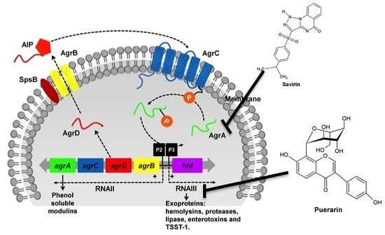
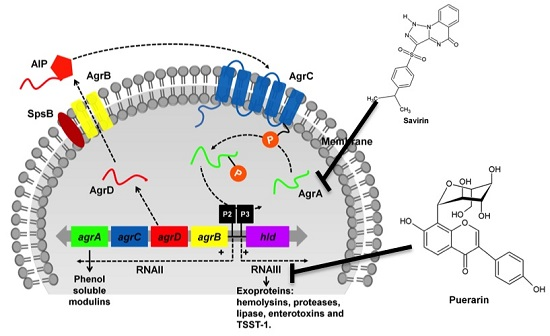
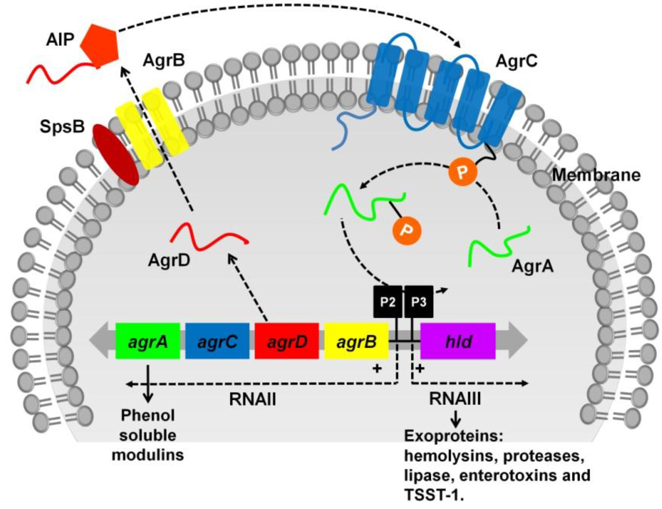
3.1. The Two-Component Regulatory Systems—Agr and Sae

The agr is currently the most extensively studied global regulator of virulence determinant production in S. aureus. The production of vital S. aureus toxins such as α-, β- and γ-hemolysins, TSST-1, enterotoxin B, D, and C, exfoliatin A and B and PVL is positively regulated by the Agr system [43,44,45,46,47,48]. In addition to toxins, the secretion of various S. aureus enzymes such as serine protease, V8 protease, staphylokinase, glycerol ester hydrolase, nuclease, lipase, PI-phospholipase C and fibrinolysin was also shown to require a functional agr system for maximal expression [39,49,50,51]. Genes regulated by agr were validated by transcription profiling of an agr mutant [52]. The agr-dependent exoprotein secretion occurs at the end of the exponential phase and continues on into stationary phase [39].
The agr locus is part of the S. aureus core genome and as outlined in Figure 1, is composed of two adjacent but divergent transcriptional units directed by two promoters, P2 and P3 [53]. P2 controls the transcription of RNAII, an operon of four genes, agrBDCA, encoding the core machinery of the agr system while P3 drives the transcription of RNAIII, a regulatory RNA of the agr system. The transmembrane endopeptidase AgrB, together with SpsB, processes and cyclizes AgrD, the peptide precursor of the autoinducer peptide, AIP, into an octapeptide, and exports it via AgrB [54]. AgrC and AgrA are homologs of histidine protein kinase sensors and response regulators that form a classical bacterial two-component signal transduction system [55,56]. At the threshold concentration, AIP binds to the extracellular receptor on AgrC. AgrC will undergo autophosphorylation and subsequently phosphorylates AgrA into its active form which, in turn, triggers P2 and P3-driven transcription followed by translation of RNAIII [57] which leads to an up-regulation of toxins and enzymes such as hemolysins, proteases, lipase, enterotoxins, and TSST-1. Binding of AgrA to the promoters also enhances the transcription of RNAII, creating a positive feedback loop to the agrBDCA operon [53,56]. Additionally, AgrA can turn on transcription of the two promoters for expression of PSMs independent of RNAIII [58]. RNAIII controls the expression of virulence factors at both transcriptional and translational levels [56]. The coding region of the δ-hemolysin encoding gene hld, is embedded within the region encoding RNAIII [59] (Figure 1).
The sae locus encodes a second major two-component regulatory system which is involved in the control of S. aureus toxins and other exoproteins expression [42]. The two genes saeR and saeS encode a response regulator and a histidine protein kinase, respectively [60]. Microarray analysis comparing the transcriptomic profile of wild-type S. aureus and a saeR/S mutant revealed a subset of 80 genes and 133 genes which were up- and downregulated, respectively, in the saeR/S deletion mutant [61]. Among the virulence factor-encoding genes down-regulated in the saeR/S mutant were genes encoding α- and γ-hemolysins, PVL, staphylococcal exotoxin, staphylococcal enterotoxin, proteases, nuclease, coagulase, and fibrinogen-binding proteins [61,62,63]. The saeR/S system was deemed to be essential for full pathogenesis of S. aureus in multiple animal models of infection [61,62,64]. As a sae mutation did not affect the expression of agr, it is proposed that the saeR/S system modulates the production of virulence factors downstream of agr [65,66]. For additional discussion on the S. aureus virulence regulatory network, readers are referred to these comprehensive reviews [67,68].

3.2. The sarA and sigB

SarA is a global transcriptional regulator that has been studied in considerable detail since its discovery over two decades ago [41]. Through genome-wide transcriptional profiling, over 120 genes are known to be modulated by the sarA locus [52]. SarA-upregulated genes encode virulence factors such as β- and δ-hemolysins, staphylococcal exotoxin, and most of the cell-bound proteins including fibrinogen, fibronectin-binding proteins, and extracellular protein A; whilst genes downregulated by SarA encode for lipase, nuclease, serine protease, α-hemolysin, as well as aureolysin [41,52]. Expression of sarA is also positively correlated with S. aureus biofilm formation [69] as validated by reduced biofilm formation in sarA mutants, leading to an increase in bacterial susceptibility towards antimicrobial agents [70]. The expression of DNase and β-lactamase remains unaffected in Sar mutants [41].
The sarA locus contains a 372-bp ORF with three distinct promoters (P1, P3, and P2) driving three overlapping transcripts with different sizes coding for the SarA protein [71]. SarA can act directly by binding to the conserved sequence within the promoter region of target genes and trigger the transcription of exoproteins (toxins), as well as cell surface proteins (protein A and fibronectin-binding proteins) [72]. It can also function indirectly through a downstream effect on other regulators such as binding to the P2 promoter of the agr system [73], thereby activating the transcription of RNAII [74]. Inactivation of sarA attenuated S. aureus virulence in several experimental animal models [75,76,77].
The sigma factor in S. aureus interacts with RNA polymerase to form a holoenzyme that recognizes specific promoter sequences, leading to transcription initiation. Sigma factor B belongs to a chromosomal operon of four genes, namely rsbU, rsbV, rsbW, and sigB [78]. In unstressed conditions, SigB is bound to RsbW and, therefore, transcription is not initiated. Once the bacteria is stressed, RsbV will be dephosphorylated by RsbU prior to its interaction with RsbW; this releases SigB and activates transcription [79]. SigB activity is positively correlated with sarA expression and this trend is also observed in toxins regulated by the Sar regulator, including TSST-1 and β-hemolysin [80,81]. The inverse correlation between sigB and agr was also recently reported in regulation of TSST-1 expression [80], and this divergence in expression was also observed in staphylococcal enterotoxin B expression [82]. The role of SigB in S. aureus virulence has been demonstrated in a few infection models, including S. aureus-induced arthritis and sepsis models [83] and the rat chronic osteomyelitis model [84].

4. From Antibiotics to Anti-Virulence Therapies

The discovery of antibiotics is one of the greatest medical breakthroughs of the early twentieth century and today, treatment of bacterial infections still relies heavily on the use of antibiotics. Antibiotics disrupt the bacterial growth cycle by blocking the synthesis and assembly of key factors required for survival. Although antibiotics are highly effective, inappropriate, or overuse has led to bacterial antibiotic resistance. Uncontrolled use of antibiotics also destroys the beneficial protection provided by the gastrointestinal microflora population in the host, making the host susceptible to infection by commensal microorganisms [85]. To make matters worse, the discovery and development of new antibiotics is waning and the number of innovative drugs reaching the market has lagged far behind the growing demand.
The major events in the history of S. aureus antibiotic resistance are typified by the emergence and rampant rise of penicillin-resistant S. aureus (PRSA), the increased prevalence of MRSA including both hospital acquired- (HA-) and community acquired (CA)-MRSA, the recognition of vancomycin-intermediate S. aureus (VISA), followed by the emergence of vancomycin-resistant S. aureus (VRSA). Recently, the World Health Organization (WHO) initiated the very first World Antibiotic Awareness Week to create awareness of the global threat of antibiotic resistance, as well as to address the challenges concerning the misuse of antibiotics in clinical practice [86]. The emergence and spread of antibiotic resistance, exemplified by the emergence of new and variant pathogens, further highlights an urgent need for novel therapeutic drugs that do not exert selective pressure that leads to evolutionary adaptation of the bacteria. An alternative approach is to develop anti-virulence therapies that interfere with bacterial virulence determinants (e.g. toxins, enzymes, or surface proteins) and/or pathways that mediate virulence (e.g. two-component virulence regulatory systems, global transcriptional regulators, or quorum sensing system) that would less likely promote the development of antibiotic resistance. Extensive knowledge on how bacterial pathogens establish infection and increased understanding of genomic determinants of pathogen virulence have opened a path to tailor robust novel anti-virulence therapeutics.
Anti-virulence compounds offer an attractive option to conventional antibiotics and hold great promise as a new therapeutic paradigm. One of the pivotal characteristics of an anti-toxin or anti-virulence compound is that the compound does not affect bacterial viability or growth. Conceptually, anti-virulence therapies that do not target bacterial viability are likely to target nonessential genes and impose reduced selective pressure minimizing the probability of resistance development [87]. Since the compound does not target bacterial growth, this strategy ensures the preservation of the host endogenous microflora population. It is believed that by inhibiting bacterial virulence traits, the bacteria are less able to colonize the host and this may allow the host natural immunity to eradicate the attenuated pathogen.
A study by Mellbye and Schuster noted that anti-virulence drugs can potentially suppress bacterial resistance whereby in pathogens that rely on cell-to-cell communication (such as S. aureus and Pseudomonas aeruginosa), resistant strains will not outcompete susceptible strains, thereby allowing anti-virulence therapies that target this social communication network to be effective even when resistance arises [88]. Allen et al. [89] proposed that a combination of anti-virulence drugs that target different types of virulence factors together with a proper treatment environment would be a more effective solution.

4.1. Targeting S. aureus Toxins: A Direct Approach

Targeting toxins has been a successful approach in combating infections against a number of pathogens including Bacillus anthracis, Bordetella pertussis, and Clostridia spp. In this particular section, we discuss the potential therapeutic agents that inhibit S. aureus hemolysins, leukotoxins, and enterotoxins via various modes of action. A number of S. aureus-specific anti-toxins have shown promise in animal models of infection. However, to date, none have been successful in clinical trials, although ample pre-clinical evidence exists that anti-toxin neutralizing agents are highly effective in preventing or treating a wide range of diseases caused by S. aureus.

4.1.1. Targeting S. aureus Hemolysins

The approval for the use of monoclonal antibodies (mAbs) against anthrax toxin by the FDA in 2009 led to studies exploring the potential of mAbs as a therapeutic option for S. aureus toxins. As described above, hemolysins are major S. aureus toxins expressed by most S. aureus strains. Hemolysin, in particular α-hemolysin (Hla), has received substantial attention as a target for anti-toxin neutralizing antibodies. Anti-α-hemolysin antibodies confer a high degree of protection against lethal staphylococcal pneumonia caused by diverse S. aureus clinical isolates in experimental animals [90,91] and significantly reduced abscess formation in a S. aureus dermonecrosis model [92,93]. Moreover, the therapeutic efficacy of antibodies appears to be additive or synergistic when applied in combination with clinically used antibiotics (vancomycin or linezolid) [91,93], signifying that the presence of these antibodies makes treatment by antibiotics more efficacious. A β-hemolysin neutralizing single-domain antibody that inhibits Hlb hemolytic activity in vitro has been isolated by antibody phage display [94]. Recently, a single antibody with high affinity and cross-reactivity towards α-hemolysin and 4 bi-component leukocidins was shown to prevent destruction of multiple human cells by both these toxins [95].
In addition to neutralizing antibodies, a number of molecules or compounds that block the hemolytic activity of S. aureus α-hemolysin have been discovered over the last few years. One example is β-cyclodextrin derivatives, small molecules that have been reported to inhibit α-hemolysis in vitro [96] and protect the host during a S. aureus infection [97,98]. The use of in silico tools and simulation program have facilitated the discovery of other α-hemolysin inhibitors [99,100,101,102]. The availability of the α-hemolysin crystal structure has enabled high throughput virtual screening of peptide or compound libraries for potent inhibitors [103,104]. For toxins with known host receptors, therapies can be designed to antagonize the receptor. ADAM10, the zinc-dependent metalloprotease receptor for Hla [12], is an attractive target for the design of an inhibitor to block toxin-receptor binding [105] to reduce lesions and the severity of recurrent skin and soft tissue infections [106]. Nevertheless, ADAM10 is a member of a large family and the role of ADAM10 has not been fully understood for other conditions, cautioning the use of an ADAM10 inhibitor.

4.1.2. Targeting S. aureus Leukotoxins

Although its contribution in CA-MRSA pathogenesis is still controversial, PVL remains as one of the main targets for the development of anti-toxin therapies. The commercially available human intravenous polyclonal immunoglobulin preparations (IVIg) contain PVL-specific antibodies and exhibit anti-PVL leukotoxicity in a dose-dependent manner, probably through interfering with PVL-neutrophil binding [107]. Recently, in a 3D human lung tissue model, IVIg, abrogated recombinant PVL and α-toxin-mediated damage in epithelial cells [108]. Proper antibiotic treatment in combination with IVIg anti-toxin therapy markedly improved the condition of patients with severe necrotizing pneumonia caused by PVL-positive S. aureus strains [109], further supporting the use of IVIg in halting disease progression especially in highly lethal S. aureus infections associated with PVL production. Leventie et al. produced humanized heavy-chain-only mAbs against LukS-PV and LukF-PV, as well as a bispecific tetravalent anti-PVL antibody that bound both LukS-PV and LukF-PV subunits [110]. These antibodies inhibit PVL binding to polymorphonuclear leukocytes and prevent the formation of new pores in vitro; similarly, in a non-infectious rabbit model of endophthalmitis, a decrease in inflammatory response and tissue injury was observed, with the tetravalent anti-PVL antibody showing a more promising effect [110]. It is worth noting that the anti-LukS-PV antibody was also able to cross-react and neutralize the HlgC subunit in γ-hemolysin, owing to the high degree of similarity in protein sequence within leukotoxins [110]. This suggests that broad-spectrum anti-toxin therapies can be generated against more than one member of the leukotoxin family. A polyclonal antibody generated against an attenuated LukS-PV mutant [111] neutralizes various leukotoxins including PVL, HlgAB, HlgCB, and LukED [112]. While an epidemiological correlation between PVL and CA-MRSA has been reported in some studies [113,114], the presence of high levels of neutralizing antibodies did not confer resistance towards skin and soft tissue-infections caused by PVL-positive MRSA [115]. Thus, the effective utility of anti-toxin antibodies against PVL requires further evaluation. In a recent study, an antimicrobial peptide (AMP) isolated from human neutrophils was identified as an anti-PVL candidate that protects leukocytes from the cytotoxic effect of PVL [116].

4.1.3. Targeting Staphylococcal Enterotoxins

Staphylococcal enterotoxin B (SEB) is one of the most-studied enterotoxins and its classification as a select agent by the United States Department of Health and Human Services makes it an attractive target for the development of anti-toxin neutralizing antibodies. SEB is relatively stable, highly resistant to denaturation and broadly classified as a superantigen [117] that can trigger increased secretion of pro-inflammatory cytokines, such as IFN-γ and TNF-α [118]. Most of the experimental therapies that target SEB are mAbs that directly bind to and neutralize SEB’s interaction with host cell receptors, leading to a reduction in pro-inflammatory cytokine production and protective immunity against the toxic and lethal effects of the toxin [119,120,121,122]. The human monoclonal antibody HuMAb-154 [119] exhibits high specificity and binding activity towards SEB. This antibody is also able to neutralize SEB-induced cytokines and prolong the survival of SEB-challenged mice, implying its potential as a prophylactic and therapeutic measure during a S. aureus infection [119]. A murine immunoglobulin IgG1 (mAb 20B1) also demonstrated potent neutralization of SEB [123]; it protected the host from lethal MRSA sepsis, as well as reduced the bacterial burden and abscess formation in superficial skin and deep-tissue infection models [122].
More recently, utilizing both NMR and crystallography techniques, the underlying mechanism and exact interaction of mAb 20B1 and SEB have been fully elucidated [121]. SEB superantigens bind to the variable region of the T-cell receptor β chain (Vβ). Buonpane et al. generated high-affinity soluble Vβ proteins as Vβ antagonists that compete with the Vβ region on the host T-cell receptor for binding to SEB, thus neutralizing SEB-mediated T-cell activation both in vitro and in vivo [124]. Subsequently, a broad spectrum neutralizing agent effective against both SEB and TSST-1 was engineered and proven to be active in vitro [125], paving the way for a single agent that is active against multiple toxins. In addition, targeting the toxin-induced host inflammatory response with known anti-inflammatory drugs is a quick approach to mitigate the toxic effects triggered by SEB via inhibition of T-cell proliferation and release of pro-inflammatory mediators [126]. Table 1 summarizes the potential anti-S. aureus toxin candidates discussed in this review and their respective modes of action.

4.2. Targeting Pathways that Govern Toxin Production: An Indirect Approach

The diverse and functionally redundant S. aureus virulence factors, including toxins and enzymes, are regulated by an intricate network of two-component regulatory systems and global transcriptional regulators. The coordinated and temporal expression of various virulence factors is important for the bacteria to adhere to host cells, establish infection and cause tissue damage in the host. As the pathogenesis of S. aureus infection cannot be attributed to a single virulence determinant, attenuation or neutralization of individual virulence factors may not confer meaningful protection to the host. In addition to the approach of direct neutralization of toxins described in the previous sections, targeting S. aureus toxins can also be done indirectly via disrupting the regulatory mechanisms that control virulence expression. As discussed in Section 3, the major virulence regulators in S. aureus are the two-component signal transduction systems (Agr and Sae) and SarA transcriptional regulator. agr and sarA knockout mutants were significantly attenuated in animal models [52,75], underscoring the importance of targeting these vital regulons to render S. aureus avirulent or less virulent.
In lieu of its role as a key virulence regulator in S. aureus, the Agr system is a robust target for the development of anti-virulence therapy. The two main proteins in the Agr system are the AgrA response regulator and the AgrC histidine kinase. In response to increasing cell density or quorum-sensing signals, AIPs will be produced to bind to and activate AgrC, directing the subsequent activation of AgrA (Figure 1). AgrA binds to P2 and P3 promoter regions, stimulating the transcription of RNAII and RNAIII which are responsible for downstream toxin gene expression. As RNAIII is the effector molecule that controls the expression of a large number of virulence factors, inhibiting RNAIII is a favorable approach to reduce the production of toxins and other virulence factors. RNAIII-inhibiting heptapeptide (RIP) interferes with quorum sensing, reduces S. aureus virulence and reverses the overall pathology in various animal infection models [127,128]. Due to its strong effect in attenuating S. aureus virulence, various RIP derivatives have been synthesized and examined. Two novel RIP derivatives (RIP-V and RIP-L) were recently found to significantly prolong the survival of mice and improve the pathological injuries in an MRSA sepsis model without affecting bacterial viability [129]. In the presence of RIPs, downregulation of RNAIII expression diminished the production of hemolysin. In combination with clinically used antibiotics, RIP accelerated wound healing and minimized the lethality rate in a S. aureus-induced sepsis mice model compared to treatment with antibiotics alone [130,131]
A number of bioactive components isolated from natural products, including isorhamnetin, chrysin, capsaicin, and puerarin (Figure 2), decreased RNAIII expression and subsequently hla expression which successfully protected the host from pneumonia caused by both MRSA and MSSA [132,133,134,135]. A significant reduction in agrA and hla transcript levels was detected in post-exponential S. aureus culture upon treatment with sub-inhibitory concentrations of a natural compound, naringenin (Figure 2). This compound also reduced the production of α-hemolysin and protected mice from S. aureus-provoked pneumonia [136]. With the exception of capsaicin, all other compounds are flavonoids that consist of a 2-phenyl-1,4-benzopyrone (flavone) backbone (Figure 2). Although the underlying molecular mechanism of these chemical entities has yet to be established, these compounds represent potential new scaffolds for the development of viable leads from natural products as anti-virulence agents in a S. aureus infection.
High-throughput virtual screening of small molecule libraries for AgrA inhibitors identified a number of compounds that did not affect bacterial growth, but, were able to repress the expression of α-hemolysin, PSM, and to some extent RNAIII, by preventing specific binding of the AgrA response regulator to the P3 promoter [137]. In another high-throughput screen utilizing a transgenic S. aureus strain carrying agr::P3 fused to a GFP reporter, Sully et al. identified a highly potent anti-virulence compound termed savirin (Figure 3) [138]. Savirin disrupted the Agr system by targeting the transcriptional function of AgrA, leading to downregulation of agr-regulated genes (including genes encoding hemolysin, PSM, PVL, lipase as well as protease) and production of secreted virulence factors. More importantly, savirin demonstrated high efficacy in two murine models of skin infection and repeated exposure to this compound had minimal impact on the development of drug resistance or tolerance [138]. The group extended the search for agr-quorum sensing inhibitors from their collection of natural bioactive compounds and successfully identified a second compound, ω-hydroxyemodin (OHM) (Figure 3), isolated from the fungus Penicillium restrictum [139]. This compound limited abscess and ulcer formation in a mouse model of skin and soft-tissue infection by binding to Agr and preventing AgrA from interacting with agr promoters, causing a reduction in secreted toxins and other virulence factors.
In the search for new anti-virulence compounds that interfere with agr activity, Mansson et al. isolated two desipeptides, Solonamide A and Solonamide B (Figure 4), from a marine Photobacterium strain that increased the expression of S. aureus protein A and decreased the expression of hla and RNAIII [140]. The solonamides are structurally similar to AIPs and are thought to antagonize the agr-quorum sensing system in S. aureus. Further studies showed that Solonamide B reduced the transcription and activity of α-hemolysin by competition with AIPs for the binding to AgrC histidine kinase, thus compromising the activation of AgrC in various S. aureus strains, including the highly virulent CA-MRSA strain USA300 [141]. At the same time, this compound also dramatically suppressed the transcription and production of PSM, which is under the control of AgrA [141]. Other active ingredients isolated from marine microorganisms also influence the expression of agr-mediated S. aureus virulence genes, albeit to a lesser extent than solonamides [142,143].
A screen of over 12 flavonoids for anti-virulence activity against S. aureus demonstrated that flavone, the backbone compound of flavonoids, significantly suppressed the production of golden pigment staphyloxanthin and α-hemolysin [144]. Transcriptional analysis of flavone-treated S. aureus cells further validated the reduction in the expression of hla encoding α-hemolysin. Furthermore, flavone repressed the expression of sae, whilst elevating agr transcription but did not affect the transcript level of sigB and sar, suggesting that flavone exerts its anti-virulence effect via the regulation of sae and agr activity [144]. Proteomics and genomics studies of manuka honey-treated MRSA disclosed a downregulation of virulence-associated genes encoding α- and γ-hemolysin, enterotoxin C, lipase, and fibronectin-binding proteins, as well as a general reduction in the expression level of global virulence regulators, such as agr, sae, and sarV [145]. These findings offer the possibility of using medicinal compounds from natural sources to alter pathogenicity of antibiotic-resistant bacteria and future studies should focus on elucidating the anti-virulence mechanism of manuka honey in an animal model.
SarA is one of the most extensively studied S. aureus transcriptional regulators. Apart from agr- system inhibitors, targeting SarA-DNA interaction is a novel approach to combat S. aureus infections. Using a structure-assisted drug design approach, Arya et al. designed and synthesized a SarA inhibitor, SarABI (Figure 5) [146]. Interestingly, despite having no anti-bacterial activity, this compound demonstrated anti-biofilm effect, reduced hemolytic activity, and attenuated bacterial adherence to host cells. Furthermore, SarABI also drastically decreased the expression of RNAIII, hld and the fibronectin encoding gene fnbA and prevented vascular graft infection by S. aureus in vivo [146]. The selective binding of SarABI to the DNA binding domain of SarA was confirmed by docking analysis [146]. Collectively, this study highlights the potential of targeting SarA as a contemporary approach for development of a novel anti-virulence agent against S. aureus.

5. Caenorhabditis elegans as a Model for the Discovery of Novel Anti-Virulence Molecules

Targeting S. aureus toxins or virulence factors appears to be a key approach for future drug design. However, a major constraint is the difficulty in probing virulence factors and infection processes within the context of a human infection. In recent years, the establishment and description of a wide range of bacterial and fungal pathogen infections in C. elegans have prompted laboratories to adopt worms as an in vivo model for the discovery of anti-infectives. Infection of C. elegans by both MSSA and MRSA has been used as a robust platform to decipher the mechanisms of host-pathogen interaction [147,148]. The virulence determinants of S. aureus thought to be important in mammalian pathogenesis are also required for pathogenicity in C. elegans [147]. S. aureus mutants for crucial mammalian virulence factors, such as agr and sarA, are attenuated in C. elegans killing, indicating that these regulators and their downstream targets, for example hemolysin and V8 protease, are required for full pathogenesis in the C. elegans model. This also suggests that key aspects of S. aureus pathogenesis are highly conserved, irrespective of the host. To identify new therapeutic molecules towards S. aureus with a mode of action that is distinct from conventional antibiotics, our laboratory developed an in vivo C. elegans—S. aureus liquid-based screen to identify compounds that do not kill the pathogen but, in turn, abrogate S. aureus virulence in the context of a whole organism [149]. Screening of a collection of natural extracts and synthetic compounds identified a subset of viable leads that promoted the extended survival of both MSSA- and MRSA-infected nematodes without conferring anti-bacterial effects on the pathogen.
The advantages of using worms in a rapid screen are its simplicity and genetic tractability as an intact host model, inexpensive to cultivate as compared to other host models, easy manipulation, and short life cycle. The nematodes are easy to manipulate because they are small enough to fit into standard 96- or 384-well microtiter plates, making them easily adaptable for high-throughput screening of compound libraries. Furthermore, the C. elegans whole-animal approach provides early and direct assessment of drug efficacy in vivo, thereby filtering out compounds that are toxic to the host or with poor pharmacokinetic properties, at the initial stage of the screen [150]. Unlike the use of murine or rabbit models for drug testing, using the C. elegans model for drug discovery does not raise any ethical concerns. With an increasing understanding of host-pathogen interactions and bacterial pathogenesis, the C. elegans model presents an advantage in being able to simultaneously detect compounds that target bacterial virulence, as well as infection processes that are only manifested when the complex host-pathogen relationship is intact. As the underlying innate immunity signaling pathways recognized in C. elegans are evolutionarily conserved, it is conceivable that findings from the nematode model could be extended to higher organisms, including humans [151].

6. Concluding Remarks

Although there is currently no anti-virulence agent approved for clinical use against bacterial infections, an emerging body of evidence has shed light on the advantages and usefulness of these therapeutic agents for fighting infections; in particular, infections by antibiotic resistant strains. Direct or indirect approaches to target S. aureus toxins should lead to the development of new adjuvants or adjunct therapies to complement the use of antibiotics. Although efforts to exploit novel anti-virulence candidates have been met with varying degrees of success, there is a lack of pharmacology and toxicology data for the promising leads. Future work should focus on selected highly-effective candidates for detailed mechanistic analysis. The possible side effects of these anti-toxin or anti-virulence candidates should also be evaluated. One potential shortfall of anti-virulence agents is the potential narrow spectrum efficacy as these drug candidates target specific virulence-mediated pathways in selected pathogens, which may restrict its application in the clinical setting. On the other hand, this could be advantageous in terms of limiting the elimination of the beneficial microflora population (for a related review, see reference [152]). Hence, the application of anti-virulence therapy in humans, and its ability to impede the problem of drug resistance without imposing strong selective pressure upon the bacterial population, requires much more extensive evaluation.

Acknowledgments

The authors acknowledge the Ministry of Science, Technology and Innovation, Malaysia (grant numbers 06-05-16-MB003 and 02-05-20-SF11117) for supporting the research utilizing C. elegans as a model for the study of host-pathogen interaction and discovery of novel anti-infectives.

Author Contributions

C.K., H.M.N. and S.N. conceived the review and wrote the manuscript.

Conflicts of Interest

The authors declare no conflicts of interest. The funding sponsors had no role in the writing of the manuscript.

References

  1. Antibiotic Resistance Threats in the United States. 2013. Available online: http://www.cdc.gov/drugresistance/threat-report-2013/ (accessed on 11 November 2015).
  2. Lowy, F.D. Staphylococcus aureus infections. N. Engl. J. Med. 1998, 339, 520–532. [Google Scholar] [CrossRef] [PubMed]
  3. McAdow, M.; DeDent, A.C.; Emolo, C.; Cheng, A.G.; Kreiswirth, B.N.; Missiakas, D.M.; Schneewind, O. Coagulases as determinants of protective immune responses against Staphylococcus aureus. Infect. Immun. 2012, 80, 3389–3398. [Google Scholar] [CrossRef] [PubMed]
  4. Jusko, M.; Potempa, J.; Kantyka, T.; Bielecka, E.; Miller, H.K.; Kalinska, M.; Dubin, G.; Garred, P.; Shaw, L.N.; Blom, A.M. Staphylococcal proteases aid in evasion of the human complement system. J. Innate Immun. 2014, 6, 31–46. [Google Scholar] [CrossRef] [PubMed]
  5. Foster, T.J.; Geoghegan, J.A.; Ganesh, V.K.; Hook, M. Adhesion, invasion and evasion: The many functions of the surface proteins of Staphylococcus aureus. Nat. Rev. Microbiol. 2014, 12, 49–62. [Google Scholar] [CrossRef] [PubMed]
  6. Vazquez, V.; Liang, X.; Horndahl, J.K.; Ganesh, V.K.; Smeds, E.; Foster, T.J.; Hook, M. Fibrinogen is a ligand for the Staphylococcus aureus microbial surface components recognizing adhesive matrix molecules (MSCRAMM) bone sialoprotein-binding protein (Bbp). J. Biol. Chem. 2011, 286, 29797–29805. [Google Scholar] [CrossRef] [PubMed]
  7. Corrigan, R.M.; Miajlovic, H.; Foster, T.J. Surface proteins that promote adherence of Staphylococcus aureus to human desquamated nasal epithelial cells. BMC Microbiol. 2009, 9, 22. [Google Scholar] [CrossRef] [PubMed]
  8. Zecconi, A.; Scali, F. Staphylococcus aureus virulence factors in evasion from innate immune defenses in human and animal diseases. Immunol. Lett. 2013, 150, 12–22. [Google Scholar] [CrossRef] [PubMed]
  9. Chavakis, T.; Preissner, K.T.; Herrmann, M. The anti-inflammatory activities of Staphylococcus aureus. Trends Immunol. 2007, 28, 408–418. [Google Scholar] [CrossRef] [PubMed]
  10. Powers, M.E.; Bubeck Wardenburg, J. Igniting the fire: Staphylococcus aureus virulence factors in the pathogenesis of sepsis. PLoS Pathog. 2014, 10, e1003871. [Google Scholar] [CrossRef] [PubMed]
  11. Valeva, A.; Walev, I.; Pinkernell, M.; Walker, B.; Bayley, H.; Palmer, M.; Bhakdi, S. Transmembrane beta-barrel of staphylococcal alpha-toxin forms in sensitive but not in resistant cells. Proc. Natl. Acad. Sci. USA 1997, 94, 11607–11611. [Google Scholar] [CrossRef] [PubMed]
  12. Wilke, G.A.; Bubeck Wardenburg, J. Role of a disintegrin and metalloprotease 10 in Staphylococcus aureus alpha-hemolysin-mediated cellular injury. Proc. Natl. Acad. Sci. USA 2010, 107, 13473–13478. [Google Scholar] [CrossRef] [PubMed]
  13. Powers, M.E.; Becker, R.E.; Sailer, A.; Turner, J.R.; Bubeck Wardenburg, J. Synergistic action of Staphylococcus aureus alpha-toxin on platelets and myeloid lineage cells contributes to lethal sepsis. Cell. Host Microbe 2015, 17, 775–787. [Google Scholar] [CrossRef] [PubMed]
  14. Walev, I.; Weller, U.; Strauch, S.; Foster, T.; Bhakdi, S. Selective killing of human monocytes and cytokine release provoked by sphingomyelinase (beta-toxin) of Staphylococcus aureus. Infect. Immun. 1996, 64, 2974–2979. [Google Scholar] [PubMed]
  15. Vandenesch, F.; Lina, G.; Henry, T. Staphylococcus aureus hemolysins, bi-component leukocidins, and cytolytic peptides: A redundant arsenal of membrane-damaging virulence factors? Front. Cell. Infect. Microbiol. 2012, 2, 12. [Google Scholar] [CrossRef] [PubMed]
  16. Meyer, F.; Girardot, R.; Piemont, Y.; Prevost, G.; Colin, D.A. Analysis of the specificity of Panton-Valentine leucocidin and gamma-hemolysin F component binding. Infect. Immun. 2009, 77, 266–273. [Google Scholar] [CrossRef] [PubMed]
  17. Olson, R.; Nariya, H.; Yokota, K.; Kamio, Y.; Gouaux, E. Crystal structure of staphylococcal LukF delineates conformational changes accompanying formation of a transmembrane channel. Nat. Struct. Biol. 1999, 6, 134–140. [Google Scholar] [PubMed]
  18. Verdon, J.; Girardin, N.; Lacombe, C.; Berjeaud, J.M.; Hechard, Y. Delta-hemolysin, an update on a membrane-interacting peptide. Peptides 2009, 30, 817–823. [Google Scholar] [CrossRef] [PubMed]
  19. Surewaard, B.G.; de Haas, C.J.; Vervoort, F.; Rigby, K.M.; DeLeo, F.R.; Otto, M.; van Strijp, J.A.; Nijland, R. Staphylococcal alpha-phenol soluble modulins contribute to neutrophil lysis after phagocytosis. Cell. Microbiol. 2013, 15, 1427–1437. [Google Scholar] [CrossRef] [PubMed]
  20. Otto, M. Phenol-soluble modulins. Int. J. Med. Microbiol. 2014, 304, 164–169. [Google Scholar] [CrossRef] [PubMed]
  21. Zivkovic, A.; Sharif, O.; Stich, K.; Doninger, B.; Biaggio, M.; Colinge, J.; Bilban, M.; Mesteri, I.; Hazemi, P.; Lemmens-Gruber, R.; et al. TLR 2 and CD14 mediate innate immunity and lung inflammation to staphylococcal Panton-Valentine leukocidin in vivo. J. Immunol. 2011, 186, 1608–1617. [Google Scholar] [CrossRef] [PubMed]
  22. Inden, K.; Kaneko, J.; Miyazato, A.; Yamamoto, N.; Mouri, S.; Shibuya, Y.; Nakamura, K.; Aoyagi, T.; Hatta, M.; Kunishima, H.; et al. Toll-like receptor 4-dependent activation of myeloid dendritic cells by leukocidin of Staphylococcus aureus. Microbes Infect. 2009, 11, 245–253. [Google Scholar] [CrossRef] [PubMed]
  23. Alonzo, F., III; Kozhaya, L.; Rawlings, S.A.; Reyes-Robles, T.; DuMont, A.L.; Myszka, D.G.; Landau, N.R.; Unutmaz, D.; Torres, V.J. CCR5 is a receptor for Staphylococcus aureus leukotoxin ED. Nature 2013, 493, 51–55. [Google Scholar] [PubMed]
  24. DuMont, A.L.; Yoong, P.; Day, C.J.; Alonzo, F., III; McDonald, W.H.; Jennings, M.P.; Torres, V.J. Staphylococcus aureus LukAB cytotoxin kills human neutrophils by targeting the CD11b subunit of the integrin Mac-1. Proc. Natl. Acad. Sci. USA 2013, 110, 10794–10799. [Google Scholar] [PubMed]
  25. Spaan, A.N.; Henry, T.; van Rooijen, W.J.; Perret, M.; Badiou, C.; Aerts, P.C.; Kemmink, J.; de Haas, C.J.; van Kessel, K.P.; Vandenesch, F.; et al. The staphylococcal toxin Panton-Valentine leukocidin targets human C5a receptors. Cell. Host Microbe 2013, 13, 584–594. [Google Scholar] [CrossRef] [PubMed]
  26. Otto, M. Basis of virulence in community-associated methicillin-resistant Staphylococcus aureus. Annu. Rev. Microbiol. 2010, 64, 143–162. [Google Scholar] [CrossRef] [PubMed]
  27. Bukowski, M.; Wladyka, B.; Dubin, G. Exfoliative toxins of Staphylococcus aureus. Toxins 2010, 2, 1148–1165. [Google Scholar] [CrossRef] [PubMed]
  28. Eyre, R.W.; Stanley, J.R. Human autoantibodies against a desmosomal protein complex with a calcium-sensitive epitope are characteristic of pemphigus foliaceus patients. J. Exp. Med. 1987, 165, 1719–1724. [Google Scholar] [CrossRef] [PubMed]
  29. Hanakawa, Y.; Schechter, N.M.; Lin, C.; Garza, L.; Li, H.; Yamaguchi, T.; Fudaba, Y.; Nishifuji, K.; Sugai, M.; Amagai, M.; et al. Molecular mechanisms of blister formation in bullous impetigo and staphylococcal scalded skin syndrome. J. Clin. Invest. 2002, 110, 53–60. [Google Scholar] [CrossRef] [PubMed]
  30. Monday, S.R.; Vath, G.M.; Ferens, W.A.; Deobald, C.; Rago, J.V.; Gahr, P.J.; Monie, D.D.; Iandolo, J.J.; Chapes, S.K.; Davis, W.C.; et al. Unique superantigen activity of staphylococcal exfoliative toxins. J. Immunol. 1999, 162, 4550–4559. [Google Scholar] [PubMed]
  31. Hennekinne, J.A.; De Buyser, M.L.; Dragacci, S. Staphylococcus aureus and its food poisoning toxins: Characterization and outbreak investigation. FEMS Microbiol. Rev. 2012, 36, 815–836. [Google Scholar] [CrossRef] [PubMed]
  32. Lin, C.F.; Chen, C.L.; Huang, W.C.; Cheng, Y.L.; Hsieh, C.Y.; Wang, C.Y.; Hong, M.Y. Different types of cell death induced by enterotoxins. Toxins 2010, 2, 2158–2176. [Google Scholar] [CrossRef] [PubMed]
  33. Balaban, N.; Rasooly, A. Staphylococcal enterotoxins. Int. J. Food Microbiol. 2000, 61, 1–10. [Google Scholar] [CrossRef]
  34. Betley, M.J.; Schlievert, P.M.; Bergdoll, M.S.; Bohach, G.A.; Iandolo, J.J.; Khan, S.A.; Pattee, P.A.; Reiser, R.R. Staphylococcal gene nomenclature. ASM News 1990, 56, 182. [Google Scholar]
  35. Berkley, S.F.; Hightower, A.W.; Broome, C.V.; Reingold, A.L. The relationship of tampon characteristics to menstrual toxic shock syndrome. JAMA 1987, 258, 917–920. [Google Scholar] [CrossRef] [PubMed]
  36. Otto, M. Staphylococcus aureus toxins. Curr. Opin. Microbiol. 2014, 17, 32–37. [Google Scholar] [CrossRef] [PubMed]
  37. Stach, C.S.; Herrera, A.; Schlievert, P.M. Staphylococcal superantigens interact with multiple host receptors to cause serious diseases. Immunol. Res. 2014, 59, 177–181. [Google Scholar] [CrossRef] [PubMed]
  38. Larkin, S.M.; Williams, D.N.; Osterholm, M.T.; Tofte, R.W.; Posalaky, Z. Toxic shock syndrome: Clinical, laboratory, and pathologic findings in nine fatal cases. Ann. Intern. Med. 1982, 96, 858–864. [Google Scholar] [CrossRef] [PubMed]
  39. Recsei, P.; Kreiswirth, B.; O’Reilly, M.; Schlievert, P.; Gruss, A.; Novick, R.P. Regulation of exoprotein gene expression in Staphylococcus aureus by agar. Mol. Gen. Genet. 1986, 202, 58–61. [Google Scholar] [CrossRef] [PubMed]
  40. Roux, A.; Todd, D.A.; Velazquez, J.V.; Cech, N.B.; Sonenshein, A.L. Cody-mediated regulation of the Staphylococcus aureus Agr system integrates nutritional and population density signals. J. Bacteriol. 2014, 196, 1184–1196. [Google Scholar] [CrossRef] [PubMed]
  41. Cheung, A.L.; Koomey, J.M.; Butler, C.A.; Projan, S.J.; Fischetti, V.A. Regulation of exoprotein expression in Staphylococcus aureus by a locus (sar) distinct from agr. Proc. Natl. Acad. Sci. USA 1992, 89, 6462–6466. [Google Scholar] [CrossRef] [PubMed]
  42. Giraudo, A.T.; Cheung, A.L.; Nagel, R. The sae locus of Staphylococcus aureus controls exoprotein synthesis at the transcriptional level. Arch. Microbiol. 1997, 168, 53–58. [Google Scholar] [CrossRef] [PubMed]
  43. Gaskill, M.E.; Khan, S.A. Regulation of the enterotoxin B gene in Staphylococcus aureus. J. Biol. Chem. 1988, 263, 6276–6280. [Google Scholar] [PubMed]
  44. Bayles, K.W.; Iandolo, J.J. Genetic and molecular analyses of the gene encoding staphylococcal enterotoxin D. J. Bacteriol 1989, 171, 4799–4806. [Google Scholar] [PubMed]
  45. Regassa, L.B.; Couch, J.L.; Betley, M.J. Steady-state staphylococcal enterotoxin type C mRNA is affected by a product of the accessory gene regulator (agr) and by glucose. Infect. Immun. 1991, 59, 955–962. [Google Scholar] [PubMed]
  46. Vojtov, N.; Ross, H.F.; Novick, R.P. Global repression of exotoxin synthesis by staphylococcal superantigens. Proc. Natl. Acad. Sci. USA 2002, 99, 10102–10107. [Google Scholar] [CrossRef] [PubMed]
  47. Mallonee, D.H.; Glatz, B.A.; Pattee, P.A. Chromosomal mapping of a gene affecting enterotoxin A production in Staphylococcus aureus. Appl. Environ. Microbiol. 1982, 43, 397–402. [Google Scholar] [PubMed]
  48. Kreiswirth, B.N.; Lofdahl, S.; Betley, M.J.; O’Reilly, M.; Schlievert, P.M.; Bergdoll, M.S.; Novick, R.P. The toxic shock syndrome exotoxin structural gene is not detectably transmitted by a prophage. Nature 1983, 305, 709–712. [Google Scholar] [CrossRef] [PubMed]
  49. Arvidson, S.; Tegmark, K. Regulation of virulence determinants in Staphylococcus aureus. Int J. Med. Microbiol. 2001, 291, 159–170. [Google Scholar] [CrossRef] [PubMed]
  50. Said-Salim, B.; Dunman, P.M.; McAleese, F.M.; Macapagal, D.; Murphy, E.; McNamara, P.J.; Arvidson, S.; Foster, T.J.; Projan, S.J.; Kreiswirth, B.N. Global regulation of Staphylococcus aureus genes by Rot. J. Bacteriol. 2003, 185, 610–619. [Google Scholar] [CrossRef] [PubMed]
  51. Smeltzer, M.S.; Hart, M.E.; Iandolo, J.J. Phenotypic characterization of xpr, a global regulator of extracellular virulence factors in Staphylococcus aureus. Infect. Immun. 1993, 61, 919–925. [Google Scholar] [PubMed]
  52. Dunman, P.M.; Murphy, E.; Haney, S.; Palacios, D.; Tucker-Kellogg, G.; Wu, S.; Brown, E.L.; Zagursky, R.J.; Shlaes, D.; Projan, S.J. Transcription profiling-based identification of Staphylococcus aureus genes regulated by the agr and/or sarA loci. J. Bacteriol. 2001, 183, 7341–7353. [Google Scholar] [CrossRef] [PubMed]
  53. Novick, R.P.; Projan, S.J.; Kornblum, J.; Ross, H.F.; Ji, G.; Kreiswirth, B.; Vandenesch, F.; Moghazeh, S. The agr P2 operon: An autocatalytic sensory transduction system in Staphylococcus aureus. Mol. Gen. Genet. 1995, 248, 446–458. [Google Scholar] [CrossRef] [PubMed]
  54. Thoendel, M.; Horswill, A.R. Identification of Staphylococcus aureus AgrD residues required for autoinducing peptide biosynthesis. J. Biol. Chem. 2009, 284, 21828–21838. [Google Scholar] [CrossRef] [PubMed]
  55. Lina, G.; Jarraud, S.; Ji, G.; Greenland, T.; Pedraza, A.; Etienne, J.; Novick, R.P.; Vandenesch, F. Transmembrane topology and histidine protein kinase activity of AgrC, the agr signal receptor in Staphylococcus aureus. Mol. Microbiol. 1998, 28, 655–662. [Google Scholar] [CrossRef] [PubMed]
  56. Novick, R.P.; Ross, H.F.; Projan, S.J.; Kornblum, J.; Kreiswirth, B.; Moghazeh, S. Synthesis of staphylococcal virulence factors is controlled by a regulatory RNA molecule. EMBO J. 1993, 12, 3967–3975. [Google Scholar] [PubMed]
  57. Koenig, R.L.; Ray, J.L.; Maleki, S.J.; Smeltzer, M.S.; Hurlburt, B.K. Staphylococcus aureus AgrA binding to the RNAIII-agr regulatory region. J. Bacteriol. 2004, 186, 7549–7555. [Google Scholar] [CrossRef] [PubMed]
  58. Queck, S.Y.; Jameson-Lee, M.; Villaruz, A.E.; Bach, T.H.; Khan, B.A.; Sturdevant, D.E.; Ricklefs, S.M.; Li, M.; Otto, M. RNAIII-independent target gene control by the agr quorum-sensing system: Insight into the evolution of virulence regulation in Staphylococcus aureus. Mol. Cell. 2008, 32, 150–158. [Google Scholar] [CrossRef] [PubMed]
  59. Balaban, N.; Novick, R.P. Translation ofRNAIII, the Staphylococcus aureus agr regulatory RNA molecule, can be activated by a 3’-end deletion. FEMS Microbiol. Lett. 1995, 133, 155–161. [Google Scholar] [PubMed]
  60. Giraudo, A.T.; Calzolari, A.; Cataldi, A.A.; Bogni, C.; Nagel, R. The sae locus of Staphylococcus aureus encodes a two-component regulatory system. FEMS Microbiol. Lett. 1999, 177, 15–22. [Google Scholar] [CrossRef] [PubMed]
  61. Voyich, J.M.; Vuong, C.; DeWald, M.; Nygaard, T.K.; Kocianova, S.; Griffith, S.; Jones, J.; Iverson, C.; Sturdevant, D.E.; Braughton, K.R.; et al. The SaeR/S gene regulatory system is essential for innate immune evasion by Staphylococcus aureus. J. Infect. Dis. 2009, 199, 1698–1706. [Google Scholar] [CrossRef] [PubMed]
  62. Date, S.V.; Modrusan, Z.; Lawrence, M.; Morisaki, J.H.; Toy, K.; Shah, I.M.; Kim, J.; Park, S.; Xu, M.; Basuino, L.; et al. Global gene expression of methicillin-resistant Staphylococcus aureus USA300 during human and mouse infection. J. Infect. Dis. 2014, 209, 1542–1550. [Google Scholar] [CrossRef] [PubMed]
  63. Giraudo, A.T.; Raspanti, C.G.; Calzolari, A.; Nagel, R. Characterization of a Tn551-mutant of Staphylococcus aureus defective in the production of several exoproteins. Can. J. Microbiol. 1994, 40, 677–681. [Google Scholar] [CrossRef] [PubMed]
  64. Cassat, J.E.; Hammer, N.D.; Campbell, J.P.; Benson, M.A.; Perrien, D.S.; Mrak, L.N.; Smeltzer, M.S.; Torres, V.J.; Skaar, E.P. A secreted bacterial protease tailors the Staphylococcus aureus virulence repertoire to modulate bone remodeling during osteomyelitis. Cell. Host Microbe 2013, 13, 759–772. [Google Scholar] [CrossRef] [PubMed]
  65. Novick, R.P.; Jiang, D. The staphylococcal saeRS system coordinates environmental signals with agr quorum sensing. Microbiology 2003, 149, 2709–2717. [Google Scholar] [CrossRef] [PubMed]
  66. Rogasch, K.; Ruhmling, V.; Pane-Farre, J.; Hoper, D.; Weinberg, C.; Fuchs, S.; Schmudde, M.; Broker, B.M.; Wolz, C.; Hecker, M.; et al. Influence of the two-component system SaeRS on global gene expression in two different Staphylococcus aureus strains. J. Bacteriol. 2006, 188, 7742–7758. [Google Scholar] [CrossRef] [PubMed]
  67. Novick, R.P. Autoinduction and signal transduction in the regulation of staphylococcal virulence. Mol. Microbiol. 2003, 48, 1429–1449. [Google Scholar] [CrossRef] [PubMed]
  68. Bronner, S.; Monteil, H.; Prevost, G. Regulation of virulence determinants in Staphylococcus aureus: Complexity and applications. FEMS Microbiol. Rev. 2004, 28, 183–200. [Google Scholar] [CrossRef] [PubMed]
  69. Beenken, K.E.; Blevins, J.S.; Smeltzer, M.S. Mutation of sarA in Staphylococcus aureus limits biofilm formation. Infect. Immun. 2003, 71, 4206–4211. [Google Scholar] [CrossRef] [PubMed]
  70. Lamichhane-Khadka, R.; Cantore, S.A.; Riordan, J.T.; Delgado, A.; Norman, A.E.; Duenas, S.; Zaman, S.; Horan, S.; Wilkinson, B.J.; Gustafson, J.E. SarA inactivation reduces vancomycin-intermediate and ciprofloxacin resistance expression by Staphylococcus aureus. Int. J. Antimicrob. Agents 2009, 34, 136–141. [Google Scholar] [CrossRef] [PubMed]
  71. Bayer, M.G.; Heinrichs, J.H.; Cheung, A.L. The molecular architecture of the sar locus in Staphylococcus aureus. J. Bacteriol 1996, 178, 4563–4570. [Google Scholar] [PubMed]
  72. Chien, Y.; Manna, A.C.; Projan, S.J.; Cheung, A.L. SarA, a global regulator of virulence determinants in Staphylococcus aureus, binds to a conserved motif essential for sar-dependent gene regulation. J. Biol. Chem. 1999, 274, 37169–37176. [Google Scholar] [CrossRef] [PubMed]
  73. Cheung, A.L.; Bayer, M.G.; Heinrichs, J.H. Sar genetic determinants necessary for transcription of RNAII and RNAIII in the agr locus of Staphylococcus aureus. J. Bacteriol. 1997, 179, 3963–3971. [Google Scholar] [PubMed]
  74. Reyes, D.; Andrey, D.O.; Monod, A.; Kelley, W.L.; Zhang, G.; Cheung, A.L. Coordinated regulation by AgrA, SarA, and SarR to control agr expression in Staphylococcus aureus. J. Bacteriol. 2011, 193, 6020–6031. [Google Scholar] [CrossRef] [PubMed]
  75. Cheung, A.L.; Eberhardt, K.J.; Chung, E.; Yeaman, M.R.; Sullam, P.M.; Ramos, M.; Bayer, A.S. Diminished virulence of a sar-/agr- mutant of Staphylococcus aureus in the rabbit model of endocarditis. J. Clin. Invest. 1994, 94, 1815–1822. [Google Scholar] [CrossRef] [PubMed]
  76. Blevins, J.S.; Elasri, M.O.; Allmendinger, S.D.; Beenken, K.E.; Skinner, R.A.; Thomas, J.R.; Smeltzer, M.S. Role of sarA in the pathogenesis of Staphylococcus aureus musculoskeletal infection. Infect. Immun. 2003, 71, 516–523. [Google Scholar] [CrossRef] [PubMed]
  77. Snowden, J.N.; Beaver, M.; Beenken, K.; Smeltzer, M.; Horswill, A.R.; Kielian, T. Staphylococcus aureus sarA regulates inflammation and colonization during central nervous system biofilm formation. PLoS ONE 2013, 8, e84089. [Google Scholar] [CrossRef] [PubMed]
  78. Wu, S.; de Lencastre, H.; Tomasz, A. Sigma-B, a putative operon encoding alternate sigma factor of Staphylococcus aureus RNA polymerase: Molecular cloning and DNA sequencing. J. Bacteriol. 1996, 178, 6036–6042. [Google Scholar] [PubMed]
  79. Zhang, S.; Reeves, A.; Woodbury, R.L.; Haldenwang, W.G. Coexpression patterns of sigma(B) regulators in Bacillus subtilis affect sigma(B) inducibility. J. Bacteriol. 2005, 187, 8520–8525. [Google Scholar] [CrossRef] [PubMed]
  80. Andrey, D.O.; Jousselin, A.; Villanueva, M.; Renzoni, A.; Monod, A.; Barras, C.; Rodriguez, N.; Kelley, W.L. Impact of the regulators sigB, rot, sarA and sarS on the toxic shock Tst promoter and TSST-1 expression in Staphylococcus aureus. PLoS ONE 2015, 10, e0135579. [Google Scholar] [CrossRef] [PubMed]
  81. Ziebandt, A.K.; Weber, H.; Rudolph, J.; Schmid, R.; Hoper, D.; Engelmann, S.; Hecker, M. Extracellular proteins of Staphylococcus aureus and the role of sarA and sigma B. Proteomics 2001, 1, 480–493. [Google Scholar] [CrossRef]
  82. Schmidt, K.A.; Donegan, N.P.; Kwan, W.A., Jr.; Cheung, A. Influences of sigma B and agr on expression of staphylococcal enterotoxin B (seb) in Staphylococcus aureus. Can. J. Microbiol. 2004, 50, 351–360. [Google Scholar] [CrossRef] [PubMed]
  83. Jonsson, I.M.; Arvidson, S.; Foster, S.; Tarkowski, A. Sigma factor B and RsbU are required for virulence in Staphylococcus aureus-induced arthritis and sepsis. Infect. Immun. 2004, 72, 6106–6111. [Google Scholar] [CrossRef] [PubMed]
  84. Tuchscherr, L.; Bischoff, M.; Lattar, S.M.; Noto Llana, M.; Pfortner, H.; Niemann, S.; Geraci, J.; Van de Vyver, H.; Fraunholz, M.J.; Cheung, A.L.; et al. Sigma factor sigB is crucial to mediate Staphylococcus aureus adaptation during chronic infections. PLoS Pathog. 2015, 11, e1004870. [Google Scholar] [CrossRef] [PubMed]
  85. Blaser, M. Antibiotic overuse: Stop the killing of beneficial bacteria. Nature 2011, 476, 393–394. [Google Scholar] [CrossRef] [PubMed]
  86. World Antibiotic Awareness Week. Available online: http://www.who.int/mediacentre/events/2015/world-antibiotic-awareness-week/event/en/ (accessed on 24 December 2015).
  87. Rasko, D.A.; Sperandio, V. Anti-virulence strategies to combat bacteria-mediated disease. Nat. Rev. Drug Discov. 2010, 9, 117–128. [Google Scholar] [CrossRef] [PubMed]
  88. Mellbye, B.; Schuster, M. The sociomicrobiology of antivirulence drug resistance: A proof of concept. MBio 2011, 2. [Google Scholar] [CrossRef] [PubMed]
  89. Allen, R.C.; Popat, R.; Diggle, S.P.; Brown, S.P. Targeting virulence: Can we make evolution-proof drugs? Nat. Rev. Microbiol. 2014, 12, 300–308. [Google Scholar] [CrossRef] [PubMed]
  90. Ragle, B.E.; Bubeck Wardenburg, J. Anti-alpha-hemolysin monoclonal antibodies mediate protection against Staphylococcus aureus pneumonia. Infect. Immun. 2009, 77, 2712–2718. [Google Scholar] [CrossRef] [PubMed]
  91. Hua, L.; Hilliard, J.J.; Shi, Y.; Tkaczyk, C.; Cheng, L.I.; Yu, X.; Datta, V.; Ren, S.; Feng, H.; Zinsou, R.; et al. Assessment of an anti-alpha-toxin monoclonal antibody for prevention and treatment of Staphylococcus aureus-induced pneumonia. Antimicrob. Agents Chemother. 2014, 58, 1108–1117. [Google Scholar] [CrossRef] [PubMed]
  92. Tkaczyk, C.; Hua, L.; Varkey, R.; Shi, Y.; Dettinger, L.; Woods, R.; Barnes, A.; MacGill, R.S.; Wilson, S.; Chowdhury, P.; et al. Identification of anti-alpha toxin monoclonal antibodies that reduce the severity of Staphylococcus aureus dermonecrosis and exhibit a correlation between affinity and potency. Clin. Vaccine Immunol. 2012, 19, 377–385. [Google Scholar] [CrossRef] [PubMed]
  93. Foletti, D.; Strop, P.; Shaughnessy, L.; Hasa-Moreno, A.; Casas, M.G.; Russell, M.; Bee, C.; Wu, S.; Pham, A.; Zeng, Z.; et al. Mechanism of action and in vivo efficacy of a human-derived antibody against Staphylococcus aureus alpha-hemolysin. J. Mol. Biol. 2013, 425, 1641–1654. [Google Scholar] [CrossRef] [PubMed]
  94. Pooja, J.; Ajit, S. Staphylococcus aureus β-hemolysin-neutralizing single-domain antibody isolated from phage display library of indian desert camel. Asian Pac. J. Trop Med. 2010, 3, 1–7. [Google Scholar]
  95. Rouha, H.; Badarau, A.; Visram, Z.C.; Battles, M.B.; Prinz, B.; Magyarics, Z.; Nagy, G.; Mirkina, I.; Stulik, L.; Zerbs, M.; et al. Five birds, one stone: Neutralization of alpha-hemolysin and 4 bi-component leukocidins of Staphylococcus aureus with a single human monoclonal antibody. MAbs 2015, 7, 243–254. [Google Scholar] [CrossRef] [PubMed]
  96. Karginov, V.A.; Nestorovich, E.M.; Schmidtmann, F.; Robinson, T.M.; Yohannes, A.; Fahmi, N.E.; Bezrukov, S.M.; Hecht, S.M. Inhibition of S. aureus alpha-hemolysin and B. anthracis lethal toxin by beta-cyclodextrin derivatives. Bioorg. Med. Chem. 2007, 15, 5424–5431. [Google Scholar] [CrossRef] [PubMed]
  97. McCormick, C.C.; Caballero, A.R.; Balzli, C.L.; Tang, A.; O’Callaghan, R.J. Chemical inhibition of alpha-toxin, a key corneal virulence factor of Staphylococcus aureus. Invest. Ophthalmol. Vis. Sci. 2009, 50, 2848–2854. [Google Scholar] [CrossRef] [PubMed]
  98. Ragle, B.E.; Karginov, V.A.; Bubeck Wardenburg, J. Prevention and treatment of Staphylococcus aureus pneumonia with a beta-cyclodextrin derivative. Antimicrob. Agents Chemother. 2010, 54, 298–304. [Google Scholar] [CrossRef] [PubMed]
  99. Dong, J.; Qiu, J.; Zhang, Y.; Lu, C.; Dai, X.; Wang, J.; Li, H.; Wang, X.; Tan, W.; Luo, M.; et al. Oroxylin a inhibits hemolysis via hindering the self-assembly of alpha-hemolysin heptameric transmembrane pore. PLoS Comput. Biol. 2013, 9, e1002869. [Google Scholar] [CrossRef] [PubMed]
  100. Qiu, J.; Wang, D.; Zhang, Y.; Dong, J.; Wang, J.; Niu, X. Molecular modeling reveals the novel inhibition mechanism and binding mode of three natural compounds to staphylococcal alpha-hemolysin. PLoS ONE 2013, 8, e80197. [Google Scholar] [CrossRef] [PubMed]
  101. Melo, M.C.; Teixeira, L.R.; Pol-Fachin, L.; Rodrigues, C.G. Inhibition of the hemolytic activity caused by Staphylococcus aureus alpha-hemolysin through isatin-schiff copper(ii) complexes. FEMS Microbiol. Lett. 2016, 363. [Google Scholar]
  102. Wang, J.; Zhou, X.; Liu, S.; Li, G.; Shi, L.; Dong, J.; Li, W.; Deng, X.; Niu, X. Morin hydrate attenuates Staphylococcus aureus virulence by inhibiting the self-assembly of alpha-hemolysin. J. Appl. Microbiol. 2015, 118, 753–763. [Google Scholar] [CrossRef] [PubMed]
  103. Rashidieh, B.; Etemadiafshar, S.; Memari, G.; Mirzaeichegeni, M.; Yazdi, S.; Farsimadan, F.; Alizadeh, S. A molecular modeling based screening for potential inhibitors to alpha hemolysin from Staphylococcus aureus. Bioinformation 2015, 11, 373–377. [Google Scholar] [CrossRef] [PubMed]
  104. Rani, N.; Saravanan, V.; Lakshmi, P.T.V.; Annamalai, A. Inhibition of pore formation by blocking the assembly of Staphylococcus aureus α-hemolysin through a novel peptide inhibitor: An in silico approach. Int. J. Pept. Res. Ther. 2014, 20, 575–583. [Google Scholar] [CrossRef]
  105. Dreymueller, D.; Uhlig, S.; Ludwig, A. Adam-family metalloproteinases in lung inflammation: Potential therapeutic targets. Am. J. Physiol Lung Cell. Mol. Physiol. 2015, 308, L325–343. [Google Scholar] [CrossRef] [PubMed]
  106. Sampedro, G.R.; DeDent, A.C.; Becker, R.E.; Berube, B.J.; Gebhardt, M.J.; Cao, H.; Bubeck Wardenburg, J. Targeting Staphylococcus aureus alpha-toxin as a novel approach to reduce severity of recurrent skin and soft-tissue infections. J. Infect. Dis. 2014, 210, 1012–1018. [Google Scholar] [CrossRef] [PubMed]
  107. Gauduchon, V.; Cozon, G.; Vandenesch, F.; Genestier, A.L.; Eyssade, N.; Peyrol, S.; Etienne, J.; Lina, G. Neutralization of Staphylococcus aureus Panton-Valentine leukocidin by intravenous immunoglobulin in vitro. J. Infect. Dis. 2004, 189, 346–353. [Google Scholar] [CrossRef] [PubMed]
  108. Mairpady Shambat, S.; Chen, P.; Nguyen Hoang, A.T.; Bergsten, H.; Vandenesch, F.; Siemens, N.; Lina, G.; Monk, I.R.; Foster, T.J.; Arakere, G.; et al. Modelling staphylococcal pneumonia in a human 3D lung tissue model system delineates toxin-mediated pathology. Dis. Model. Mech. 2015, 8, 1413–1425. [Google Scholar] [CrossRef] [PubMed]
  109. Rouzic, N.; Janvier, F.; Libert, N.; Javouhey, E.; Lina, G.; Nizou, J.Y.; Pasquier, P.; Stamm, D.; Brinquin, L.; Pelletier, C.; et al. Prompt and successful toxin-targeting treatment of three patients with necrotizing pneumonia due to Staphylococcus aureus strains carrying the Panton-Valentine leukocidin genes. J. Clin. Microbiol. 2010, 48, 1952–1955. [Google Scholar] [CrossRef] [PubMed]
  110. Laventie, B.J.; Rademaker, H.J.; Saleh, M.; de Boer, E.; Janssens, R.; Bourcier, T.; Subilia, A.; Marcellin, L.; van Haperen, R.; Lebbink, J.H.; et al. Heavy chain-only antibodies and tetravalent bispecific antibody neutralizing Staphylococcus aureus leukotoxins. Proc. Natl. Acad. Sci. USA 2011, 108, 16404–16409. [Google Scholar] [CrossRef] [PubMed]
  111. Karauzum, H.; Adhikari, R.P.; Sarwar, J.; Devi, V.S.; Abaandou, L.; Haudenschild, C.; Mahmoudieh, M.; Boroun, A.R.; Vu, H.; Nguyen, T.; et al. Structurally designed attenuated subunit vaccines for S. aureus LukS-PV and LukF-PV confer protection in a mouse bacteremia model. PLoS ONE 2013, 8, e65384. [Google Scholar] [CrossRef] [PubMed]
  112. Adhikari, R.P.; Kort, T.; Shulenin, S.; Kanipakala, T.; Ganjbaksh, N.; Roghmann, M.C.; Holtsberg, F.W.; Aman, M.J. Antibodies to S. aureus LukS-PV attenuated subunit vaccine neutralize a broad spectrum of canonical and non-canonical bicomponent leukotoxin pairs. PLoS ONE 2015, 10, e0137874. [Google Scholar] [CrossRef] [PubMed]
  113. Vandenesch, F.; Naimi, T.; Enright, M.C.; Lina, G.; Nimmo, G.R.; Heffernan, H.; Liassine, N.; Bes, M.; Greenland, T.; Reverdy, M.E.; et al. Community-acquired methicillin-resistant Staphylococcus aureus carrying Panton-Valentine leukocidin genes: Worldwide emergence. Emerg. Infect. Dis. 2003, 9, 978–984. [Google Scholar] [CrossRef] [PubMed]
  114. Diep, B.A.; Otto, M. The role of virulence determinants in community-associated MRSA pathogenesis. Trends Microbiol. 2008, 16, 361–369. [Google Scholar] [CrossRef] [PubMed]
  115. Hermos, C.R.; Yoong, P.; Pier, G.B. High levels of antibody to Panton-Valentine leukocidin are not associated with resistance to Staphylococcus aureus-associated skin and soft-tissue infection. Clin. Infect. Dis. 2010, 51, 1138–1146. [Google Scholar] [CrossRef] [PubMed]
  116. Cardot-Martin, E.; Casalegno, J.S.; Badiou, C.; Dauwalder, O.; Keller, D.; Prevost, G.; Rieg, S.; Kern, W.V.; Cuerq, C.; Etienne, J.; et al. Alpha-defensins partially protect human neutrophils against Panton-Valentine leukocidin produced by Staphylococcus aureus. Lett. Appl. Microbiol. 2015, 61, 158–164. [Google Scholar] [CrossRef] [PubMed]
  117. Pinchuk, I.V.; Beswick, E.J.; Reyes, V.E. Staphylococcal enterotoxins. Toxins 2010, 2, 2177–2197. [Google Scholar] [CrossRef] [PubMed]
  118. Marrack, P.; Blackman, M.; Kushnir, E.; Kappler, J. The toxicity of staphylococcal enterotoxin B in mice is mediated by t cells. J. Exp. Med. 1990, 171, 455–464. [Google Scholar] [CrossRef] [PubMed]
  119. Drozdowski, B.; Zhou, Y.; Kline, B.; Spidel, J.; Chan, Y.Y.; Albone, E.; Turchin, H.; Chao, Q.; Henry, M.; Balogach, J.; et al. Generation and characterization of high affinity human monoclonal antibodies that neutralize staphylococcal enterotoxin B. J. Immune Based Ther. Vaccines 2010, 8, 9. [Google Scholar] [CrossRef] [PubMed]
  120. Tilahun, M.E.; Rajagopalan, G.; Shah-Mahoney, N.; Lawlor, R.G.; Tilahun, A.Y.; Xie, C.; Natarajan, K.; Margulies, D.H.; Ratner, D.I.; Osborne, B.A.; et al. Potent neutralization of staphylococcal enterotoxin B by synergistic action of chimeric antibodies. Infect. Immun. 2010, 78, 2801–2811. [Google Scholar] [CrossRef] [PubMed]
  121. Dutta, K.; Varshney, A.K.; Franklin, M.C.; Goger, M.; Wang, X.; Fries, B.C. Mechanisms mediating enhanced neutralization efficacy of staphylococcal enterotoxin B by combinations of monoclonal antibodies. J. Biol. Chem. 2015, 290, 6715–6730. [Google Scholar] [CrossRef] [PubMed]
  122. Varshney, A.K.; Wang, X.; Scharff, M.D.; MacIntyre, J.; Zollner, R.S.; Kovalenko, O.V.; Martinez, L.R.; Byrne, F.R.; Fries, B.C. Staphylococcal enterotoxin B-specific monoclonal antibody 20B1 successfully treats diverse Staphylococcus aureus infections. J. Infect. Dis. 2013, 208, 2058–2066. [Google Scholar] [CrossRef] [PubMed]
  123. Varshney, A.K.; Wang, X.; Cook, E.; Dutta, K.; Scharff, M.D.; Goger, M.J.; Fries, B.C. Generation, characterization, and epitope mapping of neutralizing and protective monoclonal antibodies against staphylococcal enterotoxin B-induced lethal shock. J. Biol. Chem. 2011, 286, 9737–9747. [Google Scholar] [CrossRef] [PubMed]
  124. Buonpane, R.A.; Churchill, H.R.; Moza, B.; Sundberg, E.J.; Peterson, M.L.; Schlievert, P.M.; Kranz, D.M. Neutralization of staphylococcal enterotoxin B by soluble, high-affinity receptor antagonists. Nat. Med. 2007, 13, 725–729. [Google Scholar] [CrossRef] [PubMed]
  125. Yang, X.; Buonpane, R.A.; Moza, B.; Rahman, A.K.; Wang, N.; Schlievert, P.M.; McCormick, J.K.; Sundberg, E.J.; Kranz, D.M. Neutralization of multiple staphylococcal superantigens by a single-chain protein consisting of affinity-matured, variable domain repeats. J. Infect. Dis. 2008, 198, 344–348. [Google Scholar] [CrossRef] [PubMed]
  126. Krakauer, T. Sulfasalazine attenuates staphylococcal enterotoxin B-induced immune responses. Toxins 2015, 7, 553–559. [Google Scholar] [CrossRef] [PubMed]
  127. Balaban, N.; Collins, L.V.; Cullor, J.S.; Hume, E.B.; Medina-Acosta, E.; Vieira da Motta, O.; O’Callaghan, R.; Rossitto, P.V.; Shirtliff, M.E.; Serafim da Silveira, L.; et al. Prevention of diseases caused by Staphylococcus aureus using the peptide RIP. Peptides 2000, 21, 1301–1311. [Google Scholar] [CrossRef]
  128. Gov, Y.; Bitler, A.; Dell’Acqua, G.; Torres, J.V.; Balaban, N. RNAIII inhibiting peptide (RIP), a global inhibitor of Staphylococcus aureus pathogenesis: Structure and function analysis. Peptides 2001, 22, 1609–1620. [Google Scholar] [CrossRef]
  129. Ma, B.; Zhou, Y.; Li, M.; Yu, Q.; Xue, X.; Li, Z.; Da, F.; Hou, Z.; Luo, X. RIP-V improves murine survival in a sepsis model by down-regulating RNAIII expression and alpha-hemolysin release of methicillin-resistant Staphylococcus aureus. Pharmazie 2015, 70, 81–87. [Google Scholar] [PubMed]
  130. Giacometti, A.; Cirioni, O.; Ghiselli, R.; Dell’Acqua, G.; Orlando, F.; D’Amato, G.; Mocchegiani, F.; Silvestri, C.; Del Prete, M.S.; Rocchi, M.; et al. RNAIII-inhibiting peptide improves efficacy of clinically used antibiotics in a murine model of staphylococcal sepsis. Peptides 2005, 26, 169–175. [Google Scholar] [CrossRef] [PubMed]
  131. Simonetti, O.; Cirioni, O.; Ghiselli, R.; Goteri, G.; Scalise, A.; Orlando, F.; Silvestri, C.; Riva, A.; Saba, V.; Madanahally, K.D.; et al. RNAIII-inhibiting peptide enhances healing of wounds infected with methicillin-resistant Staphylococcus aureus. Antimicrob. Agents Chemother. 2008, 52, 2205–2211. [Google Scholar] [CrossRef] [PubMed]
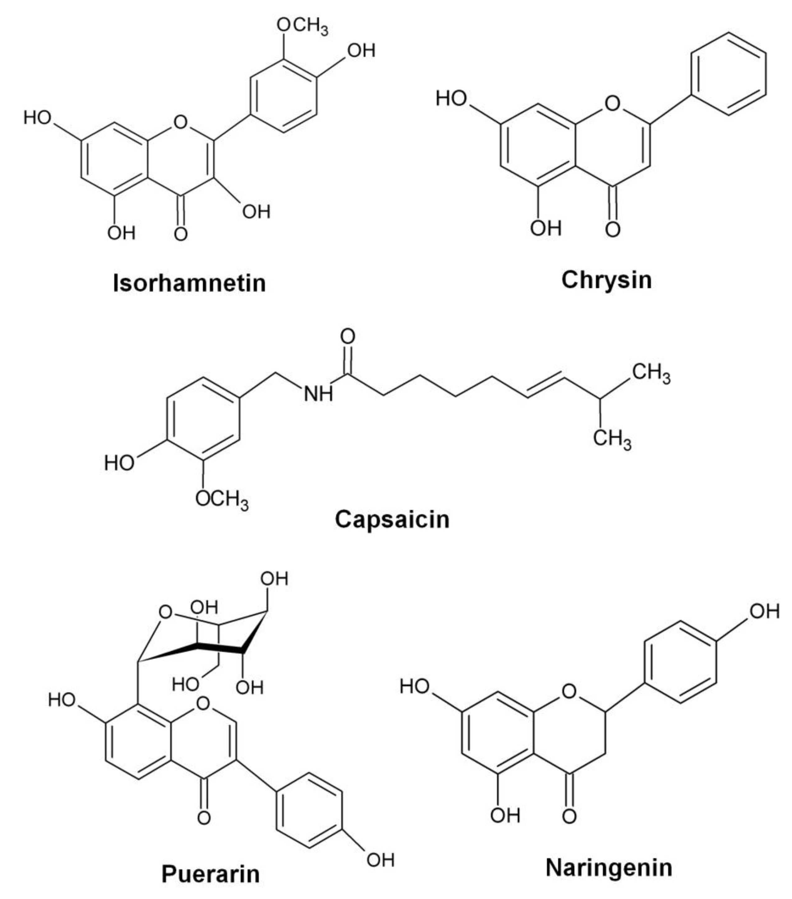
  132. Jiang, L.; Li, H.; Wang, L.; Song, Z.; Shi, L.; Li, W.; Deng, X.; Wang, J. Isorhamnetin attenuates Staphylococcus aureus-induced lung cell injury by inhibiting alpha-hemolysin expression. J. Microbiol. Biotechnol. 2015. [Google Scholar]
  133. Wang, J.; Qiu, J.; Dong, J.; Li, H.; Luo, M.; Dai, X.; Zhang, Y.; Leng, B.; Niu, X.; Zhao, S.; et al. Chrysin protects mice from Staphylococcus aureus pneumonia. J. Appl. Microbiol. 2011, 111, 1551–1558. [Google Scholar] [CrossRef] [PubMed]
  134. Tang, F.; Li, W.H.; Zhou, X.; Liu, Y.H.; Li, Z.; Tang, Y.S.; Kou, X.; Wang, S.D.; Bao, M.; Qu, L.D.; et al. Puerarin protects against Staphylococcus aureus-induced injury of human alveolar epithelial aA49 cells via downregulating alpha-hemolysin secretion. Microb. Drug Resist. 2014, 20, 357–363. [Google Scholar] [CrossRef] [PubMed]
  135. Qiu, J.; Niu, X.; Wang, J.; Xing, Y.; Leng, B.; Dong, J.; Li, H.; Luo, M.; Zhang, Y.; Dai, X.; et al. Capsaicin protects mice from community-associated methicillin-resistant Staphylococcus aureus pneumonia. PLoS ONE 2012, 7, e33032. [Google Scholar] [CrossRef] [PubMed]
  136. Zhang, Y.; Wang, J.F.; Dong, J.; Wei, J.Y.; Wang, Y.N.; Dai, X.H.; Wang, X.; Luo, M.J.; Tan, W.; Deng, X.M.; et al. Inhibition of alpha-toxin production by subinhibitory concentrations of naringenin controls Staphylococcus aureus pneumonia. Fitoterapia 2013, 86, 92–99. [Google Scholar] [CrossRef] [PubMed]
  137. Khodaverdian, V.; Pesho, M.; Truitt, B.; Bollinger, L.; Patel, P.; Nithianantham, S.; Yu, G.; Delaney, E.; Jankowsky, E.; Shoham, M. Discovery of antivirulence agents against methicillin-resistant Staphylococcus aureus. Antimicrob. Agents Chemother. 2013, 57, 3645–3652. [Google Scholar] [CrossRef] [PubMed]
  138. Sully, E.K.; Malachowa, N.; Elmore, B.O.; Alexander, S.M.; Femling, J.K.; Gray, B.M.; DeLeo, F.R.; Otto, M.; Cheung, A.L.; Edwards, B.S.; et al. Selective chemical inhibition of agr quorum sensing in Staphylococcus aureus promotes host defense with minimal impact on resistance. PLoS Pathog. 2014, 10, e1004174. [Google Scholar] [CrossRef] [PubMed]
  139. Daly, S.M.; Elmore, B.O.; Kavanaugh, J.S.; Triplett, K.D.; Figueroa, M.; Raja, H.A.; El-Elimat, T.; Crosby, H.A.; Femling, J.K.; Cech, N.B.; et al. Omega-hydroxyemodin limits Staphylococcus aureus quorum sensing-mediated pathogenesis and inflammation. Antimicrob. Agents Chemother. 2015, 59, 2223–2235. [Google Scholar] [CrossRef] [PubMed]
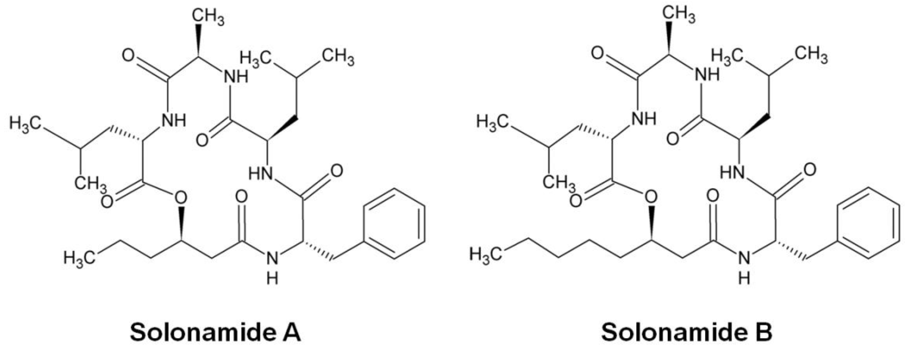
  140. Mansson, M.; Nielsen, A.; Kjaerulff, L.; Gotfredsen, C.H.; Wietz, M.; Ingmer, H.; Gram, L.; Larsen, T.O. Inhibition of virulence gene expression in Staphylococcus aureus by novel depsipeptides from a marine photobacterium. Mar. Drugs 2011, 9, 2537–2552. [Google Scholar] [CrossRef] [PubMed] [Green Version]
  141. Nielsen, A.; Mansson, M.; Bojer, M.S.; Gram, L.; Larsen, T.O.; Novick, R.P.; Frees, D.; Frokiaer, H.; Ingmer, H. Solonamide B inhibits quorum sensing and reduces Staphylococcus aureus mediated killing of human neutrophils. PLoS ONE 2014, 9, e84992. [Google Scholar] [CrossRef] [PubMed]
  142. Nielsen, A.; Mansson, M.; Wietz, M.; Varming, A.N.; Phipps, R.K.; Larsen, T.O.; Gram, L.; Ingmer, H. Nigribactin, a novel siderophore from Vibrio nigripulchritudo, modulates Staphylococcus aureus virulence gene expression. Mar. Drugs 2012, 10, 2584–2595. [Google Scholar] [CrossRef] [PubMed] [Green Version]
  143. Kjaerulff, L.; Nielsen, A.; Mansson, M.; Gram, L.; Larsen, T.O.; Ingmer, H.; Gotfredsen, C.H. Identification of four new agr quorum sensing-interfering cyclodepsipeptides from a marine Photobacterium. Mar. Drugs 2013, 11, 5051–5062. [Google Scholar] [CrossRef] [PubMed]
  144. Lee, J.H.; Park, J.H.; Cho, M.H.; Lee, J. Flavone reduces the production of virulence factors, staphyloxanthin and alpha-hemolysin, in Staphylococcus aureus. Curr. Microbiol. 2012, 65, 726–732. [Google Scholar] [CrossRef] [PubMed]
  145. Jenkins, R.; Burton, N.; Cooper, R. Proteomic and genomic analysis of methicillin-resistant Staphylococcus aureus (MRSA) exposed to manuka honey in vitro demonstrated down-regulation of virulence markers. J. Antimicrob. Chemother. 2014, 69, 603–615. [Google Scholar] [CrossRef] [PubMed]
  146. Arya, R.; Ravikumar, R.; Santhosh, R.S.; Princy, S.A. SarA based novel therapeutic candidate against Staphylococcus aureus associated with vascular graft infections. Front. Microbiol. 2015, 6, 416. [Google Scholar] [CrossRef] [PubMed]
  147. Sifri, C.D.; Begun, J.; Ausubel, F.M.; Calderwood, S.B. Caenorhabditis elegans as a model host for Staphylococcus aureus pathogenesis. Infect. Immun. 2003, 71, 2208–2217. [Google Scholar] [CrossRef] [PubMed]
  148. Wu, K.; Conly, J.; McClure, J.A.; Elsayed, S.; Louie, T.; Zhang, K. Caenorhabditis elegans as a host model for community-associated methicillin-resistant Staphylococcus aureus. Clin. Microbiol. Infect. 2010, 16, 245–254. [Google Scholar] [CrossRef] [PubMed]
  149. Kong, C.; Yehye, W.A.; Abd Rahman, N.; Tan, M.W.; Nathan, S. Discovery of potential anti-infectives against Staphylococcus aureus using a Caenorhabditis elegans infection model. BMC Complement. Altern. Med. 2014, 14, 4. [Google Scholar] [CrossRef] [PubMed]
  150. Moy, T.I.; Conery, A.L.; Larkins-Ford, J.; Wu, G.; Mazitschek, R.; Casadei, G.; Lewis, K.; Carpenter, A.E.; Ausubel, F.M. High-throughput screen for novel antimicrobials using a whole animal infection model. ACS Chem. Biol. 2009, 4, 527–533. [Google Scholar] [CrossRef] [PubMed]
  151. Irazoqui, J.E.; Urbach, J.M.; Ausubel, F.M. Evolution of host innate defence: Insights from Caenorhabditis elegans and primitive invertebrates. Nat. Rev. Immunol. 2010, 10, 47–58. [Google Scholar] [CrossRef] [PubMed]
  152. Barczak, A.K.; Hung, D.T. Productive steps toward an antimicrobial targeting virulence. Curr. Opin. Microbiol. 2009, 12, 490–496. [Google Scholar] [CrossRef] [PubMed]
Figure 1. Schematic diagram of the S. aureus agr regulatory system. The agr operon consists of two transcriptional units RNAII and RNAIII, driven by the promoters P2 and P3, respectively. RNAII is an operon of four genes, agr BDCA, encoding AgrB responsible for processing and exporting AgrD, the AIP precursor. At threshold levels of AIP, AgrC will be autophosphorylated, leading to the phosphorylation of AgrA. AgrA activates RNAIII expression, thereby increasing the secretion of S. aureus toxins and enzymes.
Toxins 08 00072 g001
Figure 2. Chemical structures of natural compounds that exhibit anti-virulence properties (redrawn from [132,133,134,135,136]).
Toxins 08 00072 g002
Figure 3. Chemical structures of savirin and OHM (redrawn from [138,139]).
Toxins 08 00072 g003
Figure 4. Chemical structure of solonamides isolated from Photobacterium sp. (redrawn from [140]).
Toxins 08 00072 g004
Figure 5. Chemical structure of SarABI (redrawn from [146]).
Toxins 08 00072 g005
Table 1. Therapeutic agents against S. aureus toxins and their proposed mechanism of action.
Toxin Target(s)TypeNameMode of ActionPhase of DevelopmentReferences
α-hemolysinMonoclonal antibodyMAbs 7B8 and 1A9Antagonizes toxin activity by inhibiting the formation of fully assembled α-hemolysin oligomer.Testing in animal model (mice pneumonia model)[90]
α-hemolysinMonoclonal antibodyMAb 2A3.1 (and affinity-optimized 2A3 variant—LC10)Neutralizes toxin and prevents toxin-mediated cell lysis via a blockade of α-toxin heptamer formation on erythrocyte membranes.Testing in animal models (S. aureus dermonecrosis murine model and mouse model of S. aureus pneumonia)[91,92]
α-hemolysinMonoclonal antibodyMAb LTM14Prevents binding of toxin to the plasma membrane of susceptible host cells.Testing in animal models (mice pneumonia, skin and bacteremia models)[93]
α-hemolysinChemical compoundβ-cyclodextrin derivativesBlocks the transmembrane pores formed by the toxin and terminates ion conductance through the pores.Testing in animal models (S. aureus pneumonia mice model)[96,98]
α-hemolysinNatural compoundOroxylin A, Oroxin A and Oroxin BBinds to the “stem” region of α-hemolysin and restricts the conformational transition of toxin from monomer to oligomer.In vitro assays[99,100]
α-hemolysinChemical compoundIsatin-Schiff copper (II) complexesPrevents the formation of ion channels by obstructing the constriction region of the α-hemolysin channel.In vitro assays[101]
α-hemolysinNatural compoundMorin hydrateInhibits self-assembly of the heptameric transmembrane pore of α-hemolysin.Testing in animals (mice pneumonia model)[102]
α-hemolysinChemical compoundADAM10 inhibitor (GI254023X)Inhibits the binding of α-hemolysin to its receptor (ADAM10).Testing in animals (mice model of recurrent skin and soft-tissue infection)[106]
β-hemolysinSingle-domain antibodydAb/SAE Cl-7-5Neutralizes S. aureus Hlb activity.In vitro assays[94]
α-hemolysin and bi-component leukocidinsMonoclonal antibodyMAb Hla-F#5Cross-neutralizes α-hemolysin and leukocidins by recognizing the conserved conformational epitope.Testing in animal models (murine models of S. aureus pneumonia and bacteremia/sepsis)[95]
PVL and α-hemolysinPolyclonal antibodyHuman intravenous polyclonal immunoglobuin (IVIg)—TegelineInhibits the lytic effect of PVL on polymorphonuclear cells and neutralizes α-hemolysin.In vitro assays for PVL; in vivo peritonitis murine model for hemolysin[107,108]
PVL and γ-hemolysinHumanized heavy chain-only antibodyBivalent and tetravalent anti-PVL mAbsBlocks binding of PVL to target cells and inhibits pore formation on target cells by γ-hemolysin.Testing in animal models (rabbit model of toxin-induced endophthalmitis)[110]
PVL and other leukotoxinsPolyclonal antibodyAnti-LukS-mut9Cross-neutralizes the lytic activity of various leukotoxins on polymorphonuclear cells.Testing in animal models (toxin-challenged mouse model)[112]
PVLAntimicrobial peptideα-defensin HNP3Binds to both LukS-PV and LukF-PV and reduces PVL-induced necrosis in human neutrophils by interfering with pore formation.In vitro assays[116]
SEBMonoclonal antibodyHuMAb-154Binds to SEB, neutralizes the toxin and inhibits SEB-induced production of proinflammatory cytokines.Testing in animal models (mice model challenged by SEB)[119]
SEBMonoclonal antibodyMAb 20B1Binds and neutralizes SEB.Testing in animal models (mice sepsis, superficial skin and deep-tissue infection models)[121,122]
SEBProteinSoluble Vβ proteinAs a receptor antagonist that offers high-affinity binding to SEB superantigens and neutralizes the toxicity of SEB.Testing in animal model (rabbit model of SEB-induced disease)[124]
SEB and TSST-1ProteinBroad spectrum Vβ proteinBinds to superantigens and neutralizes both SEB and TSST-1 activities.In vitro assays[125]
SEBFDA-approved drugSulfasalazineReverses SEB-stimulated toxic effect by inhibiting the production of proinflammatory cytokines, T-cell proliferation and NFκB activation.In vitro assays[126]

