Next Issue
Volume 8, June
Previous Issue
Volume 8, April


Toxins, Volume 8, Issue 5 (May 2016) – 38 articles

Cover Story (view full-size image): 
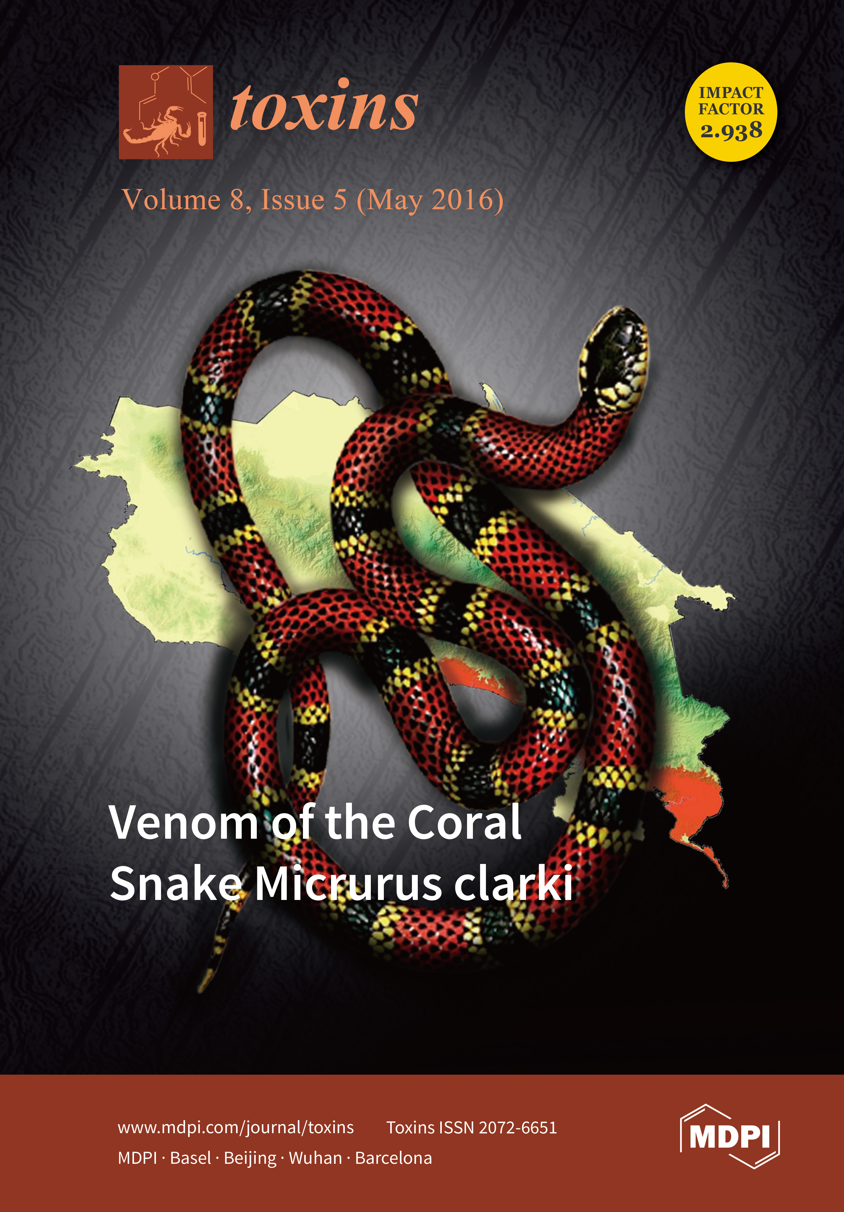
Micrurus clarki is a rare coral snake, distributed from the Southeastern Pacific region of Costa Rica (orange shade in the figure) to Western Colombia. Researchers from the University of Costa Rica and the University of Antioquia, studied its venom for the first time. By applying proteomic tools through an analytical strategy known as 'venomics', a detailed portrait of the venom protein composition was obtained. This was complemented with a characterization of toxic activities and antibody neutralization studies, together with the isolation of a novel 'three-finger toxin', clarkitoxin-I, the first protein to be sequenced from the venom of this snake species.View this article.
  • Issues are regarded as officially published after their release is announced to the table of contents alert mailing list.
  • You may sign up for e-mail alerts to receive table of contents of newly released issues.
  • PDF is the official format for papers published in both, html and pdf forms. To view the papers in pdf format, click on the "PDF Full-text" link, and use the free Adobe Reader to open them.
Order results
Result details
Section
Select all
Export citation of selected articles as:
2932 KiB  
Article
Improved Tissue-Based Analytical Test Methods for Orellanine, a Biomarker of Cortinarius Mushroom Intoxication
by Poojya Anantharam, Dahai Shao, Paula M. Imerman, Eric Burrough, Dwayne Schrunk, Tsevelmaa Sedkhuu, Shusheng Tang and Wilson Rumbeiha
Toxins 2016, 8(5), 158; https://doi.org/10.3390/toxins8050158 - 21 May 2016
Cited by 8 | Viewed by 6781
Abstract
Orellanine (OR) toxin is produced by mushrooms of the genus Cortinarius which grow in North America and in Europe. OR poisoning is characterized by severe oliguric acute renal failure, with a mortality rate of 10%–30%. Diagnosis of OR poisoning currently hinges on a [...] Read more.
Orellanine (OR) toxin is produced by mushrooms of the genus Cortinarius which grow in North America and in Europe. OR poisoning is characterized by severe oliguric acute renal failure, with a mortality rate of 10%–30%. Diagnosis of OR poisoning currently hinges on a history of ingestion of Cortinarius mushrooms and histopathology of renal biopsies. A key step in the diagnostic approach is analysis of tissues for OR. Currently, tissue-based analytical methods for OR are nonspecific and lack sensitivity. The objectives of this study were: (1) to develop definitive HPLC and LC-MS/MS tissue-based analytical methods for OR; and (2) to investigate toxicological effects of OR in mice. The HPLC limit of quantitation was 10 µg/g. For fortification levels of 15 µg/g to 50 µg/g OR in kidney, the relative standard deviation was between 1.3% and 9.8%, and accuracy was within 1.5% to 7.1%. A matrix-matched calibration curve was reproduced in this range with a correlation coefficient (r) of 0.97–0.99. The limit of detection was 20 ng/g for LC-MS/MS. In OR-injected mice, kidney OR concentrations were 97 ± 51 µg/g on Day 0 and 17 ± 1 µg/g on termination Day 3. Splenic and liver injuries were novel findings in this mouse model. The new tissue-based analytical tests will improve diagnosis of OR poisoning, while the mouse model has yielded new data advancing knowledge on OR-induced pathology. The new tissue-based analytical tests will improve diagnosis of OR poisoning, while the mouse model has yielded new data advancing knowledge on OR-induced pathology. Full article
Show Figures

Graphical abstract

3285 KiB  
Article
Comparison of Droplet Digital PCR and qPCR for the Quantification of Shiga Toxin-Producing Escherichia coli in Bovine Feces
by Bavo Verhaegen, Koen De Reu, Lieven De Zutter, Karen Verstraete, Marc Heyndrickx and Els Van Coillie
Toxins 2016, 8(5), 157; https://doi.org/10.3390/toxins8050157 - 18 May 2016
Cited by 62 | Viewed by 7656
Abstract
Cattle are considered to be the main reservoir for Shiga toxin-producing Escherichia coli (STEC) and are often the direct or indirect source of STEC outbreaks in humans. Accurate measurement of the concentration of shed STEC in cattle feces could be a key answer [...] Read more.
Cattle are considered to be the main reservoir for Shiga toxin-producing Escherichia coli (STEC) and are often the direct or indirect source of STEC outbreaks in humans. Accurate measurement of the concentration of shed STEC in cattle feces could be a key answer to questions concerning transmission of STEC, contamination sources and efficiency of treatments at farm level. Infected animals can be identified and the contamination level quantified by real-time quantitative PCR (qPCR), which has its specific limitations. Droplet digital PCR (ddPCR) has been proposed as a method to overcome many of the drawbacks of qPCR. This end-point amplification PCR is capable of absolute quantification independent from any reference material and is less prone to PCR inhibition than qPCR. In this study, the qPCR-based protocol described by Verstraete et al. (2014) for Shiga toxin genes stx1 and stx2 and the intimin gene eae quantification was optimized for ddPCR analysis. The properties of ddPCR and qPCR using two different mastermixes (EMM: TaqMan® Environmental Master Mix 2.0; UMM: TaqMan® Universal PCR Master Mix) were evaluated, using standard curves and both artificial and natural contaminated cattle fecal samples. In addition, the susceptibility of these assays to PCR-inhibitors was investigated. Evaluation of the standard curves and both artificial and natural contaminated cattle fecal samples suggested a very good agreement between qPCR using EMM and ddPCR. Furthermore, similar sensitivities and no PCR inhibition were recorded for both assays. On the other hand, qPCR using UMM was clearly prone to PCR inhibition. In conclusion, the ddPCR technique shows potential for the accurate absolute quantification of STEC on the farms, without relying on standardized reference material. Full article
Show Figures

Graphical abstract

12687 KiB  
Review
ADAM and ADAMTS Family Proteins and Snake Venom Metalloproteinases: A Structural Overview
by Soichi Takeda
Toxins 2016, 8(5), 155; https://doi.org/10.3390/toxins8050155 - 17 May 2016
Cited by 115 | Viewed by 11946
Abstract
A disintegrin and metalloproteinase (ADAM) family proteins constitute a major class of membrane-anchored multidomain proteinases that are responsible for the shedding of cell-surface protein ectodomains, including the latent forms of growth factors, cytokines, receptors and other molecules. Snake venom metalloproteinases (SVMPs) are major [...] Read more.
A disintegrin and metalloproteinase (ADAM) family proteins constitute a major class of membrane-anchored multidomain proteinases that are responsible for the shedding of cell-surface protein ectodomains, including the latent forms of growth factors, cytokines, receptors and other molecules. Snake venom metalloproteinases (SVMPs) are major components in most viper venoms. SVMPs are primarily responsible for hemorrhagic activity and may also interfere with the hemostatic system in envenomed animals. SVMPs are phylogenetically most closely related to ADAMs and, together with ADAMs and related ADAM with thrombospondin motifs (ADAMTS) family proteinases, constitute adamalysins/reprolysins or the M12B clan (MEROPS database) of metalloproteinases. Although the catalytic domain structure is topologically similar to that of other metalloproteinases such as matrix metalloproteinases, the M12B proteinases have a modular structure with multiple non-catalytic ancillary domains that are not found in other proteinases. Notably, crystallographic studies revealed that, in addition to the conserved metalloproteinase domain, M12B members share a hallmark cysteine-rich domain designated as the “ADAM_CR” domain. Despite their name, ADAMTSs lack disintegrin-like structures and instead comprise two ADAM_CR domains. This review highlights the current state of our knowledge on the three-dimensional structures of M12B proteinases, focusing on their unique domains that may collaboratively participate in directing these proteinases to specific substrates. Full article
(This article belongs to the Special Issue Snake Venom Metalloproteinases)
Show Figures

Figure 1

4865 KiB  
Article
Botulinum Neurotoxin Serotype A Recognizes Its Protein Receptor SV2 by a Different Mechanism than Botulinum Neurotoxin B Synaptotagmin
by Jasmin Weisemann, Daniel Stern, Stefan Mahrhold, Brigitte G. Dorner and Andreas Rummel
Toxins 2016, 8(5), 154; https://doi.org/10.3390/toxins8050154 - 17 May 2016
Cited by 29 | Viewed by 6494
Abstract
Botulinum neurotoxins (BoNTs) exhibit extraordinary potency due to their exquisite neurospecificity, which is achieved by dual binding to complex polysialo-gangliosides and synaptic vesicle proteins. The luminal domain 4 (LD4) of the three synaptic vesicle glycoprotein 2 isoforms, SV2A‐C, identified as protein receptors for [...] Read more.
Botulinum neurotoxins (BoNTs) exhibit extraordinary potency due to their exquisite neurospecificity, which is achieved by dual binding to complex polysialo-gangliosides and synaptic vesicle proteins. The luminal domain 4 (LD4) of the three synaptic vesicle glycoprotein 2 isoforms, SV2A‐C, identified as protein receptors for the most relevant serotype BoNT/A, binds within the 50 kDa cell binding domain HC of BoNT/A. Here, we deciphered the BoNT/A‐SV2 interactions in more detail. In pull down assays, the binding of HCA to SV2-LD4 isoforms decreases from SV2C >> SV2A > SV2B. A binding constant of 200 nM was determined for BoNT/A to rat SV2C-LD4 in GST pull down assay. A similar binding constant was determined by surface plasmon resonance for HCA to rat SV2C and to human SV2C, the latter being slightly lower due to the substitution L563F in LD4. At pH 5, as measured in acidic synaptic vesicles, the binding constant of HCA to hSV2C is increased more than 10-fold. Circular dichroism spectroscopy reveals that the quadrilateral helix of SV2C-LD4 already exists in solution prior to BoNT/A binding. Hence, the BoNT/A‐SV2C interaction is of different nature compared to BoNT/B‐Syt-II. In particular, the preexistence of the quadrilateral β-sheet helix of SV2 and its pH-dependent binding to BoNT/A via backbone–backbone interactions constitute major differences. Knowledge of the molecular details of BoNT/A‐SV2 interactions drives the development of high affinity peptides to counteract BoNT/A intoxications or to capture functional BoNT/A variants in innovative detection systems for botulism diagnostic. Full article
(This article belongs to the Section Bacterial Toxins)
Show Figures

Graphical abstract

2635 KiB  
Article
Degradation of Swainsonine by the NADP-Dependent Alcohol Dehydrogenase A1R6C3 in Arthrobacter sp. HW08
by Yan Wang, A’guan Zhai, Yanqi Zhang, Kai Qiu, Jianhua Wang and Qinfan Li
Toxins 2016, 8(5), 145; https://doi.org/10.3390/toxins8050145 - 16 May 2016
Cited by 5 | Viewed by 4363
Abstract
Swainsonine is an indolizidine alkaloid that has been found in locoweeds and some fungi. Our previous study demonstrated that Arthrobacter sp. HW08 or its crude enzyme extract could degrade swainsonie efficiently. However, the mechanism of swainsonine degradation in bacteria remains unclear. In this [...] Read more.
Swainsonine is an indolizidine alkaloid that has been found in locoweeds and some fungi. Our previous study demonstrated that Arthrobacter sp. HW08 or its crude enzyme extract could degrade swainsonie efficiently. However, the mechanism of swainsonine degradation in bacteria remains unclear. In this study, we used label-free quantitative proteomics method based on liquid chromatography-electrospray ionization-tandem mass spectrometry to dissect the mechanism of swainsonine biodegradation by Arthrobacter sp. HW08. The results showed that 129 differentially expressed proteins were relevant to swainsonine degradation. These differentially expressed proteins were mostly related to the biological process of metabolism and the molecular function of catalytic activity. Among the 129 differentially expressed proteins, putative sugar phosphate isomerase/epimerase A1R5X7, Acetyl-CoA acetyltransferase A0JZ95, and nicotinamide adenine dinucleotide phosphate (NADP)-dependent alcohol dehydrogenase A1R6C3 were found to contribute to the swainsonine degradation. Notably, NADP-dependent alcohol dehyrodgenase A1R6C3 appeared to play a major role in degrading swainsonine, but not as much as Arthrobacter sp. HW08 did. Collectively, our findings here provide insights to understand the mechanism of swainsonine degradation in bacteria. Full article
(This article belongs to the Section Plant Toxins)
Show Figures

Graphical abstract

1094 KiB  
Review
The Regulatory Networks That Control Clostridium difficile Toxin Synthesis
by Isabelle Martin-Verstraete, Johann Peltier and Bruno Dupuy
Toxins 2016, 8(5), 153; https://doi.org/10.3390/toxins8050153 - 14 May 2016
Cited by 108 | Viewed by 13734
Abstract
The pathogenic clostridia cause many human and animal diseases, which typically arise as a consequence of the production of potent exotoxins. Among the enterotoxic clostridia, Clostridium difficile is the main causative agent of nosocomial intestinal infections in adults with a compromised gut microbiota [...] Read more.
The pathogenic clostridia cause many human and animal diseases, which typically arise as a consequence of the production of potent exotoxins. Among the enterotoxic clostridia, Clostridium difficile is the main causative agent of nosocomial intestinal infections in adults with a compromised gut microbiota caused by antibiotic treatment. The symptoms of C. difficile infection are essentially caused by the production of two exotoxins: TcdA and TcdB. Moreover, for severe forms of disease, the spectrum of diseases caused by C. difficile has also been correlated to the levels of toxins that are produced during host infection. This observation strengthened the idea that the regulation of toxin synthesis is an important part of C. difficile pathogenesis. This review summarizes our current knowledge about the regulators and sigma factors that have been reported to control toxin gene expression in response to several environmental signals and stresses, including the availability of certain carbon sources and amino acids, or to signaling molecules, such as the autoinducing peptides of quorum sensing systems. The overlapping regulation of key metabolic pathways and toxin synthesis strongly suggests that toxin production is a complex response that is triggered by bacteria in response to particular states of nutrient availability during infection. Full article
Show Figures

Figure 1

667 KiB  
Review
New Insights into VacA Intoxication Mediated through Its Cell Surface Receptors
by Kinnosuke Yahiro, Toshiya Hirayama, Joel Moss and Masatoshi Noda
Toxins 2016, 8(5), 152; https://doi.org/10.3390/toxins8050152 - 13 May 2016
Cited by 14 | Viewed by 7754
Abstract
Helicobacter pylori (H. pylori), a major cause of gastroduodenal diseases, produces VacA, a vacuolating cytotoxin associated with gastric inflammation and ulceration. The C-terminal domain of VacA plays a crucial role in receptor recognition on target cells. We have previously identified [...] Read more.
Helicobacter pylori (H. pylori), a major cause of gastroduodenal diseases, produces VacA, a vacuolating cytotoxin associated with gastric inflammation and ulceration. The C-terminal domain of VacA plays a crucial role in receptor recognition on target cells. We have previously identified three proteins (i.e., RPTPα, RPTPβ, and LRP1) that serve as VacA receptors. These receptors contribute to the internalization of VacA into epithelial cells, activate signal transduction pathways, and contribute to cell death and gastric ulceration. In addition, other factors (e.g., CD18, sphingomyelin) have also been identified as cell-surface, VacA-binding proteins. Since we believe that, following interactions with its host cell receptors, VacA participates in events leading to disease, a better understanding of the cellular function of VacA receptors may provide valuable information regarding the mechanisms underlying the pleiotropic actions of VacA and the pathogenesis of H. pylori-mediated disease. In this review, we focus on VacA receptors and their role in events leading to cell damage. Full article
(This article belongs to the Special Issue Vacuolating Toxin)
Show Figures

Graphical abstract

1232 KiB  
Review
Ellagitannins in Cancer Chemoprevention and Therapy
by Tariq Ismail, Cinzia Calcabrini, Anna Rita Diaz, Carmela Fimognari, Eleonora Turrini, Elena Catanzaro, Saeed Akhtar and Piero Sestili
Toxins 2016, 8(5), 151; https://doi.org/10.3390/toxins8050151 - 13 May 2016
Cited by 84 | Viewed by 10932
Abstract
It is universally accepted that diets rich in fruit and vegetables lead to reduction in the risk of common forms of cancer and are useful in cancer prevention. Indeed edible vegetables and fruits contain a wide variety of phytochemicals with proven antioxidant, anti-carcinogenic, [...] Read more.
It is universally accepted that diets rich in fruit and vegetables lead to reduction in the risk of common forms of cancer and are useful in cancer prevention. Indeed edible vegetables and fruits contain a wide variety of phytochemicals with proven antioxidant, anti-carcinogenic, and chemopreventive activity; moreover, some of these phytochemicals also display direct antiproliferative activity towards tumor cells, with the additional advantage of high tolerability and low toxicity. The most important dietary phytochemicals are isothiocyanates, ellagitannins (ET), polyphenols, indoles, flavonoids, retinoids, tocopherols. Among this very wide panel of compounds, ET represent an important class of phytochemicals which are being increasingly investigated for their chemopreventive and anticancer activities. This article reviews the chemistry, the dietary sources, the pharmacokinetics, the evidence on chemopreventive efficacy and the anticancer activity of ET with regard to the most sensitive tumors, as well as the mechanisms underlying their clinically-valuable properties. Full article
(This article belongs to the Special Issue Dietary and Non-Dietary Phytochemicals and Cancer)
Show Figures

Graphical abstract

1325 KiB  
Article
Sensitive, Rapid, Quantitative and in Vitro Method for the Detection of Biologically Active Staphylococcal Enterotoxin Type E
by Reuven Rasooly, Paula Do and Bradley Hernlem
Toxins 2016, 8(5), 150; https://doi.org/10.3390/toxins8050150 - 13 May 2016
Cited by 7 | Viewed by 5072
Abstract
Staphylococcus aureus is a major bacterial cause of clinical infections and foodborne illnesses through its production of a group of enterotoxins (SEs) which cause gastroenteritis and also function as superantigens to massively activate T cells. In the present study, we tested Staphylococcal enterotoxin [...] Read more.
Staphylococcus aureus is a major bacterial cause of clinical infections and foodborne illnesses through its production of a group of enterotoxins (SEs) which cause gastroenteritis and also function as superantigens to massively activate T cells. In the present study, we tested Staphylococcal enterotoxin type E (SEE), which was detected in 17 of the 38 suspected staphylococcal food poisoning incidents in a British study and was the causative agent in outbreaks in France, UK and USA. The current method for detection of enterotoxin activity is an in vivo monkey or kitten bioassay; however, this expensive procedure has low sensitivity and poor reproducibility, requires many animals, is impractical to test on a large number of samples, and raises ethical concerns with regard to the use of experimental animals. The purpose of this study is to develop rapid sensitive and quantitative bioassays for detection of active SEE. We apply a genetically engineered T cell-line expressing the luciferase reporter gene under the regulation of nuclear factor of activated T-cells response element (NFAT-RE), combined with a Raji B-cell line that presents the SEE-MHC (major histocompatibility complex) class II to the engineered T cell line. Exposure of the above mixed culture to SEE induces differential expression of the luciferase gene and bioluminescence is read out in a dose dependent manner over a 6-log range. The limit of detection of biologically active SEE is 1 fg/mL which is 109 times more sensitive than the monkey and kitten bioassay. Full article
(This article belongs to the Collection Rapid Detection of Bacterial Toxins)
Show Figures

Graphical abstract

1648 KiB  
Article
Determination of Asymmetric and Symmetric Dimethylarginine in Serum from Patients with Chronic Kidney Disease: UPLC-MS/MS versus ELISA
by Jente Boelaert, Eva Schepers, Griet Glorieux, Sunny Eloot, Raymond Vanholder and Frédéric Lynen
Toxins 2016, 8(5), 149; https://doi.org/10.3390/toxins8050149 - 13 May 2016
Cited by 22 | Viewed by 5089
Abstract
Asymmetric dimethylarginine (ADMA), an endogenous inhibitor of nitric oxide (NO) synthesis, and its structural isomer symmetric dimethylarginine (SDMA) are uremic toxins accumulating in chronic kidney disease (CKD) patients. The objective of this study was to develop and validate a robust UPLC-MS/MS method for [...] Read more.
Asymmetric dimethylarginine (ADMA), an endogenous inhibitor of nitric oxide (NO) synthesis, and its structural isomer symmetric dimethylarginine (SDMA) are uremic toxins accumulating in chronic kidney disease (CKD) patients. The objective of this study was to develop and validate a robust UPLC-MS/MS method for the simultaneous determination of ADMA and SDMA in human serum. Chromatographic separation after butyl ester derivatization was achieved on an Acquity UPLC BEH C18 column, followed by tandem mass spectrometric detection. After validation, the applicability of the method was evaluated by the analysis of serum samples from 10 healthy controls and 77 CKD patients on hemodialysis (CKD5HD). Both ADMA (0.84 ± 0.19 µM vs. 0.52 ± 0.07 µM) and SDMA concentrations (2.06 ± 0.82 µM vs. 0.59 ± 0.13 µM) were significantly (p < 0.001) elevated in CKD5HD patients compared to healthy controls. In general, low degrees of protein binding were found for both ADMA and SDMA. In addition, an established commercially available ELISA kit was utilized on the same samples (n = 87) to compare values obtained both with ELISA and UPLC-MS/MS. Regression analysis between these two methods was significant (p < 0.0001) but moderate for both ADMA (R = 0.78) and SDMA (R = 0.72). Full article
Show Figures

Graphical abstract

1744 KiB  
Article
Monoclonal IgA Antibodies for Aflatoxin Immunoassays
by Özlem Ertekin, Şerife Şeyda Pirinçci and Selma Öztürk
Toxins 2016, 8(5), 148; https://doi.org/10.3390/toxins8050148 - 12 May 2016
Cited by 18 | Viewed by 4866
Abstract
Antibody based techniques are widely used for the detection of aflatoxins which are potent toxins with a high rate of occurrence in many crops. We developed a murine monoclonal antibody of immunoglobulin A (IgA) isotype with a strong binding affinity to aflatoxin B1 [...] Read more.
Antibody based techniques are widely used for the detection of aflatoxins which are potent toxins with a high rate of occurrence in many crops. We developed a murine monoclonal antibody of immunoglobulin A (IgA) isotype with a strong binding affinity to aflatoxin B1 (AFB1), aflatoxin B2 (AFB2), aflatoxin G1 (AFG1), aflatoxin G2 (AFG2) and aflatoxin M1 (AFM1). The antibody was effectively used in immunoaffinity column (IAC) and ELISA kit development. The performance of the IACs was compatible with AOAC performance standards for affinity columns (Test Method: AOAC 991.31). The total binding capacity of the IACs containing our antibody was 111 ng, 70 ng, 114 ng and 73 ng for AFB1, AFB2, and AFG1 andAFG2, respectively. Furthermore, the recovery rates of 5 ng of each AF derivative loaded to the IACs were determined as 104.9%, 82.4%, 85.5% and 70.7% for AFB1, AFB2, AFG1 and AFG2, respectively. As for the ELISA kit developed using non-oriented, purified IgA antibody, we observed a detection range of 2–50 µg/L with 40 min total test time. The monoclonal antibody developed in this research is hitherto the first presentation of quadruple antigen binding IgA monoclonal antibodies in mycotoxin analysis and also the first study of their utilization in ELISA and IACs. IgA antibodies are valuable alternatives for immunoassay development, in terms of both sensitivity and ease of preparation, since they do not require any orientation effort. Full article
(This article belongs to the Collection Biorecognition Assays for Mycotoxins)
Show Figures

Graphical abstract

2452 KiB  
Article
Withania somnifera Induces Cytotoxic and Cytostatic Effects on Human T Leukemia Cells
by Eleonora Turrini, Cinzia Calcabrini, Piero Sestili, Elena Catanzaro, Elena De Gianni, Anna Rita Diaz, Patrizia Hrelia, Massimo Tacchini, Alessandra Guerrini, Barbara Canonico, Stefano Papa, Giovanni Valdrè and Carmela Fimognari
Toxins 2016, 8(5), 147; https://doi.org/10.3390/toxins8050147 - 12 May 2016
Cited by 30 | Viewed by 7673
Abstract
Cancer chemotherapy is characterized by an elevated intrinsic toxicity and the development of drug resistance. Thus, there is a compelling need for new intervention strategies with an improved therapeutic profile. Immunogenic cell death (ICD) represents an innovative anticancer strategy where dying cancer cells [...] Read more.
Cancer chemotherapy is characterized by an elevated intrinsic toxicity and the development of drug resistance. Thus, there is a compelling need for new intervention strategies with an improved therapeutic profile. Immunogenic cell death (ICD) represents an innovative anticancer strategy where dying cancer cells release damage-associated molecular patterns promoting tumor-specific immune responses. The roots of Withania somnifera (W. somnifera) are used in the Indian traditional medicine for their anti-inflammatory, immunomodulating, neuroprotective, and anticancer activities. The present study is designed to explore the antileukemic activity of the dimethyl sulfoxide extract obtained from the roots of W. somnifera (WE). We studied its cytostatic and cytotoxic activity, its ability to induce ICD, and its genotoxic potential on a human T-lymphoblastoid cell line by using different flow cytometric assays. Our results show that WE has a significant cytotoxic and cytostatic potential, and induces ICD. Its proapoptotic mechanism involves intracellular Ca2+ accumulation and the generation of reactive oxygen species. In our experimental conditions, the extract possesses a genotoxic potential. Since the use of Withania is suggested in different contexts including anti-infertility and osteoarthritis care, its genotoxicity should be carefully considered for an accurate assessment of its risk–benefit profile. Full article
(This article belongs to the Special Issue Dietary and Non-Dietary Phytochemicals and Cancer)
Show Figures

Graphical abstract

336 KiB  
Meeting Report
Report from the 5th International Symposium on Mycotoxins and Toxigenic Moulds: Challenges and Perspectives (MYTOX) Held in Ghent, Belgium, May 2016
by Sarah De Saeger, Kris Audenaert and Siska Croubels
Toxins 2016, 8(5), 146; https://doi.org/10.3390/toxins8050146 - 12 May 2016
Cited by 11 | Viewed by 7006
Abstract
The association research platform MYTOX “Mycotoxins and Toxigenic Moulds” held the 5th meeting of its International Symposium in Ghent, Belgium on 11 May 2016.[...] Full article
4572 KiB  
Article
Two Novel Dermaseptin-Like Antimicrobial Peptides with Anticancer Activities from the Skin Secretion of Pachymedusa dacnicolor
by Daning Shi, Xiaojuan Hou, Lei Wang, Yitian Gao, Di Wu, Xinping Xi, Mei Zhou, Hang Fai Kwok, Jinao Duan, Tianbao Chen and Chris Shaw
Toxins 2016, 8(5), 144; https://doi.org/10.3390/toxins8050144 - 12 May 2016
Cited by 37 | Viewed by 6631
Abstract
The dermaseptin antimicrobial peptide family contains members of 27–34 amino acids in length that have been predominantly isolated from the skins/skin secretions of phyllomedusine leaf frogs. By use of a degenerate primer in Rapid amplification of cDNA ends (RACE) PCR designed to a [...] Read more.
The dermaseptin antimicrobial peptide family contains members of 27–34 amino acids in length that have been predominantly isolated from the skins/skin secretions of phyllomedusine leaf frogs. By use of a degenerate primer in Rapid amplification of cDNA ends (RACE) PCR designed to a common conserved domain within the 5′-untranslated regions of previously-characterized dermaseptin encoding cDNAs, two novel members of this peptide family, named dermaseptin-PD-1 and dermaseptin-PD-2, were identified in the skin secretion of the phyllomedusine frog, Pachymedusa dacnicolor. The primary structures of both peptides were predicted from cloned cDNAs, as well as being confirmed by mass spectral analysis of crude skin secretion fractions resulted from reversed-phase high-performance liquid chromatography. Chemically-synthesized replicates of dermaseptin-PD-1 and dermaseptin-PD-2 were investigated for antimicrobial activity using standard model microorganisms (Gram-positive bacteria, Gram-negative bacteria and a yeast) and for cytotoxicity using mammalian red blood cells. The possibility of synergistic effects between the two peptides and their anti-cancer cell proliferation activities were assessed. The peptides exhibited moderate to high inhibition against the growth of the tested microorganisms and cancer cell lines with low haemolytic activity. Synergistic interaction between the two peptides in inhibiting the proliferation of Escherichia coli and human neuronal glioblastoma cell line, U251MG was also manifested. Full article
(This article belongs to the Section Animal Venoms)
Show Figures

Graphical abstract

2617 KiB  
Article
The Draft Genome Sequence of the Yersinia entomophaga Entomopathogenic Type Strain MH96T
by Mark R. H. Hurst, Amy Beattie, Eric Altermann, Roger M. Moraga, Lincoln A. Harper, Joanne Calder and Aurelie Laugraud
Toxins 2016, 8(5), 143; https://doi.org/10.3390/toxins8050143 - 11 May 2016
Cited by 15 | Viewed by 6150
Abstract
Here we report the draft genome of Yersinia entomophaga type strain MH96T. The genome shows 93.8% nucleotide sequence identity to that of Yersinia nurmii type strain APN3a-cT, and comprises a single chromosome of approximately 4,275,531 bp. In silico analysis identified that, in addition [...] Read more.
Here we report the draft genome of Yersinia entomophaga type strain MH96T. The genome shows 93.8% nucleotide sequence identity to that of Yersinia nurmii type strain APN3a-cT, and comprises a single chromosome of approximately 4,275,531 bp. In silico analysis identified that, in addition to the previously documented Y. entomophaga Yen-TC gene cluster, the genome encodes a diverse array of toxins, including two type III secretion systems, and five rhs-associated gene clusters. As well as these multicomponent systems, several orthologs of known insect toxins, such as VIP2 toxin and the binary toxin PirAB, and distant orthologs of some mammalian toxins, including repeats-in-toxin, a cytolethal distending toxin, hemolysin-like genes and an adenylate cyclase were identified. The genome also contains a large number of hypothetical proteins and orthologs of known effector proteins, such as LopT, as well as genes encoding a wide range of proteolytic determinants, including metalloproteases and pathogen fitness determinants, such as genes involved in iron metabolism. The bioinformatic data derived from the current in silico analysis, along with previous information on the pathobiology of Y. entomophaga against its insect hosts, suggests that a number of these virulence systems are required for survival in the hemocoel and incapacitation of the insect host. Full article
(This article belongs to the Section Bacterial Toxins)
Show Figures

Graphical abstract

3994 KiB  
Article
Docking Simulation of the Binding Interactions of Saxitoxin Analogs Produced by the Marine Dinoflagellate Gymnodinium catenatum to the Voltage-Gated Sodium Channel Nav1.4
by Lorena M. Durán-Riveroll, Allan D. Cembella, Christine J. Band-Schmidt, José J. Bustillos-Guzmán and José Correa-Basurto
Toxins 2016, 8(5), 129; https://doi.org/10.3390/toxins8050129 - 06 May 2016
Cited by 19 | Viewed by 7127
Abstract
Saxitoxin (STX) and its analogs are paralytic alkaloid neurotoxins that block the voltage-gated sodium channel pore (Nav), impeding passage of Na+ ions into the intracellular space, and thereby preventing the action potential in the peripheral nervous system and skeletal muscle. [...] Read more.
Saxitoxin (STX) and its analogs are paralytic alkaloid neurotoxins that block the voltage-gated sodium channel pore (Nav), impeding passage of Na+ ions into the intracellular space, and thereby preventing the action potential in the peripheral nervous system and skeletal muscle. The marine dinoflagellate Gymnodinium catenatum produces an array of such toxins, including the recently discovered benzoyl analogs, for which the mammalian toxicities are essentially unknown. We subjected STX and its analogs to a theoretical docking simulation based upon two alternative tri-dimensional models of the Nav1.4 to find a relationship between the binding properties and the known mammalian toxicity of selected STX analogs. We inferred hypothetical toxicities for the benzoyl analogs from the modeled values. We demonstrate that these toxins exhibit different binding modes with similar free binding energies and that these alternative binding modes are equally probable. We propose that the principal binding that governs ligand recognition is mediated by electrostatic interactions. Our simulation constitutes the first in silico modeling study on benzoyl-type paralytic toxins and provides an approach towards a better understanding of the mode of action of STX and its analogs. Full article
(This article belongs to the Collection Marine and Freshwater Toxins)
Show Figures

Figure 1

248 KiB  
Article
Helicobacter pylori vacA Genotypes in Chronic Gastritis and Gastric Carcinoma Patients from Macau, China
by Ines Pinto-Ribeiro, Rui M. Ferreira, Sellma Batalha, Thazin Hlaing, Sio In Wong, Fatima Carneiro and Ceu Figueiredo
Toxins 2016, 8(5), 142; https://doi.org/10.3390/toxins8050142 - 05 May 2016
Cited by 18 | Viewed by 5728
Abstract
Helicobacter pylori is the major triggering factor for gastric carcinoma, but only a small proportion of infected patients develop this disease. Differences in virulence observed among H. pylori strains, namely in the vacuolating cytotoxin vacA gene, may contribute to this discrepancy. Infection with [...] Read more.
Helicobacter pylori is the major triggering factor for gastric carcinoma, but only a small proportion of infected patients develop this disease. Differences in virulence observed among H. pylori strains, namely in the vacuolating cytotoxin vacA gene, may contribute to this discrepancy. Infection with vacA s1, i1 and m1 strains increases the risk for progression of gastric premalignant lesions and for gastric carcinoma. However, in East Asian countries most of the H. pylori strains are vacA s1, regardless of the patients’ clinical status, and the significance of the vacA i1 and m1 genotypes for gastric carcinoma in this geographic area remains to be fully elucidated. The aim of the present study was to investigate this relationship in 290 patients from Macau, China. Using very sensitive and accurate genotyping methods, we detected infection with vacA i1 and with vacA m1 strains in, respectively, 85.2% and 52.6% of the patients that were infected with single genotypes. The prevalence of cagA-positive strains was 87.5%. No significant associations were observed between vacA genotypes or cagA and gastric carcinoma. It is worth noting that 37.5% of the infected patients had coexistence of H. pylori strains with different vacA genotypes. Additional studies directed to other H. pylori virulence factors should be performed to identify high risk patients in East Asia. Full article
(This article belongs to the Special Issue Vacuolating Toxin)
Show Figures

Graphical abstract

175 KiB  
Addendum
Addendum: Yang, Z., et al. Multi-Toxic Endpoints of the Foodborne Mycotoxins in Nematode Caenorhabditis elegans. Toxins (Basel), 2015, 7(12), 5224–5235
by Zhendong Yang, Kathy S. Xue, Xiulan Sun, Lili Tang and Jia-Sheng Wang
Toxins 2016, 8(5), 141; https://doi.org/10.3390/toxins8050141 - 05 May 2016
Viewed by 3204
Abstract
The authors wish to add the first affiliation address “Synergetic Innovation Center of Food Safety and Nutrition, School of Food Science, Jiangnan University” to the correspondence author, Lili Tang’s affiliations on the first page of their paper published in Toxins [1].[...] Full article
(This article belongs to the Section Mycotoxins)
1439 KiB  
Review
Toxin-Antitoxin Systems of Staphylococcus aureus
by Christopher F. Schuster and Ralph Bertram
Toxins 2016, 8(5), 140; https://doi.org/10.3390/toxins8050140 - 05 May 2016
Cited by 55 | Viewed by 11302
Abstract
Toxin-antitoxin (TA) systems are small genetic elements found in the majority of prokaryotes. They encode toxin proteins that interfere with vital cellular functions and are counteracted by antitoxins. Dependent on the chemical nature of the antitoxins (protein or RNA) and how they control [...] Read more.
Toxin-antitoxin (TA) systems are small genetic elements found in the majority of prokaryotes. They encode toxin proteins that interfere with vital cellular functions and are counteracted by antitoxins. Dependent on the chemical nature of the antitoxins (protein or RNA) and how they control the activity of the toxin, TA systems are currently divided into six different types. Genes comprising the TA types I, II and III have been identified in Staphylococcus aureus. MazF, the toxin of the mazEF locus is a sequence-specific RNase that cleaves a number of transcripts, including those encoding pathogenicity factors. Two yefM-yoeB paralogs represent two independent, but auto-regulated TA systems that give rise to ribosome-dependent RNases. In addition, omega/epsilon/zeta constitutes a tripartite TA system that supposedly plays a role in the stabilization of resistance factors. The SprA1/SprA1AS and SprF1/SprG1 systems are post-transcriptionally regulated by RNA antitoxins and encode small membrane damaging proteins. TA systems controlled by interaction between toxin protein and antitoxin RNA have been identified in S. aureus in silico, but not yet experimentally proven. A closer inspection of possible links between TA systems and S. aureus pathophysiology will reveal, if these genetic loci may represent druggable targets. The modification of a staphylococcal TA toxin to a cyclopeptide antibiotic highlights the potential of TA systems as rather untapped sources of drug discovery. Full article
(This article belongs to the Collection Staphylococcus aureus Toxins)
Show Figures

Graphical abstract

2688 KiB  
Article
Venom of the Coral Snake Micrurus clarki: Proteomic Profile, Toxicity, Immunological Cross-Neutralization, and Characterization of a Three-Finger Toxin
by Bruno Lomonte, Mahmood Sasa, Paola Rey-Suárez, Wendy Bryan and José María Gutiérrez
Toxins 2016, 8(5), 138; https://doi.org/10.3390/toxins8050138 - 05 May 2016
Cited by 34 | Viewed by 8503
Abstract
Micrurus clarki is an uncommon coral snake distributed from the Southeastern Pacific of Costa Rica to Western Colombia, for which no information on its venom could be found in the literature. Using a ‘venomics’ approach, proteins of at least nine families were identified, [...] Read more.
Micrurus clarki is an uncommon coral snake distributed from the Southeastern Pacific of Costa Rica to Western Colombia, for which no information on its venom could be found in the literature. Using a ‘venomics’ approach, proteins of at least nine families were identified, with a moderate predominance of three-finger toxins (3FTx; 48.2%) over phospholipase A2 (PLA2; 36.5%). Comparison of this venom profile with those of other Micrurus species suggests that it may represent a more balanced, ‘intermediate’ type within the dichotomy between 3FTx- and PLA2-predominant venoms. M. clarki venom was strongly cross-recognized and, accordingly, efficiently neutralized by an equine therapeutic antivenom against M. nigrocinctus, revealing their high antigenic similarity. Lethal activity for mice could be reproduced by a PLA2 venom fraction, but, unexpectedly, not by fractions corresponding to 3FTxs. The most abundant venom component, hereby named clarkitoxin-I, was identified as a short-chain (type I) 3FTx, devoid of lethal effect in mice, whose target remains to be defined. Its amino acid sequence of 66 residues shows high similarity with predicted sequences of venom gland transcripts described for M. fulvius, M. browni, and M. diastema. Full article
(This article belongs to the Special Issue Venomics, Venom Proteomics and Venom Transcriptomics)
Show Figures

Graphical abstract

3201 KiB  
Article
Toxin and Growth Responses of the Neurotoxic Dinoflagellate Vulcanodinium rugosum to Varying Temperature and Salinity
by Eric Abadie, Alexia Muguet, Tom Berteaux, Nicolas Chomérat, Philipp Hess, Emmanuelle Roque D’OrbCastel, Estelle Masseret and Mohamed Laabir
Toxins 2016, 8(5), 136; https://doi.org/10.3390/toxins8050136 - 05 May 2016
Cited by 15 | Viewed by 5586
Abstract
Vulcanodinium rugosum, a recently described species, produces pinnatoxins. The IFR-VRU-01 strain, isolated from a French Mediterranean lagoon in 2010 and identified as the causative dinoflagellate contaminating mussels in the Ingril Lagoon (French Mediterranean) with pinnatoxin-G, was grown in an enriched natural seawater [...] Read more.
Vulcanodinium rugosum, a recently described species, produces pinnatoxins. The IFR-VRU-01 strain, isolated from a French Mediterranean lagoon in 2010 and identified as the causative dinoflagellate contaminating mussels in the Ingril Lagoon (French Mediterranean) with pinnatoxin-G, was grown in an enriched natural seawater medium. We tested the effect of temperature and salinity on growth, pinnatoxin-G production and chlorophyll a levels of this dinoflagellate. These factors were tested in combinations of five temperatures (15, 20, 25, 30 and 35 °C) and five salinities (20, 25, 30, 35 and 40) at an irradiance of 100 µmol photon m−2 s−1. V. rugosum can grow at temperatures and salinities ranging from 20 °C to 30 °C and 20 to 40, respectively. The optimal combination for growth (0.39 ± 0.11 d−1) was a temperature of 25 °C and a salinity of 40. Results suggest that V. rugosum is euryhaline and thermophile which could explain why this dinoflagellate develops in situ only from June to September. V. rugosum growth rate and pinnatoxin-G production were highest at temperatures ranging between 25 and 30 °C. This suggests that the dinoflagellate may give rise to extensive blooms in the coming decades caused by the climate change-related increases in temperature expected in the Mediterranean coasts. Full article
(This article belongs to the Collection Marine and Freshwater Toxins)
Show Figures

Figure 1

3540 KiB  
Article
Activity of Palythoa caribaeorum Venom on Voltage-Gated Ion Channels in Mammalian Superior Cervical Ganglion Neurons
by Fernando Lazcano-Pérez, Héctor Castro, Isabel Arenas, David E. García, Ricardo González-Muñoz and Roberto Arreguín-Espinosa
Toxins 2016, 8(5), 135; https://doi.org/10.3390/toxins8050135 - 05 May 2016
Cited by 13 | Viewed by 5215
Abstract
The Zoanthids are an order of cnidarians whose venoms and toxins have been poorly studied. Palythoa caribaeorum is a zoanthid commonly found around the Mexican coastline. In this study, we tested the activity of P. caribaeorum venom on voltage-gated sodium channel (NaV [...] Read more.
The Zoanthids are an order of cnidarians whose venoms and toxins have been poorly studied. Palythoa caribaeorum is a zoanthid commonly found around the Mexican coastline. In this study, we tested the activity of P. caribaeorum venom on voltage-gated sodium channel (NaV1.7), voltage-gated calcium channel (CaV2.2), the A-type transient outward (IA) and delayed rectifier (IDR) currents of KV channels of the superior cervical ganglion (SCG) neurons of the rat. These results showed that the venom reversibly delays the inactivation process of voltage-gated sodium channels and inhibits voltage-gated calcium and potassium channels in this mammalian model. The compounds responsible for these effects seem to be low molecular weight peptides. Together, these results provide evidence for the potential use of zoanthids as a novel source of cnidarian toxins active on voltage-gated ion channels. Full article
(This article belongs to the Special Issue Animal Toxins and Biological Ion Channels)
Show Figures

Graphical abstract

1578 KiB  
Article
Isolation of the Binding Protein of Periplocoside E from BBMVs in Midgut of the Oriental Amyworm Mythimna separata Walker (Lepidoptera: Noctuidae) through Affinity Chromatography
by Mingxing Feng, Zhenyu He, Yuanyuan Wang, Xiufang Yan, Jiwen Zhang, Zhaonong Hu and Wenjun Wu
Toxins 2016, 8(5), 139; https://doi.org/10.3390/toxins8050139 - 04 May 2016
Cited by 5 | Viewed by 4280
Abstract
Periplocosides, which are insecticidal compounds isolated from the root bark of Periploca sepium Bunge, can affect the digestive system of insects. However, the mechanism though which periplocosides induces a series of symptoms remains unknown. In this study, affinity chromatography was conducted by coupling [...] Read more.
Periplocosides, which are insecticidal compounds isolated from the root bark of Periploca sepium Bunge, can affect the digestive system of insects. However, the mechanism though which periplocosides induces a series of symptoms remains unknown. In this study, affinity chromatography was conducted by coupling periplocoside E-semi-succinic acid ester with epoxy amino hexyl (EAH) sepharose 4B. Sodium dodecyl sulfonate-polyacrylamide gelelectrophoresis (SDS-PAGE) was performed to analyze the fraction eluted by periplocoside E. Eight binding proteins (luciferin 4-monooxygenase, aminopeptidase N, aminopeptidase N3, nicotinamide adenine dinucleotide health (NADH) dehydrogenase subunit 5, phosphatidylinositol 3-phosphate 3-phosphatase myotubularin, actin, uncharacterized family 31 glucosidase KIAA1161, and 2OG-Fe(2) oxygenase superfamily protein) were obtained and identified through liquid chromatography/quadrupole-time of flight-mass spectrometry (LC/Q-TOF-MS) analysis of the midgut epithelium cells of Mythimna separata larvae. Aminopeptidase N and N3 are potential putative targets of periplocosides. This study establishes the foundation for further research on the mechanism of action and target localization of periplocosides in agricultural pests. Full article
(This article belongs to the Section Plant Toxins)
Show Figures

Graphical abstract

1786 KiB  
Review
Immunotoxin Therapies for the Treatment of Epidermal Growth Factor Receptor-Dependent Cancers
by Nathan Simon and David FitzGerald
Toxins 2016, 8(5), 137; https://doi.org/10.3390/toxins8050137 - 04 May 2016
Cited by 43 | Viewed by 9158
Abstract
Many epithelial cancers rely on enhanced expression of the epidermal growth factor receptor (EGFR) to drive proliferation and survival pathways. Development of therapeutics to target EGFR signaling has been of high importance, and multiple examples have been approved for human use. However, many [...] Read more.
Many epithelial cancers rely on enhanced expression of the epidermal growth factor receptor (EGFR) to drive proliferation and survival pathways. Development of therapeutics to target EGFR signaling has been of high importance, and multiple examples have been approved for human use. However, many of the current small molecule or antibody-based therapeutics are of limited effectiveness due to the inevitable development of resistance and toxicity to normal tissues. Recombinant immunotoxins are therapeutic molecules consisting of an antibody or receptor ligand joined to a protein cytotoxin, combining the specific targeting of a cancer-expressed receptor with the potent cell killing of cytotoxic enzymes. Over the decades, many bacterial- or plant-based immunotoxins have been developed with the goal of targeting the broad range of cancers reliant upon EGFR overexpression. Many examples demonstrate excellent anti-cancer properties in preclinical development, and several EGFR-targeted immunotoxins have progressed to human trials. This review summarizes much of the past and current work in the development of immunotoxins for targeting EGFR-driven cancers. Full article
(This article belongs to the Collection Immunotoxins 2016)
Show Figures

Graphical abstract

2210 KiB  
Article
A Simple and High-Throughput Analysis of Amatoxins and Phallotoxins in Human Plasma, Serum and Urine Using UPLC-MS/MS Combined with PRiME HLB μElution Platform
by Shuo Zhang, Yunfeng Zhao, Haijiao Li, Shuang Zhou, Dawei Chen, Yizhe Zhang, Qunmei Yao and Chengye Sun
Toxins 2016, 8(5), 128; https://doi.org/10.3390/toxins8050128 - 04 May 2016
Cited by 45 | Viewed by 7346
Abstract
Amatoxins and phallotoxins are toxic cyclopeptides found in the genus Amanita and are among the predominant causes of fatal food poisoning in China. In the treatment of Amanita mushroom poisoning, an early and definite diagnosis is necessary for a successful outcome, which has [...] Read more.
Amatoxins and phallotoxins are toxic cyclopeptides found in the genus Amanita and are among the predominant causes of fatal food poisoning in China. In the treatment of Amanita mushroom poisoning, an early and definite diagnosis is necessary for a successful outcome, which has prompted the development of protocols for the fast and confirmatory determination of amatoxins and phallotoxins in human biological fluids. For this purpose, a simple, rapid and sensitive multiresidue UPLC-MS/MS method for the simultaneous determination of α-amanitin, β-amanitin, γ-amanitin, phalloidin (PHD) and phallacidin (PCD) in human plasma, serum and urine was developed and validated. The diluted plasma, serum and urine samples were directly purified with a novel PRiME technique on a 96-well μElution plate platform, which allowed high-throughput sample processing and low reagent consumption. After purification, a UPLC-MS/MS analysis was performed using positive electrospray ionization (ESI+) in multiple reaction monitoring (MRM) mode. This method fulfilled the requirements of a validation test, with good results for the limit of detection (LOD), lower limit of quantification (LLOQ), accuracy, intra- and inter-assay precision, recovery and matrix effects. All of the analytes were confirmed and quantified in authentic plasma, serum and urine samples obtained from cases of poisoning using this method. Using the PRiME μElution technique for quantification reduces labor and time costs and represents a suitable method for routine toxicological and clinical emergency analysis. Full article
(This article belongs to the Section Mycotoxins)
Show Figures

Figure 1

1665 KiB  
Article
Pathway for Biodegrading Nodularin (NOD) by Sphingopyxis sp. USTB-05
by Nan Feng, Fan Yang, Hai Yan, Chunhua Yin, Xiaolu Liu, Haiyang Zhang, Qianqian Xu, Le Lv and Huasheng Wang
Toxins 2016, 8(5), 116; https://doi.org/10.3390/toxins8050116 - 04 May 2016
Cited by 13 | Viewed by 4644
Abstract
Nodularin (NOD) is greatly produced by Nodularia spumigena and released into the environment when toxic cyanobacterial blooms happened in natural water body, which is seriously harmful to human and animals. The promising bacterial strain of Sphingopyxis sp. USTB-05 was found to have an [...] Read more.
Nodularin (NOD) is greatly produced by Nodularia spumigena and released into the environment when toxic cyanobacterial blooms happened in natural water body, which is seriously harmful to human and animals. The promising bacterial strain of Sphingopyxis sp. USTB-05 was found to have an ability in biodegrading NOD. Initially, 11.6 mg/L of NOD could be completely eliminated within 72 h by whole cells of USTB-05, and within 36 h by its crude enzymes (CEs) of 570 mg/L, respectively. During the enzymatic biodegradation process of NOD, two products were observed on the profiles of HPLC. Based on the analysis of m/z ratios of NOD and its two products on a rapid-resolution liquid chromatogram-mass spectrum (RRLC-MS), we suggested that at least two enzymes of USTB-05 participated in biodegrading NOD. The first enzyme hydrolyzed Arg-Adda peptide bond of cyclic NOD and converted it to linear NOD as the first product. The second enzyme was found to cut off the target peptide bond between Adda and Glu of linearized NOD, and Adda was produced as a second and dead-end product. This finding is very important in both basic research and the application of USTB-05 on the removal of NOD from a water environment. Full article
(This article belongs to the Collection Marine and Freshwater Toxins)
Show Figures

Figure 1

3816 KiB  
Review
Clostridium difficile Toxins A and B: Insights into Pathogenic Properties and Extraintestinal Effects
by Stefano Di Bella, Paolo Ascenzi, Steven Siarakas, Nicola Petrosillo and Alessandra Di Masi
Toxins 2016, 8(5), 134; https://doi.org/10.3390/toxins8050134 - 03 May 2016
Cited by 164 | Viewed by 30768
Abstract
Clostridium difficile infection (CDI) has significant clinical impact especially on the elderly and/or immunocompromised patients. The pathogenicity of Clostridium difficile is mainly mediated by two exotoxins: toxin A (TcdA) and toxin B (TcdB). These toxins primarily disrupt the cytoskeletal structure and the tight [...] Read more.
Clostridium difficile infection (CDI) has significant clinical impact especially on the elderly and/or immunocompromised patients. The pathogenicity of Clostridium difficile is mainly mediated by two exotoxins: toxin A (TcdA) and toxin B (TcdB). These toxins primarily disrupt the cytoskeletal structure and the tight junctions of target cells causing cell rounding and ultimately cell death. Detectable C. difficile toxemia is strongly associated with fulminant disease. However, besides the well-known intestinal damage, recent animal and in vitro studies have suggested a more far-reaching role for these toxins activity including cardiac, renal, and neurologic impairment. The creation of C. difficile strains with mutations in the genes encoding toxin A and B indicate that toxin B plays a major role in overall CDI pathogenesis. Novel insights, such as the role of a regulator protein (TcdE) on toxin production and binding interactions between albumin and C. difficile toxins, have recently been discovered and will be described. Our review focuses on the toxin-mediated pathogenic processes of CDI with an emphasis on recent studies. Full article
Show Figures

Graphical abstract

1440 KiB  
Article
An Immunosuppressant Peptide from the Hard Tick Amblyomma variegatum
by Yufeng Tian, Wenlin Chen, Guoxiang Mo, Ran Chen, Mingqian Fang, Gabriel Yedid and Xiuwen Yan
Toxins 2016, 8(5), 133; https://doi.org/10.3390/toxins8050133 - 03 May 2016
Cited by 19 | Viewed by 4570
Abstract
Ixodid ticks are well known for spreading transmitted tick-borne pathogens while being attached to their hosts for almost 1–2 weeks to obtain blood meals. Thus, they must secrete many immunosuppressant factors to combat the hosts’ immune system. In the present work, we investigated [...] Read more.
Ixodid ticks are well known for spreading transmitted tick-borne pathogens while being attached to their hosts for almost 1–2 weeks to obtain blood meals. Thus, they must secrete many immunosuppressant factors to combat the hosts’ immune system. In the present work, we investigated an immunosuppressant peptide of the hard tick Amblyomma variegatum. This peptide, named amregulin, is composed of 40 residues with an amino acid sequence of HLHMHGNGATQVFKPRLVLKCPNAAQLIQPGKLQRQLLLQ. A cDNA of the precursor peptide was obtained from the National Center for Biotechnology Information (NCBI, Bethesda, MD, USA). In rat splenocytes, amregulin exerts significant anti-inflammatory effects by inhibiting the secretion of inflammatory factors in vitro, such as tumor necrosis factor-alpha (TNF-α), interleukin-1 (IL-1), interleukin-8 (IL-8) and interferon-gamma (IFN-γ). In rat splenocytes, treated with amregulin, compared to lipopolysaccharide (LPS) alone, the inhibition of the above inflammatory factors was significant at all tested concentrations (2, 4 and 8 µg/mL). Amregulin shows strong free radical scavenging and antioxidant activities (5, 10 and 20 µg/mL) in vitro. Amregulin also significantly inhibits adjuvant-induced paw inflammation in mouse models in vivo. This peptide may facilitate the ticks’ successful blood feeding and may lead to host immunotolerance of the tick. These findings have important implications for the understanding of tick-host interactions and the co-evolution between ticks and the viruses that they bear. Full article
(This article belongs to the Special Issue Arthropod Venoms)
Show Figures

Graphical abstract

866 KiB  
Communication
Zebrafish Sensitivity to Botulinum Neurotoxins
by Kamalakar Chatla, Patricia S. Gaunt, Lora Petrie-Hanson, Lorelei Ford and Larry A. Hanson
Toxins 2016, 8(5), 132; https://doi.org/10.3390/toxins8050132 - 03 May 2016
Cited by 11 | Viewed by 5193
Abstract
Botulinum neurotoxins (BoNT) are the most potent known toxins. The mouse LD50 assay is the gold standard for testing BoNT potency, but is not sensitive enough to detect the extremely low levels of neurotoxin that may be present in the serum of [...] Read more.
Botulinum neurotoxins (BoNT) are the most potent known toxins. The mouse LD50 assay is the gold standard for testing BoNT potency, but is not sensitive enough to detect the extremely low levels of neurotoxin that may be present in the serum of sensitive animal species that are showing the effects of BoNT toxicity, such as channel catfish affected by visceral toxicosis of catfish. Since zebrafish are an important animal model for diverse biomedical and basic research, they are readily available and have defined genetic lines that facilitate reproducibility. This makes them attractive for use as an alternative bioassay organism. The utility of zebrafish as a bioassay model organism for BoNT was investigated. The 96 h median immobilizing doses of BoNT/A, BoNT/C, BoNT/E, and BoNT/F for adult male Tübingen strain zebrafish (0.32 g mean weight) at 25 °C were 16.31, 124.6, 4.7, and 0.61 picograms (pg)/fish, respectively. These findings support the use of the zebrafish-based bioassays for evaluating the presence of BoNT/A, BoNT/E, and BoNT/F. Evaluating the basis of the relatively high resistance of zebrafish to BoNT/C and the extreme sensitivity to BoNT/F may reveal unique functional patterns to the action of these neurotoxins. Full article
(This article belongs to the Section Bacterial Toxins)
Show Figures

Figure 1

4415 KiB  
Article
Regulatory T Cells Contribute to the Inhibition of Radiation-Induced Acute Lung Inflammation via Bee Venom Phospholipase A2 in Mice
by Dasom Shin, Gihyun Lee, Sung-Hwa Sohn, Soojin Park, Kyung-Hwa Jung, Ji Min Lee, Jieun Yang, Jaeho Cho and Hyunsu Bae
Toxins 2016, 8(5), 131; https://doi.org/10.3390/toxins8050131 - 30 Apr 2016
Cited by 20 | Viewed by 5433
Abstract
Bee venom has long been used to treat various inflammatory diseases, such as rheumatoid arthritis and multiple sclerosis. Previously, we reported that bee venom phospholipase A2 (bvPLA2) has an anti-inflammatory effect through the induction of regulatory T cells. Radiotherapy is [...] Read more.
Bee venom has long been used to treat various inflammatory diseases, such as rheumatoid arthritis and multiple sclerosis. Previously, we reported that bee venom phospholipase A2 (bvPLA2) has an anti-inflammatory effect through the induction of regulatory T cells. Radiotherapy is a common anti-cancer method, but often causes adverse effects, such as inflammation. This study was conducted to evaluate the protective effects of bvPLA2 in radiation-induced acute lung inflammation. Mice were focally irradiated with 75 Gy of X-rays in the lung and administered bvPLA2 six times after radiation. To evaluate the level of inflammation, the number of immune cells, mRNA level of inflammatory cytokine, and histological changes in the lung were measured. BvPLA2 treatment reduced the accumulation of immune cells, such as macrophages, neutrophils, lymphocytes, and eosinophils. In addition, bvPLA2 treatment decreased inflammasome-, chemokine-, cytokine- and fibrosis-related genes’ mRNA expression. The histological results also demonstrated the attenuating effect of bvPLA2 on radiation-induced lung inflammation. Furthermore, regulatory T cell depletion abolished the therapeutic effects of bvPLA2 in radiation-induced pneumonitis, implicating the anti-inflammatory effects of bvPLA2 are dependent upon regulatory T cells. These results support the therapeutic potential of bvPLA2 in radiation pneumonitis and fibrosis treatments. Full article
(This article belongs to the Section Animal Venoms)
Show Figures

Figure 1

Previous Issue
Next Issue
Back to TopTop