Article

Toxin and Growth Responses of the Neurotoxic Dinoflagellate Vulcanodinium rugosum to Varying Temperature and Salinity

1
Institut Français de Recherche pour l’Exploitation de la Mer (IFREMER), Laboratoire Environnement Ressources du Languedoc-Roussillon, Centre for Marine Biodiversity, Exploitation and Conservation (MARBEC), CS30171 Sète Cedex 03 34200, France
2
Institut Français de Recherche pour l’Exploitation de la Mer (IFREMER), Laboratoire Environnement Ressources de Bretagne Occidentale, Place de la Croix, Concarneau 29900, France
3
Institut Français de Recherche pour l’Exploitation de la Mer (IFREMER), Laboratoire Phycotoxines (DYNECO/PHYC), Rue de l’Ile d’Yeu, BP 21105 Nantes Cedex 3 44311, France
4
Center for Marine Biodiversity, Exploitation and Conservation (MARBEC), Université de Montpellier (UM), Institut de Recherche pour le Développement (IRD), Ifremer, Centre National de la Recherche Scientifique (CNRS), Place E. Bataillon, CC93, Montpellier Cedex 5 34095, France
*
Author to whom correspondence should be addressed.
Toxins 2016, 8(5), 136; https://doi.org/10.3390/toxins8050136
Submission received: 24 February 2016 / Revised: 17 April 2016 / Accepted: 18 April 2016 / Published: 5 May 2016
(This article belongs to the Collection Marine and Freshwater Toxins)

Abstract

:
Vulcanodinium rugosum, a recently described species, produces pinnatoxins. The IFR-VRU-01 strain, isolated from a French Mediterranean lagoon in 2010 and identified as the causative dinoflagellate contaminating mussels in the Ingril Lagoon (French Mediterranean) with pinnatoxin-G, was grown in an enriched natural seawater medium. We tested the effect of temperature and salinity on growth, pinnatoxin-G production and chlorophyll a levels of this dinoflagellate. These factors were tested in combinations of five temperatures (15, 20, 25, 30 and 35 °C) and five salinities (20, 25, 30, 35 and 40) at an irradiance of 100 µmol photon m−2 s−1. V. rugosum can grow at temperatures and salinities ranging from 20 °C to 30 °C and 20 to 40, respectively. The optimal combination for growth (0.39 ± 0.11 d−1) was a temperature of 25 °C and a salinity of 40. Results suggest that V. rugosum is euryhaline and thermophile which could explain why this dinoflagellate develops in situ only from June to September. V. rugosum growth rate and pinnatoxin-G production were highest at temperatures ranging between 25 and 30 °C. This suggests that the dinoflagellate may give rise to extensive blooms in the coming decades caused by the climate change-related increases in temperature expected in the Mediterranean coasts.

1. Introduction

Many dinoflagellate species are responsible for Harmful Algal Blooms (HABs) with a negative impact on economic activity and human health [1]. Results obtained within the frame of the monitoring network, “REPHY” (French phytoplankton and phycotoxins monitoring network), created in 1984, have indicated an increase in the frequency of HABs and their spread along French coastal areas. In French Mediterranean lagoons, Alexandrium catenella, which produces paralytic shellfish poisoning toxins, has often been associated with shellfish intoxications and farm closures in Thau lagoon [2,3,4,5], e.g., caused by high cell concentrations in situ having reached 14 × 106 cells L−1 in 2004. In 2007, mussels collected in the Ingril Lagoon (Mediterranean, France) presented atypical toxicity, i.e., mouse deaths that could not be explained by the presence of known toxins. Mice death symptoms were not due to regulated lipophilic toxins (okadaic acid, dinophysistoxins, yessotoxins, azaspiracids) but were related to neurotoxins [6]. This atypical toxicity had been observed in this lagoon for several years. In 2010, the mouse bioassay was replaced by liquid chromatography coupled with tandem mass spectrometry (LC-MS/MS) which allowed detailed measurement of the lipophilic toxins in shellfish confirming that the above mentioned lipophilic toxins could not be incriminated. The fast acting atypical toxicity corresponded to a toxic imine group, pinnatoxins, which caused the observed death of mussels in the Ingril Lagoon. These neurotoxins, first isolated from Pinna muricata in Japan [7,8], have been described as potent shellfish poisons. A new dinoflagellate was found in the Japanese waters in 2010 and described as a pinnatoxin producer [9]. In 2010, a dinoflagellate species was isolated in the Ingril Lagoon and identified as a new species named Vulcanodinium rugosum [10]. The species was suspected to be the producer of pinnatoxins and thus responsible for the atypical toxicity observed in mice. Recent studies have clearly shown that pinnatoxin accumulation in shellfish is related to the presence of V. rugosum in situ [11,12]. In the Ingril Lagoon, pinnatoxin G (PnTX-G) was found in mussels and clams at high levels ranging from 37 to 459 µg PnTX-G kg−1 in mussels and 17 to 95 µg PnTX-G kg−1 in clams [6]. These authors also showed that a strain of V. rugosum, isolated from the Ingril Lagoon (IFR-VRU-01) when grown in culture, produced PnTX-G (4.7 pg cell−1).
The development of HAB species and the resulting blooms are controlled by complex biotic and abiotic factors. Among them, water temperature and salinity are believed to greatly influence the biology and physiology of dinoflagellates and thus their population dynamics. Laboratory and environmental studies have demonstrated the role of water temperature and, to a lesser extent, salinity on the growth of harmful algal species, and therefore on the formation and decline of blooms [2,13,14,15,16,17,18]. Water temperature and salinity fluctuations could impact many physiological processes in HABs, such photosynthesis, toxin production and growth [14,19,20,21,22,23]. Water temperature significantly affects the dynamics of HABs by regulating the formation rate of the dormant stage. Water temperature also affected excystment and thus their ability to inoculate the water column [24,25] as well as the growth of resulting vegetative cells [2,26]. Studies found that salinity was correlated with cell toxin concentrations [2,26,27,28,29,30,31,32,33]. However, most of the studies were performed on planktonic dinoflagellates such as species of the genus Alexandrium with only a few studies being carried out on benthic dinoflagellates [34,35]. To our knowledge, no studies have investigated the effects of temperature and salinity on the growth and toxin production of V. rugosum a species described as benthic by Nézan & Chomérat [10] and Zeng et al. [12]. Data on environmental factors affecting the toxin content in benthic and bentho-pelagic dinoflagellates from various geographic regions and particularly from the Mediterranean Sea are still scarce.
This work aimed at determining the effects of different degrees of salinity and different temperatures tested in combination on the growth and toxin production of V. rugosum. The strain tested, IFR-VRU-01 was isolated from Ingril Lagoon in 2010 and grown in nutrient-replete laboratory culture, to obtain the information needed regarding the physiology of this dinoflagellate to better understand its population dynamics in a natural environment.

2. Results

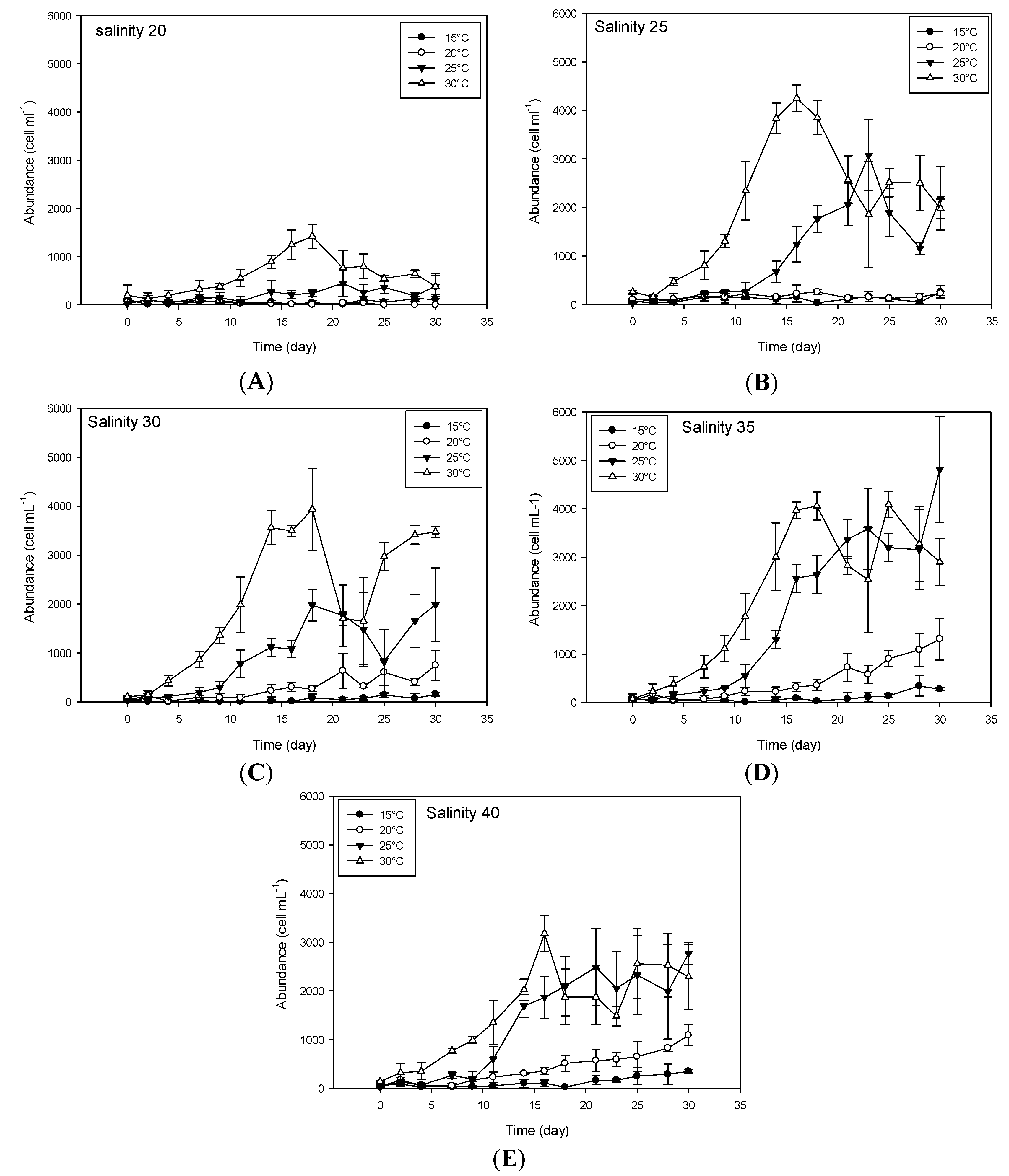
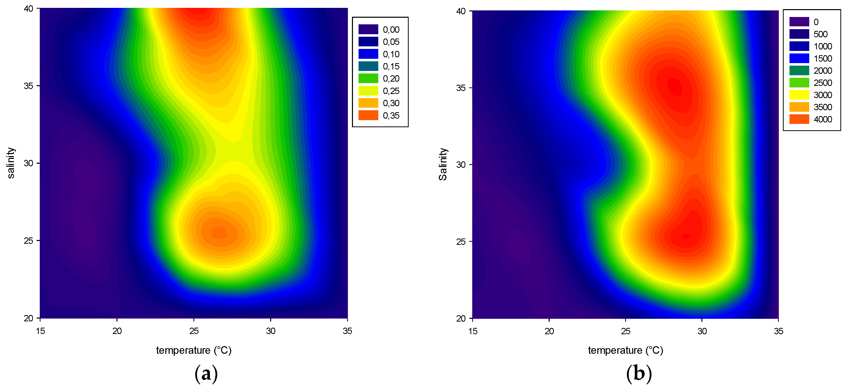
2.1. Effect of Environmental Factors on Growth

2.1.1. Temperature

In this study we observed that V. rugosum was able to grow at temperatures ranging from 20 to 30 °C, while it was not able to grow at 35 °C, the highest temperature tested (Figure 1 and Figure 2). V. rugosum did also not grow at 15 °C irrespective of the salinity of the medium. The dinoflagellate showed the lowest growth rate (0.10 d−1) at 20 °C compared to the values obtained at 25 °C and 30 °C. Also the maximum cell density (cell yield) was the lowest (maximum 1311 cells mL−1) at 20 °C (Figure 1). The optimal temperatures for V. rugosum growth were 25 and 30 °C with a maximum cell density of 3585 cells mL−1 and 4252 cells mL−1, and a maximum growth rate of 0.39 d−1 and 0.2 d−1, respectively. The maximum cell density was reached earlier at 30 °C for all the salinities tested.

2.1.2. Salinity

The most favorable salinity conditions ranged between 25 and 40 at temperatures of 25 °C and 30 °C. Under these conditions, growth rate (µ) ranged between 0.21 d−1 and 0.39 d−1 and cell density between 1952 cells mL−1 and 4252 cells mL−1 (Figure 1 and Figure 2). At a salinity of 20, the cell growth was only observed at 30 °C (µ = 0.19 d−1), but with a low cell yield (1422 cells mL−1). We observed a linear correlation between growth rates and salinity at a temperature of 25 °C and salinity between 30 and 40 (r2 = 0.627, p = 0.011, µ = −0.327 + (0.0177 × salinity)).

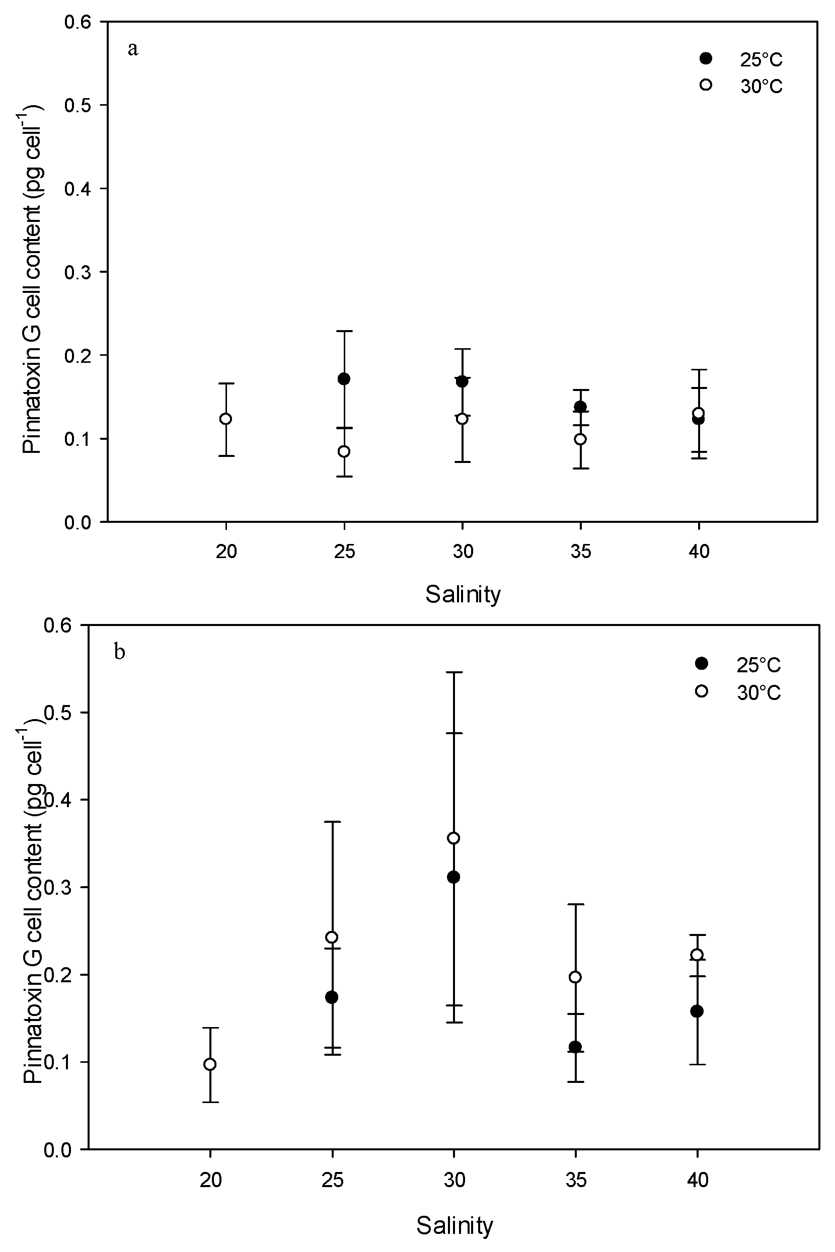
2.2. Effect of Environmental Factors on PnTX-G Production

V. rugosum did not grow at either 15 or 35 °C. At 20 °C, the exponential and stationary phases were not significantly different. For these reasons, the toxin contents of cells were analyzed for the all salinities tested (only for temperatures of 25 °C and 30 °C). PnTX-G concentrations were dependent on the culture conditions. The lowest concentration (0.08 ± 0.03 pg cell−1) was found at a temperature of 30 °C and a salinity of 25 (exponential phase), while the highest concentration (0.36 ± 0.19 pg cell−1) was found at a temperature of 30 °C and a salinity of 30 (stationary phase). PnTX-G concentrations at a temperature of 25 °C and during the exponential phase seemed to decrease in relation to salinity (0.17 pg cell−1 at a salinity of 25 to 0.12 pg cell−1 at a salinity of 40) (Figure 3). In the stationary phase, PnTX-G concentrations appeared to vary as a function of salinity. At the temperature of 25 °C, PnTX-G cell contents increased at a salinity between 25 and 30 (0.13 to 0.31 pg cell−1) and decreased at a salinity between 35 and 40 (0.12 and 0.16 pg cell−1; Figure 3). At the temperature of 30 °C, similar variations in toxin content were observed as a function of salinity (0.10 to 0.36 pg cell−1 at a salinity between 20 and 30; 0.20 and 0.22 pg cell−1 at a salinity between 35 and 40). Differences between toxin contents during the different growth phases were more visible at a temperature of 30 °C (salinity > 20 , p < 0.05) than at 25 °C (difference only for a salinity of 30, p = 0.644). In general, PnTX-G cell contents were lower during the exponential phase than during the stationary phase (p < 0.05), particularly at 30 °C (Figure 3).

2.3. Effect of Environmental Factors on Chlorophyll Production

Pigment concentrations were measured only in cultures grown at 25 °C and 30 °C and at all of the tested salinities (except a salinity of 20 at a temperature of 25 °C due to lack of growth) (Figure 4 and Figure 5). During the exponential phase of growth, higher chlorophyll a concentrations were observed at 25 °C compared to 30 °C (Figure 4, ANOVA p = 0.033; 37.8 ± 7.8 pg cell−1 at a salinity of 25 to 22.4 ± 2.3 pg cell−1 at a salinity of 40 at a temperature of 25 °C and 19.9 ± 5.9 pg cell−1 at a salinity of 25 to 25.5 ± 12.9 pg cell−1 at a salinity of 40 at a temperature of 30 °C).
During the stationary phase, no significant difference was found between Chl a concentrations at the temperatures tested (p = 0.315) (29.9 ± 5.6 pg cell−1 at a salinity of 35 to 57.3 ± 7.64 pg cell−1 at a salinity of 25 at a temperature of 25 °C and 41.6 ± 15.2 pg cell−1 at a salinity of 35 to 64.4 ± 23.4 pg cell−1 at a salinity of 25 at a temperature of 30 °C). Chlorophyll a production was higher during the stationary phase (p < 0.002). When measured during the exponential phase of growth, the phaeopigment concentration was higher for cultures grown at 25 °C compared to those grown at 30 °C (p < 0.001) at all the salinities tested. During the stationary phase, these differences were only observed at salinities of 25 and 30 (Figure 5 p < 0.016).

3. Discussion and Conclusions

3.1. Physical and Chemical Parameters Modulate Growth and Toxin Content of Vulcanodinium Rugosum

To our knowledge this is the first time that growth rate and toxin content of V. rugosum have been determined over an extensive range of the two main environmental parameters, temperature and salinity tested in combination. In Ingril Lagoon, the rain pattern is characterized by significant inter-annual variability (200–1000 mm year−1), and the temperature and salinity also vary widely as a function of season and water depth: for example, they ranged from 0 to 29 °C and from 17 to 42, respectively, between 2000 and 2013 (REPHY monitoring program, Figure 6). These variations are comparatively high, which is related to the low depth of the water column in this lagoon. Indeed, the monthly average temperature in this lagoon was > 20 °C between May and September and the monthly average salinity ranged between 32 and 39 (with very important monthly variations). Our results showed that V. rugosum from Ingril Lagoon could not grow at temperatures ≤15 °C and ≥35 °C. At 20 °C, the dinoflagellate did develop, but at a relatively low rate (0.13 d−1) and with a long lag phase (18 days) when the salinity was ≥30. V. rugosum showed the highest growth rates and maximum cell densities when it was grown at 25 °C (0.21 to 0.39 d−1) and 30 °C (0.16 to 0.26 d−1), and when salinities ranged from 25 to 40, which suggests that the species is thermophile. As a comparison, Alexandrium catenella from the Thau lagoon was found to grow at temperatures and salinities ranging from 15 to 27 °C and from 31 to 40, respectively [21,29,36,37]. The optimal temperature and salinity for V. rugosum growth were close to the values reported for other epiphytic or benthopelagic dinoflagellates such as Gambierdiscus spp. [34,35] and Ostreopsis spp. from Thailand, which showed high growth rates (0.47 to 0.56 d−1) at temperatures and salinities ranging between 25 °C and 30 °C and between 25 and 35, respectively [38]. V. rugosum from Ingril Lagoon showed growth values lower than those of A. catenella isolated from Thau lagoon which reached up to 1.0 d−1. The growth rate of V. rugosum was closer to those of Ostreopsis spp. (0.17 d−1 [39] 0.34 d−1 [40] and 0.49 d−1 [41]) and Prorocentrum lima (0.34 d−1 [42]). The linear regression between growth rate and salinity observed with V. rugosum is usually not described for other dinoflagellates. This relationship has been shown either for both Gambierdiscus [34] and Prorocentrum donghaiense [23].
Nutrient concentration and ratio, temperature and salinity may all affect cellular toxin content in many dinoflagellate species [21,37,43,44,45,46,47]. Abadie et al. [48] showed that PnTX-G content of V. rugosum cells was significantly lower with urea as a nitrogen source compared to nitrate- and ammonium-based cultures. The IFR-VRU-01 strain which was isolated in the Ingril Lagoon and used for our laboratory experiments produces only PnTX-G with a minimum and a maximum amounts of 0.08 pg cell−1 and 0.36 pg cell−1, respectively. Here, V. rugosum cell toxin content was lower during exponential phase compared to that observed during stationary phase for the tested temperature (p = 0.012). This could be explained by the increase of the energy allocated to toxin synthesis instead of cell division and an increase in cell size often correlated with an increase in cell toxin content. Similarly, Guerrini et al. [40] showed that the toxin content in two strains of O. ovata was higher during the stationary phase. In contrast, toxin content was higher during the exponential phase in Alexandrium catenella and Pyrodinium bahamense [26,49].
The cell concentrations of PnTX-G found for the French Mediterranean V. rugosum strain (IFR-VRU-01) are particularly low (0.08–0.36 pg cell−1) when compared to those previously observed for other strains of the same species developing in Pacific and Indian oceans, i.e., 11.9 pg cell−1 for CAWD188 [9] and 87 pg cell−1 for CAWD180 [50]. The differences did not seem to be related to culture conditions including temperature and salinity (Table 1) but to the intraspecific variability within this species. Interestingly, in this study, the PnTX-G cell content of the IFR-VRU-01 strain from the Ingril Lagoon was relatively low compared to the toxin content determined for the same strain by Hess et al. [6] (4.7 pg cell−1) in 2012. The cell content of PnTXs might decrease with culture age. The IFR-VRU-01 strain was isolated in 2010 and maintained in the laboratory through successive culturing which may have led to genetic mutations and alteration of the bacterial flora. Similarly, the dinoflagellate Alexandrium minutum had been shown to become less toxic after prolonged culturing [51]. Likewise, another clone of a strain of the paralytic shellfish toxin producer Alexandrium lusitanicum (synonym of Alexandrium minutum Halim) completely lost its toxicity in culture [52].

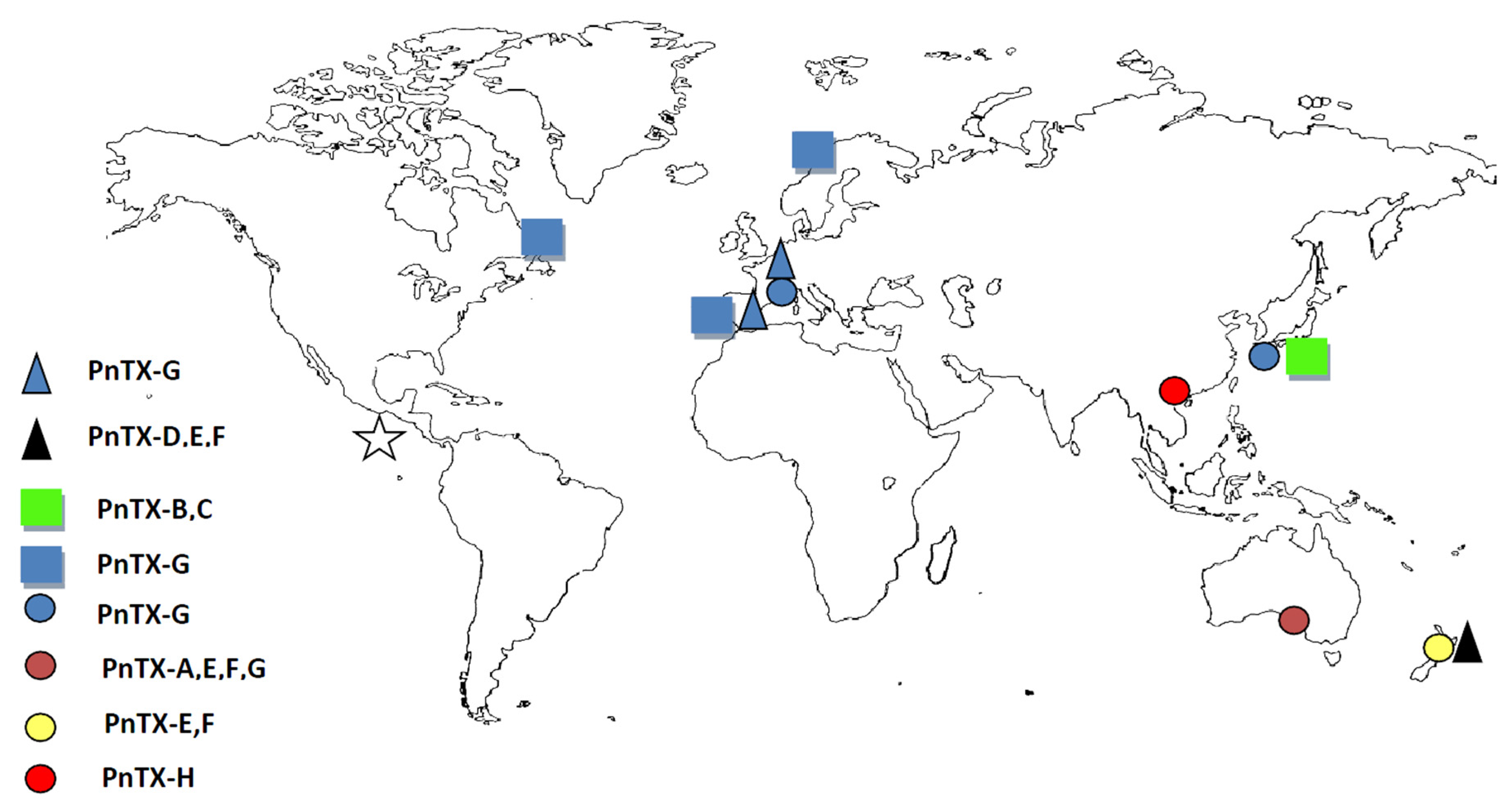
3.2. Worldwide Distribution of V. rugosum and Related Toxins

Recent works show that V. rugosum is widely distributed around the world suggesting that it could be considered as a cosmopolitan species [6,9,12,54,57,58] (Figure 7, Table 1). Natural or human-assisted dispersion (ballast waters, shellfish relocation ...) or the development of endemic populations could explain this geographic distribution [59]. Further studies have to focus on the phylogeny of isolated strains of V. rugosum to better understand the geographic distribution of this probably expanding harmful dinoflagellate. Concerning the toxins produced by the dinoflagellate, data so far show important toxin diversity varying as a function of the strains colonizing each of the oceans. In the Pacific, PnTX-E and F dominated in the strains originating from New Zealand (CAWD163–178), whereas PnTX-G was the only toxin detected in Japanese strains (CAWD188, 190), and the Chinese strain CAWD198 produces only PnTX-H. The Australian strains produce PnTX-G and H (CAWD180, 183) (Table 1 and references therein). Selwood, et al. [55] discovered the presence of portimine, a new cyclic imine, in a V. rugosum strain isolated from Northland, New Zealand. Regarding isolates from the Southern Ocean, pinnatoxins A, E, F and G were found in two strains (CAWD180 and CAWD183) isolated from Franklin Harbour, Australia. In contrast, the strain IFR-VRU-01 from Mediterranean waters has been shown to produce only PnTX-G [6]. As suggested for Alexandrium [60], the production of toxins by V. rugosum may be genetically driven for each clonal strain. In addition, pinnatoxins were found in passive sampling devices (SPATT) and shellfish in various marine ecosystems, despite V. rugosum cells not being reported in those same studies. In Mediterranean waters (Catalonia, Spain), PnTX-G was detected by solid phase adsorption toxin tracking (SPATT) [61]. This passive sampling technique revealed the presence of PnTX-E and F and traces of PnTX-A, D and G in New Zealand waters [62]. PnTX-B and C were detected in the Okinawan bivalve Pinna muricata [63]. PnTX-G was also detected in different shellfish species from the Eastern Canadian coast and Norway [64,65]. On the other hand, V. rugosum was identified in Mexican waters but no toxin analyses were performed [57]. Interestingly, summarized data relating to toxin analyses of V. rugosum cultures maintained in different laboratories (Table 1, Figure 8) showed that PnTX-G was the main pinnatoxin form in Mediterranean and Atlantic waters. Asian and Pacific waters are characterized by higher pinnatoxin diversity as all the PnTX analogues were detected in the related ecosystems (PnTX-A, E, F, G and H). PnTX B and C were found only in the Okinawan (Japan) bivalve Pinna muricata. This suggests that V. rugosum may well be an introduced species in the Mediterranean and Atlantic seas. Surprisingly, these PnTXs (B and C) were not found in mussels and clams from Ingril Lagoon (unpublished data) whereas PnTX-A was present in these mollusks at non-quantifiable traces [6]. Overall, these data matched [66] suggestions about the biotransformation of algal metabolites PnTXs-E, -F and -G into shellfish metabolites PnTXs-A, -B, -C and -D. Quantification of PnTXs by LC-MS/MS revealed significant variability in V. rugosum cell contents (PnTX-E: 0.4–10 pg cell−1, PnTX-F: 2.3–41 pg cell−1, PnTX-G: 0.14–87 pg cell−1) which may be directly related to the strain or to culture conditions. Pinnatoxins exhibit fast-acting toxicity when injected intraperitoneally (i.p.) into mice [58]. The high intrinsic toxicity of PnTXs is indicated by low LD50 values following intraperitoneal injection in mice. LD50 values varied substantially as a function of the V. rugosum strain and the intracellular toxin injected (CAWD167–PnTX-E and F: 1.33 mg kg−1, CAW183–PnTX-G: 42.7–48 µg kg−1, CAWD198–PnTX-H: 67 µg kg−1) (Table 1).

3.3. Conclusions

Laboratory data on the ecophysiology of V. rugosum are coherent with the fact that this dinoflagellate typically develops during summer time in the Mediterranean.
In 2013, the cell density of V. rugosum was found to be at its highest in the summer, between June and September (Abadie et al. [67] unpublished data), which confirmed the growth results from our laboratory experiments. PnTX-G concentrations were high in mussels (Mytilus galloprovincialis) between June and September 2013. V. rugosum has been shown in this study to be a euryhaline dinoflagellate which suggests that this harmful species could spread to other Mediterranean lagoons and confined water systems.
Some studies suggest that there may be relationships between shellfish toxicity and large-scale patterns of climate variability. For example, there was an attempt to explain the increase of Alexandrium blooming in the Puget Sound by El Niño which increased the temperature of the water column and produced stratification more suitable to dinoflagellates [68]. Moore et al. [69] suggested that aspects of the local climate (daily to seasonal timescales) such as air temperature and stream flow could be more important in determining oceanographic variability in the Puget Sound than large-scale climate variations such as the El Niño Southern Oscillation (=ENSO, i.e., inter-annual to inter-decadal timescales). However, no significant correlations were identified [69] for Alexandrium in the Puget Sound marine system. It is undeniable that marine ecosystems are facing a global temperature increase [70,71,72,73]. For example, 2014 was ranked as the warmest year since the beginning of weather records in France. Southwestern Mediterranean lagoons are characterized by the relatively low depth of their water columns (e.g., Ingril Lagoon: 0–1.7 m) which means that water temperature could increase more rapidly and stay warm for longer periods following any warming. Our data showed that V. rugosum is thermophile like other benthic dinoflagellates and its growth is mainly driven by temperature which suggests that this dinoflagellate may be responsible for blooms in the coming decades caused by the climate change-related expected increase in temperature of the Mediterranean area.

4. Experimental Section

4.1. Vulcanoidinum rugosum: Origin and Culture

The IFR-VRU-01 strain of V. rugosum was isolated from the Ingril Lagoon (French Mediterranean coast) in 2010 by Nézan & Chomérat [10] (Figure 8). IFR-VRU-01 was shown to produce PnTX-G [6]. The non-axenic culture of V. rugosum was maintained at a temperature of 25 °C, a salinity of 35 and an irradiance of 100 µmol photon m2 s−1 in an Enriched Natural Sea Water medium (ENSW) [74] in the MARBEC laboratory (Montpellier, France).
As described by Rhodes et al. [11], the life cycle of V. rugosum is characterized by typical, motile vegetative cells and unornamented, non-motile cells (30–32 mm diameter). For each experiment testing the effect of temperature and salinity, the flasks were inoculated only with healthy motile cells. Our microscopic observations clearly showed that the cultures were dominated largely by motile cells (>90%) until the stationary phase when non motile cells appeared and settled against the flask walls. Growth rate was calculated for the exponential phase based only on cell counts of motile cells. PnTX-G and Chlorophyll a (Chl a) levels were measured during the exponential phase and at the beginning of the stationary phase when motile cells still dominated.

4.2. Experimental Conditions

Seventy-five 250 mL-flasks (Cellstar® Cell Culture Flasks, Greiner Bio-One, Courtaboeuf, France) were inoculated. The flasks were filled with ENSW medium adjusted at five different salinities (20, 25, 30, 35 and 40). Each flask was inoculated at initial concentration of 400 cells mL−1 in a total volume of 200 mL. For each salinity, three flasks (replicates) were placed in four incubators (SANYO Incubator, Firlabo, Panasonic Healthcare Company, Etten-Leur, the Netherlands, Meditest 600/1300, Panasonic MIR-554, Panasonic Healthcare Company, Etten-Leur, the Netherlands, and one temparated-controled room) at different temperatures (15, 20, 25, 30 and 35 °C). The test at 35 °C was separately conducted after the first four tests. Cultures were exposed to an irradiance of 100 µmol photon m2 s−1 using cool white fluorescent lights and a 12:12 h light:dark cycle. Temperature of culture chambers were monitored throughout the experiment by a temperature probe (Proges Plus 22 L). Light irradiance was controlled using a PAR sensor (PQS 1 KIPP ZONEN, Delft, The Netherlands). The salinity of each medium was checked during the experiment with a WTW LF 197 probe (WTW Wissenschaftlich-Technische Werkstätten GmbH, Weilheim, Germany).

4.3. Cell Concentrations and Growth Rate Measurements

The cell density of each flask was monitored every two days for thirty days. After gentle homogenization to avoid any stress to the cultures, 1350 μL aliquots of culture were withdrawn sterilely with a micropipette and counted with a Nageotte cell (counting chamber) under an inverted light microscope (Zeiss Axiovert 25, Carl Zeiss SAS, Marly Le Roy, France). In accordance with Guillard [75], the growth rate (μ; expressed in day−1) was calculated from the slope of a linear regression line over the entire exponential phase of growth by the least squares fit of the straight line to the data after logarithmic transformation; μ = Ln (N1) – Ln (N0) /T1T0 in units of day−1 where N1 and N0 were cell densities at times T1 and T0, respectively, during the linear portion of exponential growth phase.

4.4. Chlorophyll a Analysis

Chlorophyll a concentrations were determined for each flask when the cultures were in the exponential and stationary phases. For Chl a measurements, culture (15 mL) was withdrawn and filtered on a 25 mm-diameter GF/F Whatman filter at a low pressure (<100 mbar) to avoid cell lysis. Filters were placed in 12 mL-polypropylene-tubes and stored at −20 °C. Aqueous acetone (5 mL, acetone:water 9:1) was used for extraction and filters were decomposed using an ultrasonic rod (Vibra cell™ 7518, Sonics & Materials, Inc., Newtown, CT, USA, 130 Watt—50 kHz). The mix was centrifuged at 2750× g and 4 °C for 30 min. The supernatants were analyzed by spectrofluorimetry (Perkin Elmer, PerkinElmer Inc., Waltham, MA, USA, LS50B) using the method described by Neuveux [76]. Pigment concentration was expressed in pg cell−1. To calculate the toxin concentration per cell, the cell density of the chemically analyzed sample was used.

4.5. Pinnatoxin G Measurement

For the toxin analyses, we took a 10 mL sample from each culture during the exponential and stationary growth phases. The samples were centrifuged (3000× g, 15 min, 4 °C) and the supernatants removed carefully. Methanol (100%, 2 mL) was added to the remaining pellet and the samples stored at −20 °C until toxin extraction [54]. Extraction of the pinnatoxins (Figure 9) was carried using three consecutive sonication steps lasting 10 min each, followed by filtration of the extracts over a 0.2 µm membrane (Whatman Mini-UniPrepTM, GE Healthcare Europe GmbH, Velizy-Villacoublay, France). The filtered extracts were stored at −24 °C until quantification. Quantification of PnTX-G was carried out using liquid chromatography coupled to tandem mass spectrometry (LC-MS/MS), using external calibrants over the range of 0.25 to 5 ng mL−1. A C8 column (Phenomenex, Phenomenex Inc., Torrance, CA, USA) was used at 25 °C for analysis (injection volume 5 µL). The analysis was conducted at a flow rate of 0.8 mL min−1 [6]. The limit of detection (LD) and the limit of quantification (LQ) values were 0.08 and 0.27 ng mL−1, respectively.

4.6. Statistics

For each set of data, differences between means were analyzed by Student’s t-test or the ANOVA test (Sigmaplot 12.5, Systat Software Inc., San Jose, CA, USA). The appropriate statistical test was conducted after the verification of the normality of the data (Shapiro–Wilk). If the test of the normality failed, a Kruskal–Wallis One Way Analysis of Variance on Ranks (non parametric test) was performed.
Values were expressed for each condition as the mean (m) of the triplicate and Standard.

Acknowledgments

Thanks to EC2CO Ecosphère Continentale et Côtière DRIL (VULCANO project) and TOTAL Foundation (LAGUNOTOX project) for funding this work. We would also to thank the Agence de l’Eau Rhone Méditerranée Corse (AERMC—Rhone Mediterranean Corsica Water Agency) for their financial help. Thanks to the IRD (Institut National pour la Recherche et le Développement—National Institute for Research and Development) for funding Mohamed Laabir’s stay in Tunisia.

Author Contributions

Eric Abadie and Alexia Muguet performed the experiments. Eric Abadie and Mohamed Laabir conceived, designed and analyzed the data and wrote the paper. Tom Berteaux contributed to the analysis of the toxins. Philipp Hess, Estelle Masseret, Nicolat Chomérat and Emmanuelle Roque D’Orbcastel contributed to correcting and improving the manuscript.

Conflicts of Interest

The authors declare no conflict of interest.

References

  1. Smayda, T.J. Harmful algal blooms: Their ecophysiology and general relevance to phytoplankton blooms in the sea. Limnol. Oceanogr. 1997, 42, 1137–1153. [Google Scholar] [CrossRef]
  2. Laabir, M.; Jauzein, C.; Genovesi, B.; Masseret, E.; Grzebyk, D.; Cecchi, P.; Vaquer, A.; Perrin, Y.; Collos, Y. Influence of temperature, salinity and irradiance on the growth and cell yield of the harmful red tide dinoflagellate Alexandrium catenella colonizing Mediterranean waters. J. Plankton Res. 2011, 33, 1550–1563. [Google Scholar] [CrossRef]
  3. Abadie, E.; Amzil, Z.; Belin, C.; Comps, M.A.; Elziere-Papayanni, P.; Lassus, P.; Le Bec, C.; Marcaillou-Le Baut, C.; Nezan, E.; Poggi, R. Contamination de l’etang de Thau par Alexandrium Tamarense. Episode de Novembre a Decembre 1998; Ifremer: Plouzané, France, 1999. [Google Scholar]
  4. Lilly, E.L.; Halanych, K.M.; Anderson, D.M. Species boundaries and global biogeography of the Alexandrium tamarense complex (Dinophyceae). J. Phycol. 2007, 43, 1329–1338. [Google Scholar] [CrossRef]
  5. Genovesi, B.; Mouillot, D.; Vaquer, A.; Laabir, M.; Pastoureaud, A. Towards an optimal sampling strategy for Alexandrium catenella (Dinophyceae) benthic resting cysts. Harmful Algae 2007, 6, 837–848. [Google Scholar] [CrossRef]
  6. Hess, P.; Abadie, E.; Herve, F.; Berteaux, T.; Sechet, V.; Araoz, R.; Molgo, J.; Zakarian, A.; Sibat, M.; Rundberget, T.; et al. Pinnatoxin G is responsible for atypical toxicity in mussels (Mytilus galloprovincialis) and clams (Venerupis decussata) from Ingril, a French Mediterranean Lagoon. Toxicon 2013, 75, 16–26. [Google Scholar] [CrossRef] [PubMed]
  7. Uemura, D.; Chou, T.; Haino, T.; Nagatsu, A.; Fukuzawa, S.; Zheng, S.Z.; Chen, H.S. Pinnatoxin-A—A toxic amphoteric macrocycle from the okinawan bivalve Pinna muricata. J. Am. Chem. Soc. 1995, 117, 1155–1156. [Google Scholar] [CrossRef]
  8. Chou, T.; Haino, T.; Kuramoto, M.; Uemura, D. Isolation and structure of pinnatoxin D, a new shellfish poison from the okinawan bivalve Pinna muricata. Tetrahedron Lett. 1996, 37, 4027–4030. [Google Scholar] [CrossRef]
  9. Smith, K.F.; Rhodes, L.L.; Suda, S.; Selwood, A.I. A dinoflagellate producer of pinnatoxin G, isolated from sub-tropical Japanese waters. Harmful Algae 2011, 10, 702–705. [Google Scholar] [CrossRef]
  10. Nézan, E.; Chomérat, N. Vulcanodinium rugosum gen. Nov., sp. Nov. (Dinophyceae): A new marine dinoflagellate from the French Mediterranean Coast. Cryptogam. Algologie 2011, 32, 3–18. [Google Scholar] [CrossRef]
  11. Rhodes, L.; Smith, K.; Selwood, A.; McNabb, P.; Munday, R.; Suda, S.; Molenaar, S.; Hallegraeff, G. Dinoflagellate Vulcanodinium rugosum identified as the causative organism of pinnatoxins in Australia, New Zealand and Japan. Phycologia 2011, 50, 624–628. [Google Scholar] [CrossRef]
  12. Zeng, N.; Gu, H.; Smith, K.F.; Rhodes, L.L.; Selwood, A.I.; Yang, W. The first report of Vulcanodinium rugosum (Dinophyceae) from the South China Sea with a focus on the life cycle. N. Z. J. Mar. Freshw. Res. 2012, 46, 511–521. [Google Scholar] [CrossRef]
  13. Matsubara, T.; Nagasoe, S.; Yamasaki, Y.; Shikata, T.; Shimasaki, Y.; Oshima, Y.; Honjo, T. Effects of temperature, salinity, and irradiance on the growth of the dinoflagellate Akashiwo sanguinea. J. Exp. Mar. Biol. Ecol. 2007, 342, 226–230. [Google Scholar] [CrossRef]
  14. Nagasoe, S.; Kim, D.-I.; Shimasaki, Y.; Oshima, Y.; Yamaguchi, M.; Honjo, T. Effects of temperature, salinity and irradiance on the growth of the red tide dinoflagellate Gyrodinium instriatum Freudenthal et Lee. Harmful Algae 2006, 5, 20–25. [Google Scholar] [CrossRef]
  15. Yamaguchi, M.; Honjo, T. Effects of temperature, salinity and irradiance on the growth of the noxious red tide flagellate Gymnodinium nagasakiense (Dinophycae). Nippon Suisan Gakk. 1989, 55, 2029–2036. [Google Scholar] [CrossRef]
  16. Yamaguchi, M.; Imai, I.; Honjo, T. Effects of temperature, salinity and irradiance on the growth-rates of the noxious red tide flagellates Chattonella antiqua and C. marina (Raphidophycae). Nippon Suisan Gakk. 1991, 57, 1277–1284. [Google Scholar] [CrossRef]
  17. Yamaguchi, M.; Shigeru, I.; Nagasaki, K.; Matsuyama, Y.; Uchida, T.; Imai, I. Effects of temperature and salinity on the growth of the red tide flagellates Heterocapsa circularisquama (Dinophyceae) and Chattonella verruculosa (Raphidophyceae). J. Plankton Res. 1997, 19, 1167–1174. [Google Scholar] [CrossRef]
  18. Yamamoto, T.; Oh, S.J.; Kataoka, Y. Effects of temperature, salinity and irradiance on the growth of the toxic dinoflagellate Gymnodinium catenatum (Dinophyceae) isolated from Hiroshima Bay, Japan. Fish. Sci. 2002, 68, 356–363. [Google Scholar] [CrossRef]
  19. Anderson, D.M.; Cembella, A.D.; Hallegraeff, G.M. Progress in understanding harmful algal blooms: Paradigm shifts and new technologies for research, monitoring, and management. Annu. Rev. Mar. Sci. 2012, 4, 143–176. [Google Scholar] [CrossRef] [PubMed] [Green Version]
  20. Fu, F.-X.; Zhang, Y.; Warner, M.E.; Feng, Y.; Sun, J.; Hutchins, D.A. A comparison of future increased CO2 and temperature effects on sympatric Heterosigma akashiwo and Prorocentrum minimum. Harmful Algae 2008, 7, 76–90. [Google Scholar] [CrossRef]
  21. Laabir, M.; Collos, Y.; Masseret, E.; Grzebyk, D.; Abadie, E.; Savar, V.; Sibat, M.; Amzil, Z. Influence of environmental factors on the paralytic shellfish toxin content and profile of Alexandrium catenella (Dinophyceae) isolated from the Mediterranean Sea. Mar. Drugs 2013, 11, 1583–1601. [Google Scholar] [CrossRef] [PubMed]
  22. Pan, Y.; Cembella, A.D.; Quilliam, M.A. Cell cycle and toxin production in the benthic dinoflagellate Prorocentrum lima. Mar. Biol. 1999, 134, 541–549. [Google Scholar] [CrossRef]
  23. Xu, N.; Duan, S.; Li, A.; Zhang, C.; Cai, Z.; Hu, Z. Effects of temperature, salinity and irradiance on the growth of the harmful dinoflagellate Prorocentrum donghaiense Lu. Harmful Algae 2010, 9, 13–17. [Google Scholar] [CrossRef]
  24. Genovesi, B.; Laabir, M.; Masseret, E.; Collos, Y.; Vaquer, A.; Grzebyk, D. Dormancy and germination features in resting cysts of Alexandrium tamarense species complex (Dinophyceae) can facilitate bloom formation in a shallow lagoon (Thau, Southern France). J. Plankton Res. 2009, 31, 1209–1224. [Google Scholar] [CrossRef]
  25. Triki, H.Z.; Daly-Yahia, O.K.; Malouche, D.; Komiha, Y.; Deidun, A.; Brahim, M.; Laabir, M. Distribution of resting cysts of the potentially toxic dinoflagellate Alexandrium pseudogonyaulax in recently-deposited sediment within Bizerte Lagoon (Mediterranean Coast, Tunisia). Mar. Pollut. Bull. 2014, 84, 172–181. [Google Scholar] [CrossRef] [PubMed]
  26. Navarro, J.M.; Munoz, M.G.; Contreras, A.M. Temperature as a factor regulating growth and toxin content in the dinofiagellate Alexandrium catenella. Harmful Algae 2006, 5, 762–769. [Google Scholar] [CrossRef]
  27. Band-Schmidt, C.J.; Bustillos-Guzman, J.J.; Hernandez-Sandoval, F.E.; Nunez-Vasquez, E.J.; Lopez-Cortés, D.J. Effect of temperature on growth and paralytic toxin profiles in isolates of Gymnodinium catenatum (Dinophyceae) from the Pacific Coast of Mexico. Toxicon 2014, 90, 199–212. [Google Scholar] [CrossRef] [PubMed]
  28. Giacobbe, M.G.; Oliva, F.D.; Maimone, G. Environmental factors and seasonal occurrence of the dinoflagellate Alexandrium minutum, a PSP potential producer, in a Mediterranean Lagoon. Estuar. Coast. Shelf Sci. 1996, 42, 539–549. [Google Scholar] [CrossRef]
  29. Grzebyk, D.; Bechemin, C.; Ward, C.J.; Verite, C.; Codd, G.A.; Maestrini, S.Y. Effects of salinity and two coastal waters on the growth and toxin content of the dinoflagellate Alexandrium minutum. J. Plankton Res. 2003, 25, 1185–1199. [Google Scholar] [CrossRef]
  30. Hwang, D.F.; Lu, Y.H. Influence of environmental and nutritional factors on growth, toxicity, and toxin profile of dinoflagellate Alexandrium minutum. Toxicon 2000, 38, 1491–1503. [Google Scholar] [CrossRef]
  31. Kim, D.-I.; Matsuyama, Y.; Nagasoe, S.; Yamaguchi, M.; Yoon, Y.-H.; Oshima, Y.; Imada, N.; Honjo, T. Effects of temperature, salinity and irradiance on the growth of the harmful red tide dinoflagellate Cochlodinium polykrikoides margalef (Dinophyceae). J. Plankton Res. 2004, 26, 61–66. [Google Scholar] [CrossRef]
  32. Parkhill, J.P.; Cembella, A.D. Effects of salinity, light and inorganic nitrogen on growth and toxigenicity of the marine dinoflagellate Alexandrium tamarense from Northeastern Canada. J. Plankton Res. 1999, 21, 939–955. [Google Scholar] [CrossRef]
  33. Wang, D.-Z.; Hsieh, D.P.H. Growth and toxin production in batch cultures of a marine dinoflagellate Alexandrium tamarense HK9301 isolated from the South China Sea. Harmful Algae 2005, 4, 401–410. [Google Scholar] [CrossRef]
  34. Kibler, S.R.; Litaker, R.W.; Holland, W.C.; Vandersea, M.W.; Tester, P.A. Growth of eight Gambierdiscus (Dinophyceae) species: Effects of temperature, salinity and irradiance. Harmful Algae 2012, 19, 1–14. [Google Scholar] [CrossRef]
  35. Yoshimatsu, T.; Yamaguchi, H.; Iwamoto, H.; Nishimura, T.; Adachi, M. Effects of temperature, salinity and their interaction on growth of Japanese Gambierdiscus spp. (Dinophyceae). Harmful Algae 2014, 35, 29–37. [Google Scholar] [CrossRef]
  36. Aguilera-Belmonte, A.; Inostroza, I.; Carrillo, K.S.; Franco, J.M.; Riobo, P.; Gomez, P.I. The combined effect of salinity and temperature on the growth and toxin content of four chilean strains of Alexandrium catenella (Whedon and Kofoid) balech 1985 (Dinophyceae) isolated from an outbreak occurring in Southern Chile in 2009. Harmful Algae 2013, 23, 55–59. [Google Scholar] [CrossRef]
  37. Etheridge, S.M.; Roesler, C.S. Effects of temperature, irradiance, and salinity on photosynthesis, growth rates, total toxicity, and toxin composition for Alexandrium fundyense isolates from the gulf of maine and bay of fundy. Deep Sea Res. Part II Top. Stud. Oceanogr. 2005, 52, 2491–2500. [Google Scholar] [CrossRef]
  38. Tawong, W.; Yoshimatsu, T.; Yamaguchi, H.; Adachi, M. Effects of temperature, salinity and their interaction on growth of benthic dinoflagellates Ostreopsis spp. from Thailand. Harmful Algae 2015, 44, 37–45. [Google Scholar] [CrossRef]
  39. Pistocchi, R.; Pezzolesi, L.; Guerrini, F.; Vanucci, S.; Dell’Aversano, C.; Fattorusso, E. A review on the effects of environmental conditions on growth and toxin production of Ostreopsis ovata. Toxicon 2011, 57, 421–428. [Google Scholar] [CrossRef] [PubMed]
  40. Guerrini, F.; Pezzolesi, L.; Feller, A.; Riccardi, M.; Ciminiello, P.; Dell’Aversano, C.; Tartaglione, L.; Iacovo, E.D.; Fattorusso, E.; Forino, M.; et al. Comparative growth and toxin profile of cultured Ostreopsis ovata from the Tyrrhenian and Adriatic Seas. Toxicon 2010, 55, 211–220. [Google Scholar] [CrossRef] [PubMed]
  41. Morton, S.L.; Norris, D.R.; Bomber, J.W. Effect of temperature, salinity and light-intensity on the growth and seasonality of toxic dinoflagellates associeted with ciguatera. J. Exp. Mar. Biol. Ecol. 1992, 157, 79–90. [Google Scholar] [CrossRef]
  42. Vale, P.; Veloso, V.; Amorim, A. Toxin composition of a Prorocentrum lima strain isolated from the Portuguese Coast. Toxicon 2009, 54, 145–152. [Google Scholar] [CrossRef] [PubMed]
  43. Hamasaki, K.; Horie, M.; Tokimitsu, S.; Toda, T.; Taguchi, S. Variability in toxicity of the dinoflagellate Alexandrium tamarense isolated from hiroshima bay, western japan, as a reflection of changing environmental conditions. J. Plankton Res. 2001, 23, 271–278. [Google Scholar] [CrossRef]
  44. Lee, T.C.-H.; Kwok, O.-T.; Ho, K.-C.; Lee, F.W.-F. Effects of different nitrate and phosphate concentrations on the growth and toxin production of an Alexandrium tamarense strain collected from drake passage. Mar. Environ. Res. 2012, 81, 62–69. [Google Scholar] [CrossRef] [PubMed]
  45. Leong, S.C.Y.; Murata, A.; Nagashima, Y.; Taguchi, S. Variability in toxicity of the dinoflagellate Alexandrium tamarense in response to different nitrogen sources and concentrations. Toxicon 2004, 43, 407–415. [Google Scholar] [CrossRef] [PubMed]
  46. Lim, P.-T.; Leaw, C.-P.; Kobiyama, A.; Ogata, T. Growth and toxin production of tropical Alexandrium minutum halim (Dinophyceae) under various nitrogen to phosphorus ratios. J. Appl. Phycol. 2010, 22, 203–210. [Google Scholar] [CrossRef]
  47. Xu, J.; Ho, A.Y.T.; He, L.; Yin, K.; Hung, C.; Choi, N.; Lam, P.K.S.; Wu, R.S.S.; Anderson, D.M.; Harrison, P.J. Effects of inorganic and organic nitrogen and phosphorus on the growth and toxicity of two Alexandrium species from Hong Kong. Harmful Algae 2012, 16, 89–97. [Google Scholar] [CrossRef]
  48. Abadie, E.; Kaci, L.; Berteaux, T.; Hess, P.; Sechet, V.; Masseret, E.; Rolland, J.; Laabir, M. Effect of nitrate, ammonium and urea on growth and pinnatoxin G production of Vulcanodinium rugosum. Mar. Drugs 2015, 13, 5642. [Google Scholar] [CrossRef] [PubMed]
  49. Gedaria, A.I.; Luckas, B.; Reinhardt, K.; Azanza, R.V. Growth response and toxin concentration of cultured Pyrodinium bahamense var. Compressum to varying salinity and temperature conditions. Toxicon 2007, 50, 518–529. [Google Scholar] [CrossRef] [PubMed]
  50. Rhodes, L.; Smith, K.; Selwood, A.; McNabb, P.; Molenaar, S.; Munday, R.; Wilkinson, C.; Hallegraeff, G. Production of pinnatoxins E, F and G by scrippsielloid dinoflagellates isolated from Franklin Harbour, South Australia. N. Z. J. Mar. Freshw. Res. 2011, 45, 703–709. [Google Scholar] [CrossRef]
  51. Hansen, G.; Daugbjerg, N.; Franco, J.M. Morphology, toxin composition and lsu rdna phylogeny of Alexandrium minutum (Dinophyceae) from Denmark, with some morphological observations on other European strains. Harmful Algae 2003, 2, 317–335. [Google Scholar] [CrossRef]
  52. Martins, C.A.; Kulis, D.; Franca, S.; Anderson, D.M. The loss of psp toxin production in a formerly toxic Alexandrium lusitanicum clone. Toxicon 2004, 43, 195–205. [Google Scholar] [CrossRef] [PubMed]
  53. Satta, C.T.; Anglès, S.; Lugliè, A.; Guillén, J.; Sechi, N.; Camp, J.; Garcés, E. Studies on dinoflagellate cyst assemblages in two estuarine Mediterranean Bays: A useful tool for the discovery and mapping of harmful algal species. Harmful Algae 2013, 24, 65–79. [Google Scholar] [CrossRef]
  54. Rhodes, L.; Smith, K.; Selwood, A.; McNabb, P.; van Ginkel, R.; Holland, P.; Munday, R. Production of pinnatoxins by a peridinoid dinoflagellate isolated from Northland, New Zealand. Harmful Algae 2010, 9, 384–389. [Google Scholar] [CrossRef]
  55. Selwood, A.I.; Wilkins, A.L.; Munday, R.; Shi, F.; Rhodes, L.L.; Holland, P.T. Portimine: A bioactive metabolite from the benthic dinoflagellate Vulcanodinium rugosum. Tetrahedron Lett. 2013, 54, 4705–4707. [Google Scholar] [CrossRef]
  56. Selwood, A.I.; Wilkins, A.L.; Munday, R.; Gu, H.F.; Smith, K.F.; Rhodes, L.L.; Rise, F. Pinnatoxin H: A new pinnatoxin analogue from a South China Sea Vulcanodinium rugosum isolate. Tetrahedron Letters 2014, 55, 5508–5510. [Google Scholar] [CrossRef]
  57. Hernandez-Becerril, D.U.; Rodriguez-Palacio, M.C.; Lozano-Ramirez, C. Morphology and life stages of the potentially pinnatoxin-producing thecate dinoflagellate Vulcanodinium rugosum from the tropical Mexican Pacific. Bot. Mar. 2013, 56, 535–540. [Google Scholar] [CrossRef]
  58. Munday, R.; Selwood, A.I.; Rhodes, L. Acute toxicity of pinnatoxins E, F and G to mice. Toxicon 2012, 60, 995–999. [Google Scholar] [CrossRef] [PubMed]
  59. Hallegraeff, G.M. A review of harmful algal blooms and their apparent global increase. Phycologia 1993, 32, 79–99. [Google Scholar] [CrossRef]
  60. Anderson, D.M.; Kulis, D.M.; Sullivan, J.J.; Hall, S.; Lee, C. Dynamics and physiology of saxitoxin production by the dinoflagellates Alexandrium spp. Mar. Biol. 1990, 104, 511–524. [Google Scholar] [CrossRef]
  61. Garcia-Altares, M.; Casanova, A.; Bane, V.; Diogène, J.; Furey, A.; de la Iglesia, P. Confirmation of pinnatoxins and spirolides in shellfish and passive samplers from Catalonia (Spain) by liquid chromatography coupled with triple quadrupole and high-resolution hybrid tandem mass spectrometry. Mar. Drugs 2014, 12, 3706–3732. [Google Scholar] [CrossRef] [PubMed]
  62. MacKenzie, L.A.; Selwood, A.I.; McNabb, P.; Rhodes, L. Benthic dinoflagellate toxins in two warm-temperate estuaries: Rangaunu and Parengarenga Harbours, Northland, New Zealand. Harmful Algae 2011, 10, 559–566. [Google Scholar] [CrossRef]
  63. Takada, N.; Umemura, N.; Suenaga, K.; Chou, T.; Nagatsu, A.; Haino, T.; Yamada, K.; Uemura, D. Pinnatoxins B and C, the most toxic components in the pinnatoxin series from the okinawan bivalve Pinna muricata. Tetrahedron Lett. 2001, 42, 3491–3494. [Google Scholar] [CrossRef]
  64. McCarron, P.; Rourke, W.A.; Hardstaff, W.; Pooley, B.; Quilliam, M.A. Identification of pinnatoxins and discovery of their fatty acid ester metabolites in mussels (Mytilus edulis) from Eastern Canada. J. Agric. Food Chem. 2012, 60, 1437–1446. [Google Scholar] [CrossRef] [PubMed]
  65. Rundberget, T.; Aasen, J.A.B.S.; Selwood, A.I.; Miles, C.O. Pinnatoxins and spirolides in norwegian blue mussels and seawater. Toxicon 2011, 58, 700–711. [Google Scholar] [CrossRef] [PubMed]
  66. Selwood, A.I.; Miles, C.O.; Wilkins, A.L.; van Ginkel, R.; Munday, R.; Rise, F.; McNabb, P. Isolation, structural determination and acute toxicity of pinnatoxins E, F and G. J. Agric. Food Chem. 2010, 58, 6532–6542. [Google Scholar] [CrossRef] [PubMed]
  67. Abadie, E.; Rhodes, L.; Chomerat, N.; Chiantella, C.; Crottier, A.; Laabir, M. Occurrence of the neurotoxic dinoflagellate Vulcanodinium rugosum in mediterranean: What are the driving environnemental factors of its dynamic. J. Plankton Res. Submission in progress.
  68. Moore, S.K.; Mantua, N.J.; Hickey, B.M.; Trainer, V.L. The relative influences of el niño-southern oscillation and pacific decadal oscillation on paralytic shellfish toxin accumulation in Northwest Pacific shellfish. Limnol. Oceanogr. 2010, 55, 2262–2274. [Google Scholar] [CrossRef]
  69. Moore, S.K.; Mantua, N.J.; Hickey, B.M.; Trainer, V.L. Recent trends in paralytic shellfish toxins in puget sound, relationships to climate, and capacity for prediction of toxic events. Harmful Algae 2009, 8, 463–477. [Google Scholar] [CrossRef]
  70. Hiscock, K.; Southward, A.; Tittley, I.; Hawkins, S. Effects of changing temperature on benthic marine life in Britain and Ireland. Aquat. Conserv. Mar. Freshw. Ecosyst. 2004, 14, 333–362. [Google Scholar] [CrossRef]
  71. Hoegh-Guldberg, O.; Bruno, J.F. The impact of climate change on the world’s marine ecosystems. Science 2010, 328, 1523–1528. [Google Scholar] [CrossRef] [PubMed]
  72. Philippart, C.J.M.; Anadon, R.; Danovaro, R.; Dippner, J.W.; Drinkwater, K.F.; Hawkins, S.J.; Oguz, T.; O’Sullivan, G.; Reid, P.C. Impacts of climate change on european marine ecosystems: Observations, expectations and indicators. J. Exp. Mar. Biol. Ecol. 2011, 400, 52–69. [Google Scholar] [CrossRef]
  73. Wernberg, T.; Smale, D.A.; Thomsen, M.S. A decade of climate change experiments on marine organisms: Procedures, patterns and problems. Glob. Chang. Biol. 2012, 18, 1491–1498. [Google Scholar] [CrossRef]
  74. Harrison, P.J.; Waters, R.E.; Taylor, F.J.R. A broad-spectrum artificial seawater medium for coastal and open ocean phytoplankton. J. Phycol. 1980, 16, 28–35. [Google Scholar] [CrossRef]
  75. Guillard, R.R.L. Division Rates. In Handbook of Phycological Methods: Culture Methods and Growth Measurements; Janet R. Stein, Cambridge University Press: Cambridge, United Kingdom, 1973; Volume 1, pp. 289–311. [Google Scholar]
  76. Neuveux, J.; Lantoine, F. Spectrofluorometric assay for chlorophylls phaeopigments using the least squares approximation technique. Deep Sea Res. 1993, 40, 1747–1765. [Google Scholar] [CrossRef]
Figure 1. Growth pattern of Vulcanodinium rugosum IFR-VRU-01 strain exposed to different temperature and salinity conditions. The curve (A) represent the tested temperatures for salinity 20; the curve (B) represent the tested temperatures for salinity 25; the curve (C) represent the tested temperatures for salinity 30; the curve (D) represent the tested temperatures for salinity 35 and the curve (E) represent the tested temperatures for salinity 40. Bars represent the standard deviation of the triplicates.
Toxins 08 00136 g001
Figure 2. Vulcanodinium rugosum IFR-VRU-01 strain growth rate in day−1 (a) and cell yield in cells mL−1 (b) in relation to the tested salinity and temperature.
Toxins 08 00136 g002
Figure 3. Pinnatoxin G cell content of Vulcanodinium rugosum IFR-VRU-01 strain exposed to different salinities and two temperatures. The dots represent PnTX-G concentrations in cell (pg cell−1) during the exponential (a) and stationary (b) phases of growth. Bars represent the standard deviation of the triplicates.
Toxins 08 00136 g003
Figure 4. Chlorophyll a content in Vulcanodinium rugosum IFR-VRU strain cells exposed to different salinities and two temperatures, harvested during the exponential (a) and stationary (b) phases of growth. The dots represent chlorophyll a concentrations in cell (pg cell−1). Bars represent the standard deviation of the triplicates.
Toxins 08 00136 g004aToxins 08 00136 g004b
Figure 5. Vulcanodinium rugosum phaeopigment a contents of cultures grown at 25 and 30 °C and at salinities ranging from 25 to 40 determined during the exponential (a) and stationary (b) growth phases.
Toxins 08 00136 g005aToxins 08 00136 g005b
Figure 6. Monthly variation of the water temperature (a) and salinity (b) of Ingril Lagoon REPHY monitoring 2000–2013.
Toxins 08 00136 g006
Figure 7. Worldwide distribution of pinnatoxins and/or Vulcanodinium rugosum (V. rugosum). The symbols presented below are plotted in different colors to indicate which pinnatoxin (PnTX A to H) was found and in which geographic location. Pinnatoxins were detected in V. rugosum cells isolated in the studied area (circle), only in shellfish (square), only in SPATT (passive sampling device) (triangle). V. rugosum cells were identified in the studied area but no toxin analysis was performed (star).
Toxins 08 00136 g007
Figure 8. Ingril Lagoon (French Mediterranean coast).
Toxins 08 00136 g008
Figure 9. Chemical structure of pinnatoxin A and G.
Toxins 08 00136 g009
Table 1. Summary of Vulcanodinium rugosum presence in various marine ecosystems, culture conditions, detected toxins and toxicity are specified when available.
Strain and Studied AreaTemperature (°C)SalinityIrradiance (µmol photons m−2 s−1)Culture MediumDetected Toxins (Amount in pg cell−1)Toxicity (LD 50 mice)Reference
Mediterranean waters
IFR-VRU-01(Ingril Lagoon France)25–3030–35100ENSWPnTX-G (0.14–0.36 ) Portimine This study, Abadie et al. (2015) [48]
IFR-VRU-01(Ingril Lagoon France)1838200L1PnTX-G (4.7) Hess et al. (2013) [6]
** (Ebre Delta Spain)****** ** Satta et al. (2013) [53]
Pacific waters
CAWD163 (Rangaunu Harbour NZ)25**70–100KPnTX-E (0.8) PnTX-F (5.1) Rhodes et al. (2010) [54]
CAWD166 (Rangaunu Harbour NZ)25**70–100KPnTX-E (3.7) PnTX-F (20.1) Rhodes et al. (2010) [54]
CAWD167 (Rangaunu Harbour NZ)25**70–100KPnTX-E (1.4) PnTX-F (8.4) Rhodes et al. (2010) [54]
CAWD168 (Rangaunu Harbour NZ)25**70–100KPnTX-E (0.8) PnTX-F (4.6)IP 1.33 mg/kg gavage 2.33 mg/kgRhodes et al. (2010) [54]
CAWD170 (Rangaunu Harbour NZ)25**70–100KPnTX-E (2.4) PnTX-F (13.6) Rhodes et al. (2010) [54]
CAWD171 (Rangaunu Harbour NZ)25**70–100KPnTX-E (0.5) PnTX-F (3.5) Rhodes et al. (2010) [54]
CAWD178 (Rangaunu Harbour NZ)25**70–100KPnTX-E (0.4) PnTX-F (2.3) Rhodes et al. (2010) [54]
** (Rangaunu Harbour NZ)****KPortimineIP: 1570 µg/kgSelwood et al. (2013) [55]
CAWD188 (Ishigakijima Island Okinawa Japan)*****KPnTX-G (11.9) Smith et al. (2011) [9]
CAWD190 (Ishigakijima Island Okinawa Japan)*****KPnTX-G (15) Smith et al. (2011) [9]
G65 (South China Sea)20**90f2new PnTX (20) Zeng et al. (2012) [12]
CAWD198 (South China Sea)25**100KPnTX-HIP 67 µg/kg gavage 163 µg/kgSelwood et al. (2014) [56]
** (Lazaro Cardenas Michoacan Mexico)18–20**90–167L1SE** Hernandez-Becerril et al. (2013) [57]
Indian Ocean
CAWD180 (Franklin Harbour Australia)25 KPnTX-G (87) PnTX-E (10) PnTX-F (41) PnTX-A (1.3) Rhodes et al. (2011) [50]
CAWD180 (Franklin Harbour Australia)25 KPnTX-G (13) Rhodes et al. (2011) [50]
CAWD183 (Franklin Harbour Australia)25****KPnTX-GIP Fed: 48.0 µg/kg IP Fasted: 42.7 µg/kgMunday et al. (2012) [58]

