Article

Isolation of the Binding Protein of Periplocoside E from BBMVs in Midgut of the Oriental Amyworm Mythimna separata Walker (Lepidoptera: Noctuidae) through Affinity Chromatography

1
Key Laboratory of Botanical Pesticide R & D in Shaanxi Province, Yangling 712100, Shaanxi, China
2
Institute of Pesticide Science, College of Plant Protection, Northwest A & F University, Yangling 712100, Shaanxi, China
*
Author to whom correspondence should be addressed.
These authors contributed equally to this work.
Toxins 2016, 8(5), 139; https://doi.org/10.3390/toxins8050139
Submission received: 4 March 2016 / Revised: 26 April 2016 / Accepted: 27 April 2016 / Published: 4 May 2016
(This article belongs to the Section Plant Toxins)

Abstract

:
Periplocosides, which are insecticidal compounds isolated from the root bark of Periploca sepium Bunge, can affect the digestive system of insects. However, the mechanism though which periplocosides induces a series of symptoms remains unknown. In this study, affinity chromatography was conducted by coupling periplocoside E-semi-succinic acid ester with epoxy amino hexyl (EAH) sepharose 4B. Sodium dodecyl sulfonate-polyacrylamide gelelectrophoresis (SDS-PAGE) was performed to analyze the fraction eluted by periplocoside E. Eight binding proteins (luciferin 4-monooxygenase, aminopeptidase N, aminopeptidase N3, nicotinamide adenine dinucleotide health (NADH) dehydrogenase subunit 5, phosphatidylinositol 3-phosphate 3-phosphatase myotubularin, actin, uncharacterized family 31 glucosidase KIAA1161, and 2OG-Fe(2) oxygenase superfamily protein) were obtained and identified through liquid chromatography/quadrupole-time of flight-mass spectrometry (LC/Q-TOF-MS) analysis of the midgut epithelium cells of Mythimna separata larvae. Aminopeptidase N and N3 are potential putative targets of periplocosides. This study establishes the foundation for further research on the mechanism of action and target localization of periplocosides in agricultural pests.

Graphical Abstract

1. Introduction

Global pesticide production should be highly effective, confer low toxicity and high safety coefficient, and exhibit satisfactory selectivity and environmental compatibility. Botanical pesticides have been widely investigated and applied because of their desirable properties [1]. New natural biological active compounds must also be discovered to promote the development of environmentally friendly pesticides [2]. As such, the use of biological active ingredients [3] or action targets [4] as guides is an important aspect in research and development of botanical pesticides.
Periploca sepium Bunge, a traditional Chinese herbal medicinal plant belonging to the Asclepiadaceae family, exhibits medicinal and insecticidal properties [5,6]. P. sepium secondary metabolites contain periplocosides, which are pregnane glycosides with evident insecticidal activity against several insect species [7]. These compounds possess a special mode of action that involves obvious swelling of the abdomen, and, thus far, no other modes of insecticidal action have been recorded [8]. Fluorescence localization analysis indicated that periplocoside NW (PSNW) can bind to the midgut cells of Mythimna separata larvae [9]. Ultrastructural observations showed that microvilli, organelle, and cytomembrane are destructed in the midgut cells of M. separata larvae [10]. Research on histopathological effects indicated that brush-border membrane vesicles (BBMVs) from the midgut epithelium cells of M. separata larvae are the initial action site; one or more binding sites may also exist in the larvae [11]. However, the specific binding sites and insecticidal mechanism of periplocosides in M. separata larval midgut cells remain unclear.
The cellular targets of bioactive compounds can be identified using several approaches. Affinity purification is a classic and commonly used technique to identify receptors of insecticidal active substances [12]. In contrast to other approaches, affinity-based approaches are employed to identify directly small-molecule binding proteins.
In this research, although periplocoside E has relatively low insecticidal activity or is inactive compared with periplocoside P and T [13], periplocoside E was selected based on the structure and high consumption of experimental materials to determine the binding protein of periplocosides compounds. The chemical structure of periplocoside E is similar to those of periplocoside P and T in terms of the common features except for one or two substituents (Figure 1). Thus, we separated the binding protein of periplocoside E from the BBMVs from the midgut epithelium of M. separata larvae through affinity chromatography to elucidate the mode of action of the compound.

2. Results and Discussion

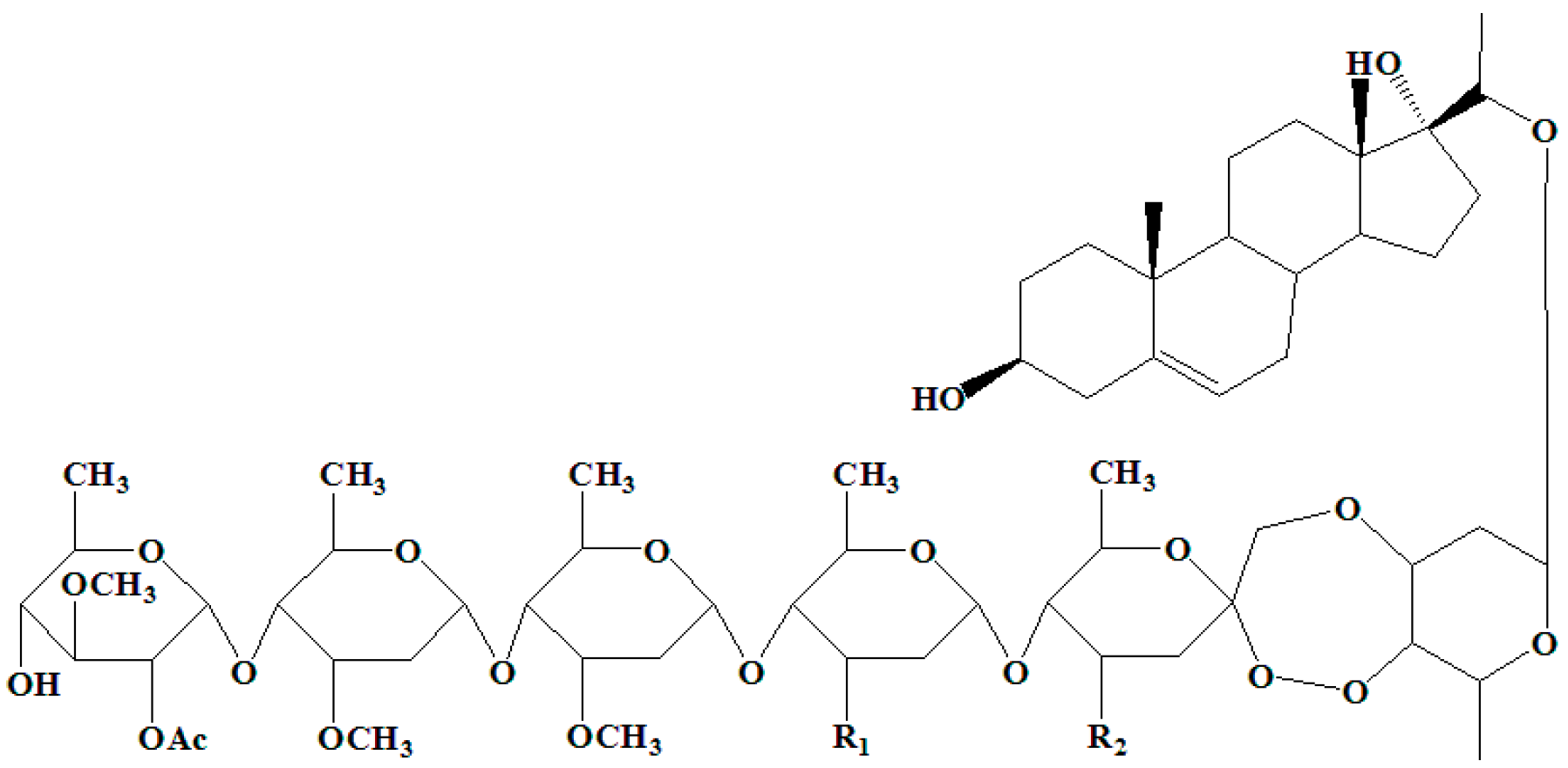
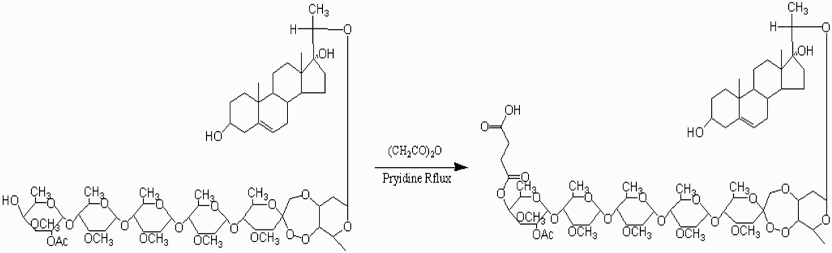
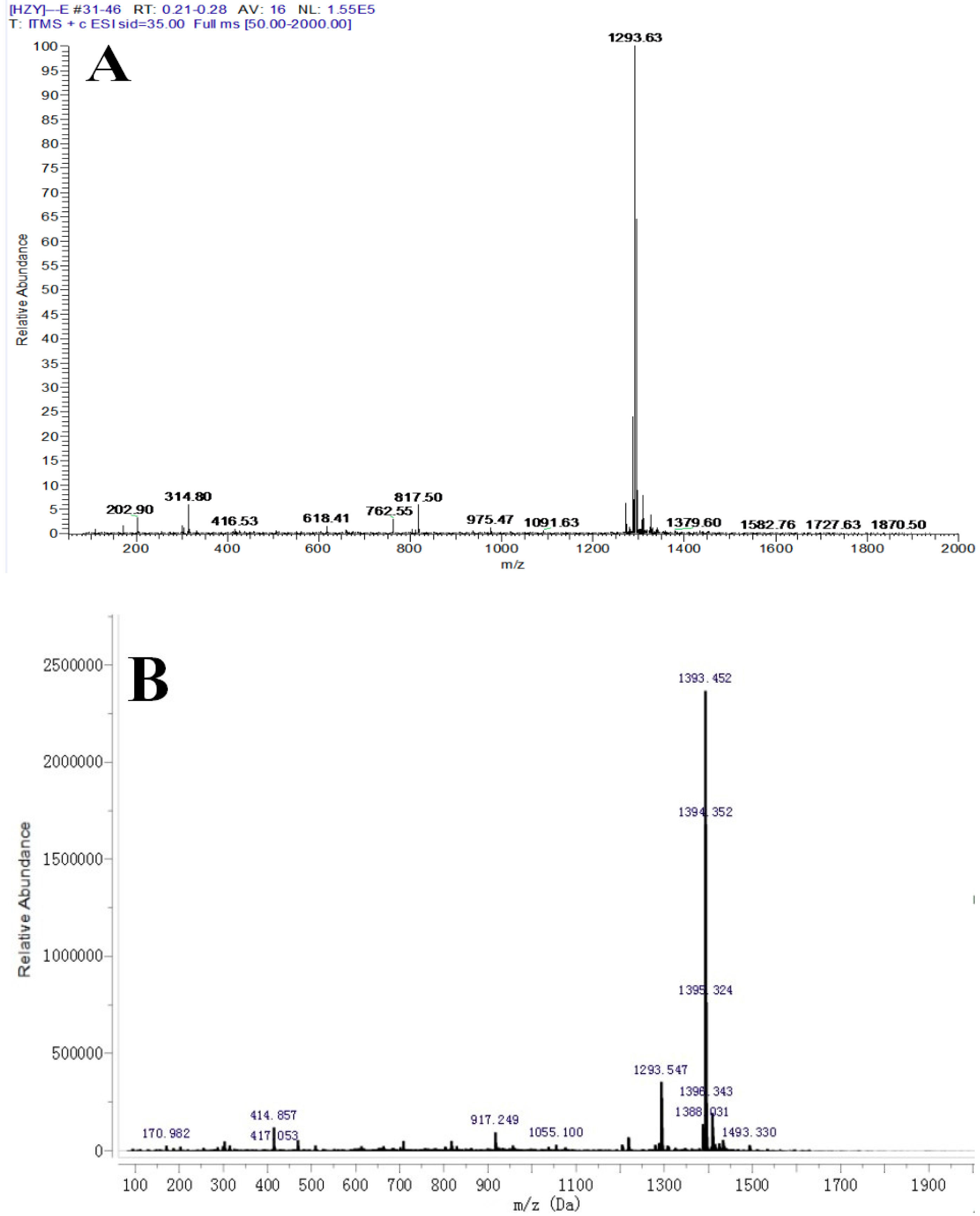
2.1. Identification of the Ligand (Periplocoside E-Semi-Succinic Acid Ester)

The ligand, periplocoside E-semi-succinic acid ester, was synthesized (Figure 2) and identified by electrospray ionization-mass spectrometry (ESI-MS) and nuclear magnetic resonance (NMR) analysis.
According to ESI-MS identification, the quasi-molecular ion peaks [M + Na] + of periplocoside E was located at m/z 1293.48 (Figure 3A), and the quasi-molecular ion peak [M + Na] + of periplocoside E-semi-succinic acid ester was found at m/z 1393.56 (Figure 3B). The ESI-MS value of periplocoside E-semi-succinic acid ester is consistent with the theoretical value. Further analysis by NMR identification showed that periplocoside E-semi-succinic acid ester (Figure 4B) contains four more lines than that of periplcoside E (Figure 4A). The NMR spectra of these lines are δC 174.59, 173.35, 28.39, and 28.34, respectively. Among these lines, δC 174.59 and 173.35 represent two carbonyl carbon atoms in the succinic anhydride structure, and δC 28.39 and 28.34 are assigned to two methylene carbon atoms. Hence, periplocoside E-semi-succinic acid ester was successfully synthesized.

2.2. Isolation of Binding Proteins

Our preliminary results revealed that the BBMVs from the midgut epithelium cells of M. separata larvae possess the initial action site of periplocosides [11]. Therefore, the BBMVs of M. separata larvae was isolated and subjected to affinity chromatography to separate the binding proteins of periplocosides. Two fractions, namely, Fractions 1 (Phosphate buffer (PB), unbound protein eluted with a binding buffer) and 2 (I, eluted with periplocoside E dissolved in the binding buffer) were collected (Figure 5A).
SDS-PAGE (Figure 5B) and LC/Q-TOF-MS analysis was conducted to resolve and evaluate proteins eluted by excess periplocoside E. Table 1 shows the binding proteins obtained from the NCBI database.
Table 1 shows that eight binding proteins of periplocoside E (luciferin 4-monooxygenase, aminopeptidase N, aminopeptidase N3, NADH dehydrogenase subunit 5, phosphatidylinositol 3-phosphate 3-phosphatase myotubularin, actin, uncharacterized family 31 glucosidase KIAA1161, and 2OG-Fe(2) oxygenase superfamily protein) were obtained from the M. separata midgut through affinity chromatography. Based on previous observations of symptoms, a preliminary research mechanism, the function of the obtained proteins, and the determination results of enzyme activity (data not published), we speculate that aminopeptidase N and aminopeptidase N3 are likely to be the putative target of periplocosides.
Symptoms of insect poisoning by periplocosides are characterized by a cessation of feeding and apparent abdomen swelling [8]. Further ultrastructural observation showed that microvilli on midgut epithelial cells are severely damaged [10]. We hypothesized that periplocosides may disrupt microvilli on the epithelial cells of the midgut of insects by interacting with proteins on the microvillar membrane or those associated with the membrane; these interactions may affect the normal functions of insects. Similar to periplocosides, the Bt toxin [14] and celangulins [15] act on the midgut of insects. Thus, periplocosides may have the same target protein as that of the Bt toxin and celangulins.
Research on the Bt toxin indicated that aminopeptidase N (APN) [16], cadherin-like protein[17], alkaline phosphatase (ALP) [18], and actin [19] are the receptors of the detoxification function. V-ATPase is a potential putative target protein of celangulins [20]. Through comparison with the receptors of the Bt toxin and celangulins, we found actin and aminopeptidase N (APN) in the present research. Actin is an abundant protein in eukaryotic cells [21]. Actin’s main function is to form microfilaments to maintain cell morphology; actin also exhibits other multiple functions, such as cell motility, cell division, and organelle movement [22,23]. Many of these processes are mediated by extensive and intimate interactions of actin and cellular membranes [24]. However, in terms of selectivity, actin has less possibility of becoming a target protein of pesticides because of its highly conservative nature in insect cells. Further studies on actin must be performed to exclude and validate this conjecture.
Aminopeptidase-N (APN) is an exopeptidase that is abundant in the BBMVs of many lepidopteran and dipteran insects. APN catalyzes the removal of N-terminal neutral amino acids [25]. APN is a common component of lepidoptera larval midgut and is used as a marker enzyme to assess the purity of BBMVs from insect larval midguts [26]. APN belongs to the family of zinc-dependent enzyme peptide with the zinc finger structure and N-glycosylation sites (Asn-X-Ser/Thr). The phosphatidyl inositol signal sequence is located in the C-terminal, and the signal peptide sequence is situated in the N-terminal. APN is connected to BBMVs in the insect midgut through the glycosylated phosphatidylinositol [27].
The most studied and reported aspect of APN is the Bt toxin. APN is the first toxin receptor protein that plays a role in the mechanism of the Bt toxin [16]. Knight et al. [28] and Gill et al. [29] cloned a 120-kD receptor-related protein gene combined with the Cry1Ac of Manduca sexta and Heliothis virescen; these studies applied the method of cDNA library and showed that APN is the encoding protein. Moreover, binding experiments showed that APN is the receptor of Lymantria dispar, Plutella xylostella, and Trichoplusia ni combined with Cry1Ac [30,31]. Gill et al. [32] indicated that the APN of Manduca sexta in the midguts of Drosophila melanogaster larvae is not sensitive to Cry1Ac; they found that this structure led to the presence of D. melanogaster larvae, which is sensitive to Cry1Ac. Furthermore, multiple aminopeptidase-N protein sequence alignments showed that the first 40 residues of human, rat, pig, and mouse enzymes are characterized by signal sequence, which may function as a membrane anchor [33,34]. Some reports showed that human and pig coronaviruses also use aminopeptidase N as receptor in their target tissues [35,36]. APN has a long history of existence as a target receptor.
In conclusion, the eight proteins isolated through affinity chromatography are potential binding proteins that interact with periplocosides. Aminopeptidase-N may be the target protein of periplocosides because it induces a series of symptoms to destroy the digestive system. However, further in-depth studies are needed to qualify and validate whether APN is the receptor of periplocosides. The presence of periplocosides in the midgut of insects, which leads to the death of the insect, should be evaluated using the interaction of periplocosides with APN. This work will serve as an important guide in elucidating the mechanism of periplocosides.

3. Conclusions

Eight potential binding proteins of periplocoside E were isolated from the midgut BBMVs of M. separata larvae through affinity chromatography. These proteins include luciferin 4-monooxygenase, aminopeptidase N, aminopeptidase N3, NADH dehydrogenase subunit 5, phosphatidylinositol 3-phosphate 3-phosphatase myotubularin, actin, uncharacterized family 31 glucosidase KIAA1161, and 2OG-Fe(2) oxygenase superfamily protein. The functions of some of the important proteins linked to the symptoms caused by periplocosides were also analyzed. We speculate that aminopeptidase N (APN) is the putative target protein of periplocosides. However, further studies are needed to verify the accuracy of this assumption.

4. Materials and Methods

4.1. Insects

The fifth instar larvae of M. separata were provided by the Institute of Pesticide Science, Northwest A & F University (NWAFU) (Yangling, China). A colony of M. separata was cultured in the laboratory for 20 years at 25 °C, 70% relative humidity, and a photoperiod of 16 h of light, with 8 h of darkness under periodic introduction of field-collected insects.

4.2. Chemicals

Periplocoside E was extracted and purified from P. sepium Bunge (root barks) in the Institute of Pesticide Science, Northwest A & F University (NWAFU, Yangling, China) [37]. The chemical structure of PS-E is shown in Figure 1. PS-E has a purity of over 98% based on high performance liquid chromatography (HPLC) analysis. EAH sepharose 4B was purchased from general electronics (GE) Healthcare (Beijing, China). Tris, NaCl, HCl, acetic acid, sodium acetate, and all other chemicals were purchased from Solarbio (Beijing, China).

4.3. Preparation of Brush Border Membrane Vesicles (BBMV)

The fifth instar larvae of M. separata Walker were starved for 12 h before the experiment. The midguts were then dissected. Peritrophic matrix and gut contents were removed. The remaining tissues were washed in an ice-cold 0.7% NaCl solution. The cleaned tissue was dried on filter paper, weighed, and used to separate the BBMVs according to the differential magnesium precipitation method with some modifications [38]. The final BBMV pellet was dissolved in an ice-cold dissolution buffer (150 mM NaCl, 5 mM EGTA, 1 mM PMSF, 20 mM Tris-HCl, and 1% CHAPS). Protein concentration was quantified by Bradford Assay, and the prepared BBMVs were stored at −80 °C until analysis.

4.4. Synthesis of Ligand

The ligand, periplocoside E-semi-succinic acid ester, was synthesized using periplocoside E and succinic anhydride (Figure 2).
Briefly, 100 mg of periplocoside E (0.08 mmol) and 100 mg of succinic anhydride (1 mmol) were placed in a reaction vessel and dissolved in 20 mL of anhydrous pyridine. The mixture was reacted at 120 °C through a heating reflux method. Subsequently, 20 mL of methanol was added to terminate the completed reaction (surveillance by thin layer chromatography (TLC)). The solvent was evaporated at 60 °C by using rotary evaporators (Thermo Fisher, Beijing, China). The remaining dry powder was dissolved in methyl alcohol and further separated by column chromatography. The structure was characterized by MS and 13C-NMR and MS analyses.

4.5. Preparation of the Medium and Coupling of the Ligand

The medium, namely, epoxy amino hexyl (EAH)-activated sepharose 4B, was prepared according to the operation manual. The lyophilized powder of EAH Sepharose 4B (2.5 g) was sufficiently swelled in 100 mL of 20% ethanol. When the medium swelled, the ethanol solution was decanted, and the required amount of the matrix was washed five times on a glass filter (porosity G3) with distilled water (pH 4.5). The matrix was subjected to sedimentation using 80 mL of 0.5 mol/L NaCl.
The carbodiimide method was used for ligand coupling. Periplocoside E-semi-succinic acid ester (ligand) was dissolved in the coupling solution (50% ethylene glycol solution) and added to the matrix (matrix:ligand = 1:1) to form a suspension. Briefly, 0.1 mol/L carbodiimide was added to the suspension. The superfluous medium was eluted with three cycles of an alternating pH elution buffer (buffer 1: 0.1 mol/L acetic acid/sodium acetate containing 0.5 mol/L NaCl, pH 4.0; buffer 2: 0.1 mol/L Tris-HCl containing 0.5 mol/L NaCl, pH 8.0). The volumes of each elution buffer were at least five times the medium volumes.

4.6. Affinity Chromatography

The prepared EAH sepharose 4B was packed to the column of an AKTA protein purification system (GE Healthcare, Beijing, China). The packing flow rate was maintained for three bed volumes after reaching a constant bed height was reached.
After loading the BBMVs of the M. separata, the medium was washed with the binding buffer (0.1 mol/L NaH2PO4/Na2HPO4, pH 7.4 containing 0.5 mol/L NaCl) until the base line become stable. For competitive elution, periplocoside E was dissolved in the binding buffer. The elution peak was collected by washing the medium with the elution buffer. The affinity medium was then washed with an alternating high pH (0.1 mol/L Tris-HCl, pH 8.0 containing 0.5 mol/L NaCl) and low pH (0.1 mol/L acetic acid/ sodium acetate, pH4.0 containing 0.5 mol/L NaCl) buffer solution to regenerate the medium. This cycle was repeated three times.

4.7. SDS-PAGE and LC/Q-TOF-MS Analysis

The collected binding protein solution was dialyzed to decrease its high salt concentration. The samples were then lyophilized. SDS-PAGE (12% separating gel and 5% spacer gel) was conducted by dissolving the sample in the Tris-HCl (pH 7.2). The whole visible lane was obtained for protein MS identification. Protein MS was implemented in Beijing Protein Institute (Beijing, China) by using LC/Q-TOF-MS.

Acknowledgments

This study was partly sponsored by the Grants of the National Natural Science Foundation of China (31171868), the National Key Basic Research Program (973 Program, 2010CB126100) for Science and Technology, Ministry of China and Special Fund for Agro-scientific Research in the Public Interest (200903052).

Author Contributions

The experiments were conceived and designed by Zhaonong Hu and Wenjun Wu. The experiments were performed by Mingxing Feng, Zhenyu He, Yuanyuan Wang and Xiufang Yan. Data was analyzed by Mingxing Feng, Zhenyu He, and Jiwen Zhang. The paper was written by Mingxing Feng and Zhaonong Hu.

Conflicts of Interest

The authors declare no conflict of interest.

References

  1. Senthil-Nathan, S. A review of biopesticides and their mode of action against insect pests. In Environmental Sustainability; Springer: New Delhi, India, 2015; pp. 49–63. [Google Scholar]
  2. Mouhssen, L. Screening of natural products for drug discovery. Expert Opin. Drug Discov. 2007, 2, 697–705. [Google Scholar]
  3. Zhang, K.; Pugliese, M.; Pugliese, A.; Passantino, A. Biological active ingredients of traditional Chinese herb astragalus membranaceus on treatment of diabetes: A systematic review. Mini Rev. Med. Chem. 2015, 15, 315–329. [Google Scholar] [PubMed]
  4. Chen, B.; Butte, A.J. Leveraging big data to transform target selection and drug discovery. Clin. Pharmacol. Ther. 2016, 99, 285–297. [Google Scholar] [CrossRef] [PubMed]
  5. Li, T.X.; Zhang, C.Y.; Li, G.H.; Li, Q.H.; Zhang, Y.J. Ecological and biological characteristics of Periploca sepium. Drugs Clin. 2013, 28, 730–733. [Google Scholar]
  6. Tokiwa, T.; Harada, K.; Matsumura, T.; Tukiyama, T. Oriental medicinal herb, Periploca sepium, extract inhibits growth and IL-6 production of human synovial fibroblast-like cells. Biol. Pharm. Bull. 2004, 27, 1691–1693. [Google Scholar] [CrossRef] [PubMed]
  7. Li, Y.; Zhang, J.W.; Yang, H.; Li, J.H.; Qian, Y.; Wu, W.J. Studies on insecticidal constituents from Periploca sepium Bunge. Acta Boreali-Occident. Sin. 2006, 15, 620–623. [Google Scholar]
  8. Li, Y.; Zeng, X.N.; Wang, W.Z.; Luo, C.H.; Yan, Q.; Tian, M. Chemical constituents from the roots of Periploca sepium with insecticidal activity. J. Asian Nat. Prod. Res. 2012, 14, 811–816. [Google Scholar] [CrossRef] [PubMed]
  9. Feng, M.X.; Zhao, J.; Zhang, J.W.; Hu, Z.N.; Wen, W.J. Fluorescence localization and comparative ultrastructural study of periplocoside NW from Periploca sepium Bunge in the midgut of the Oriental Amyworm, Mythimna separata Walker (Lepidoptera: Noctuidae). Toxins 2014, 6, 1575–1585. [Google Scholar] [CrossRef] [PubMed]
  10. Zuo, J.N.; Sun, W.C.; Feng, M.X.; Hu, Z.N.; Wen, W.J. Immuno-electron microscopic localization research of periplocoside T from Periploca sepium Bunge on midgut of larvae in the Oriental Amyworm, Mythimna separata Walker (Lepidoptera: Noctuidae). J. Northwest A F Univ. 2015, 44, 118–122. [Google Scholar]
  11. Feng, M.X.; Shi, B.J.; Zhao, Y.C.; Hu, Z.N.; Wen, W.J. Histopathological effects and immunolocalization of periplocoside NW from Periploca sepium Bunge on the midgut epithelium of Mythimna separata Walker larvae. Pestic. Biochem. Physiol. 2014, 115, 67–72. [Google Scholar] [CrossRef] [PubMed]
  12. Kool, J.; Jonker, N.; Irth, H.; Niessen, W.M.A. Studying protein-protein affinity and immobilized ligand-protein affinity interactions using MS-based methods. Anal. Bioanal. Chem. 2011, 401, 1109–1125. [Google Scholar] [CrossRef] [PubMed]
  13. Gao, L.T. Separation and Preparation of Polyclonal Antibody of Perplocoside T; Northwest A & F University: Yangling, China, 2012. [Google Scholar]
  14. Dirk, D.; Fereshte, A.S. Botulinum toxin: Mechanisms of action. Eur. Neurol. 2005, 63, 3–9. [Google Scholar]
  15. Yang, R.Y.; Liu, H.X.; Hu, Z.N.; Zhang, L.S.; Tian, X.M. Study on the mechanism of action of celangulin V. J. Northwest Agric. Univ. 2000, 28, 18–21. [Google Scholar]
  16. Knight, P.J.K.; Crickmore, N.; Ellar, D.J. The receptor for Bacillus thuringiensis CrylA(c) delta-endotoxin in the brush border membrane of the lepidopteran Manduca sexta is aminopeptidase N. Mol. Microbiol. 1994, 11, 429–436. [Google Scholar] [CrossRef] [PubMed]
  17. Francis, B.R.; Bulla, L.A. Further characterization of BT-R1, the cadherin-like receptor for Cry1Ab toxin in tobacco hornworm (Manduca sexta) Midguts. Insect Biochem. Mol. Biol. 1997, 27, 541–550. [Google Scholar] [CrossRef]
  18. Chen, W.B.; Liu, C.X.; Xiao, Y.T.; Zhang, D.D.; Zhang, Y.D.; Li, X.C.; Tabashnik, B.E.; Wu, K.M. A toxin-binding alkaline phosphatase fragment synergizes Bt toxin Cry1Ac against susceptible and resistant Helicoverpa armigera. PLoS ONE 2015, 10, e0126288. [Google Scholar] [CrossRef] [PubMed]
  19. Vega-Cabrera, A.; Cancino-Rodezno, A.; Porta, H.; Pardo-Lopez, L. Aedes aegypti Mos20 cells internalizes cry toxins by endocytosis, and actin has a role in the defense against Cry11Aa toxin. Toxins 2014, 6, 464–487. [Google Scholar] [CrossRef]
  20. Lu, L.N.; Qi, Z.J.; Zhang, J.W.; Wu, W.J. Separation of binding protein of Celangulin V from the midgut of Mythimna separata Walker by affinity chromatography. Toxins 2015, 7, 1738–1748. [Google Scholar] [CrossRef] [PubMed]
  21. Mounier, N.; Sparrw, J.C. Muscle actin genes in insects. Comp. Biochem. Physiol. Part B 1993, 105, 231–238. [Google Scholar] [CrossRef]
  22. Doherty, G.J.; McMahon, H.T. Mediation, modulation, and consequences of membrane-cytoskeleton interactions. Annu. Rev. Biophys. 2008, 37, 65–95. [Google Scholar] [CrossRef] [PubMed]
  23. Pollard, T.D.; Cooper, J.A. Actin, a central player in cell shape and movement. Science 2009, 326, 1208–1212. [Google Scholar] [CrossRef] [PubMed]
  24. Rescher, U.; Ruhe, D.; Ludwig, C.; Zobiack, N.; Gerke, V. Annexin 2 is a phosphatidylinositol (4,5)-bisphosphate binding protein recruited to actin assembly sites at cellular membranes. J. Cell Sci. 2004, 117, 3473–3480. [Google Scholar] [CrossRef] [PubMed]
  25. Hafkenscheid, J.C.M. Aminopeptidases and amino acid arylamidases. Methods Enzym. Anal. 1984, 5, 2–34. [Google Scholar]
  26. Wolfersberger, M.G. Enzymology of plasma membranes of insect intestinal cells. Am. Zool. 1984, 24, 187–197. [Google Scholar] [CrossRef]
  27. Taylor, A. Aminopeptidases: Structure and function. FASEB J. 1993, 7, 290–298. [Google Scholar] [PubMed]
  28. Knight, P.J.K.; Carroll, J.; Ellar, D.J. Analysis of glycan structures on the 120 kDa aminopeptidase N of Manduca sexta and their interactions with Bacillus thuringiensis Cry1Ac toxin. Insect Biochem. Mol. Biol. 2004, 34, 101–112. [Google Scholar] [CrossRef] [PubMed]
  29. Gill, S.S.; Cowles, E.A.; Francis, V. Identification, isolation, and cloning of a Bacillus thuringiensis CryIAc toxin-binding protein from the midgut of the lepidoptera insect Heliothis virescens. J. Biol. Chem. 1995, 270, 27277–27282. [Google Scholar] [CrossRef] [PubMed]
  30. Valaitis, A.P.; Mi, K.L.; Rajamohan, F.; Dean, D.H. Brush border membrane aminopeptidase-N in the midgut of the gypsy moth serves as the receptor for the CryIA (c) δ-endotoxin of Bacillus thuringiensis. Insect Biochem. Mol. Biol. 1995, 25, 1143–1151. [Google Scholar] [CrossRef]
  31. Lee, M.K.; You, T.H.; Young, B.A.; Cotrill, J.A.; Valaitis, A.P.; Dean, D.H. Aminopeptidase N purified from gypsy moth brush border membrane vesicles is a specific receptor for Bacillus thuringiensis CryIAc toxin. Appl. Environ. Microbiol. 1996, 62, 2845–2849. [Google Scholar] [PubMed]
  32. Gill, M.; Ellar, D. Transgenic Drosophila reveals a functional in vivo receptor for the Bacillus thuringiensis toxin Cry1Ac1. Insect Mol. Biol. 2002, 11, 619–625. [Google Scholar] [CrossRef] [PubMed]
  33. Semenza, G. Anchoring and biosynthesis of stalked brush border membrane proteins: Glycosidases and peptidases of enterocytes and renal tubuli. Annu. Rev. Cell Biol. 1986, 2, 255–307. [Google Scholar] [CrossRef] [PubMed]
  34. Gierasch, L.M. Signal sequences. Biochemistry 1989, 28, 923–930. [Google Scholar] [CrossRef] [PubMed]
  35. Delmas, B.; Gelfi, J.; L’Haridon, R.; Vogel, L.K.; Sjöström, H.; Norén, O.; Laude, H. Aminopeptidase N is a major receptor for the entero-pathogenic coronavirus TGEV. Nature 1992, 357, 417–420. [Google Scholar] [CrossRef] [PubMed]
  36. Yeager, C.L.; Ashmun, R.A.; Williams, R.K.; Cardellichio, C.B.; Shapiro, L.H.; Look, A.T.; Holmes, K.V. Human aminopeptidase N is a receptor for human coronavirus 229E. Nature 1992, 357, 420–422. [Google Scholar] [CrossRef] [PubMed]
  37. Shi, B.J. Studies on the Insecticidal Constituent of the Root Bark of Periploca sepium and Tripterygium hypoglaucum; Northwest A & F University: Yangling, China, 2009. [Google Scholar]
  38. Wolfersberger, M.; Luethy, P.; Maurer, A.; Parenti, P.; Sacchi, F.V.; Giordana, B.; Hanozet, G.M. Preparation and partial characterization of amino acid transporting brush border membrane vesicles from the larval midgut of the cabbage butterfly (Pieris brassicae). Comp. Biochem. Physiol. Part A 1987, 86, 301–308. [Google Scholar] [CrossRef]
Figure 1. Structure of periplocoside E, P, and T. Periplocoside E: R1 = OCH3, R2 = OCH3. Periplocoside P: R1 = OH, R2 = OCH3. Periplocoside T: R1 = OH, R2 = OH.
Toxins 08 00139 g001
Figure 2. Synthetic route of periplocoside E-semi-succinic acid ester.
Toxins 08 00139 g002
Figure 3. Mass spectrum (MS) spectrum of periplocoside E (A) and periplocoside E-semi-succinic acid ester (B).
Toxins 08 00139 g003
Figure 4. Nuclear magnetic resonance (NMR) spectra of periplocoside E (A) and periplocoside E-semi-succinic acid ester (B).
Toxins 08 00139 g004
Figure 5. Affinity chromatography using periplocoside E-Sepharose of brush-border membrane vesicle (BBMV) proteins extracted from M. separata larvae. (A) Phosphate buffer (PB) is the unbound protein eluted with a binding buffer; I is eluted with periplocoside E dissolved in the binding buffer; (B) 12% sodium dodecyl sulfonate-polyacrylamide gelelectrophoresis (SDS-PAGE) of fractions; M: protein marker.
Toxins 08 00139 g005
Table 1. Binding proteins of periplocoside E from M. separata midgut recognized by affinity chromatography.
NumberProtein IDProtein MassProtein
1gi|51291809363095Luciferin 4-monooxygenase
2gi|15212557114719Aminopeptidase N
3gi|21327773107153Aminopeptidase N3
4gi|25380766266764Nicotinamide adenine dinucleotide health (NADH) dehydrogenase subunit 5
5gi|52083688778123Phosphatidylinositol 3-phosphate 3-phosphatase myotubularin
6gi|32343532631100Actin
7gi|51290112773062Uncharacterized family 31 glucosidase KIAA1161
8gi|399942986860252OG-Fe(2) oxygenase superfamily protein

