Article

Helicobacter pylori vacA Genotypes in Chronic Gastritis and Gastric Carcinoma Patients from Macau, China

1
i3S—Instituto de Investigação e Inovação em Saúde, Universidade do Porto, Porto 4200-135, Portugal
2
Ipatimup—Institute of Molecular Pathology and Immunology of the University of Porto, Porto 4200-135, Portugal
3
Centro Hospitalar Conde São Januário, Macau, China
4
Department of Pathology and Oncology, Faculty of Medicine of the University of Porto, Porto 4200-319, Portugal
5
Department of Pathology, Centro Hospitalar São João, Porto 4200-319, Portugal
*
Author to whom correspondence should be addressed.
These authors contributed equally to this work.
Toxins 2016, 8(5), 142; https://doi.org/10.3390/toxins8050142
Submission received: 28 February 2016 / Revised: 11 April 2016 / Accepted: 29 April 2016 / Published: 5 May 2016
(This article belongs to the Special Issue Vacuolating Toxin)

Abstract

:
Helicobacter pylori is the major triggering factor for gastric carcinoma, but only a small proportion of infected patients develop this disease. Differences in virulence observed among H. pylori strains, namely in the vacuolating cytotoxin vacA gene, may contribute to this discrepancy. Infection with vacA s1, i1 and m1 strains increases the risk for progression of gastric premalignant lesions and for gastric carcinoma. However, in East Asian countries most of the H. pylori strains are vacA s1, regardless of the patients’ clinical status, and the significance of the vacA i1 and m1 genotypes for gastric carcinoma in this geographic area remains to be fully elucidated. The aim of the present study was to investigate this relationship in 290 patients from Macau, China. Using very sensitive and accurate genotyping methods, we detected infection with vacA i1 and with vacA m1 strains in, respectively, 85.2% and 52.6% of the patients that were infected with single genotypes. The prevalence of cagA-positive strains was 87.5%. No significant associations were observed between vacA genotypes or cagA and gastric carcinoma. It is worth noting that 37.5% of the infected patients had coexistence of H. pylori strains with different vacA genotypes. Additional studies directed to other H. pylori virulence factors should be performed to identify high risk patients in East Asia.

Graphical Abstract

1. Introduction

Gastric carcinoma is the fifth most incident and the third most common cause of cancer-related death worldwide. Half of the cases have occurred in East Asian countries, mainly in China, which has the highest estimated mortality rates (24 per 100,000 in men, 9.8 per 100,000 in women) [1].
The major risk factor for the development of gastric carcinoma is persistent Helicobacter pylori infection [2]. H. pylori induces chronic superficial gastritis that may evolve to chronic atrophic gastritis, intestinal metaplasia, dysplasia and finally to gastric carcinoma [3]. Only a small number of infected individuals will progress towards carcinoma, and it has been shown that disease development depends on the interplay between host genetic susceptibility, environmental factors, and bacterial virulence factors [4,5,6].
H. pylori strains are genetically very heterogeneous and genetic variation within genes encoding virulence factors contributes to differences in the pathogenicity of strains [7]. One of the most studied H. pylori virulence-associated genes is cagA, encoding the CagA effector protein that is translocated into the host cells by a type IV secretion system [8,9]. In Western countries, approximately 70% of the H. pylori strains contain the cagA gene, and infection with this type of strain has been associated with increased risk for gastric atrophy, intestinal metaplasia and gastric carcinoma [4,10,11]. In East Asian countries, however, the prevalence of cagA-positive strains is higher than 90%, making it difficult to disclose its impact in gastric carcinoma [12,13].
The vacA gene, encoding the vacuolating cytotoxin A (VacA), is another H. pylori virulence-associated gene that exhibits variation among strains [14]. Although vacA is present in all H. pylori strains, it shows allelic variation in three main regions: the signal (s)-region that contains the s1 (s1a, s1b, and s1c) or s2 alleles, the intermediate (i)-region that contains the i1 or i2 alleles, and the middle (m)-region that contains the m1 or m2 (m2a and m2b) alleles [15,16,17]. Different combinations of vacA s, i and m alleles may occur, resulting in VacA toxins with distinct abilities to induce vacuolation in epithelial cells [15]. It has been shown that while vacA s1/m1 strains are vacuolating and vacA s2/m2 are non-vacuolating, the vacA s1/m2 strains may or not induce vacuolation depending on the i-region genotype. Therefore, vacA s1/m2 strains that contain the i1 allele are vacuolating, whereas strains that contain the i2 allele are non-vacuolating [15,16,18]. Numerous studies have shown that in Western countries, H. pylori vacA s1 and m1 strains are associated with enhanced gastric mucosal inflammation and with increased risk for atrophy, intestinal metaplasia and carcinoma in comparison with vacA s2 and m2 strains [11,19,20]. Additional studies including patients from Western countries also found associations between infection with strains containing the vacA i1 allele and increased risk for progression of gastric precancerous lesions and for gastric carcinoma [18,21,22,23].
In East Asian countries, however, the relationship between H. pylori vacA s-region genotypes and gastric carcinoma has been difficult to ascertain, as virtually all strains are vacA s1 [12,13]. In contrast, the vacA m-region appears to be more variable among East Asian strains, with the m1 allele predominating in Japan and South Korea, and the m2 allele predominating in Southeast Asia, including countries such as Vietnam [24]. Few studies have evaluated the clinical significance of the vacA i region in gastric carcinoma in East Asia [25,26], namely in China. Therefore, we investigated the relationship between H. pylori vacA i- and m-region genotypes and gastric carcinoma development in patients living in Macau, China.

2. Results

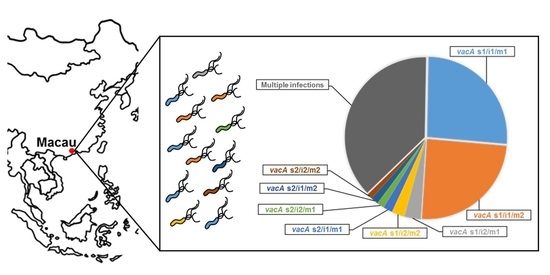
2.1. H. pylori vacA Genotypes and cagA Status

Two hundred and eighty-one (96.9%) from a total of 290 patients included in the study were positive for H. pylori as shown by PCR.
The characterization of the vacA i-region was obtained in 272 (96.8%) of H. pylori-infected patients, in which 236 (86.8%) patients were infected with a single strain, while the remaining 36 (13.2%) were infected with multiple H. pylori strains. Of the patients with single infections, 201 (85.2%) were infected with vacA i1 strains and the remaining 35 (14.8%) patients were infected with i2 strains (Table 1).
Characterization of the vacA m-region was successful in all but one case. Two hundred and eleven (75.4%) samples contained a single allele: 111 (52.6%) were genotyped as m1 and 100 (47.4%) as m2 (Table 1); 93.0% of the m2 strains were of the m2a subtype, and the remaining 7.0% of strains were m2b. In 69 (24.6%) patients, more than one m allele was detected, and among these the m1/m2a combination was the most frequent (n = 65, 94.2%).
Approximately 95% of H. pylori-infected patients were successfully characterized for the vacA s-region. Multiple vacA s-region genotypes were observed in 27 (9.8%) patients, and within single infections the vacA s1 allele was detected in 227 (91.2%) cases and the s2 allele in 22 (8.8%) cases (Table 1). Most of the vacA s1 strains were of the s1c subtype (71.4%), while the s1a and the s1b alleles were detected in 33 (14.5%) and eight (3.5%) strains, respectively. In 24 (10.6%) samples, two vacA s subtypes were simultaneously detected: s1a/s1b (n = 4), s1a/s1c (n = 17), and s1b/s1c (n = 3), and these samples were considered as vacA s1 for the remaining analyses. Among 27 patients infected with multiple strains, the following combinations of s alleles were found: s1a/s2 (n = 6; 22.2%), s1c/s2 (n = 11; 40.8%); s1a/s1b/s2 (n = 2; 7.4%); and s1a/s1c/s2 (n = 8; 29.6%).
Considering all vacA regions together, the following genotype combinations were observed: s1/i1/m1 (n = 71, 41.8%), s1/i1/m2 (n = 67, 39.4%), s1/i2/m1 (n = 9, 5.3%), s1/i2/m2 (n = 7, 4.1%), s2/i1/m1 (n = 5, 2.9%), s2/i2/m1(n = 4, 2.4%), s2/i1/m2 (n = 4, 2.4%), and s2/i2/m2 (n = 3, 1.8%). It is noteworthy that a total of 102 (37.5%) patients were infected with multiple strains (Table 1).
The prevalence of cagA-positive strains was 87.5% (Table 1). Combining the vacA genotypes with the cagA status, 13 different genotypic combinations were found. The most prevalent ones were vacA s1/i1/m1/cagA-positive (n = 67, 39.4%) and vacA s1/i1/m2/cagA-positive (n = 64, 37.6%). The vacA s2/i2/m1/cagA-negative genotype was the least prevalent (n = 1, 0.6%), and vacA s2/i2/m2/cagA-negative strains were not found.

2.2. Relationship between vacA i-Region Genotypes, s- and m-Region Genotypes and cagA Status

Regarding the relationship between vacA genotypes and cagA, vacA i1 strains were frequently s1 (89.6%) and cagA-positive (90.9%), while vacA i2 strains were more commonly s2 (43.8%) and cagA-negative (56.2%; p = 0.002 and p < 0.001, respectively; Table 2). The prevalence of the i1 allele was similarly detected among m1 (85.4%) and m2 (87.7%) strains, and no relationship was observed between the genotypes of these vacA regions (Table 2).

2.3. Relationship between H. pylori Genotypes and Gastric Carcinoma

The 281 H. pylori-infected patients comprised 234 patients with chronic gastritis (mean age 58.0 ± 15.1 years; female:male ratio of 1.13:1) and 47 with gastric carcinoma (mean age 66.6 ± 14.1 years; male:female ratio of 2.4:1).
H. pylori vacA i1, s1, and m1, as well as cagA-positive strains, were found with similar frequencies in patients with chronic gastritis and patients with gastric carcinoma, and consequently, no significant relationships could be disclosed between vacA or cagA genotypes and gastric carcinoma (Table 1). Also, no significant differences were observed between vacA or cagA genotypes and degree of atrophy, presence of intestinal metaplasia, or dysplasia. Overall, 37.5% of the patients were infected with multiple strains as assessed by vacA genotyping. No relationship was observed between infection with single/multiple H. pylori strains and gastric carcinoma (Table 3).

3. Discussion

Gastric carcinoma is one of the most incident and deadly cancers worldwide. The incidence of gastric carcinoma is particularly high in East Asian countries, where the prevalence of H. pylori infection is also high [1,2,27,28]. H. pylori strain variation, especially that related to vacA and cagA virulence factor-encoding genes, has been proposed as a means to identify strains with the highest degrees of pathogenicity and, consequently, individuals with the highest risk of disease [29].
This study constitutes the largest case-control study in East Asia to address H. pylori virulence factor genotyping and gastric carcinoma. Here, we have characterized the H. pylori vacA i- and m-regions in Chinese patients with chronic gastritis and with gastric carcinoma from Macau. In the vacA i-region, the i1 allele was dominant and no differences in allelic prevalence were observed between chronic gastritis and gastric carcinoma patients. These results contrast with those of earlier studies in Iranian, Italian, Portuguese, Spanish, Belgian, and UK populations, where the vacA i1 allele has been associated with increased risk for atrophic gastritis, intestinal metaplasia, and gastric carcinoma [16,18,21,22,23,30]. However, our results in the Chinese population of Macau are consistent with those of studies that include patients from East Asian countries, namely Japan, South Korea, and China, and that report a prevalence of over 95% of the vacA i1 allele [25,26,31,32]. Therefore, although the vacA i-region genotyping is useful to identify patients at high risk of gastric carcinoma in Western countries, it may have limitations in East Asian countries.
We have observed a similar frequency distribution of the two vacA m-region alleles, with 52.6% of the strains in Macau being m1 and the remaining 47.4% being m2. Nevertheless, the frequency distribution of these alleles was similar in chronic gastritis and gastric carcinoma patients, and therefore no relationship between vacA m-region genotypes and gastric carcinoma could be established. It has been shown that the m-region genotypes have regional geographic variation between East Asian countries and it has been suggested that the m-region may play a role in the regional differences in gastric carcinoma incidence [24]. The patients from Japan and South Korea are mainly infected with vacA m1 strains [12,26], while m2 strains are more prevalent in patients from Vietnam or Hong Kong [33,34], possibly reflecting the higher incidence of gastric carcinoma in the former than in the latter. Within mainland China, a significant geographic diversity of the vacA m-region has also been also described, and the m1 allele frequency was higher in Guangxi, an area with a high gastric carcinoma incidence, than in Beijing [13]. Although these data are in favor of vacA m1 strains being more virulent than m2 strains, based on our data, the vacA m-region genotypes do not appear to be useful for identifying high-risk individuals in Macau.
Similarly to what was found in the vacA i-region, in the s-region the great majority of the strains in Macau contained the s1 allele, with s1c being the most prevalent subtype. Likewise, a very high frequency of cagA-positive strains was detected in this population. These observations concur with previous descriptions from Chinese, Korean, and Japanese populations showing a strong predominance of vacA s1 and cagA-positive strains [12,13,25,35,36]. Due to the high frequency of vacA s1 and cagA-positive strains in Macau, and in contrast with other geographic areas, including Western, Middle Eastern, Latin American, and African countries [19,20,37,38,39], no relationships with disease outcome could be disclosed.
While the great majority of H. pylori strains presented the most virulent genotypic combination of vacA s1/i1/cagA-positive, the less virulent vacA s2/i2/m2/cagA-negative strains were not found. An association between the vacA i1 and s1 genotypes and between the latter and the presence of cagA was observed in this study. The associations between these vacA loci and cagA have been reported both in Western and in other East Asian populations [18,21,26,31]. The reasons underlying these associations are not very clear. The vacA and cagA genes are located distantly in the chromosome, and therefore genotype association cannot be due to clonality, nor to genetic linkage, as H. pylori is highly recombinatorial [40,41]. Interestingly, it has been proposed that VacA and CagA interplay with each other with the purpose of regulating excessive damage promoted by each of the virulence factors individually, which would probably destroy the H. pylori niche [42].
The genotyping of vacA directly in gastric biopsy specimens allowed us to identify a high number of patients (37.5%) with coexisting strains with different vacA types, which were considered as multiple H. pylori infections. The prevalence of multiple infections has now been reported for numerous geographic locations [13,19,43,44,45,46], and in general it appears to be higher in countries where the risk for H. pylori infection is also high [47]. Whether the coexistence of strains with different vacA genotypes reflects the high prevalence of H. pylori in Macau or genotype adaptation to specific niches within a gastric biopsy remains to be disclosed. Nevertheless, the natural history and consequences of the acquisition of the infections with two (or more) strains requires further studies, as there may be clinical/therapeutic implications.
In conclusion, the great majority of the H. pylori strains found in Macau were vacA s1/i1/cagA-positive, independent of the clinical outcome of the infection. Therefore, these factors cannot be used for identifying high-risk patients in this geographic region. Other regions of variation in vacA, such as the d-region [25], in other H. pylori virulence-associated factors, namely in the cagA gene promoter [48,49] or EPIYA-encoding region [36,50], may represent alternative markers of H. pylori virulence in East Asian populations.

4. Materials and Methods

4.1. Patients and Study Population

A total of 290 paraffin-embedded gastric tissue specimens from patients living in Macau were characterized for H. pylori vacA genotypes and for the presence of the cagA gene. Samples included 238 cases of chronic gastritis (mean age 60.8 ± 14.2 years; female:male ratio of 1.1:1), comprising patients with superficial gastritis (n = 82) and atrophic gastritis (n = 156); and 52 cases of gastric carcinoma (mean age 66.6 ± 13.6 years; male:female ratio of 2.2:1). Within the atrophic gastritis group, 18 patients had dysplastic lesions (n = 15 had mild dysplasia and n = 3 had moderate dysplasia). Evidence of epithelial erosions/ulcerations was present in 79 cases with chronic gastritis. Patients were selected consecutively from the registry of Centro Hospitalar Conde de São Januário (CHCSJ), Macau, China. The study population was composed by Chinese (n = 282; 97.2%), Macanese (n = 6; 2.1%), and Asian (n = 2; 0.7%) living in Macau. Samples were delinked and unidentified from their donors. The study was approved by the institutional review board of CHCSJ, registry of entry no. 5875, approved on 18 August 2008.

4.2. Histopathology

Two gastric biopsy specimens from the antrum (one from the greater curvature and one from the incisura angularis) and one from the corpus were immersed in 10% formalin and embedded in paraffin. Sections were stained with hematoxylin and eosin, Alcian blue-periodic acid-Schiff, and modified Giemsa. Only cases with adequate sized biopsy specimens of both antral and corpus mucosa were accepted for histological assessment by an experienced pathologist, who was blinded with respect to the clinical information of each patient. The histopathological parameters were classified according to the criteria described in the updated Sydney’s classification system [51]. Gastric carcinoma cases were categorized according to Lauren’s and Carneiro’s classification [52,53].

4.3. DNA Isolation

H. pylori detection and characterization was performed in DNA extracted from paraffin-embedded gastric tissue. Cuts of 10 μm of formalin-fixed and paraffin-embedded blocks were obtained from each biopsy specimen, changing the blade after each block to avoid cross-contamination. Total DNA was isolated after digestion in a solution containing 10 mM Tris-HCl (pH 8.0), 5 mM EDTA, 0.1% sodium dodecyl sulphate (SDS), and 0.1 mg/mL proteinase K for at least 12 h at 55 °C. Proteinase K was inactivated by incubation for 10 min at 95 °C.
DNA isolation from H. pylori strains 60190 (ATCC 49503; vacA s1/i1/m1 and cagA-positive) and Tx30a (ATCC 51932; vacA s2/i2/m2 and cagA-negative) was performed using the GRS Genomic DNA kit-Bacteria (GRISP, Porto, Portugal), following the manufacturer’s instructions. DNAs from these strains were used as controls for genotyping.

4.4. H. pylori vacA Genotyping and cagA Gene Detection

H. pylori vacA i-region was genotyped by allele-specific polymerase chain reaction (PCR) using the forward primer VacIABF and the reverse primers C1R, and C2R, as previously reported [16,23]. PCR mixtures were prepared in a volume of 25 µL, containing 1x PCR buffer (Applied Biosystems, Foster City, CA, USA), 2.5 mM MgCl2 (Applied Biosystems), 1 mM concentrations of deoxynucleotide triphosphates (dNTP, Nzytech, Lisbon, Portugal), 0.5 U of AmpliTaq Gold (Applied Biosystems), and 0.5 µM concentrations of forward and reverse primers. PCR was performed with a 9 min predenaturation at 95 °C, followed by 45 cycles of 30 s at 95 °C, 45 s at 50 °C, and 45 s at 72 °C. Final extension was performed for 10 min at 72 °C. PCR of the human glyceraldehyde3-phosphate dehydrogenase (GAPDH) housekeeping gene was used to monitor the DNA isolation procedure. PCR products were electrophoresed on 2% agarose gels and examined under UV light, according to standard procedures. Genotyping of vacA s- and m-regions and detection of cagA were performed by PCR followed by reverse hybridization on a line probe assay (LiPA), as previously described [19,54]. Patients were considered to be infected with multiple H. pylori strains, unless otherwise stated, when vacA typing detected more than one allele at each of the s-, i-, or m-regions.

4.5. Statistical Analyses

Associations between vacA genotypes and cagA, and the relationships between genotypes and disease were assessed using the Fisher’s exact test. Age and gender frequencies between groups of patients were compared with Student’s t-test and with Fisher’s exact test, respectively.
Statistical analyses were computed with the software Statview for Windows (Version 5; SAS Institute, Cary, USA, 1998). Differences were considered to be statistically significant at p values < 0.05.

Acknowledgments

This study was funded by Fundo Europeu de Desenvolvimento Regional (FEDER) through Programa Operacional Factores de Competitividade (COMPETE) and by FCT—Fundação para a Ciência e a Tecnologia in the framework of Project FCOMP-01-0124-FEDER-021251 (PTDC/SAU-SAP/120024/2010). IPR and RMF are supported by FCT fellowships SFHRH/BD/110803/2015 and SFRH/BPD/84084/2012, respectively, and by Programa Operacional Capital Humano (POCH). We would like to acknowledge Che Sio Sin, Lao Ut and Tam Sze Shuen at the Department of Pathology of Centro Hospitalar Conde São Januário for technical support.

Author Contributions

C.F., F.C. and S.B. conceived and designed the study; S.B., T.H., S.I.W. and F.C. contributed to sample collection and to histopathological analyses; I.P.R. and R.M.F. performed the genotyping experiments; I.P.R., R.M.F., S.B. and CF analyzed the data; I.P.R., R.M.F. and C.F. wrote the paper.

Conflicts of Interest

The authors declare no conflict of interest.

References

  1. Ferlay, J.; Soerjomataram, I.; Ervik, M.; Dikshit, R.; Eser, S.; Mathers, C.; Rebelo, M.; Parkin, D.M.; Forman, D.; Bray, F. Globocan 2012 v1.0, Cancer Incidence and Mortality Worldwide: Iarc Cancerbase no. 11; International Agency for Research on Cancer: Lyon, France, 2013; Available online: http://globocan.iarc.fr (accessed on 14 March 2016).
  2. Polk, D.B.; Peek, R.M., Jr. Helicobacter pylori: Gastric cancer and beyond. Nat. Rev. Cancer 2010, 10, 403–414. [Google Scholar] [CrossRef] [PubMed]
  3. Correa, P. Human gastric carcinogenesis: A multistep and multifactorial process—First american cancer society award lecture on cancer epidemiology and prevention. Cancer Res. 1992, 52, 6735–6740. [Google Scholar] [PubMed]
  4. Figueiredo, C.; Machado, J.C.; Pharoah, P.; Seruca, R.; Sousa, S.; Carvalho, R.; Capelinha, A.F.; Quint, W.; Caldas, C.; van Doorn, L.J.; et al. Helicobacter pylori and interleukin 1 genotyping: An opportunity to identify high-risk individuals for gastric carcinoma. J. Natl. Cancer Inst. 2002, 94, 1680–1687. [Google Scholar] [CrossRef] [PubMed]
  5. El-Omar, E.M.; Rabkin, C.S.; Gammon, M.D.; Vaughan, T.L.; Risch, H.A.; Schoenberg, J.B.; Stanford, J.L.; Mayne, S.T.; Goedert, J.; Blot, W.J.; et al. Increased risk of noncardia gastric cancer associated with proinflammatory cytokine gene polymorphisms. Gastroenterology 2003, 124, 1193–1201. [Google Scholar] [CrossRef]
  6. Machado, J.C.; Figueiredo, C.; Canedo, P.; Pharoah, P.; Carvalho, R.; Nabais, S.; Castro Alves, C.; Campos, M.L.; Van Doorn, L.J.; Caldas, C.; et al. A proinflammatory genetic profile increases the risk for chronic atrophic gastritis and gastric carcinoma. Gastroenterology 2003, 125, 364–371. [Google Scholar] [CrossRef]
  7. Dorer, M.S.; Talarico, S.; Salama, N.R. Helicobacter pylori’s unconventional role in health and disease. PLoS Pathog. 2009, 5, e1000544. [Google Scholar] [CrossRef] [PubMed]
  8. Odenbreit, S.; Puls, J.; Sedlmaier, B.; Gerland, E.; Fischer, W.; Haas, R. Translocation of helicobacter pylori caga into gastric epithelial cells by type iv secretion. Science 2000, 287, 1497–1500. [Google Scholar] [CrossRef] [PubMed]
  9. Backert, S.; Tegtmeyer, N.; Fischer, W. Composition, structure and function of the helicobacter pylori cag pathogenicity island encoded type iv secretion system. Fut. Microbiol. 2015, 10, 955–965. [Google Scholar] [CrossRef] [PubMed]
  10. Peek, R.M., Jr.; Miller, G.G.; Tham, K.T.; Perez-Perez, G.I.; Zhao, X.; Atherton, J.C.; Blaser, M.J. Heightened inflammatory response and cytokine expression in vivo to caga+ helicobacter pylori strains. Lab. Investig. J. Tech. Methods Pathol. 1995, 73, 760–770. [Google Scholar]
  11. Nogueira, C.; Figueiredo, C.; Carneiro, F.; Gomes, A.T.; Barreira, R.; Figueira, P.; Salgado, C.; Belo, L.; Peixoto, A.; Bravo, J.C.; et al. Helicobacter pylori genotypes may determine gastric histopathology. Am. J. Pathol. 2001, 158, 647–654. [Google Scholar] [CrossRef]
  12. Yamaoka, Y.; Kodama, T.; Gutierrez, O.; Kim, J.G.; Kashima, K.; Graham, D.Y. Relationship between helicobacter pylori icea, caga, and vaca status and clinical outcome: Studies in four different countries. J. Clin. Microbiol. 1999, 37, 2274–2279. [Google Scholar] [PubMed]
  13. Wang, J.; van Doorn, L.J.; Robinson, P.A.; Ji, X.; Wang, D.; Wang, Y.; Ge, L.; Telford, J.L.; Crabtree, J.E. Regional variation among vaca alleles of helicobacter pylori in china. J. Clin. Microbiol. 2003, 41, 1942–1945. [Google Scholar] [CrossRef] [PubMed]
  14. Cover, T.L.; Tummuru, M.K.; Cao, P.; Thompson, S.A.; Blaser, M.J. Divergence of genetic sequences for the vacuolating cytotoxin among helicobacter pylori strains. J. Biol. Chem. 1994, 269, 10566–10573. [Google Scholar] [PubMed]
  15. Atherton, J.C.; Cao, P.; Peek, R.M., Jr.; Tummuru, M.K.; Blaser, M.J.; Cover, T.L. Mosaicism in vacuolating cytotoxin alleles of helicobacter pylori. Association of specific vaca types with cytotoxin production and peptic ulceration. J. Biol. Chem. 1995, 270, 17771–17777. [Google Scholar] [PubMed]
  16. Rhead, J.L.; Letley, D.P.; Mohammadi, M.; Hussein, N.; Mohagheghi, M.A.; Eshagh Hosseini, M.; Atherton, J.C. A new helicobacter pylori vacuolating cytotoxin determinant, the intermediate region, is associated with gastric cancer. Gastroenterology 2007, 133, 926–936. [Google Scholar] [CrossRef] [PubMed]
  17. van Doorn, L.J.; Figueiredo, C.; Sanna, R.; Pena, S.; Midolo, P.; Ng, E.K.; Atherton, J.C.; Blaser, M.J.; Quint, W.G. Expanding allelic diversity of helicobacter pylori vaca. J. Clin. Microbiol. 1998, 36, 2597–2603. [Google Scholar] [PubMed]
  18. Ferreira, R.M.; Machado, J.C.; Letley, D.; Atherton, J.C.; Pardo, M.L.; Gonzalez, C.A.; Carneiro, F.; Figueiredo, C. A novel method for genotyping the helicobacter pylori vaca intermediate region directly in gastric biopsy specimens. J. Clin. Microbiol. 2012, 50, 3983–3989. [Google Scholar] [CrossRef] [PubMed]
  19. Gonzalez, C.A.; Figueiredo, C.; Lic, C.B.; Ferreira, R.M.; Pardo, M.L.; Ruiz Liso, J.M.; Alonso, P.; Sala, N.; Capella, G.; Sanz-Anquela, J.M. Helicobacter pylori caga and vaca genotypes as predictors of progression of gastric preneoplastic lesions: A long-term follow-up in a high-risk area in spain. Am. J. Gastroenterol. 2011, 106, 867–874. [Google Scholar] [CrossRef] [PubMed]
  20. Matos, J.I.; de Sousa, H.A.; Marcos-Pinto, R.; Dinis-Ribeiro, M. Helicobacter pylori caga and vaca genotypes and gastric phenotype: A meta-analysis. Eur. J. Gastroenterol. Hepatol. 2013, 25, 1431–1441. [Google Scholar] [CrossRef] [PubMed]
  21. Basso, D.; Zambon, C.F.; Letley, D.P.; Stranges, A.; Marchet, A.; Rhead, J.L.; Schiavon, S.; Guariso, G.; Ceroti, M.; Nitti, D.; et al. Clinical relevance of helicobacter pylori caga and vaca gene polymorphisms. Gastroenterology 2008, 135, 91–99. [Google Scholar] [CrossRef] [PubMed]
  22. Memon, A.A.; Hussein, N.R.; Miendje Deyi, V.Y.; Burette, A.; Atherton, J.C. Vacuolating cytotoxin genotypes are strong markers of gastric cancer and duodenal ulcer-associated helicobacter pylori strains: A matched case-control study. J. Clin. Microbiol. 2014, 52, 2984–2989. [Google Scholar] [CrossRef] [PubMed]
  23. Ferreira, R.M.; Figueiredo, C.; Bonet, C.; Pardo, M.L.; Liso, J.M.; Alonso, P.; Sala, N.; Capella, G.; Sanz-Anquela, J.M.; Gonzalez, C.A. Helicobacter pylori vaca intermediate region genotyping and progression of gastric preneoplastic lesions. Am. J. Gastroenterol. 2012, 107, 145–146. [Google Scholar] [CrossRef] [PubMed]
  24. Yamaoka, Y. Mechanisms of disease: Helicobacter pylori virulence factors. Nat. Rev. Gastroenterol. Hepatol. 2010, 7, 629–641. [Google Scholar] [CrossRef] [PubMed]
  25. Ogiwara, H.; Sugimoto, M.; Ohno, T.; Vilaichone, R.K.; Mahachai, V.; Graham, D.Y.; Yamaoka, Y. Role of deletion located between the intermediate and middle regions of the helicobacter pylori vaca gene in cases of gastroduodenal diseases. J. Clin. Microbiol. 2009, 47, 3493–3500. [Google Scholar] [CrossRef] [PubMed]
  26. Jang, S.; Jones, K.R.; Olsen, C.H.; Joo, Y.M.; Yoo, Y.J.; Chung, I.S.; Cha, J.H.; Merrell, D.S. Epidemiological link between gastric disease and polymorphisms in vaca and caga. J. Clin. Microbiol. 2010, 48, 559–567. [Google Scholar] [CrossRef] [PubMed]
  27. Azevedo, N.F.; Guimaraes, N.; Figueiredo, C.; Keevil, C.W.; Vieira, M.J. A new model for the transmission of helicobacter pylori: Role of environmental reservoirs as gene pools to increase strain diversity. Crit. Rev. Microbiol. 2007, 33, 157–169. [Google Scholar] [CrossRef] [PubMed] [Green Version]
  28. Uemura, N.; Okamoto, S.; Yamamoto, S.; Matsumura, N.; Yamaguchi, S.; Yamakido, M.; Taniyama, K.; Sasaki, N.; Schlemper, R.J. Helicobacter pylori infection and the development of gastric cancer. N. Engl. J. Med. 2001, 345, 784–789. [Google Scholar] [CrossRef] [PubMed]
  29. Ferreira, R.M.; Machado, J.C.; Figueiredo, C. Clinical relevance of helicobacter pylori vaca and caga genotypes in gastric carcinoma. Best Pract. Res. Clin. Gastroenterol. 2014, 28, 1003–1015. [Google Scholar] [CrossRef] [PubMed]
  30. Winter, J.A.; Letley, D.P.; Cook, K.W.; Rhead, J.L.; Zaitoun, A.A.; Ingram, R.J.; Amilon, K.R.; Croxall, N.J.; Kaye, P.V.; Robinson, K.; et al. A role for the vacuolating cytotoxin, vaca, in colonisation and helicobacter pylori-induced metaplasia in the stomach. J. Infect. Dis. 2014, 210, 954–963. [Google Scholar] [CrossRef] [PubMed]
  31. Chung, C.; Olivares, A.; Torres, E.; Yilmaz, O.; Cohen, H.; Perez-Perez, G. Diversity of vaca intermediate region among helicobacter pylori strains from several regions of the world. J. Clin. Microbiol. 2010, 48, 690–696. [Google Scholar] [CrossRef] [PubMed]
  32. Kim, J.Y.; Kim, N.; Nam, R.H.; Suh, J.H.; Chang, H.; Lee, J.W.; Kim, Y.S.; Kim, J.M.; Choi, J.W.; Park, J.G.; et al. Association of polymorphisms in virulence factor of helicobacter pylori and gastroduodenal diseases in south korea. J. Gastroenterol. Hepatol. 2014, 29, 984–991. [Google Scholar] [CrossRef] [PubMed]
  33. Yamaoka, Y.; Orito, E.; Mizokami, M.; Gutierrez, O.; Saitou, N.; Kodama, T.; Osato, M.S.; Kim, J.G.; Ramirez, F.C.; Mahachai, V.; et al. Helicobacter pylori in north and south america before columbus. FEBS Lett. 2002, 517, 180–184. [Google Scholar] [CrossRef]
  34. Wong, B.C.; Yin, Y.; Berg, D.E.; Xia, H.H.; Zhang, J.Z.; Wang, W.H.; Wong, W.M.; Huang, X.R.; Tang, V.S.; Lam, S.K. Distribution of distinct vaca, caga and icea alleles in helicobacter pylori in hong kong. Helicobacter 2001, 6, 317–324. [Google Scholar] [CrossRef] [PubMed]
  35. Pan, Z.J.; van der Hulst, R.W.; Feller, M.; Xiao, S.D.; Tytgat, G.N.; Dankert, J.; van der Ende, A. Equally high prevalences of infection with caga-positive helicobacter pylori in chinese patients with peptic ulcer disease and those with chronic gastritis-associated dyspepsia. J. Clin. Microbiol. 1997, 35, 1344–1347. [Google Scholar] [PubMed]
  36. Sahara, S.; Sugimoto, M.; Vilaichone, R.K.; Mahachai, V.; Miyajima, H.; Furuta, T.; Yamaoka, Y. Role of helicobacter pylori caga epiya motif and vaca genotypes for the development of gastrointestinal diseases in southeast asian countries: A meta-analysis. BMC Infect. Dis. 2012, 12, 223. [Google Scholar] [CrossRef] [PubMed]
  37. Sugimoto, M.; Yamaoka, Y. The association of vaca genotype and helicobacter pylori-related disease in latin american and african populations. Clin. Microbiol. Infect. 2009, 15, 835–842. [Google Scholar] [CrossRef] [PubMed]
  38. Sugimoto, M.; Zali, M.R.; Yamaoka, Y. The association of vaca genotypes and helicobacter pylori-related gastroduodenal diseases in the middle east. Eur. J. Clin. Microbiol. Infect. Dis. 2009, 28, 1227–1236. [Google Scholar] [CrossRef] [PubMed]
  39. Plummer, M.; van Doorn, L.J.; Franceschi, S.; Kleter, B.; Canzian, F.; Vivas, J.; Lopez, G.; Colin, D.; Munoz, N.; Kato, I. Helicobacter pylori cytotoxin-associated genotype and gastric precancerous lesions. J. Natl. Cancer Inst. 2007, 99, 1328–1334. [Google Scholar] [CrossRef] [PubMed]
  40. Alm, R.A.; Ling, L.S.; Moir, D.T.; King, B.L.; Brown, E.D.; Doig, P.C.; Smith, D.R.; Noonan, B.; Guild, B.C.; deJonge, B.L.; et al. Genomic-sequence comparison of two unrelated isolates of the human gastric pathogen helicobacter pylori. Nature 1999, 397, 176–180. [Google Scholar] [CrossRef] [PubMed]
  41. Salama, N.; Guillemin, K.; McDaniel, T.K.; Sherlock, G.; Tompkins, L.; Falkow, S. A whole-genome microarray reveals genetic diversity among helicobacter pylori strains. Proc. Natl. Acad. Sci. USA 2000, 97, 14668–14673. [Google Scholar] [CrossRef] [PubMed]
  42. Argent, R.H.; Thomas, R.J.; Letley, D.P.; Rittig, M.G.; Hardie, K.R.; Atherton, J.C. Functional association between the helicobacter pylori virulence factors vaca and caga. J. Med. Microbiol. 2008, 57, 145–150. [Google Scholar] [CrossRef] [PubMed]
  43. Figueiredo, C.; Van Doorn, L.J.; Nogueira, C.; Soares, J.M.; Pinho, C.; Figueira, P.; Quint, W.G.; Carneiro, F. Helicobacter pylori genotypes are associated with clinical outcome in portuguese patients and show a high prevalence of infections with multiple strains. Scand. J. Gastroenterol. 2001, 36, 128–135. [Google Scholar] [CrossRef] [PubMed]
  44. Morales-Espinosa, R.; Castillo-Rojas, G.; Gonzalez-Valencia, G.; Ponce de Leon, S.; Cravioto, A.; Atherton, J.C.; Lopez-Vidal, Y. Colonization of mexican patients by multiple helicobacter pylori strains with different vaca and caga genotypes. J. Clin. Microbiol. 1999, 37, 3001–3004. [Google Scholar] [PubMed]
  45. Wong, B.C.; Wang, W.H.; Berg, D.E.; Fung, F.M.; Wong, K.W.; Wong, W.M.; Lai, K.C.; Cho, C.H.; Hui, W.M.; Lam, S.K. High prevalence of mixed infections by helicobacter pylori in hong kong: Metronidazole sensitivity and overall genotype. Aliment. Pharmacol. Ther. 2001, 15, 493–503. [Google Scholar] [CrossRef] [PubMed] [Green Version]
  46. Ben Mansour, K.; Fendri, C.; Battikh, H.; Garnier, M.; Zribi, M.; Jlizi, A.; Burucoa, C. Multiple and mixed helicobacter pylori infections: Comparison of two epidemiological situations in tunisia and france. Infect. Genet. Evolut. 2016, 37, 43–48. [Google Scholar] [CrossRef] [PubMed]
  47. Krebes, J.; Didelot, X.; Kennemann, L.; Suerbaum, S. Bidirectional genomic exchange between helicobacter pylori strains from a family in coventry, united kingdom. Int. J. Med. Microbiol. IJMM 2014, 304, 1135–1146. [Google Scholar] [CrossRef] [PubMed]
  48. Loh, J.T.; Shaffer, C.L.; Piazuelo, M.B.; Bravo, L.E.; McClain, M.S.; Correa, P.; Cover, T.L. Analysis of caga in helicobacter pylori strains from colombian populations with contrasting gastric cancer risk reveals a biomarker for disease severity. Cancer Epidemiol. Biomark. Prev. 2011, 20, 2237–2249. [Google Scholar] [CrossRef] [PubMed]
  49. Ferreira, R.M.; Pinto-Ribeiro, I.; Wen, X.; Marcos-Pinto, R.; Dinis-Ribeiro, M.; Carneiro, F.; Figueiredo, C. Helicobacter pylori caga promoter region sequences influence caga expression and interleukin 8 secretion. J. Infect. Dis. 2016, 213, 669–673. [Google Scholar] [CrossRef] [PubMed]
  50. Mueller, D.; Tegtmeyer, N.; Brandt, S.; Yamaoka, Y.; De Poire, E.; Sgouras, D.; Wessler, S.; Torres, J.; Smolka, A.; Backert, S. C-src and c-abl kinases control hierarchic phosphorylation and function of the caga effector protein in western and east asian helicobacter pylori strains. J. Clin. Investig. 2012, 122, 1553–1566. [Google Scholar] [CrossRef] [PubMed]
  51. Dixon, M.F.; Genta, R.M.; Yardley, J.H.; Correa, P. Classification and grading of gastritis. The updated sydney system. International workshop on the histopathology of gastritis, houston 1994. Am. J. Surg. Pathol. 1996, 20, 1161–1181. [Google Scholar] [CrossRef] [PubMed]
  52. Carneiro, F.; Seixas, M.; Sobrinho-Simoes, M. New elements for an updated classification of the carcinomas of the stomach. Pathol. Res. Pract. 1995, 191, 571–584. [Google Scholar] [CrossRef]
  53. Lauren, P. The two histological main types of gastric carcinoma: Diffuse and so-called intestinal-type carcinoma. An attempt at a histo-clinical classification. Acta Pathol. Microbiol. Scand. 1965, 64, 31–49. [Google Scholar] [PubMed]
  54. Van Doorn, L.J.; Figueiredo, C.; Rossau, R.; Jannes, G.; van Asbroek, M.; Sousa, J.C.; Carneiro, F.; Quint, W.G. Typing of helicobacter pylori vaca gene and detection of caga gene by pcr and reverse hybridization. J. Clin. Microbiol. 1998, 36, 1271–1276. [Google Scholar] [PubMed]
Table 1. H. pylori vacA genotypes and cagA status in patients from Macau, China.
Chronic Gastritis (n = 234)Gastric Carcinoma (n = 47)p-ValueTotal
vacA i-region 1
i1172 (85.1%)29 (85.3%)>0.999201 (85.2%)
i230 (14.9%)5 (14.7%)35 (14.8%)
vacA m-region 2
m194 (53.7%)17 (47.2%)0.583111 (52.6%)
m281 (46.3%)19 (52.8%)100 (47.4%)
vacA s-region 3
s1189 (91.7%)38 (88.4%)0.553227 (91.2%)
s217 (8.3%)5 (11.6%)22 (8.8%)
cagA status
positive208 (88.9%)38 (80.9%)0.146246 (87.5%)
negative26 (11.1%)9 (19.1%)35 (12.5%)
1 Nine cases (three gastritis and six carcinomas) could not be genotyped and multiple strains were detected in 36 cases (29 gastritis and seven carcinomas); 2 One carcinoma case could not be genotyped and multiple strains were detected in 69 cases (59 gastritis and 10 carcinomas); 3 Five cases of gastritis could not be genotyped and multiple strains were detected in 27 cases (23 gastritis and four carcinomas).
Table 2. Relationship between vacA i-region genotypes with s- and m-region genotypes, and with cagA status.
vacA i-RegionvacA s-RegionvacA m-RegioncagA Status
s1s2m1m2PositiveNegative
vacA i1138 (89.6%)9 (56.3%)76 (85.4%)71 (87.7%)140 (90.9%)7 (43.8%)
vacA i216 (10.4%)7 (43.8%)13 (14.6%)10 (12.3%)14 (9.1%)9 (56.2%)
p-value0.0020.823<0.001
Only samples completely genotyped and with single genotypes for the vacA s-, m- and i-regions are included.
Table 3. Relationship between single and multiple H. pylori infections and gastric carcinoma.
H. pyloriChronic GastritisGastric Carcinomap-ValueTotal
Single infections 1142 (62.0%)28 (65.1%)0.735170 (62.5%)
Multiple infections 287 (38.0%)15 (34.9%)102 (37.5%)
1 Nine samples could not be genotyped; 2 As evaluated by the presence of more than one allele at each of the vacA s-, i-, or m-regions.

