Article

Impact of a Single Oral Acute Dose of Aflatoxin B1 on Liver Function/Cytokines and the Lymphoproliferative Response in C57Bl/6 Mice

by
Angélica Tieme Ishikawa
1,*,
Elisa Yoko Hirooka
2,
Paula Leonello Alvares e Silva
1,
Ana Paula Frederico Rodrigues Loureiro Bracarense
3,
Karina Keller Marques da Costa Flaiban
3,
Claudia Yuri Akagi
1,
Osamu Kawamura
4,
Marcio Carvalho da Costa
5 and
Eiko Nakagawa Itano
1,*
1
Department of Pathological Sciences, State University of Londrina, P.O. Box 10.011, Londrina 86057-970, Paraná, Brazil
2
Department of Food Science and Technology, State University of Londrina, P.O. Box 10.011, Londrina 86057-970, Paraná, Brazil
3
Department of Preventive Veterinary Medicine, State University of Londrina, P.O. Box 10.011, Londrina 86051-970, Paraná, Brazil
4
Food Hygiene Laboratory, Faculty of Agriculture, Kagawa University, Miki-cho 761-0795, Kagawa, Japan
5
Department of Veterinary Biomedicine, University of Montreal, 3200 Rue Sicotte, St-Hyacinthe, QC J2S 2M2, Canada
*
Authors to whom correspondence should be addressed.
Toxins 2017, 9(11), 374; https://doi.org/10.3390/toxins9110374
Submission received: 16 October 2017 / Revised: 15 November 2017 / Accepted: 15 November 2017 / Published: 17 November 2017
(This article belongs to the Special Issue Impact of Human Metabolism on the Toxicological Effects of Mycotoxins)

Abstract

:
Aflatoxin B1 (AFB1), a mycotoxin found in food and feed, exerts harmful effects on humans and animals. The liver is the earliest target of AFB1, and its effects have been evaluated in animal models exposed to acute or chronic doses. Considering the possibility of sporadic ingestion of AFB1-contaminated food, this study investigated the impact of a single oral dose of AFB1 on liver function/cytokines and the lymphoproliferative response in mice. C57BL/6 mice were treated with a single oral AFB1 dose (44, 442 or 663 μg AFB1/kg of body weight) on the first day. Liver function (ALT, γ-GT, and total protein), cytokines (IL-4, IFN-γ, and IL-17), histopathology, and the spleen lymphoproliferative response to mitogens were evaluated on the 5th day. Although AFB1 did not produce any significant changes in the biochemical parameters, 663 μg AFB1/kg-induced hepatic upregulation of IL-4 and IFN-γ, along with liver tissue injury and suppression of the lymphoproliferative response to ConA (p < 0.05). In conclusion, a single oral dose of AFB1 exposure can induce liver tissue lesions, liver cytokine modulation, and immune suppression in C57BL/6 mice.

1. Introduction

Aflatoxins (AFs) are mycotoxins produced by Aspergillus flavus and A. parasiticus that contaminate agricultural commodities under harvest and post-harvest conditions. Human and animal health issues caused by ingestion of food contaminated with AFs are considered a permanent risk and a worldwide problem [1,2]. The mammalian liver, an early target of AFs, is the major drug-detoxifying organ, responsible for the metabolic activation and elimination of toxic components [3].
Among the AFs, aflatoxin B1 (AFB1) is generally predominant and is considered the most toxic analog [4]. Ingestion of AFB1-contaminated products can result in immunosuppressive, teratogenic, mutagenic, and carcinogenic effects. The International Agency for Research on Cancer classifies AFB1 as Group 1, i.e., carcinogenic to humans [5,6]. Furthermore, AFB1 exerts many immunotoxic effects, ranging from alterations in innate immunity or antigen-presenting cells [7,8,9] to changes in adaptive immunity, resulting in a reduced number of circulating lymphocytes, the inhibition of lymphocyte blastogenesis, and the alteration of cytokine expression in animals of various species [9,10].
In studies of the different effects and impact of AFB1 exposure, animals such as mice have been exposed to acute or chronic AFB1 doses for an extended period [11,12,13,14]. Considering the possibility of sporadic ingestion of AFB1-contaminated food, this study investigated the impact of a single oral dose of AFB1 (with low doses; <to ~1% of the median lethal dose (LD50)) on liver function/cytokines and the lymphoproliferative response in C57Bl/6 mice.

2. Results

2.1. Evaluation of the Effect of Aflatoxin B1 on Serum ALT, γ-GT, and Total Protein Levels

Oral administration of AFB1 did not produce any significant change in the ALT and γ-GT serum enzymes or protein levels (p > 0.05). Additionally, the vehicle group treated with saline:ethanol (95:5) presented similar results to those obtained for the water-treated controls (p > 0.05) (Table 1).
Means values within column with different superscript letters were statistically significant (p < 0.05), as determined through Tukey’s test. The reference values for mice serum enzymes are 23.00 ± 4.92 units/L of ALT, 7.57 ± 4.2 units/L of γ-GT, and of 5.07 ± 0.2 g/dL of total protein [15,16].

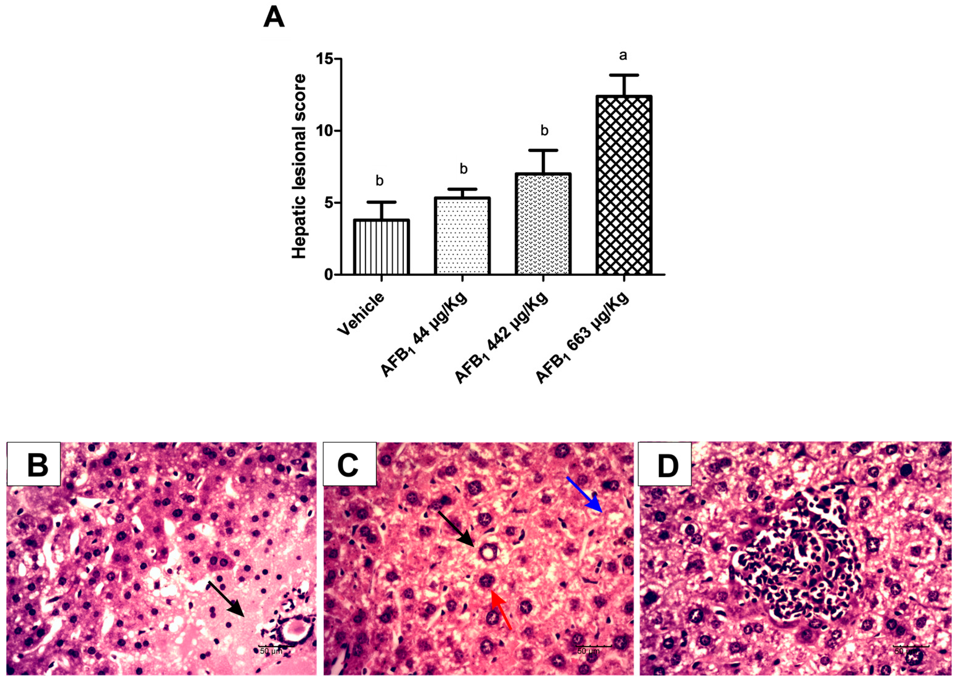
2.2. Evaluation of Aflatoxin B1 on Histological Lesions in Liver Tissue

Cytoplasmic hepatocyte vacuolation, megalocytosis, nuclear vacuolation, inflammatory infiltrate, and necrosis were present in the liver of treated animals; the last two lesions were more frequently observed. Lesions recorded in the liver were considered moderate to severe in mice treated with AFB1. A significant increase in the lesional score was observed in animals exposed to 663 μg of AFB1/kg of body weight (b.w.) compared with animals exposed to vehicle (p = 0.001 (Figure 1). Animals treated with 663 μg of AFB1/kg of b.w. showed more pronounced intensity of megalocytosis, nuclear vacuolation, and necrosis than animals treated with vehicle, whereas the main lesions in the animals treated with 44 and 442 μg of AFB1/kg of b.w. were necrosis.

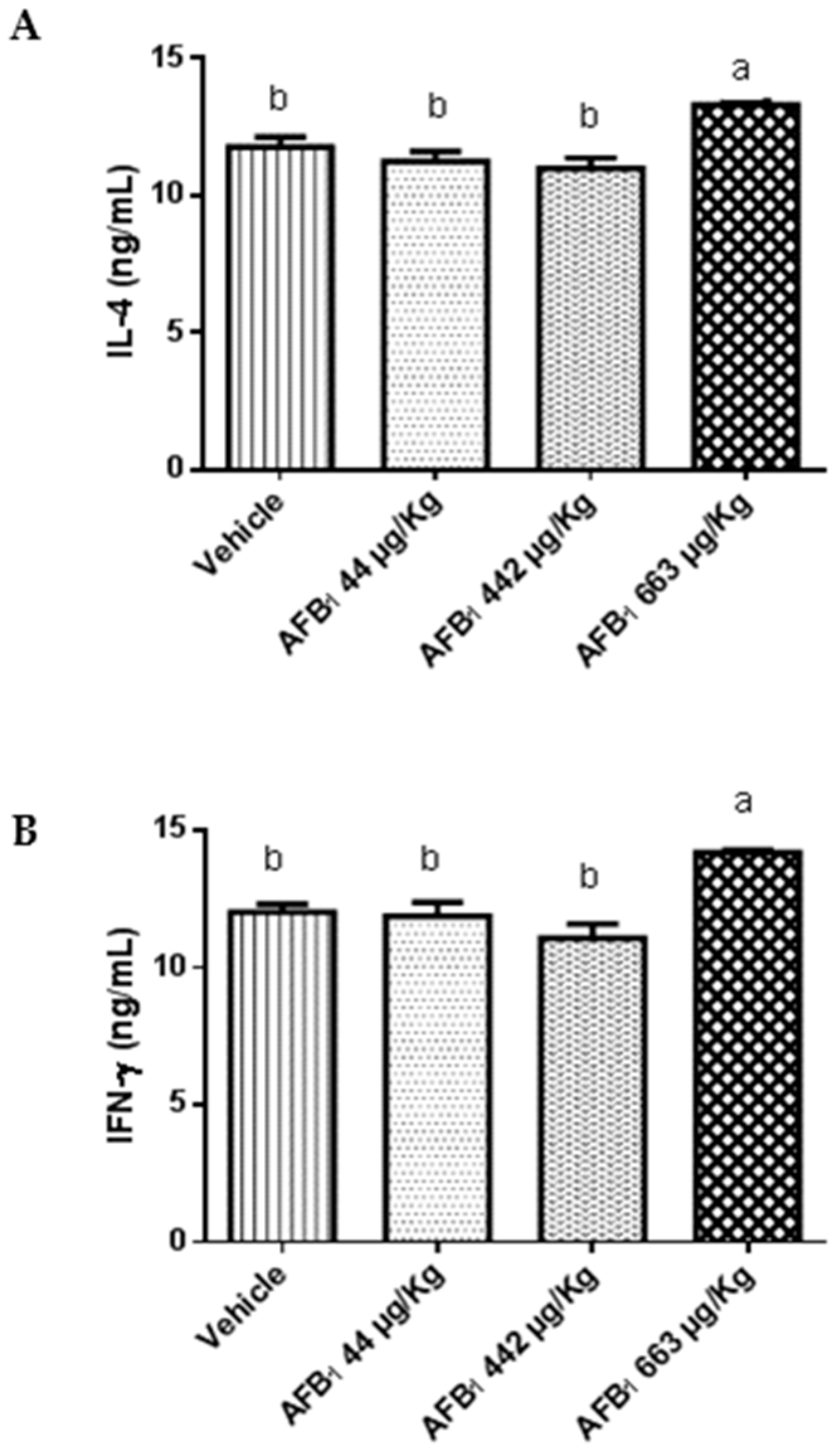
2.3. Effects of Aflatoxin B1 on Cytokine Expression in the Liver

Mice treated with 663 μg of AFB1/kg b.w. showed significant upregulation of IL-4 and IFN-γ (p = 0.002) compared with the vehicle group (Figure 2). There was no difference in the IL-17 cytokine levels between animals treated with AFB1 and untreated animals (p > 0.05). In contrast, there were differences between animals treated with 44 and 442 μg of AFB1/kg of b.w. and animals treated with 663 μg of AFB1/kg of b.w. (p < 0.05).

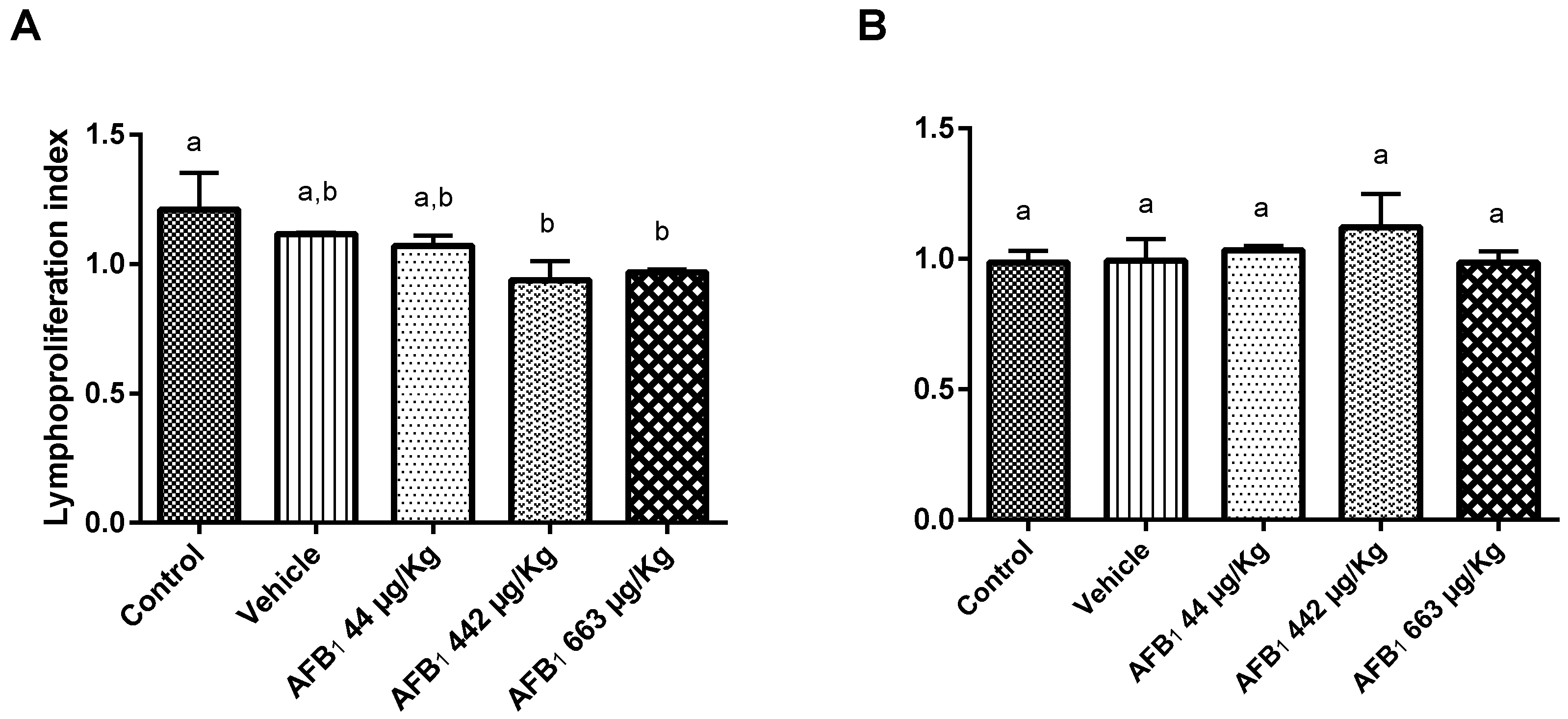
2.4. Effects of Aflatoxin B1 on Lymphoproliferation Assay

Significant suppression of the proliferative response was observed for concanavalin A (ConA)-stimulated lymphocytes under treatment with 442 and 663 μg AFB1/kg of b.w. (p < 0.05). The control animals showed a lymphoproliferation index of 1.21 ± 0.14, whereas the experimental animals treated with doses of 442 μg and 663 μg AFB1/kg of b.w. showed indexes of 0.94 ± 0.07 and 0.97 ± 0.01, respectively. However, no significant depression of the proliferative assay was observed in lipopolysaccharide (LPS)-stimulated lymphocytes when the data were compared with those of the control animals (Figure 3).

3. Discussion

Among mycotoxins, AFs are of major concern worldwide in terms of their risks to human and animal health. Their harmful effects have generally been demonstrated by administering repeated AF doses over long periods of exposure in animal models. In this study, we investigated the effects of exposure to single oral doses of AFB1 in C57Bl/6 mice. AFB1 did not have a major impact on the investigated hepatic biochemical parameters in the mice 5 days after administration. It is possible that the time of sampling was too long and the measurement of liver functions levels in earlier days was be different in animals treated with AFB1 compared to the control group. However, such results are in accordance with the results of a study conducted by Almeida et al. [15], who did not detect differences in the alkaline phosphatase levels in the serum of C57Bl/6 mice after 168 h of AF treatment (60 mg/kg animal weight). Moreover, our results are consistent with those observed by Baptista et al. [17], who fed albino rats 400 μg AFB1/kg of b.w. over 28 days and did not observe any significant differences in ALT, AST, ALP, or γ-GT enzyme activities or albumin levels.
The sensitivity degree and toxicity of AFB1 varies between species due to differences in its biotransformation. Some animals, such as sheep, dogs, pigs, and rats, are considered extremely susceptible to AFB1, whereas others, such as monkeys, chickens and mice, are considered resistant species [18]. The LD50 described in the literature for mice is variable, with values ranging from 9 to 60 mg of AFB1/kg of b.w. [15]. The doses of AFB1 used in this experiment (44, 442, and 663 μg/kg of b.w.) might appear high when applied to humans; however, aflatoxicosis cases have been reported to occur at similar or higher consumption levels of AFB1 [19,20].
Changes in the hepatic cellular architecture and organization were detected by histopathological analysis. The extent of liver damage was directly correlated with the concentration of AFB1 and the duration of the exposure [17]. In this study, a 7.7-fold increase in the lesional score was observed in AFB1-exposed animals. Notably, this study constitutes the first evaluation of the effects of a subclinical, single AFB1 exposure in mice, and the hepatic lesions observed here are consistent with those observed in other studies that used different doses or frequencies of exposure [21].
In the present study, upregulation of the production of the hepatic inflammatory cytokine IFN-γ, along with increased expression of the anti-inflammatory cytokine IL-4, were observed with higher doses of AFB1. There is no consensus regarding the cytokine responses induced by AF exposure [22,23,24]. Here, the difference obtained in anti- and pro-inflammatory cytokines levels might be due to time, a single and higher dose (663 μg of AFB1/kg of b.w.), or different organs and species. Because the evaluations performed in this study were at early stages (5 days) prior to the adaptive response, increases in IL-4 and IFN-γ expression were attributed to innate immune cell activation. Additionally, many innate immune cell populations produce IL-17 in response to stress, injury, or pathogens [25]. Our results indicate that IL-17 levels were significantly different among the highest and lowest doses of AFB1, but no differences were detected between the treated and control groups. Thus, even a single dose of AFB1 might induce hepatic cytokine immunomodulation, but whether all of the cytokines evaluated are involved in hepatic injury remains uncertain, and further studies are required.
AFB1 has a selective effect on cell-mediated immunity, with a relatively weak effect on the humoral immune system [21]. Consistently, in this study, we detected the inhibitory effects of AFB1 on ConA-stimulated lymphoblastogenesis (ConA is a lectin widely used as a polyclonal T-cell activator) but not on LPS-stimulated lymphoblastogenesis. Reddy & Sharma [26] reported the inhibitory effects of AFB1 on both LPS- and ConA-stimulated lymphoblastogenesis in animals exposed to low, repeated doses of AFB1. The difference in the LPS response detected in this investigation could be due to the use of a single dose instead of repeated doses.
According to a review by Peraica et al. [27], the acute hepatotoxic effects of AFs recorded in humans have mostly been observed among adults in rural populations with poor nutritional levels. The same authors cited the case of a young woman who ingested a total of 5.5 mg of AFB1 over 2 days, and whose laboratory examinations were normal and suggested that the hepatotoxicity of AFB1 might be lower in well-nourished persons.
In this study, the biochemical parameters of hepatic functions were not altered in mice exposed to AFB1. However, several other important parameters, such as liver tissue injury, cytokine levels, and cellular responses, were altered even with a single dose. Moreover, in a previous study, we verified that a single AFB1 exposure induces changes in the gut microbiota in C57Bl/6 mice [28].
It is important to investigate the effects of a single dose of AFs due to the possibility of sporadic ingestion of aflatoxin-contaminated food. In this study, harmful effects were observed even with one low oral dose of AFB1 (~1% of the median lethal dose (LD50) for mice [26]). These effects might be temporary but could contribute to exacerbation in cases of patients with liver disease or other diseases associated with immunosuppression, requiring further studies.

4. Conclusions

In conclusion, even a single, oral, low dose of AFB1 can induce liver tissue lesions, liver cytokine modulation and immune suppression in C57BL/6 mice.

5. Material and Methods

5.1. Aflatoxin B1 Standard, Dose Criteria, and Duration of Exposure

The AFB1 standard from Aspergillus flavus (A663, Sigma, St. Louis, MO, USA) was quantified according to the methods indicated by Instituto Adolfo Lutz [29]. The molar absorptivity of AFB1 considered for calculating AFB1 concentrations in methanol was 21,800 at 360 nm. The AFB1 solution was dried under gaseous N2, and the standard was dissolved in a 95:5 saline:ethanol solution for experiments.
The lowest AFB1 dose (44 μg/kg of b.w.) was calculated based on the maximum level allowed (5 μg/kg) in both rice and beans [30,31], according to the average bean (183 g/day) and rice (160 g/day) consumption levels, which are components of the traditional diet in Brazil [32]. The highest tested AFB1 dose (663 μg/kg of b.w.) was based on the chronic AFB1 dose used in other studies [13,33] and represents approximately 1% of the LD50 for mice. The duration of exposure was based on the plasma half-life of AFB1 in male rats, which is approximately 92 h [34].

5.2. Animals, Housing, and Experimental Design

A total of 25 male C57Bl/6 mice (10 weeks of age, average weight: 22.55 ± 0.89 g) were obtained from the University of São Paulo—Ribeirão Preto City, Brazil. C57Bl/6 mice were selected for this study because they are highly susceptible to the acute effects of aflatoxin B1 [15]. The mice were acclimatized for 3 weeks and housed in polyethylene boxes with a bedding of wood shavings. They were maintained under standard conditions, which included a temperature of approximately 25 °C with a regular 12 h light/12 h dark cycle. All mice were given standard rodent pellet food and water ad libitum.
The experimental design used in this study was randomized with five repetitions (each animal represented one repetition) for each group. Group 1 consisted of untreated control mice; Group 2 received only the vehicle (saline:ethanol, 95:5) on the first day; Group 3 received a single dose of 44 μg AFB1/kg b.w. on the first day; Group 4 received a single dose of 442 μg AFB1/kg b.w.; and Group 5 received a single dose of 663 μg AFB1/kg b.w. AFB1 suspended in saline:ethanol (95:5) was administered via oral gavage (0.1 mL per 10 g of body weight). After 5 days, the animals were bled and euthanized, and their organs (liver and spleen) were then removed.
Biochemical parameters were analyzed from the serum, and a lymphoproliferation assay was performed using splenocytes. This study was approved by Committee of Animal Ethics of State University of Londrina (CEUA n° 26362.2014.65 process, 18 December 2014).

5.3. Measurements of Liver Function from Serum

ALT and γ-GT enzymes and total protein levels in serum were evaluated with a Dimension® Clinical Chemistry System (Siemens, Newark, NJ, USA).

5.4. Histopathological Analysis

Liver samples were fixed in 10% buffered formalin solution, dehydrated in increasing alcohol concentrations, and embedded in paraffin for histological analysis. The tissue samples were sectioned at 5-μm thickness, stained with hematoxylin and eosin (HE), and mounted with coverslips. Histological changes were evaluated using an adapted tissue score based on the intensity and severity of lesions as previously described by Gerez et al. [35]. Briefly, the criteria used to establish the lesional score were hepatocyte megalocytosis, inflammatory infiltrate, hepatocyte nuclear vacuolation, hepatocyte cytoplasmic vacuolation, and necrosis. The extent of each lesion was scored as follows, megalocytosis (the mean of five fields per histological section): 1–10 = 0, 1–20 = 1+, 2–30 = 2+, and 3–40 = 3+; inflammatory infiltrate and nuclear vacuolation (histological section): absent = 0, 1 = 1+, and >2 = 2+; hepatic cell vacuolation (histological section): mild = 0, and moderate = 1+; and necrosis (histological section): absent = 0, mild = 1+, moderate = 2+, and severe = 3+. For each type of lesion, the score of the extent was multiplied by the severity factor [35].

5.5. Cytokine Assays

The liver samples were macerated in an adjusted volume of 0.1 M phosphate buffered saline (PBS), pH 7.4 (1:5000 dilution). The IL-4, IFN-γ, and IL-17 levels in the liver were determined by commercial sandwich ELISA kits (BioSource International, Inc., Camarillo, CA, USA), according to the manufacturer’s instructions.

5.6. Lymphoproliferation Assay

The spleens were removed from the mice aseptically, and their erythrocytes were lysed with Tris-ammonium chloride solution. In 96-well flat-bottom culture plates, 100 μL of the splenocytes (1 × 10⁵ cells/mL) from each mouse were cultured in duplicate wells in RPMI 1640 medium (containing l-glutamine and, 10% fetal calf serum), with 0.5 μg/mL LPS (Sigma, St. Louis, MO, USA) or with 0.5 μg/mL ConA (Gibco Life Technologies, Grand Island, NY, USA). The cells were cultured for 84 h at 37 °C with 5% CO2, and 100 μL of RPMI medium and 10 μL of 3-(4,5-dimethylthiazol-2-ly)-2,5-diphenyltetrazolium bromide (5 mg/mL MTT in PBS, Sigma, St. Louis, MO, USA) were then added to each well. The plates were then further incubated for 4 h at 37 °C, and formazan crystals were subsequently solubilized by adding 200 μL of dimethyl sulfoxide (DMSO). The optical density was subsequently measured with an ELISA microplate reader (iMarkTM, Bio-Rad, Hercules, CA, USA) at 550 nm, and the proliferation index (P. I.) of the stimulated/nonstimulated cells was calculated in duplicate [36].

5.7. Statistical Analysis

The biochemical parameter and lymphoproliferative assay data were analyzed using Statistica software (version 7.0, 2004, Stat Soft, Tulsa, OK, USA) and are presented as the mean ± standard deviation. Before the analysis, homogeneity of variance (Levene’s test) and the normality of the data distribution (Shapiro-Wilk’s test) were tested. One-way analysis of variance (ANOVA) followed by Tukey’s test was performed, and p values < 0.05 were considered statistically significant.

Acknowledgments

This work was supported by CAPES (the Brazilian government organization for grant aid and fellowship to Brazilian researchers)—Nanobiotec Network, n°04/2008, CNPq and the Araucária Foundation (Paraná State grant).

Author Contributions

A.T.I., E.Y.H. and E.N.I. conceived and designed the experiments; A.T.I., P.L.A.S., C.Y.A. and E.N.I. performed the experiments; A.T.I. and E.I.N. analyzed the data; E.Y.H., A.P.F.R.L.B., K.K.M.d.C.F., O.K., M.C.C. and E.N.I. contributed reagents/materials/analysis tools; A.T.I., A.P.F.R.L.B. and E.I.N. wrote the paper.

Conflicts of Interest

The authors declare no conflict of interest.

Abbreviations

AFB1aflatoxin B1
AFsaflatoxins
ALPalkaline phosphatase
ALTalanine aminotransferase
ASTaspartate aminotransferase
b.w.body weight
ConAconcanavalin A
DMSOdimethyl sulfoxide
ELISAenzyme-linked immunosorbent assay
LD50median lethal dose
LPSlipopolysaccharide
MTT3-(4,5-dimethylthiazol-2-yl)-2,5-diphenyltetrazolium bromide
P. I.proliferation index
PBSphosphate buffered saline
RPMIRoswell Park Memorial Institute
γ-GTγ-glutamyl transpeptidase

References

  1. Picinin, L.C.A.; Cerqueira, M.M.O.P.; Vargas, E.A.; Lana, A.M.Q.; Toaldo, I.M.; Bordignon-Luiz, M.T. Influence of climate conditions on aflatoxin M1 contamination in raw milk from Minas Gerais State, Brazil. Food Control 2013, 31, 419–424. [Google Scholar] [CrossRef]
  2. Santili, A.B.N.; Camargo, A.C.; Nunes, R.S.R.; Glória, E.M.; Machado, P.F.; Cassoli, L.D.; Dias, C.T.S.; Calori-Domingues, M.A. Aflatoxin M1 in raw mik from different regions of São Paulo State—Brazil. Food Addit. Contam. Part B 2015, 25, 1–8. [Google Scholar]
  3. Liska, D.J. The detoxification enzyme systems. Altern. Med. Rev. 1998, 3, 187–198. [Google Scholar] [PubMed]
  4. Liang, N.; Wang, F.; Peng, X.; Fang, J.; Cui, H.; Chen, Z.; Lai, W.; Zhou, Y.; Geng, Y. Effect of sodium selenite on pathological changes and renal functions in broilers fed a diet containing aflatoxin B1. Int. J. Res. Public Health 2015, 12, 11196–11208. [Google Scholar] [CrossRef] [PubMed]
  5. International Agency for Research on Cancer (IARC). Chemical Agents and Related Occupations; World Health Organization: Lyon, France, 2012; Volume 100F. [Google Scholar]
  6. Diao, E.; Hou, H.; Chen, B.; Shan, C.; Dong, H. Ozonolysis efficiency and safety evaluation of aflatoxin B1 in peanuts. Food Chem. Toxicol. 2013, 55, 519–525. [Google Scholar] [CrossRef] [PubMed]
  7. Mehrzar, J.; Devriendt, B.; Baert, K.; Cox, E. Aflatoxin B1 interferes with the antigen-presenting capacity of porcine dendritic cells. Toxicol. In Vitro 2014, 28, 521–537. [Google Scholar]
  8. Mohammadi, A.; Mehrzad, J.; Mahmoudi, M.; Scheneider, M. Environmentally relevant level of aflatoxin B1 dysregulates human dendritic cells through signaling on key Toll-Like Receptors. Int. J. Toxicol. 2014, 33, 175–186. [Google Scholar] [CrossRef] [PubMed]
  9. Bianco, G.; Russo, R.; Marzocco, S.; Velotto, S.; Autore, G.; Severino, L. Modulation of macrophage activity by aflatoxins B1 and B2 and their metabolites aflatoxins M1 and M2. Toxicon 2012, 59, 644–650. [Google Scholar] [CrossRef] [PubMed] [Green Version]
  10. Grenier, B.; Applegate, T.J. Modulation of intestinal functions following mycotoxin ingestion: Meta-analysis of published experiments in animals. Toxins 2013, 5, 396–430. [Google Scholar] [CrossRef] [PubMed]
  11. Ilic, Z.; Crawford, D.; Egner, P.A.; Sell, S. Glutathione-S-transferase A3 knockout mice are sensitive to acute cytotoxic and genotoxic effects of aflatoxin B1. Toxicol. Appl. Pharmacol. 2010, 242, 241–246. [Google Scholar] [CrossRef] [PubMed]
  12. Abdulrazzaq, Y.M.; Padmanabhan, R.; Bastaki, S.; Kochyil, J.; Shafiullah, M. Teratogenic effects of aflatoxin B1 in mice exposed in early and late gestation. Pediatr. Res. 2011, 70, 405. [Google Scholar] [CrossRef]
  13. Jha, A.; Krithika, R.; Manjeet, D.; Verma, R.J. Protective effect of black tea infusion on aflatoxin-induced hepatotoxicity in mice. J. Clin. Exp. Hepatol. 2013, 3, 29–36. [Google Scholar] [CrossRef] [PubMed]
  14. Mulder, J.E.; Bondy, G.S.; Mehta, R.; Massey, T.E. The impact of chronic aflatoxin B1 exposure and p53 genotype on base excision. Mutat. Res. 2015, 773, 63–68. [Google Scholar] [CrossRef] [PubMed]
  15. Almeida, R.M.A.; Correa, B.; Xavier, J.G.; Mallozzi, M.A.B.; Gambale, W.; Paula, C.R. Acute effect of aflatoxin B1 on different inbred mouse strains. Mycopathologia 1996, 133, 23–29. [Google Scholar] [CrossRef] [PubMed]
  16. Fernández, I.; Peña, A.; Del Teso, N.; Pérez, V.; Rodrígues-Cuesta, J. Clinical Biochemistry Parameters in C57Bl/6 mice after blood collection from the submandibular vein and retroorbital plexus. J. Am. Assoc. Lab. Anim. Sci. 2010, 49, 202–206. [Google Scholar] [PubMed]
  17. Baptista, A.S.; Abdalla, A.L.; Aguiar, C.L.; Baptista, A.A.S.; Micheluchi, D.; Zampronio, A.C.; Pires, D.S.; Glória, E.M.; Calori-Domingues, M.A.; Walder, J.M.M.; et al. Utilization of diets amended with yeast and amino acids for the control of aflatoxicosis. World J. Microbiol. Biotechnol. 2008, 24, 2547–2554. [Google Scholar] [CrossRef]
  18. Bbosa, G.S.; Kitya, D.; Lubega, A.; Ogwal-Okeng, J.; Anokbonggo, W.W.; Kyegombe, D.B. Review of the biological and health effects of aflatoxins on body organs and body systems. In Aflatoxins-Recent Advances and Future Prospects; Intech: London, UK, 2013; Chapter 12. [Google Scholar]
  19. Williams, J.H.; Phillips, T.D.; Jolly, P.E.; Stiles, J.K.; Jolty, C.M.; Aggarwal, D. Humans aflatoxicosis in developing countries: A review of toxicology, exposure, potential health consequences, and interventions. Am. J. Clin. Nutr. 2004, 80, 1106–1122. [Google Scholar] [PubMed]
  20. Barrett, J.R. Liver cancer and aflatoxin: New information from the Kenyan Outbreak. Environ. Health Perspect. 2005, 113, A837–A838. [Google Scholar] [CrossRef]
  21. Colakoglu, F.; Donmez, H.H. Effects of aflatoxin on liver and protective effectiveness of esterified glucomannan in merino rams. Sci. World J. 2012, 2012, 462925. [Google Scholar] [CrossRef] [PubMed]
  22. Li, Y.; Ma, Q.-G.; Zhao, L.-H.; Wei, H.; Duan, G.-X.; Zhang, J.-Y.; Ji, C. Effects of lipoic acid on immune function, the antioxidant defense system, and inflammation-related genes expression of broiler chickens fed aflatoxin contaminated diets. Int. J. Mol. Sci. 2014, 15, 5649–5662. [Google Scholar] [CrossRef] [PubMed]
  23. Qian, G.; Tang, L.; Guo, X.; Wang, F.; Massey, M.E.; Su, J.; Guo, T.L.; Williams, J.H.; Phillips, T.D.; Wang, J.-S. Aflatoxin B1 modulates the expression of phenotypic markers and cytokines by splenic lymphocytes of male F344 rats. J. Appl. Toxicol. 2014, 34, 241–249. [Google Scholar] [CrossRef] [PubMed]
  24. Monson, M.S.; Coulombe, R.A.; Reed, K.M. Aflatoxicosis: Lessons from toxicity and responses to aflatoxin B1 in poultry. Agriculture 2015, 5, 742–777. [Google Scholar] [CrossRef]
  25. Cua, D.J.; Tato, C.M. Innate IL-17-producing cells: The sentinels of the immune system. Nat. Rev. Immunol. 2010, 10, 479–489. [Google Scholar] [CrossRef] [PubMed]
  26. Reddy, R.V.; Sharma, R.P. Effects of aflatoxin B1 on murine lymphocytic functions. Toxicology 1989, 54, 31–44. [Google Scholar] [CrossRef]
  27. Peraica, M.; Radic, B.; Lucic, A.; Pavlovic, M. Toxic effects of mycotoxins in humans. Bull. World Health Organ. Suppl. 1999, 77, 754–766. [Google Scholar]
  28. Ishikawa, A.T.; Weese, J.S.; Bracarense, A.P.F.R.L.; Alfieri, A.A.; Oliveira, G.G.; Kawamura, O.; Hirooka, E.Y.; Itano, E.N.; Costa, M.C. Single aflatoxin B1 exposure induces changes in gut microbiota community in C57Bl/6 mice. World Mycotoxin J. 2017, 10, 249–254. [Google Scholar] [CrossRef]
  29. Instituto Adolfo Lutz (IAL). Métodos Físico-Químicos Para Análise de Alimentos; Instituto Adolfo Lutz: São Paulo, Brazil, 2008; 1020p.
  30. European Community (EC). Commission Regulation (EC) n°165/2010 of 26 February of 2010 amending regulation (EC) n°1881/2006 setting maximum levels for certain contaminants in foodstuffs as regards aflatoxins. Off. J. Eur. Union 2010, L50, 8–12. [Google Scholar]
  31. Agência Nacional de Vigilância Sanitária (ANVISA). Resolution of Board of Directors-RDC 07/2011, Technical Regulation on Maximum Tolerated for Mycotoxins in Food; Editora do Ministério da Saúde: Brasília, Brazil, 2011.
  32. Instituto Brasileiro de Geografia e Estatística (IBGE). Pesquisa de Orçamentos Familiares 2008–2009, Análise do Consumo Alimentar Pessoal no Brasil; IBGE: Rio de Janeiro, Brazil, 2011; 150p. [Google Scholar]
  33. Feitah, H.A.; Dessouki, A.A.; Hassanin, A.A.I.; Tahan, A.S. Toxopathological and cytogenic effects of aflatoxin B1 (AFB1) on pregnant rats. Pathol. Res. Pract. 2014, 12, 1079–1089. [Google Scholar] [CrossRef] [PubMed]
  34. Coulombe, R.A., Jr.; Sharma, R.P. Clearance and excretion of intratracheally and orally administered aflatoxin B1 in the rat. Food Chem. Toxicol. 1985, 23, 827–830. [Google Scholar] [CrossRef]
  35. Gerez, J.R.; Pinton, P.; Callu, P.; Grosjean, F.; Oswald, I.P.; Bracarense, A.P.F.L. Deoxynivalenol alone or in combination with nivalenol and zearalenone induce systemic histological changes in pigs. Exp. Toxicol. Pathol. 2015, 67, 89–98. [Google Scholar] [CrossRef] [PubMed]
  36. Massuda, T.Y.C.; Nagashima, L.A.; Leonello, P.C.; Kaminami, M.S.; Mantovani, M.S.; Sano, A.; Uno, J.; Venancio, E.J.; Camargo, Z.P.; Itano, E.N. Cyclosporin A treatment and decreaded fungal load/antigenemia in experimental murine paracoccidioidomycosis. Mycoptahologia 2011, 171, 161–169. [Google Scholar] [CrossRef] [PubMed]
Figure 1. Effect of aflatoxin B1 on the livers of mice exposed to 44 μg, 442 μg, and 663 μg of AFB1/kg b.w. at 5 days after exposure. (A) Lesional score. The data are expressed as the mean ± SD, n = 5. Means without a common letter were statistically significant (p < 0.05), as demonstrated by Tukey’s test. The liver lesions observed in animals treated with AFB1 were (B) focal necrosis of hepatocytes (arrow), HE, 40×, 50 μm; (C) vacuolar hepatocyte degeneration (nuclear (black arrow) and cytoplasmic (blue arrow)) and megalocytosis (red arrow), HE, 40×, 50 μm; and (D) centrolobular inflammatory infiltrate, HE, 40×, 50 μm.
Toxins 09 00374 g001
Figure 2. Effect of aflatoxin B1 on the expression levels of (A) interleukin 4 (IL-4), (B) interferon γ (IFN-γ), and (C) interleukin 17 (IL-17) in the liver at 5 days post-aflatoxin exposure. Data are expressed as the mean ± SD, n = 5. a,b columns with different superscript letters were statistically significant (p < 0.05), as determined by Tukey’s test.
Toxins 09 00374 g002aToxins 09 00374 g002b
Figure 3. Effects of aflatoxin B1 on mouse splenocyte proliferative responses in the presence of (A) concanavalin A or (B) lipopolysaccharide 5 days after aflatoxin treatment. Significance levels were determined based on the comparison of experimental animal data with control animal data. The data are expressed as the mean ± standard deviation (SD) of the proliferative index (optical density (OD) of the test well/OD of control well) for five animals. a,b columns with different superscript letters were statistically significant (p < 0.05), as determined by Tukey’s test.
Toxins 09 00374 g003
Table 1. Effects of aflatoxin B1 on alanine aminotransferase (ALT), gamma glutamyl transpeptidase (γ-GT), and total protein levels in mice serum five days after a single oral dose of aflatoxin B1.
GroupParameters
ALT (U/L)γ-GT (U/L)Total Protein (g/dL)
Control26.67 ± 9.37 a6.83 ± 0.75 a5.63 ± 0.49 a
Vehicle43.25 ± 15.52 a6.5 ± 0.71 a5.46 ± 0.54 a
AFB1 44 μg/kg37.33 ± 8.64 a6.3 ± 1.03 a5.83 ± 0.27 a
AFB1 442 μg/kg28.33 ± 7.23 a5.50 ± 0.71 a5.12 ± 0.34 a
AFB1 663 μg/kg22.80 ± 3.70 a7.00 ± 2.00 a5.46 ± 0.30 a
a p < 0.05.

