Self-Assembled Mucin-Containing Microcarriers via Hard Templating on CaCO3 Crystals

 and
and
Abstract
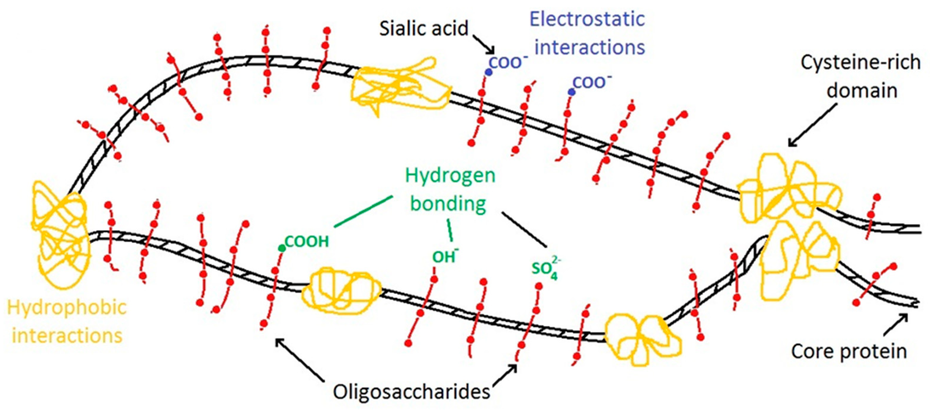
:1. Introduction
2. Materials and Methods
2.1. Analytical Determination of Mucin
2.2. Purification of Mucin via Chromatography
2.3. Synthesis of Mucin-FITC
2.4. Synthesis of Desialylated Mucin
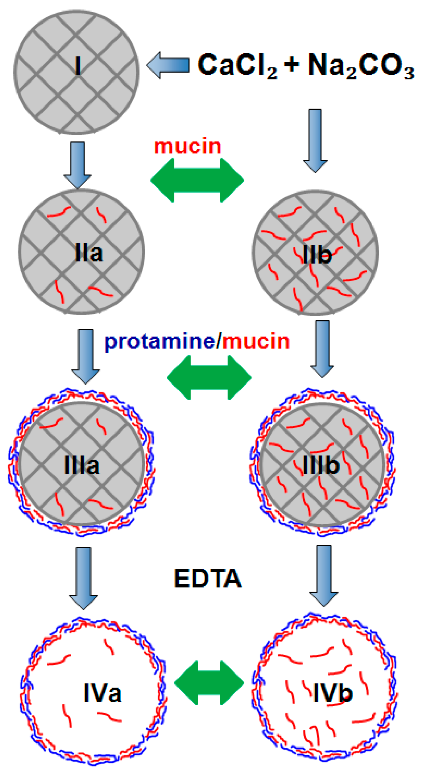
2.5. Mucin Loading into the CaCO3 Crystals by Adsorption
2.6. Mucin and Aprotinin Loading into the CaCO3 Crystals by Co-Synthesis
2.7. Preparation of Polyelectrolyte Microcapsules
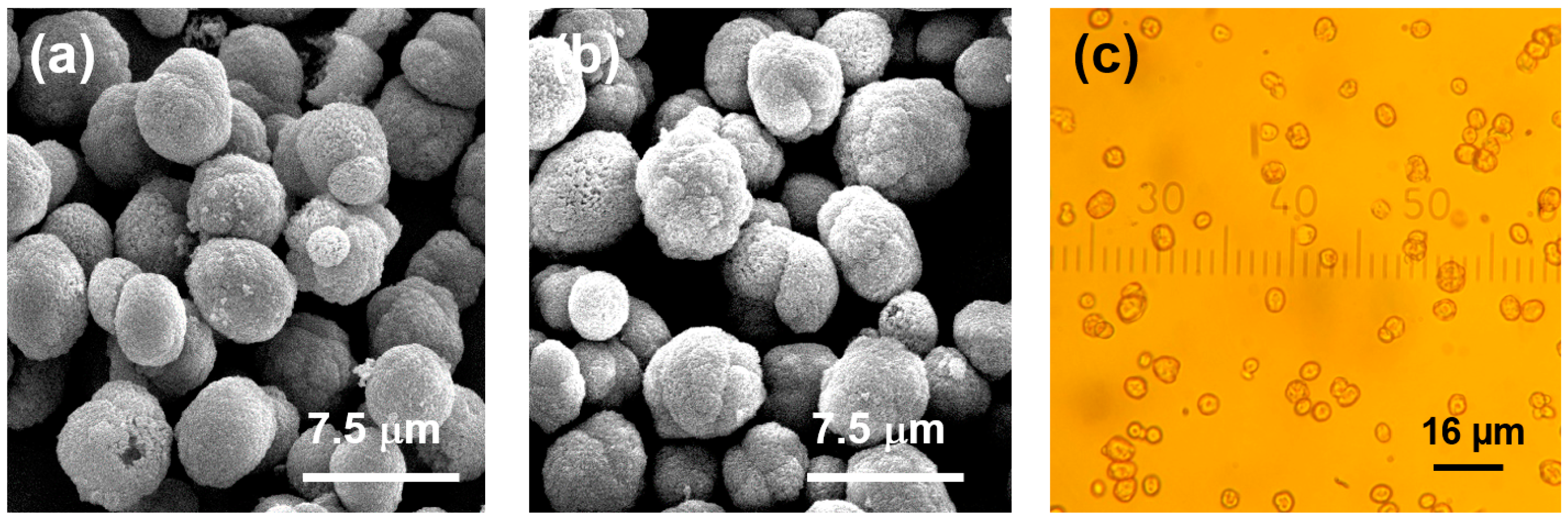
2.8. Characterisation of the Crystals and Microcapsules
3. Results and Discussion
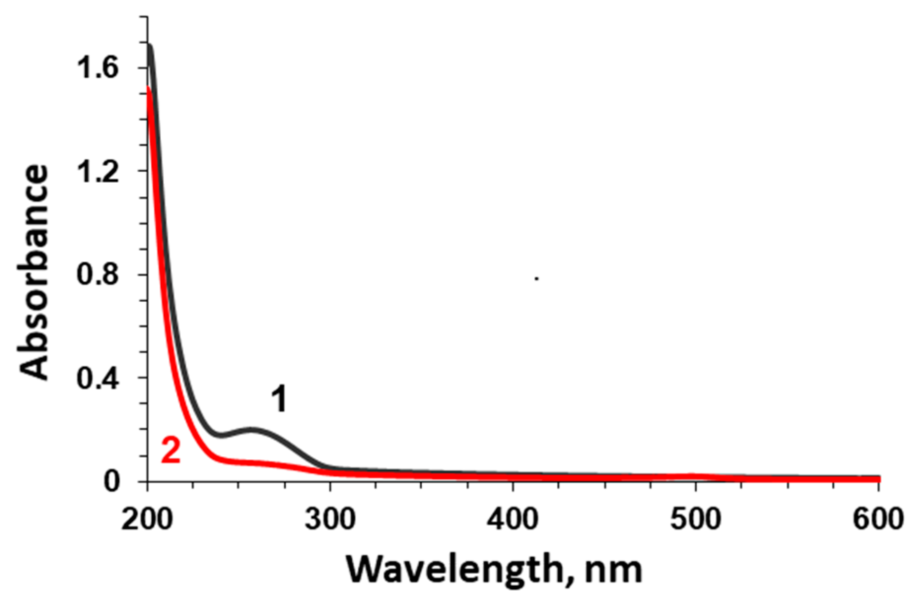
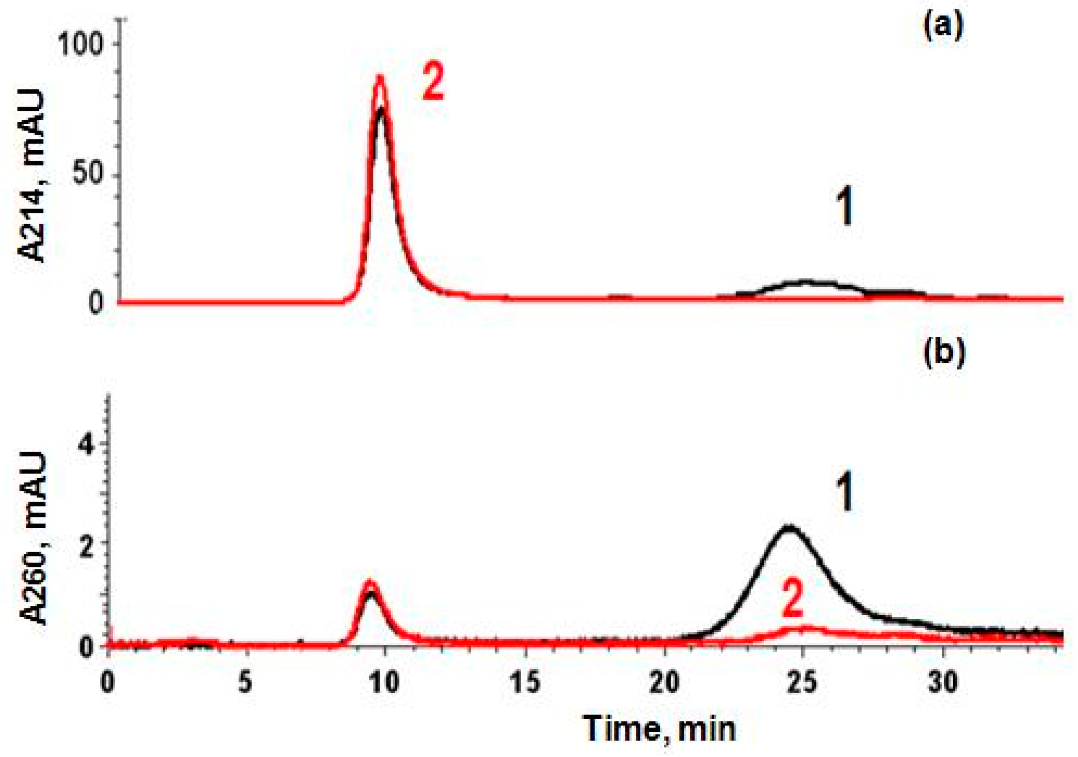
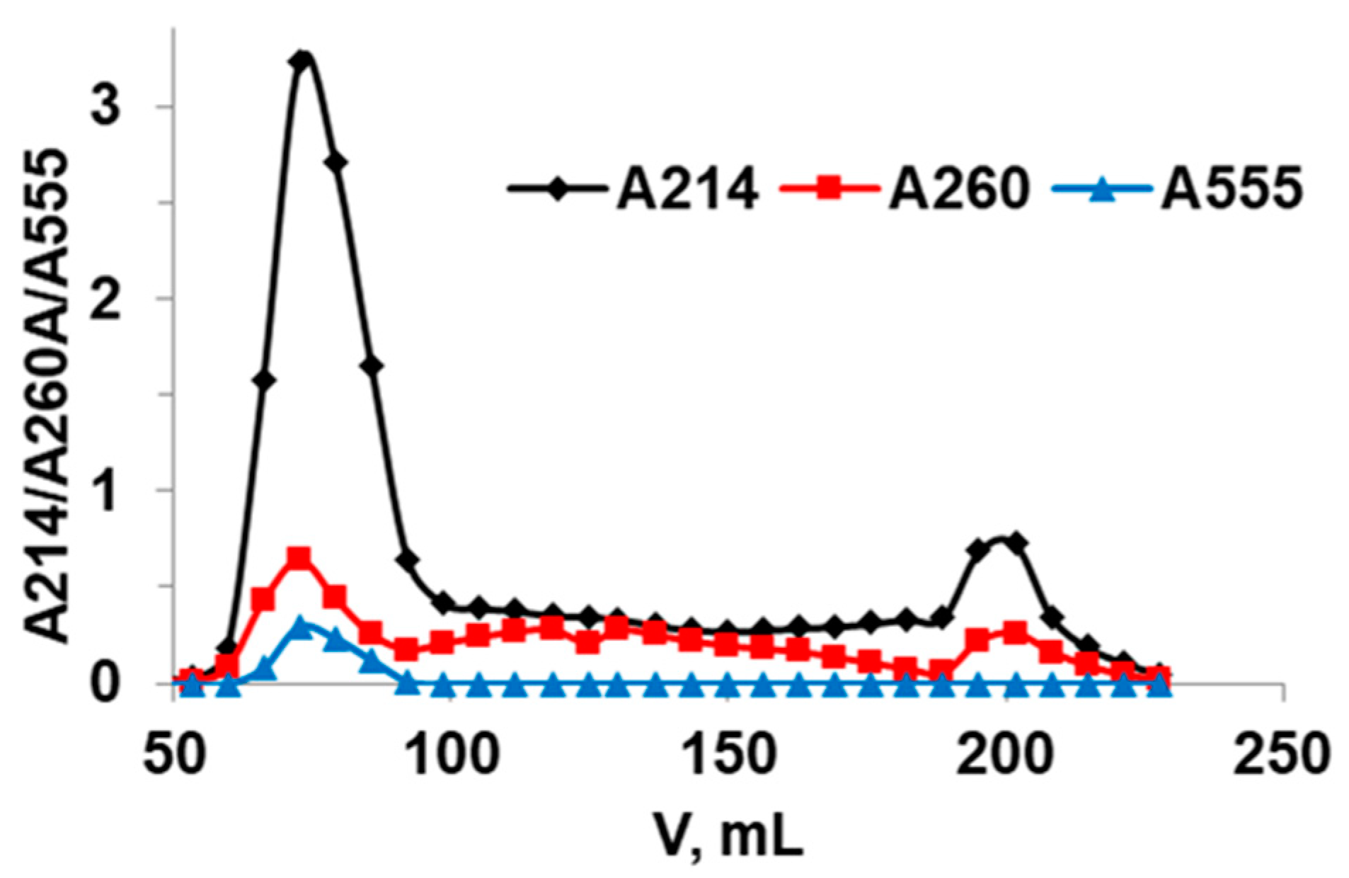
3.1. Analysis of Mucin Purity via Permeation Gel Chromatography
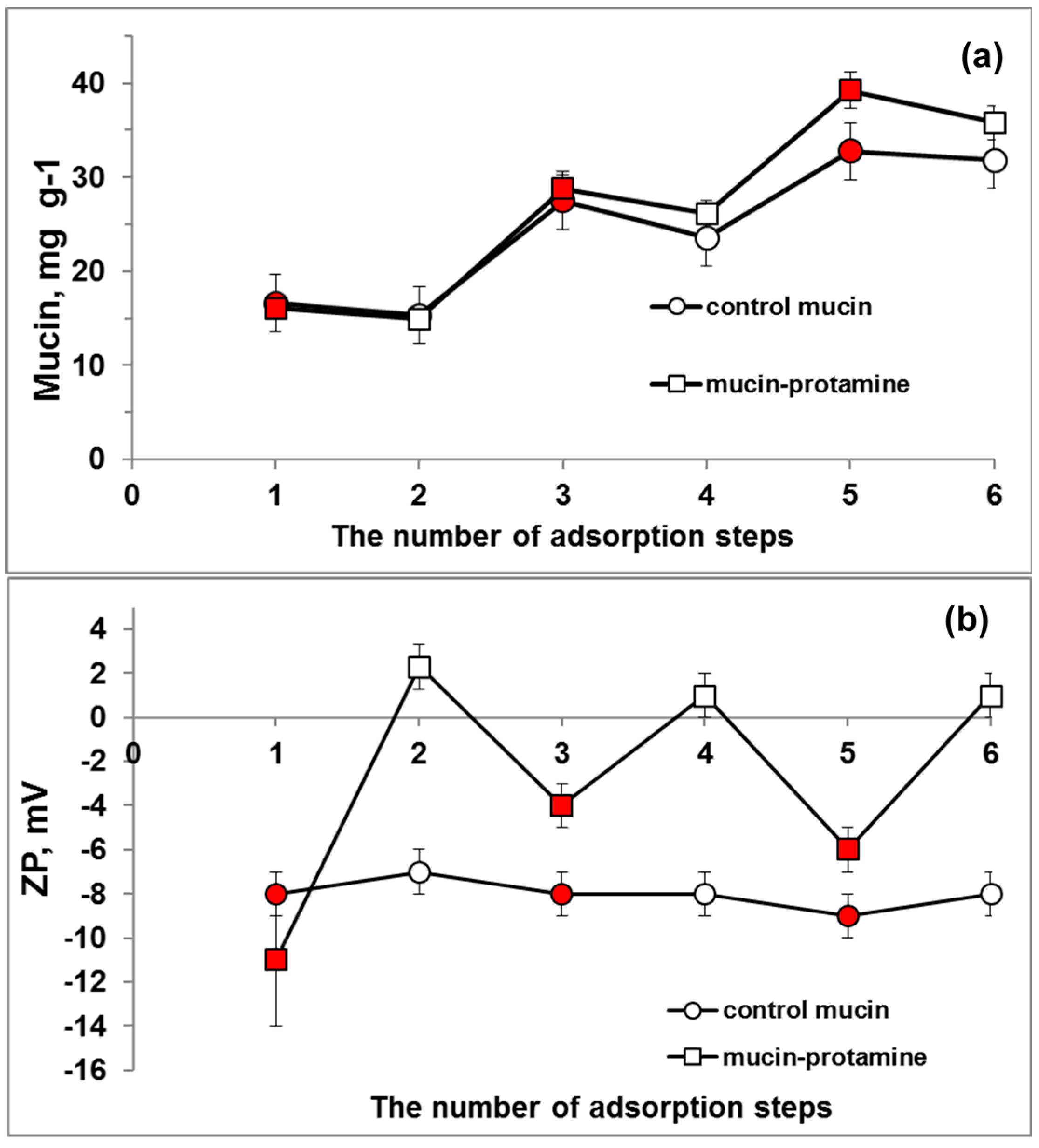
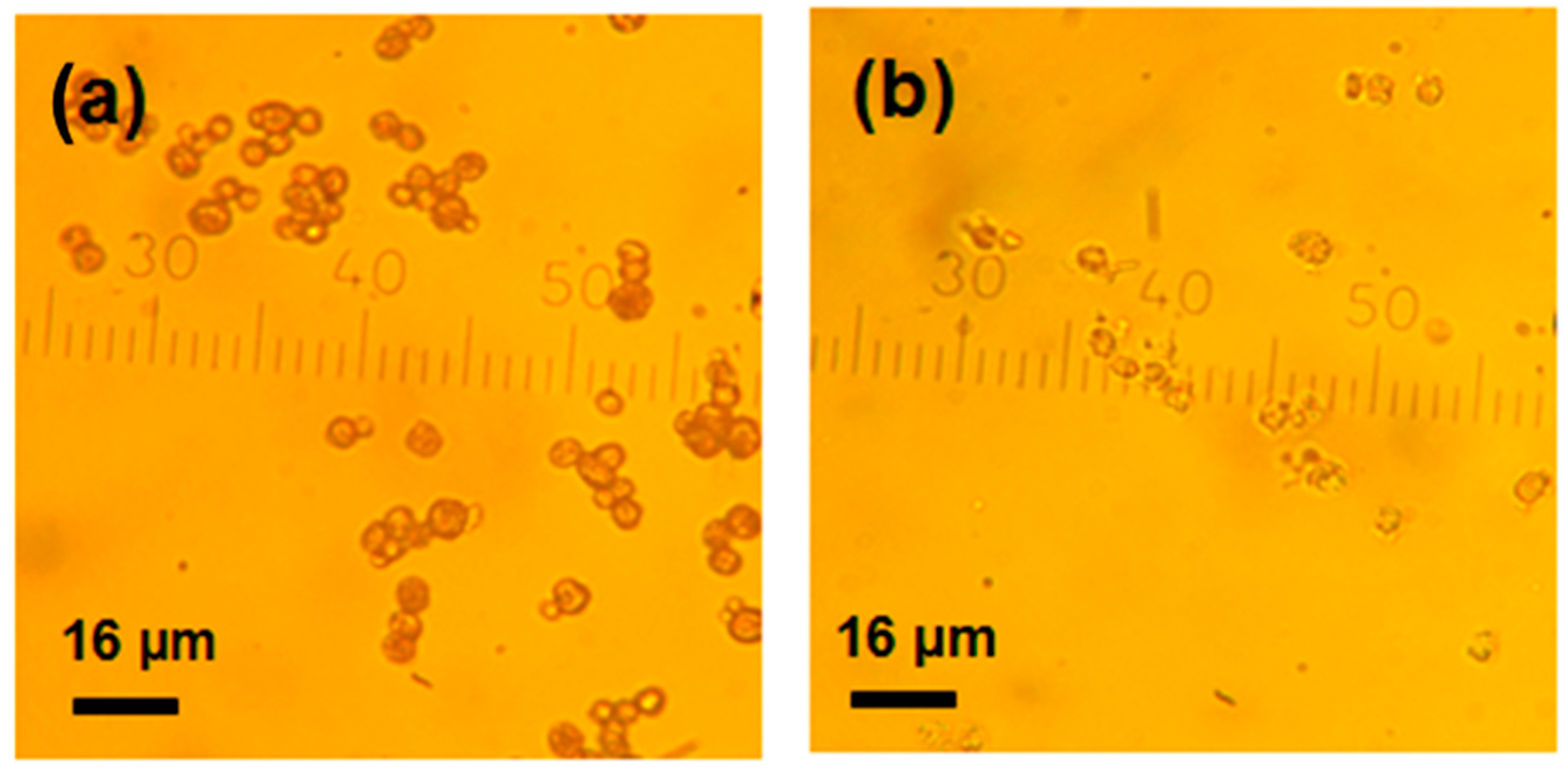
3.2. Loading of Mucin into CaCO3 Crystals (Adsorption and Co-Synthesis)
3.3. Encapsulation of Aprotinin into Mucin-Containing CaCO3 Crystals
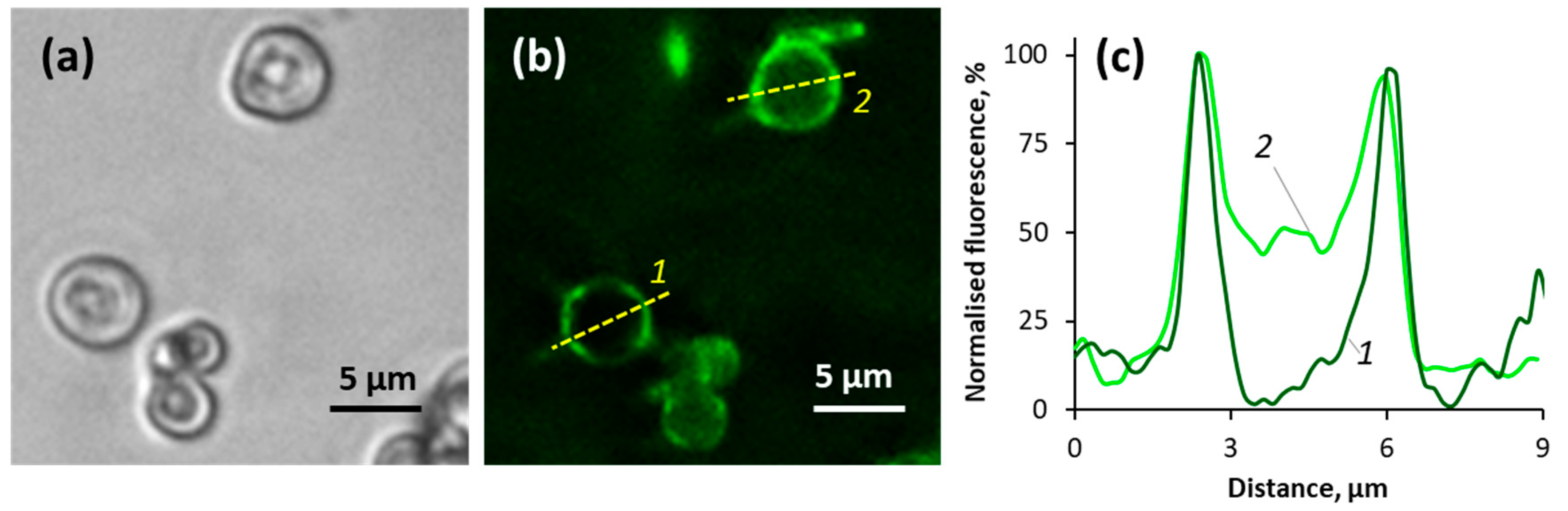
3.4. Mucin-Containing Polymer Multilayer Capsules
3.5. BAC Loading into Mucin-Containing Microparticles
4. Conclusions
Supplementary Materials
Author Contributions
Acknowledgments
Conflicts of Interest
References
- Delcea, M.; Mohwald, H.; Skirtach, A.G. Stimuli-responsive LbL capsules and nanoshells for drug delivery. Adv. Drug Deliv. Rev. 2011, 63, 730–747. [Google Scholar] [CrossRef] [PubMed]
- Volodkin, D.; Skirtach, A.; Möhwald, H. LbL Films as Reservoirs for Bioactive Molecules Bioactive Surfaces; Börner, H.G., Lutz, J.-F., Eds.; Springer: Berlin/Heidelberg, Germany, 2011; Volume 240, pp. 135–161. [Google Scholar]
- Ariga, K.; Lvov, Y.M.; Kawakami, K.; Ji, Q.M.; Hill, J.P. Layer-by-Layer Self-assembled Shells for Drug Delivery. Adv. Drug Deliv. Rev. 2011, 63, 762–771. [Google Scholar] [CrossRef] [PubMed]
- Balabushevich, N.G.; Izumrudov, V.A.; Larionova, N.I. Protein microparticles with controlled stability prepared via layer-by-layer adsorption of biopolyelectrolytes. Polym. Sci. Ser. A 2012, 54, 540–551. [Google Scholar] [CrossRef]
- Volodkin, D.V.; Larionova, N.I.; Sukhorukov, G.B. Protein Encapsulation via Porous CaCO3 Microparticles Templating. Biomacromolecules 2004, 5, 1962–1972. [Google Scholar] [CrossRef] [PubMed]
- Volodkin, D.V.; Petrov, A.I.; Prevot, M.; Sukhorukov, G.B. Matrix Polyelectrolyte Microcapsules: New System for Macromolecule Encapsulation. Langmuir 2004, 20, 3398–3406. [Google Scholar] [CrossRef] [PubMed]
- Volodkin, D. CaCO3 templated micro-beads and-capsules for bioapplications. Adv. Colloid. Interface Sci. 2014, 207, 306–324. [Google Scholar] [CrossRef] [PubMed]
- Díez-Pascual, A.M.; Shuttleworth, P.S. Layer-by-Layer Assembly of Biopolyelectrolytes onto Thermo/pH-Responsive Micro/Nano-Gels. Materials 2014, 7, 7472–7512. [Google Scholar] [CrossRef] [PubMed] [Green Version]
- Ariga, K.; McShane, M.; Lvov, Y.M.; Ji, Q.M.; Hill, J.P. Layer-by-layer assembly for drug delivery and related applications. Expert Opin. Drug Deliv. 2011, 8, 633–644. [Google Scholar] [CrossRef] [PubMed]
- Dekker, J.; Rossen, J.; Buller, H.; Einerhand, A. The MUC family: An obituary. Trends Biochem. Sci. 2002, 27, 126–131. [Google Scholar] [CrossRef]
- Lee, S.; Muller, M.; Rezwan, K.; Spencer, N.D. Porcine gastric mucin (PGM) at the water/poly(dimethylsiloxane) (PDMA) interface: Influence of pH and ionic strength on its conformation, adsorption and aqueous lubrication properties. Langmuir 2005, 21, 8344–8353. [Google Scholar] [CrossRef] [PubMed]
- Bansil, R.; Turner, B.S. Mucin structure, aggregation, physiological functions and biomedical applications. Curr. Opin. Colloid Interface Sci. 2006, 11, 164–170. [Google Scholar] [CrossRef]
- Sandberg, T.; Blom, H.; Caldwell, K.D. Potential use of mucins as biomaterial coatings. I. Fractionation, characterization, and model adsorption of bovine, porcine, and human mucins. J. Biomed. Mater. Res. A 2009, 91, 762–772. [Google Scholar] [CrossRef] [PubMed]
- Leal, J.; Smyth, H.D.C.; Ghosh, D. Physicochemical properties of mucus and their impact on ransmucosal drug delivery. Int. J. Pharm. 2017, 532, 555–572. [Google Scholar] [CrossRef] [PubMed]
- Bansil, R.; Turner, B.S. The biology of mucus: Composition, synthesis and organization. Adv. Drug Deliv. Rev. 2018, 124, 3–15. [Google Scholar] [CrossRef] [PubMed]
- Yang, X.; Forier, K.; Steukers, L.; Vlierberghe, S.; Dubruel, P.; Braeckmans, K.; Glorieu, S.; Nauwynck, H.J. Immobilization of Pseudorabies Virus in Porcine Tracheal Respiratory Mucus Revealed by Single Particle Tracking. PLoS ONE 2012, 7, e51054. [Google Scholar] [CrossRef] [PubMed] [Green Version]
- Cao, X.; Bansil, R.; Bhaskar, K.; Turner, B.; LaMont, J.; Niu, N.; Afdhal, N.H. pHdependent conformational change of gastric mucin leads to sol–gel transition. Biophys. J. 1999, 76, 1250–1258. [Google Scholar] [CrossRef]
- Taylor, C.; Allen, A.; Dettmar, P.; Pearson, J. The gel matrix of gastric mucus is maintained by a complex interplay of transient and nontransient associations. Biomacromolecules 2003, 4, 922–927. [Google Scholar] [CrossRef] [PubMed]
- Nikogeorgos, N.; Madsen, J.B.; Lee, S. Influence of impurities and contact scale on the lubricating propertiesof bovine submaxillary mucin (BSM) films on a hydrophobic surface. Colloids Surf. B Biointerfaces 2014, 122, 760–766. [Google Scholar] [CrossRef] [PubMed]
- Svensson, O.; Arnebrant, T. Mucin layers and multilayers—Physicochemical properties and applications. Curr. Opin. Colloid Interface Sci. 2010, 15, 395–405. [Google Scholar] [CrossRef]
- Shi, L.; Caldwell, K.D. Mucin Adsorption to Hydrophobic Surfaces. J. Colloid Interface Sci. 2000, 224, 372–381. [Google Scholar] [CrossRef] [PubMed]
- Svensson, O.; Lindh, L.; Cardenas, M.; Arnebrant, T. Layer-by-layer assembly of mucin and itosan—Influence of surface properties, concentration and type of mucin. J. Colloid Interface Sci. 2006, 299, 608–616. [Google Scholar] [CrossRef] [PubMed]
- Ahn, J.; Crouzier, T.; Ribbeck, K.; Rubher, M.F.; Cohen, R.E. Turning the properties of mucin via layer-by-layer assembly. Biomacromolecules 2015, 16, 228–235. [Google Scholar] [CrossRef] [PubMed]
- Lindh, L.; Svendsen, I.E.; Svensson, O.; Cárdenas, M.; Arnebrant, T. The salivary mucin MUC5B and lactoperoxidase can be used for layer-by-layer film formation. J. Colloid Interface Sci. 2007, 310, 74–82. [Google Scholar] [CrossRef] [PubMed]
- Nikogeorgos, N.; Patil, N.J.; Zappone, B.; Lee, S. Interaction of porcine gastric mucin with various polycations and its influence on the boundary lubrication properties. Polymer 2016, 100, 158–168. [Google Scholar] [CrossRef]
- Nowald, C.; Penk, A.; Chiu, H.-Y.; Bein, T.; Huster, D.; Lieleg, O. A Selective ucin/Methylcellulose Hybrid Gel with Tailored Mechanical Properties. Macromol. Biosci. 2016, 16, 567–579. [Google Scholar] [CrossRef] [PubMed]
- Berg, A.A.; Buul, J.D.; Tytgat, G.N.J.; Groen, A.K.J.; Ostrow, D. Mucins and calcium phosphate precipitates additively stimulate cholesterol crystallization. J. Lipid Res. 1998, 39, 1744–1751. [Google Scholar] [PubMed]
- Raynal, B.D.; Hardingham, T.E.; Sheehan, J.K.; Thornton, D.J. Calcium-dependent protein interactions in MUC5B provide reversible cross-links in salivary mucus. J. Biol. Chem. 2003, 278, 28703–28710. [Google Scholar] [CrossRef] [PubMed]
- Su, Y.; Xu, Y.; Yang, L.; Wenga, S.; Soloway, R.D.; Wang, D.; Wua, J. Spectroscopic studies of the effect of the metal ions on the structure of mucin. J. Mol. Struct. 2009, 920, 8–13. [Google Scholar] [CrossRef]
- Amborta, D.; Johanssona, M.E.V.; Gustafssona, J.K.; Nilssonb, H.E.; Ermunda, A.; Johanssona, B.R.; Koeckb, P.J.B.; Hebertb, H.; Hanssona, G.C. Calcium and pH-dependent packing and release of the gel-forming MUC2 mucin. PNAS 2012, 109, 5645–5650. [Google Scholar] [CrossRef] [PubMed] [Green Version]
- Yamasaki, T.; Chijiiwa, K.; Endo, M. Isolation of mucin from human hepatic bile and its induced effects on precipitation of cholesterol and calcium carbonate in vivo. Deg. Dis. Sci. 1993, 38, 909–915. [Google Scholar] [CrossRef]
- Turner, B.S.; Bhaskar, K.R.; Hadzopoulou-Cladaras, M.; LaMont, J.T. Cysteine-rich regions of pig gastric mucin contain von Willebrand factor and cystine knot domains at the carboxyl terminal. Biochim. Biophys. Acta-Gene Struct. Expr. 1999, 1447, 77–92. [Google Scholar] [CrossRef]
- Balabushevich, N.G.; Guerenu, A.V.; Feoktistova, N.A.; Volodkin, D. Protein loading into porous CaCO3 microspheres: Adsorption equilibrium and bioactivity retention. Phys. Chem. Chem. Phys. 2015, 17, 2523–2530. [Google Scholar] [CrossRef] [PubMed]
- Balabushevich, N.G.; Lopez de Guerenu, A.V.; Feoktistova, N.A.; Volodkin, D. Protein-Containing Multilayer Capsules by Templating on Mesoporous CaCO3 Particles: POST- and PRE-Loading Approaches. Macromol. Biosci. 2016, 16, 95–105. [Google Scholar] [CrossRef] [PubMed]
- Mantle, M.; Allen, A. A colorimetric assay for glycoproteins based on the periodic acid/Schiff stain. Biochem. Soc. Trans. 1978, 6, 607–609. [Google Scholar] [CrossRef] [PubMed]
- Fields, R. The rapid determination of amino groups with TNBS. Methods Enzymol. 1972, 25, 464–468. [Google Scholar] [PubMed]
- Hess, E.L.; Coburn, A.F.; Bates, R.C.; Murphy, P. A New Method for Measuring Sialic Acid Levels in Serum and Its Application to Rheumatic Fever. J. Clin. Investig. 1957, 36, 449–455. [Google Scholar] [CrossRef] [PubMed]
- Balabushevitch, N.G.; Kildeyeva, N.R.; Moroz, N.A.; Trusova, S.P.; Virnik, A.D.; Khromov, G.L.; Larionova, N.I. Regulating aspects of biosoluble and insoluble film release systems containing protein proteinase inhibitor. Appl. Biochem. Biotechnol. 1996, 61, 129–138. [Google Scholar] [CrossRef]
- Shomig, V.J.; Kasdorf, B.T.; Scholz, K.; Bidmon, K.; Lieleg, O.; Berensmeier, S. An optimazid purification process for porcine gastric mucin with preservation of its natural functional properties. RSC Adv. 2016, 6, 44932–44943. [Google Scholar] [CrossRef]
- Feoktistova, N.; Rose, J.; Prokopovic, V.Z.; Vikulina, A.S.; Skirtach, A.; Volodkin, D. Controlling the vaterite CaCO3 crystal pores. Design of tailor-made polymer based microcapsules by hard templating. Langmuir 2016, 32, 4229–4238. [Google Scholar] [CrossRef] [PubMed]
- Paulraj, T.; Feoktistova, N.; Velk, N.; Uhlig, K.; Duschl, C.; Volodkin, D. Microporous Polymeric 3D Scaffolds Templated by the Layer-by-Layer Self-Assembly. Macromol. Rapid Commun. 2014, 35, 1408–1413. [Google Scholar] [CrossRef] [PubMed]
- Vikulina, A.S.; Feoktistova, N.A.; Balabushevich, N.G.; Skirtach, A.G.; Volodkin, D.V. The mechanism of catalase loading into porous vaterite CaCO3 crystals by co-synthesis. Phys. Chem. Chem. Phys. 2018, 20, 8822–8831. [Google Scholar] [CrossRef] [PubMed]
- Fritz, H.; Wunderer, G. Biochemistry and Applications of Aprotinin, the Kallikrein Inhibitor from Bovine Organs. Arzneim. Forsch./Drug Res. 1983, 33, 479–494. [Google Scholar]
- Balhorn, R. The protamine family of sperm nuclear proteins. Genome Biol. 2007, 8, 227–234. [Google Scholar] [CrossRef] [PubMed]
- Balabushevich, N.G.; Zimina, E.P.; Larioniva, N.I. Encapsulation of catalase in polyelectrolyte microspheres composed of melamine formaldehyde, dextran sulfate, and protamine. Biochem. Mosc. 2004, 69, 763–769. [Google Scholar] [CrossRef]
- Ellman, G.L. Tissue sulfhydryl groups. Arch. Biochem. Biophys. 1959, 82, 70–77. [Google Scholar] [CrossRef]
- Boateng, J.S.; Pawar, H.V.; Tetteh, J. Evaluation of in vitro wound adhesion characteristics of composite film and wafer based dressings using texture analysis and FTIR spectroscopy: A chemometrics factor analysis approach. RSC Adv. 2015, 5, 107064–107075. [Google Scholar] [CrossRef]
- Sandberg, T.; Karlsson, O.M.; Carlsson, J.; Feiler, A.; Caldwell, K.D. Potential use of mucins as biomaterial coatings. II. Mucin coatings affect the conformation and neutrophil-activating properties of adsorbed host proteins—Toward a mucosal mimic. J. Biomed. Mater. Res. A 2009, 91, 773–785. [Google Scholar] [CrossRef] [PubMed]
- Lechanteur, A.; Neves, J.; Sarmento, B. The role of mucus in cell-based models used to screen mucosal drug delivery. Adv. Drug Del. Rev. 2018, 124, 50–63. [Google Scholar] [CrossRef] [PubMed] [Green Version]
- Builders, P.F.; Kunle, O.O.; Okpaku, L.C.; Builders, M.I.; Attama, A.A.; Adikwu, M.U. Preparation and evaluation of mucinated sodium alginate microparticles for oral delivery of insulin. Eur. J. Pharm. Biopharm. 2008, 70, 777–783. [Google Scholar] [CrossRef] [PubMed]
- Uhlig, K.; Madaboosi, N.; Schmidt, S.; Jager, M.S.; Rose, J.; Duschl, C.; Volodkin, D.V. 3d localization and diffusion of proteins in polyelectrolyte multilayers. Soft Matter 2012, 8, 11786–11789. [Google Scholar] [CrossRef]
- Sustr, D.; Hlavacek, A.; Duschl, C.; Volodkin, D. Multi-Fractional Analysis of Molecular Diffusion in Polymer Multilayers by FRAP. A New Simulation-Based Approach. J. Phys. Chem B 2018, 122, 1323–1333. [Google Scholar] [CrossRef] [PubMed]
- Behra, M.; Schmidt, S.; Hartmann, J.; Volodkin, D.V.; Hartmann, L. Synthesis of Porous PEG Microgels Using CaCO3 Microspheres as Hard Templates. Macromol. Rapid Commun. 2012, 33, 1049–1054. [Google Scholar] [CrossRef] [PubMed]
- Behra, M.; Azzouz, N.; Schmidt, S.; Volodkin, D.V.; Mosca, S.; Chanana, M.; Seeberger, P.H.; Hartmann, L. Magnetic porous sugar-functionalized PEG microgels for efficient isolation and removal of bacteria from solution. Biomacromolecules 2013, 14, 1927–1935. [Google Scholar] [CrossRef] [PubMed]
- Feoktistova, N.; Stoychev, G.; Ionov, L.; Volodkin, D. Porous thermo-responsive pNIPAM microgels. Eur. Polym. J. 2015, 68, 650–656. [Google Scholar] [CrossRef]
- Sergeeva, A.; Feoktistova, N.; Prokopovic, V.; Gorin, D.; Volodkin, D. Design of porous alginate hydrogels by sacrificial CaCO3 templates: Pore formation mechanism. Adv. Mater. Interfaces 2016, 2, 1500386. [Google Scholar] [CrossRef]
- Sergeeva, A.; Sergeev, R.; Lengert, E.; Zakharevich, A.; Parakhonskiy, B.; Gorin, D.; Sergeev, S.; Volodkin, D. Composite magnetite and protein containing CaCO3 crystals. External manipulation and vaterite calcite recrystallization-mediated release performance. ACS Appl. Mater. Interfaces 2015, 7, 21315–21325. [Google Scholar] [CrossRef] [PubMed]
- Sergeeva, A.S.; Gorin, D.A.; Volodkin, D.V. In-situ assembly of Ca-alginate gels with controlled pore loading/release capability. Langmuir 2015, 13, 10813–10821. [Google Scholar] [CrossRef] [PubMed]
- Volodkin, D.V.; Balabushevitch, N.G.; Sukhorukov, G.B.; Larionova, N.I. Model systems for controlled protein release: PH-sensitive polyelectrolyte microparticles. S.T.P. Pharma Sci. 2003, 13, 163–170. [Google Scholar]
- Balabushevich, N.G.; Pechenkin, M.A.; Zorov, I.N.; Shibanova, E.D.; Larionova, N.I. Mucoadhesive Polyelectrolyte Microparticles Containing Recombinant Human Insulin and Its Analogs Aspart and Lispro. Biochem. Mosc. 2011, 76, 327–331. [Google Scholar] [CrossRef]
- Balabushevich, N.G.; Pechenkin, M.A.; Shibanova, E.D.; Volodkin, D.V.; Mikhalchik, E.V. Multifunctional Polyelectrolyte Microparticles for Oral Insulin Delivery. Macromol. Biosci. 2013, 13, 1379–1388. [Google Scholar] [CrossRef] [PubMed]
- Parakhonskiy, B.V.; Yashchenok, A.M.; Donatan, S.; Volodkin, D.V.; Tessarolo, F.; Antolini, R.; Möhwald, H.; Skirtach, A.G. Macromolecule Loading into Spherical, Elliptical, Star-Like and Cubic Calcium Carbonate Carriers. Chem. Phys. Chem. 2014, 15, 2817–2822. [Google Scholar] [CrossRef] [PubMed]


| Loading Method | Efficiency of Mucin Loading, % | Release after Washing, % of Loaded | ZP, mV | |
|---|---|---|---|---|
| Spectrophotometry | Analytical Chromatography | |||
| Adsorption | 12 ± 2 | 10 ± 1 | 11 ± 1 | −(15 ± 3) |
| Co-synthesis | 22 ± 3 | 18 ± 2 | 5 ± 1 | −(11 ± 2) |
© 2018 by the authors. Licensee MDPI, Basel, Switzerland. This article is an open access article distributed under the terms and conditions of the Creative Commons Attribution (CC BY) license (http://creativecommons.org/licenses/by/4.0/).
Share and Cite
Balabushevich, N.G.; Sholina, E.A.; Mikhalchik, E.V.; Filatova, L.Y.; Vikulina, A.S.; Volodkin, D. Self-Assembled Mucin-Containing Microcarriers via Hard Templating on CaCO3 Crystals. Micromachines 2018, 9, 307. https://doi.org/10.3390/mi9060307
Balabushevich NG, Sholina EA, Mikhalchik EV, Filatova LY, Vikulina AS, Volodkin D. Self-Assembled Mucin-Containing Microcarriers via Hard Templating on CaCO3 Crystals. Micromachines. 2018; 9(6):307. https://doi.org/10.3390/mi9060307
Chicago/Turabian StyleBalabushevich, Nadezhda G., Ekaterina A. Sholina, Elena V. Mikhalchik, Lyubov Y. Filatova, Anna S. Vikulina, and Dmitry Volodkin. 2018. "Self-Assembled Mucin-Containing Microcarriers via Hard Templating on CaCO3 Crystals" Micromachines 9, no. 6: 307. https://doi.org/10.3390/mi9060307