Next Issue
Volume 10, May
Previous Issue
Volume 10, March


Cancers, Volume 10, Issue 4 (April 2018) – 42 articles

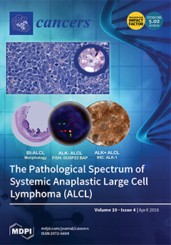
Cover Story (view full-size image): Anaplastic large cell lymphoma (ALCL) is a T-cell neoplasm with unique morphology and phenotype, which is subdivided into four different entities depending on clinical and molecular features: systemic ALK-positive ALCL, systemic ALK-negative ALCL, primary cutaneous ALCL, and breast implant-associated ALCL. While ALK fusion proteins are the genetic hallmark of ALK-positive ALCL and present a paradigm of a dominant oncogenic driver mutation, the genetic landscape of ALK-negative ALCL has only recently been explored further, deepening our understanding of its clinical behavior and pathogenesis and providing new targets to develop specific therapies. View this paper
  • Issues are regarded as officially published after their release is announced to the table of contents alert mailing list.
  • You may sign up for e-mail alerts to receive table of contents of newly released issues.
  • PDF is the official format for papers published in both, html and pdf forms. To view the papers in pdf format, click on the "PDF Full-text" link, and use the free Adobe Reader to open them.
Order results
Result details
Section
Select all
Export citation of selected articles as:
11 pages, 1776 KiB  
Article
Responses to the Selective Bruton’s Tyrosine Kinase (BTK) Inhibitor Tirabrutinib (ONO/GS-4059) in Diffuse Large B-cell Lymphoma Cell Lines
by Ryohei Kozaki, Meike Vogler, Harriet S. Walter, Sandrine Jayne, David Dinsdale, Reiner Siebert, Martin J.S. Dyer and Toshio Yoshizawa
Cancers 2018, 10(4), 127; https://doi.org/10.3390/cancers10040127 - 23 Apr 2018
Cited by 26 | Viewed by 8027
Abstract
Bruton’s tyrosine kinase (BTK) is a key regulator of the B-cell receptor signaling pathway, and aberrant B-cell receptor (BCR) signaling has been implicated in the survival of malignant B-cells. However, responses of the diffuse large B-cell lymphoma (DLBCL) to inhibitors of BTK (BTKi) [...] Read more.
Bruton’s tyrosine kinase (BTK) is a key regulator of the B-cell receptor signaling pathway, and aberrant B-cell receptor (BCR) signaling has been implicated in the survival of malignant B-cells. However, responses of the diffuse large B-cell lymphoma (DLBCL) to inhibitors of BTK (BTKi) are infrequent, highlighting the need to identify mechanisms of resistance to BTKi as well as predictive biomarkers. We investigated the response to the selective BTKi, tirabrutinib, in a panel of 64 hematopoietic cell lines. Notably, only six cell lines were found to be sensitive. Although activated B-cell type DLBCL cells were most sensitive amongst all cell types studied, sensitivity to BTKi did not correlate with the presence of activating mutations in the BCR pathway. To improve efficacy of tirabrutinib, we investigated combination strategies with 43 drugs inhibiting 34 targets in six DLBCL cell lines. Based on the results, an activated B-cell-like (ABC)-DLBCL cell line, TMD8, was the most sensitive cell line to those combinations, as well as tirabrutinib monotherapy. Furthermore, tirabrutinib in combination with idelalisib, palbociclib, or trametinib was more effective in TMD8 with acquired resistance to tirabrutinib than in the parental cells. These targeted agents might be usefully combined with tirabrutinib in the treatment of ABC-DLBCL. Full article
(This article belongs to the Special Issue Tyrosine Kinase Signaling Pathways in Cancer)
Show Figures

Figure 1

15 pages, 3343 KiB  
Article
Hypoxia-Induced Cisplatin Resistance in Non-Small Cell Lung Cancer Cells Is Mediated by HIF-1α and Mutant p53 and Can Be Overcome by Induction of Oxidative Stress
by Christophe Deben, Vanessa Deschoolmeester, Jorrit De Waele, Julie Jacobs, Jolien Van den Bossche, An Wouters, Marc Peeters, Christian Rolfo, Evelien Smits, Filip Lardon and Patrick Pauwels
Cancers 2018, 10(4), 126; https://doi.org/10.3390/cancers10040126 - 21 Apr 2018
Cited by 44 | Viewed by 7246
Abstract
The compound APR-246 (PRIMA-1MET) is a known reactivator of (mutant) p53 and inducer of oxidative stress which can sensitize cancer cells to platinum-based chemotherapeutics. However, the effect of a hypoxic tumor environment has been largely overlooked in this interaction. This study [...] Read more.
The compound APR-246 (PRIMA-1MET) is a known reactivator of (mutant) p53 and inducer of oxidative stress which can sensitize cancer cells to platinum-based chemotherapeutics. However, the effect of a hypoxic tumor environment has been largely overlooked in this interaction. This study focusses on the role of hypoxia-inducible factor-1α (HIF-1α) and the p53 tumor suppressor protein in hypoxia-induced cisplatin resistance in non-small cell lung cancer (NSCLC) cells and the potential of APR-246 to overcome this resistance. We observed that hypoxia-induced cisplatin resistance only occurred in the p53 mutant NCI-H2228Q331* cell line, and not in the wild type A549 and mutant NCI-H1975R273H cell lines. Cisplatin reduced HIF-1α protein levels in NCI-H2228Q331* cells, leading to a shift in expression from HIF-1α-dependent to p53-dependent transcription targets under hypoxia. APR-246 was able to overcome hypoxia-induced cisplatin resistance in NCI-H2228Q331* cells in a synergistic manner without affecting mutant p53Q331* transcriptional activity, but significantly depleting total glutathione levels more efficiently under hypoxic conditions. Synergism was dependent on the presence of mutant p53Q331* and the induction of reactive oxygen species, with depletion of one or the other leading to loss of synergism. Our data further support the rationale of combining APR-246 with cisplatin in NSCLC, since their synergistic interaction is retained or enforced under hypoxic conditions in the presence of mutant p53. Full article
(This article belongs to the Special Issue p53 Signaling in Cancers)
Show Figures

Figure 1

13 pages, 2093 KiB  
Brief Report
Selective Inhibition of Histone Deacetylation in Melanoma Increases Targeted Gene Delivery by a Bacteriophage Viral Vector
by Samuel Campbell, Keittisak Suwan, Sajee Waramit, Eric Ofori Aboagye and Amin Hajitou
Cancers 2018, 10(4), 125; https://doi.org/10.3390/cancers10040125 - 21 Apr 2018
Cited by 3 | Viewed by 4411
Abstract
The previously developed adeno-associated virus/phage (AAVP) vector, a hybrid between M13 bacteriophage (phage) viruses that infect bacteria only and human Adeno-Associated Virus (AAV), is a promising tool in targeted gene therapy against cancer. AAVP can be administered systemically and made tissue specific through [...] Read more.
The previously developed adeno-associated virus/phage (AAVP) vector, a hybrid between M13 bacteriophage (phage) viruses that infect bacteria only and human Adeno-Associated Virus (AAV), is a promising tool in targeted gene therapy against cancer. AAVP can be administered systemically and made tissue specific through the use of ligand-directed targeting. Cancer cells and tumor-associated blood vessels overexpress the αν integrin receptors, which are involved in tumor angiogenesis and tumor invasion. AAVP is targeted to these integrins via a double cyclic RGD4C ligand displayed on the phage capsid. Nevertheless, there remain significant host-defense hurdles to the use of AAVP in targeted gene delivery and subsequently in gene therapy. We previously reported that histone deacetylation in cancer constitutes a barrier to AAVP. Herein, to improve AAVP-mediated gene delivery to cancer cells, we combined the vector with selective adjuvant chemicals that inhibit specific histone deacetylases (HDAC). We examined the effects of the HDAC inhibitor C1A that mainly targets HDAC6 and compared this to sodium butyrate, a pan-HDAC inhibitor with broad spectrum HDAC inhibition. We tested the effects on melanoma, known for HDAC6 up-regulation, and compared this side by side with a normal human kidney HEK293 cell line. Varying concentrations were tested to determine cytotoxic levels as well as effects on AAVP gene delivery. We report that the HDAC inhibitor C1A increased AAVP-mediated transgene expression by up to ~9-fold. These findings indicate that selective HDAC inhibition is a promising adjuvant treatment for increasing the therapeutic value of AAVP. Full article
(This article belongs to the Special Issue Histone Modification in Cancer)
Show Figures

Figure 1

19 pages, 1180 KiB  
Review
Oncolytic Virotherapy versus Cancer Stem Cells: A Review of Approaches and Mechanisms
by Shyambabu Chaurasiya, Nanhai G. Chen and Susanne G. Warner
Cancers 2018, 10(4), 124; https://doi.org/10.3390/cancers10040124 - 19 Apr 2018
Cited by 37 | Viewed by 7061
Abstract
A growing body of evidence suggests that a subset of cells within tumors are resistant to conventional treatment modalities and may be responsible for disease recurrence. These cells are called cancer stem cells (CSC), which share properties with normal stem cells including self-renewal, [...] Read more.
A growing body of evidence suggests that a subset of cells within tumors are resistant to conventional treatment modalities and may be responsible for disease recurrence. These cells are called cancer stem cells (CSC), which share properties with normal stem cells including self-renewal, pluripotency, drug resistance, and the ability to maintain quiescence. While most conventional therapies can efficiently destroy rapidly dividing cancer cells comprising the bulk of a tumor, they often fail to kill the less abundant and quiescent CSCs. Furthermore, killing of only differentiated cells in the tumor may actually allow for enrichment of CSCs and thereby portend a bad prognosis. Therefore, targeting of CSCs is important to achieve long-term success in cancer therapy. Oncolytic viruses represent a completely different class of therapeutics that can kill cancer cells in a variety of ways, which differ from those of conventional therapies. Hence, CSCs that are inherently resistant to conventional therapies may be susceptible to oncolytic virus-mediated killing. Recent studies have shown that oncolytic viruses can efficiently kill CSCs in many types of cancer. Here, we discuss the mechanism through which CSCs can escape conventional therapies and how they may still be susceptible to different classes of oncolytic viruses. Furthermore, we provide a summary of recent studies that have tested oncolytic viruses on CSCs of different origins and discuss possible future directions for this fascinating subset of oncolytic virus research. Full article
(This article belongs to the Special Issue Oncolytic Virotherapy)
Show Figures

Figure 1

29 pages, 853 KiB  
Review
Innovative Diagnostic Methods for Early Prostate Cancer Detection through Urine Analysis: A Review
by Carmen Bax, Gianluigi Taverna, Lidia Eusebio, Selena Sironi, Fabio Grizzi, Giorgio Guazzoni and Laura Capelli
Cancers 2018, 10(4), 123; https://doi.org/10.3390/cancers10040123 - 18 Apr 2018
Cited by 54 | Viewed by 7655
Abstract
Prostate cancer is the second most common cause of cancer death among men. It is an asymptomatic and slow growing tumour, which starts occurring in young men, but can be detected only around the age of 40–50. Although its long latency period and [...] Read more.
Prostate cancer is the second most common cause of cancer death among men. It is an asymptomatic and slow growing tumour, which starts occurring in young men, but can be detected only around the age of 40–50. Although its long latency period and potential curability make prostate cancer a perfect candidate for screening programs, the current procedure lacks in specificity. Researchers are rising to the challenge of developing innovative tools able of detecting the disease during its early stage that is the most curable. In recent years, the interest in characterisation of biological fluids aimed at the identification of tumour-specific compounds has increased significantly, since cell neoplastic transformation causes metabolic alterations leading to volatile organic compounds release. In the scientific literature, different approaches have been proposed. Many studies focus on the identification of a cancer-characteristic “odour fingerprint” emanated from biological samples through the application of sensorial or senso-instrumental analyses, others suggest a chemical characterisation of biological fluids with the aim of identifying prostate cancer (PCa)-specific biomarkers. This paper focuses on the review of literary studies in the field of prostate cancer diagnosis, in order to provide an overview of innovative methods based on the analysis of urine, thereby comparing them with the traditional diagnostic procedures. Full article
Show Figures

Figure 1

10 pages, 1465 KiB  
Commentary
The Hippo-YAP Pathway Regulates 3D Organ Formation and Homeostasis
by Erika Ishihara and Hiroshi Nishina
Cancers 2018, 10(4), 122; https://doi.org/10.3390/cancers10040122 - 17 Apr 2018
Cited by 9 | Viewed by 5681
Abstract
The vertebrate body shape is formed by the specific sizes and shapes of its resident tissues and organs, whose alignments are essential for proper functioning. To maintain tissue and organ shape, and thereby function, it is necessary to remove senescent, transformed, and/or damaged [...] Read more.
The vertebrate body shape is formed by the specific sizes and shapes of its resident tissues and organs, whose alignments are essential for proper functioning. To maintain tissue and organ shape, and thereby function, it is necessary to remove senescent, transformed, and/or damaged cells, which impair function and can lead to tumorigenesis. However, the molecular mechanisms underlying three-dimensional (3D) organ formation and homeostasis are not fully clear. Yes-associated protein (YAP) is a transcriptional co-activator that is involved in organ size control and tumorigenesis. Recently, we reported that YAP is essential for proper 3D body shape through regulation of cell tension by using a unique medaka fish mutant, hirame (hir). In Madin–Darby canine kidney (MDCK) epithelial cells, active YAP-transformed cells are eliminated apically when surrounded by normal cells. Furthermore, in a mosaic mouse model, active YAP-expressing damaged hepatocytes undergo apoptosis and are eliminated from the liver. Thus, YAP functions in quantitative and quality control in organogenesis. In this review, we describe the various roles of YAP in vertebrates, including in the initiation of liver cancer. Full article
Show Figures

Figure 1

21 pages, 5735 KiB  
Review
Ubiquitin-Dependent Regulation of the Mammalian Hippo Pathway: Therapeutic Implications for Cancer
by Thanh Hung Nguyen and Jan-Michael Kugler
Cancers 2018, 10(4), 121; https://doi.org/10.3390/cancers10040121 - 17 Apr 2018
Cited by 18 | Viewed by 5110
Abstract
The Hippo pathway serves as a key barrier for oncogenic transformation. It acts by limiting the activity of the proto-oncogenes YAP and TAZ. Reduced Hippo signaling and elevated YAP/TAZ activities are frequently observed in various types of tumors. Emerging evidence suggests that the [...] Read more.
The Hippo pathway serves as a key barrier for oncogenic transformation. It acts by limiting the activity of the proto-oncogenes YAP and TAZ. Reduced Hippo signaling and elevated YAP/TAZ activities are frequently observed in various types of tumors. Emerging evidence suggests that the ubiquitin system plays an important role in regulating Hippo pathway activity. Deregulation of ubiquitin ligases and of deubiquitinating enzymes has been implicated in increased YAP/TAZ activity in cancer. In this article, we review recent insights into the ubiquitin-mediated regulation of the mammalian Hippo pathway, its deregulation in cancer, and possibilities for targeting the Hippo pathway through the ubiquitin system. Full article
Show Figures

Figure 1

5 pages, 1387 KiB  
Perspective
Perspective: Contribution of Epstein–Barr virus (EBV) Reactivation to the Carcinogenicity of Nasopharyngeal Cancer Cells
by Chung-Chun Wu, Chih-Yeu Fang, Sheng-Yen Huang, Shih-Hsin Chiu, Chia-Huei Lee and Jen-Yang Chen
Cancers 2018, 10(4), 120; https://doi.org/10.3390/cancers10040120 - 17 Apr 2018
Cited by 17 | Viewed by 5979
Abstract
Nasopharyngeal carcinoma (NPC) is a squamous cell carcinoma derived from the epithelium of the post-nasal cavity, with a unique geographic and ethnic distribution. Epstein–Barr virus (EBV) is an etiological agent of NPC, but how it contributes to carcinogenesis is not completely clear. Although [...] Read more.
Nasopharyngeal carcinoma (NPC) is a squamous cell carcinoma derived from the epithelium of the post-nasal cavity, with a unique geographic and ethnic distribution. Epstein–Barr virus (EBV) is an etiological agent of NPC, but how it contributes to carcinogenesis is not completely clear. Although it is thought that EBV latency participates in the development of NPC, increasing evidence reveals that the lytic cycle also plays an important role in the carcinogenic process. In this review, we summarize our recent studies on how EBV reactivation causes genomic instability and accelerates tumorigenesis in epithelial cells. The roles of three lytic genes, namely, BRLF1, BGLF5 and BALF3, in this process are also introduced. Moreover, blocking EBV reactivation using natural compounds may help delay the progression of NPC tumorigenesis. These studies provide a new insight into NPC carcinogenesis and raise the possibility that inhibition of EBV reactivation may be a novel approach to prevent the relapse of NPC. Full article
(This article belongs to the Special Issue Epstein–Barr Virus Associated Cancers)
Show Figures

Figure 1

14 pages, 17089 KiB  
Article
NF-κB Signaling Regulates Epstein–Barr Virus BamHI-Q-Driven EBNA1 Expression
by Rob J. A. Verhoeven, Shuang Tong, Jingfeng Zong, Yixin Chen, Sai-Wah Tsao, Jianji Pan and Honglin Chen
Cancers 2018, 10(4), 119; https://doi.org/10.3390/cancers10040119 - 16 Apr 2018
Cited by 13 | Viewed by 5683
Abstract
Epstein–Barr virus (EBV) nuclear antigen 1 (EBNA1) is one of the few viral proteins expressed by EBV in nasopharyngeal carcinoma (NPC), most likely because of its essential role in maintaining the viral genome in EBV-infected cells. In NPC, EBNA1 expression is driven by [...] Read more.
Epstein–Barr virus (EBV) nuclear antigen 1 (EBNA1) is one of the few viral proteins expressed by EBV in nasopharyngeal carcinoma (NPC), most likely because of its essential role in maintaining the viral genome in EBV-infected cells. In NPC, EBNA1 expression is driven by the BamHI-Q promoter (Qp), which is regulated by both cellular and viral factors. We previously determined that the expression of another group of EBV transcripts, BamHI-A rightward transcripts (BARTs), is associated with constitutively activated nuclear factor-κB (NF-κB) signaling in NPC cells. Here, we show that, like the EBV BART promoter, the EBV Qp also responds to NF-κB signaling. NF-κB p65, but not p50, can activate Qp in vitro, and NF-κB signaling regulates Qp-EBNA1 expression in NPC cells, as well as in other EBV-infected epithelial cells. The introduction of mutations in the putative NF-κB site reduced Qp activation by the NF-κB p65 subunit. Binding of p65 to Qp was shown by chromatin immunoprecipitation (ChIP) analysis, while electrophoretic mobility shift assays (EMSAs) demonstrated that p50 can also bind to Qp. Inhibition of NF-κB signaling by the IκB kinase inhibitor PS-1145 resulted in the downregulation of Qp-EBNA1 expression in C666-1 NPC cells. Since EBNA1 has been reported to block p65 activation by inhibiting IKKα/β through an unknown mechanism, we suggest that, in NPC, NF-κB signaling and EBNA1 may form a regulatory loop which supports EBV latent gene expression, while also limiting NF-κB activity. These findings emphasize the role of NF-κB signaling in the regulation of EBV latency in EBV-associated tumors. Full article
(This article belongs to the Special Issue Epstein–Barr Virus Associated Cancers)
Show Figures

Graphical abstract

11 pages, 20100 KiB  
Commentary
Roles of Polyploid/Multinucleated Giant Cancer Cells in Metastasis and Disease Relapse Following Anticancer Treatment
by Razmik Mirzayans, Bonnie Andrais and David Murray
Cancers 2018, 10(4), 118; https://doi.org/10.3390/cancers10040118 - 15 Apr 2018
Cited by 134 | Viewed by 11141
Abstract
Tumors and tumor-derived cell lines contain polyploid giant cells with significantly elevated genomic content, often with multiple nuclei. The frequency of giant cells can increase markedly following anticancer treatment. Although giant cells enter a dormant phase and therefore do not form macroscopic colonies [...] Read more.
Tumors and tumor-derived cell lines contain polyploid giant cells with significantly elevated genomic content, often with multiple nuclei. The frequency of giant cells can increase markedly following anticancer treatment. Although giant cells enter a dormant phase and therefore do not form macroscopic colonies (aggregates of ≥50 cells) in the conventional in vitro colony formation assay, they remain viable and metabolically active. The purpose of this commentary is to underscore the potential importance of polyploid/multinucleated giant cells in metastasis and cancer recurrence following exposure to anticancer agents. We also discuss the possibility that most preclinical (cell-based and animal model) drug discovery approaches might not account for delayed responses that are associated with dormant giant cells. Full article
Show Figures

Figure 1

13 pages, 4953 KiB  
Article
Public Health Care Financing and the Costs of Cancer Care: A Cross-National Analysis
by Ana Iolanda Voda and Ionel Bostan
Cancers 2018, 10(4), 117; https://doi.org/10.3390/cancers10040117 - 12 Apr 2018
Cited by 18 | Viewed by 4852
Abstract
Expenditure and financing aspects in the healthcare system in general, and in cancer care in particular, are subjects of increasing concern to the medical community. Nowadays, it is imperative for the healthcare system to respond to the challenge of universal access to quality [...] Read more.
Expenditure and financing aspects in the healthcare system in general, and in cancer care in particular, are subjects of increasing concern to the medical community. Nowadays, it is imperative for the healthcare system to respond to the challenge of universal access to quality healthcare, by measuring the financial resources within the healthcare sector. The purpose of this review is to highlight the major gaps in the healthcare expenditures for all types of care, as well as on cancer and anti-cancer drugs across 28 European Union member states. The indicators taken into account are divided into two major groups: (1) healthcare expenditures for all types of care, and (2) healthcare expenditures on cancer and anti-cancer drugs. The programs used for our analysis are SPSS Statistics V20.0 (IBM Corporation, Armonk, NY, USA) and Stat World Explorer. The overall picture confirms that there are considerable disparities between the 28 countries in relation to their expenditures on health. The trend in public expenditures for all types of care, compared to the share of healthcare expenditures as a percentage of the GDP, shows the increase of health expenses between 2010 and 2014, but a lower rise compared to the total GDP increase. Healthcare expenditure on cancer (%THE) is rather low, despite the high cost associated with anti-cancer drugs. New treatments and drugs development will be increasingly difficult to achieve if the share devoted to cancer does not increase, and the lack of funds may act as a barrier in receiving high-quality care. Full article
Show Figures

Figure 1

15 pages, 3133 KiB  
Article
Proton Beam Therapy Alone for Intermediate- or High-Risk Prostate Cancer: An Institutional Prospective Cohort Study
by Takeshi Arimura, Takashi Yoshiura, Kyoko Matsukawa, Naoaki Kondo, Ikumi Kitano and Takashi Ogino
Cancers 2018, 10(4), 116; https://doi.org/10.3390/cancers10040116 - 10 Apr 2018
Cited by 9 | Viewed by 4721
Abstract
The role of proton beam therapy (PBT) as monotherapy for localized prostate cancer (PCa) remains unclear. The purpose of this study was to evaluate the efficacy and adverse events of PBT alone for these patients. Between January 2011 and July 2014, 218 patients [...] Read more.
The role of proton beam therapy (PBT) as monotherapy for localized prostate cancer (PCa) remains unclear. The purpose of this study was to evaluate the efficacy and adverse events of PBT alone for these patients. Between January 2011 and July 2014, 218 patients with intermediate- and high-risk PCa who declined androgen deprivation therapy (ADT) were enrolled to the study and were treated with PBT following one of the following protocols: 74 Gray (GyE) with 37 fractions (fr) (74 GyE/37 fr), 78 GyE/39 fr, and 70 GyE/28 fr. The 5-year progression-free survival rate in the intermediate- and high-risk groups was 97% and 83%, respectively (p = 0.002). The rate of grade 2 or higher late gastrointestinal toxicity was 3.9%, and a significant increased incidence was noted in those who received the 78 GyE/39 fr protocol (p < 0.05). Grade 2 or higher acute and late genitourinary toxicities were observed in 23.5% and 3.4% of patients, respectively. Our results indicated that PBT monotherapy can be a beneficial treatment for localized PCa. Furthermore, it can preserve the quality of life of these patients. We believe that this study provides crucial hypotheses for further study and for establishing new treatment strategies. Full article
(This article belongs to the Special Issue Proton and Carbon Ion Therapy)
Show Figures

Figure 1

37 pages, 28465 KiB  
Review
YAP/TAZ Activation as a Target for Treating Metastatic Cancer
by Janine S. A. Warren, Yuxuan Xiao and John M. Lamar
Cancers 2018, 10(4), 115; https://doi.org/10.3390/cancers10040115 - 10 Apr 2018
Cited by 118 | Viewed by 12992
Abstract
Yes-Associated Protein (YAP) and Transcriptional Co-activator with PDZ-binding Motif (TAZ) have both emerged as important drivers of cancer progression and metastasis. YAP and TAZ are often upregulated or nuclear localized in aggressive human cancers. There is abundant experimental evidence demonstrating that YAP or [...] Read more.
Yes-Associated Protein (YAP) and Transcriptional Co-activator with PDZ-binding Motif (TAZ) have both emerged as important drivers of cancer progression and metastasis. YAP and TAZ are often upregulated or nuclear localized in aggressive human cancers. There is abundant experimental evidence demonstrating that YAP or TAZ activation promotes cancer formation, tumor progression, and metastasis. In this review we summarize the evidence linking YAP/TAZ activation to metastasis, and discuss the roles of YAP and TAZ during each step of the metastatic cascade. Collectively, this evidence strongly suggests that inappropriate YAP or TAZ activity plays a causal role in cancer, and that targeting aberrant YAP/TAZ activation is a promising strategy for the treatment of metastatic disease. To this end, we also discuss several potential strategies for inhibiting YAP/TAZ activation in cancer and the challenges each strategy poses. Full article
Show Figures

Figure 1

13 pages, 4646 KiB  
Review
Immune Response against ALK in Children with ALK-Positive Anaplastic Large Cell Lymphoma
by Serena Stadler, Vijay Kumar Singh, Fabian Knörr, Christine Damm-Welk and Wilhelm Woessmann
Cancers 2018, 10(4), 114; https://doi.org/10.3390/cancers10040114 - 10 Apr 2018
Cited by 9 | Viewed by 6448
Abstract
Patients with anaplastic lymphoma kinase (ALK)-positive anaplastic large cell lymphoma (ALCL) mount a humoral and cellular immune response against ALK. More than 90% of children and adolescents with ALK-positive ALCL have detectable anti-ALK antibodies in serum or plasma, and the antibody titer inversely [...] Read more.
Patients with anaplastic lymphoma kinase (ALK)-positive anaplastic large cell lymphoma (ALCL) mount a humoral and cellular immune response against ALK. More than 90% of children and adolescents with ALK-positive ALCL have detectable anti-ALK antibodies in serum or plasma, and the antibody titer inversely correlates with the risk of relapse. ALK-specific CD8 and CD4 T cell responses have been described in patients with ALK-positive ALCL. Vaccination with ALK DNA led to protection against lymphoma growth in a murine model. Collectively, these data suggest that the ALK-specific immune response is involved in the control of the disease. The characteristics of the humoral and cellular immune response against ALK as well as tumor immune escape mechanisms have been increasingly investigated. However, tumor and host factors contributing to the individual immune response against ALK are still largely unknown. Depending on the individual strength of the immune response and its determinants, individualized immunological approaches might be appropriate for the consolidation of ALCL patients. Strategies such as ALK vaccination could be effective for those with a pre-existing anti-tumor immunity, while an allogeneic blood stem cell transplantation or check-point inhibition could be effective for others. Full article
Show Figures

Figure 1

16 pages, 3999 KiB  
Review
ALK in Neuroblastoma: Biological and Therapeutic Implications
by Ricky M. Trigg and Suzanne D. Turner
Cancers 2018, 10(4), 113; https://doi.org/10.3390/cancers10040113 - 10 Apr 2018
Cited by 109 | Viewed by 9737
Abstract
Neuroblastoma (NB) is the most common and deadly solid tumour in children. Despite the development of new treatment options for high-risk NB, over half of patients relapse and five-year survival remains at 40–50%. Therefore, novel treatment strategies aimed at providing long-term disease remission [...] Read more.
Neuroblastoma (NB) is the most common and deadly solid tumour in children. Despite the development of new treatment options for high-risk NB, over half of patients relapse and five-year survival remains at 40–50%. Therefore, novel treatment strategies aimed at providing long-term disease remission are urgently sought. ALK, encoding the anaplastic lymphoma kinase receptor, is altered by gain-of-function point mutations in around 14% of high-risk NB and represents an ideal therapeutic target given its low or absent expression in healthy tissue postnatally. Small-molecule inhibitors of Anaplastic Lymphoma Kinase (ALK) approved in ALK fusion-positive lung cancer are currently undergoing clinical assessment in patients with ALK-mutant NB. Parallel pre-clinical studies are demonstrating the efficacy of ALK inhibitors against common ALK variants in NB; however, a complex picture of therapeutic resistance is emerging. It is anticipated that long-term use of these compounds will require combinatorial targeting of pathways downstream of ALK, functionally-related ‘bypass’ mechanisms and concomitant oncogenic pathways. Full article
Show Figures

Figure 1

17 pages, 3732 KiB  
Article
Construction and Characterization of a Humanized Anti-Epstein-Barr Virus gp350 Antibody with Neutralizing Activity in Cell Culture
by Jerome E. Tanner, Jing Hu and Caroline Alfieri
Cancers 2018, 10(4), 112; https://doi.org/10.3390/cancers10040112 - 09 Apr 2018
Cited by 8 | Viewed by 5119
Abstract
Acute Epstein-Barr virus (EBV) infection in immunosuppressed transplant patients can give rise to a malignant B-cell proliferation known as post-transplant lymphoproliferative disease (PTLD). The EBV major virion surface glycoprotein (gp)350 is a principal target of naturally occurring neutralizing antibodies and is viewed as [...] Read more.
Acute Epstein-Barr virus (EBV) infection in immunosuppressed transplant patients can give rise to a malignant B-cell proliferation known as post-transplant lymphoproliferative disease (PTLD). The EBV major virion surface glycoprotein (gp)350 is a principal target of naturally occurring neutralizing antibodies and is viewed as the best target to prevent acute infection and PTLD in at-risk transplant recipients. We have constructed a humanized (hu) version of the murine anti-gp350 neutralizing monoclonal antibody 72a1. The hu72a1 IgG1 antibody displayed no significant anti-mouse activity, recognized both gp350 and its splice variant gp220 as well as a gp350 peptide that was shown to constitute the principal EBV gp350 neutralizing epitope when tested in immunoassays. Hu72a1 antibody blocked in vitro EBV infection of B cells at a level which equaled that of a mouse-human chimeric 72a1 antibody construct. This work provides a further structural and immunological understanding of the 72a1 antibody interaction with EBV gp350, and constitutes a launch point for future anti-EBV therapeutic antibodies designed to block EBV infection and prevent PTLD while eliminating the deleterious antigenic murine features of the original 72a1 antibody. Full article
(This article belongs to the Special Issue Epstein–Barr Virus Associated Cancers)
Show Figures

Figure 1

10 pages, 7062 KiB  
Article
Proton Partial Breast Irradiation: Detailed Description of Acute Clinico-Radiologic Effects
by Valentina Ovalle, Eric A. Strom, Simona Shaitelman, Karen Hoffman, Richard Amos, George Perkins, Welela Tereffe, Benjamin D. Smith, Michael Stauder and Wendy Woodward
Cancers 2018, 10(4), 111; https://doi.org/10.3390/cancers10040111 - 07 Apr 2018
Cited by 4 | Viewed by 4039
Abstract
Introduction: Accelerated partial breast irradiation (APBI) with protons results in a very different acute effect profile than standard whole breast irradiation. We reviewed our initial experience with proton APBI and felt that a detailed description of these effects were needed to permit a [...] Read more.
Introduction: Accelerated partial breast irradiation (APBI) with protons results in a very different acute effect profile than standard whole breast irradiation. We reviewed our initial experience with proton APBI and felt that a detailed description of these effects were needed to permit a common tool to compare experience with this developing technology. Methods: Sixty sequential patients treated with proton APBI on a prospective protocol were evaluated and 43 patients with a minimum six-month follow-up underwent detailed photographic and radiologic analysis. The tumorectomy cavity plus an additional 1.5 cm clinical target volume (CTV) was treated with two or three passively-scattered proton beams to a dose of 34 Gy in 10 fractions in one week. Photographs were taken at the end of radiation, at two weeks, six weeks, and every six months thereafter. Mammography was obtained at six months after radiation and annually thereafter. All visual changes were categorized using the smallest meaningful gradations in findings and are demonstrated herein. All treatment-related mammographic findings are reported. Findings: Visual and mammographic findings showed a clear time-dependent relationship and significant variation between individuals. Peak skin reaction occurred at two to six weeks after completion of therapy. At two weeks most patients had either no visible effects and patchy erythema involving <50% of the treated skin (60%). At six weeks most patients had either patchy erythema involving <50% of the overlying skin (33%) or patchy erythema involving >50% of the treated skin (28%). Only one patient developed any moist desquamation. At six months most patients had no visible skin changes (57%) or a small, circular area of mild hyperpigmentation (33%). Mammographic changes seen at six months were regional skin thickening (40%), residual seroma (14%), localized retraction (26%), and fat necrosis (2%). A subcategorized variant on the CTCAE 4.0 was developed to foster granular recording of these findings. Full article
(This article belongs to the Special Issue Proton and Carbon Ion Therapy)
Show Figures

Figure 1

24 pages, 23433 KiB  
Review
New Challenges in Targeting Signaling Pathways in Acute Lymphoblastic Leukemia by NGS Approaches: An Update
by Adrián Montaño, Maribel Forero-Castro, Darnel Marchena-Mendoza, Rocío Benito and Jesús María Hernández-Rivas
Cancers 2018, 10(4), 110; https://doi.org/10.3390/cancers10040110 - 07 Apr 2018
Cited by 38 | Viewed by 8859
Abstract
The identification and study of genetic alterations involved in various signaling pathways associated with the pathogenesis of acute lymphoblastic leukemia (ALL) and the application of recent next-generation sequencing (NGS) in the identification of these lesions not only broaden our understanding of the involvement [...] Read more.
The identification and study of genetic alterations involved in various signaling pathways associated with the pathogenesis of acute lymphoblastic leukemia (ALL) and the application of recent next-generation sequencing (NGS) in the identification of these lesions not only broaden our understanding of the involvement of various genetic alterations in the pathogenesis of the disease but also identify new therapeutic targets for future clinical trials. The present review describes the main deletions, amplifications, sequence mutations, epigenetic lesions, and new structural DNA rearrangements detected by NGS in B-ALL and T-ALL and their clinical importance for therapeutic procedures. We reviewed the molecular basis of pathways including transcriptional regulation, lymphoid differentiation and development, TP53 and the cell cycle, RAS signaling, JAK/STAT, NOTCH, PI3K/AKT/mTOR, Wnt/β-catenin signaling, chromatin structure modifiers, and epigenetic regulators. The implementation of NGS strategies has enabled important mutated genes in each pathway, their associations with the genetic subtypes of ALL, and their outcomes, which will be described further. We also discuss classic and new cryptic DNA rearrangements in ALL identified by mRNA-seq strategies. Novel cooperative abnormalities in ALL could be key prognostic and/or predictive biomarkers for selecting the best frontline treatment and for developing therapies after the first relapse or refractory disease. Full article
(This article belongs to the Special Issue Cancer Biomarkers)
Show Figures

Figure 1

30 pages, 16898 KiB  
Review
EBNA1: Oncogenic Activity, Immune Evasion and Biochemical Functions Provide Targets for Novel Therapeutic Strategies against Epstein-Barr Virus- Associated Cancers
by Joanna B. Wilson, Evelyne Manet, Henri Gruffat, Pierre Busson, Marc Blondel and Robin Fahraeus
Cancers 2018, 10(4), 109; https://doi.org/10.3390/cancers10040109 - 06 Apr 2018
Cited by 46 | Viewed by 10337
Abstract
The presence of the Epstein-Barr virus (EBV)-encoded nuclear antigen-1 (EBNA1) protein in all EBV-carrying tumours constitutes a marker that distinguishes the virus-associated cancer cells from normal cells and thereby offers opportunities for targeted therapeutic intervention. EBNA1 is essential for viral genome maintenance and [...] Read more.
The presence of the Epstein-Barr virus (EBV)-encoded nuclear antigen-1 (EBNA1) protein in all EBV-carrying tumours constitutes a marker that distinguishes the virus-associated cancer cells from normal cells and thereby offers opportunities for targeted therapeutic intervention. EBNA1 is essential for viral genome maintenance and also for controlling viral gene expression and without EBNA1, the virus cannot persist. EBNA1 itself has been linked to cell transformation but the underlying mechanism of its oncogenic activity has been unclear. However, recent data are starting to shed light on its growth-promoting pathways, suggesting that targeting EBNA1 can have a direct growth suppressing effect. In order to carry out its tasks, EBNA1 interacts with cellular factors and these interactions are potential therapeutic targets, where the aim would be to cripple the virus and thereby rid the tumour cells of any oncogenic activity related to the virus. Another strategy to target EBNA1 is to interfere with its expression. Controlling the rate of EBNA1 synthesis is critical for the virus to maintain a sufficient level to support viral functions, while at the same time, restricting expression is equally important to prevent the immune system from detecting and destroying EBNA1-positive cells. To achieve this balance EBNA1 has evolved a unique repeat sequence of glycines and alanines that controls its own rate of mRNA translation. As the underlying molecular mechanisms for how this repeat suppresses its own rate of synthesis in cis are starting to be better understood, new therapeutic strategies are emerging that aim to modulate the translation of the EBNA1 mRNA. If translation is induced, it could increase the amount of EBNA1-derived antigenic peptides that are presented to the major histocompatibility (MHC) class I pathway and thus, make EBV-carrying cancers better targets for the immune system. If translation is further suppressed, this would provide another means to cripple the virus. Full article
(This article belongs to the Special Issue Epstein–Barr Virus Associated Cancers)
Show Figures

Figure 1

23 pages, 44166 KiB  
Review
Immunohistochemistry for Diagnosis of Metastatic Carcinomas of Unknown Primary Site
by Janick Selves, Elodie Long-Mira, Marie-Christine Mathieu, Philippe Rochaix and Marius Ilié
Cancers 2018, 10(4), 108; https://doi.org/10.3390/cancers10040108 - 05 Apr 2018
Cited by 117 | Viewed by 28279
Abstract
Immunohistochemistry has become an essential ancillary examination for the identification and classification of carcinomas of unknown primary site (CUPs). Over the last decade, the diagnostic accuracy of organ- or tumour-specific immunomarkers and the clinical validation of effective immunohistochemical panels has improved significantly. When [...] Read more.
Immunohistochemistry has become an essential ancillary examination for the identification and classification of carcinomas of unknown primary site (CUPs). Over the last decade, the diagnostic accuracy of organ- or tumour-specific immunomarkers and the clinical validation of effective immunohistochemical panels has improved significantly. When dealing with small sample sizes, diagnostic accuracy is crucial, particularly in the current era of targeted molecular and immune-based therapies. Effective systematic use of appropriate immunohistochemical panels enables accurate classification of most of the undifferentiated carcinomas as well as careful preservation of tissues for potential molecular or other ancillary tests. This review discusses the algorithmic approach to the diagnosis of CUPs using CK7 and CK20 staining patterns. It outlines the most frequently used tissue-specific antibodies, provides some pitfalls essential in avoiding potential diagnostic errors and discusses the complementary tools, such as molecular tumour profiling and mutation-specific antibodies, for the improvement of diagnosis and prediction of the treatment response. Full article
(This article belongs to the Special Issue Immunohistochemistry and Cancer Diagnosis)
Show Figures

Figure 1

22 pages, 88948 KiB  
Review
The Pathological Spectrum of Systemic Anaplastic Large Cell Lymphoma (ALCL)
by Ivonne A. Montes-Mojarro, Julia Steinhilber, Irina Bonzheim, Leticia Quintanilla-Martinez and Falko Fend
Cancers 2018, 10(4), 107; https://doi.org/10.3390/cancers10040107 - 04 Apr 2018
Cited by 53 | Viewed by 12200
Abstract
Anaplastic large cell lymphoma (ALCL) represents a group of malignant T-cell lymphoproliferations that share morphological and immunophenotypical features, namely strong CD30 expression and variable loss of T-cell markers, but differ in clinical presentation and prognosis. The recognition of anaplastic lymphoma kinase (ALK) fusion [...] Read more.
Anaplastic large cell lymphoma (ALCL) represents a group of malignant T-cell lymphoproliferations that share morphological and immunophenotypical features, namely strong CD30 expression and variable loss of T-cell markers, but differ in clinical presentation and prognosis. The recognition of anaplastic lymphoma kinase (ALK) fusion proteins as a result of chromosomal translocations or inversions was the starting point for the distinction of different subgroups of ALCL. According to their distinct clinical settings and molecular findings, the 2016 revised World Health Organization (WHO) classification recognizes four different entities: systemic ALK-positive ALCL (ALK+ ALCL), systemic ALK-negative ALCL (ALK− ALCL), primary cutaneous ALCL (pC-ALCL), and breast implant-associated ALCL (BI-ALCL), the latter included as a provisional entity. ALK is rearranged in approximately 80% of systemic ALCL cases with one of its partner genes, most commonly NPM1, and is associated with favorable prognosis, whereas systemic ALK− ALCL shows heterogeneous clinical, phenotypical, and genetic features, underlining the different oncogenesis between these two entities. Recognition of the pathological spectrum of ALCL is crucial to understand its pathogenesis and its boundaries with other entities. In this review, we will focus on the morphological, immunophenotypical, and molecular features of systemic ALK+ and ALK− ALCL. In addition, BI-ALCL will be discussed. Full article
Show Figures

Figure 1

18 pages, 363 KiB  
Review
Interplay of Viral Infection, Host Cell Factors and Tumor Microenvironment in the Pathogenesis of Nasopharyngeal Carcinoma
by Shaina Chor Mei Huang, Sai Wah Tsao and Chi Man Tsang
Cancers 2018, 10(4), 106; https://doi.org/10.3390/cancers10040106 - 04 Apr 2018
Cited by 54 | Viewed by 5486
Abstract
Undifferentiated nasopharyngeal carcinoma (NPC) is strongly associated with Epstein-Barr virus (EBV) infection. In addition, heavy infiltration of leukocytes is a common characteristic of EBV-associated NPC. It has long been suggested that substantial and interactive impacts between cancer and stromal cells create a tumor [...] Read more.
Undifferentiated nasopharyngeal carcinoma (NPC) is strongly associated with Epstein-Barr virus (EBV) infection. In addition, heavy infiltration of leukocytes is a common characteristic of EBV-associated NPC. It has long been suggested that substantial and interactive impacts between cancer and stromal cells create a tumor microenvironment (TME) to promote tumorigenesis. The coexistence of tumor-infiltrating lymphocytes with EBV-infected NPC cells represents a distinct TME which supports immune evasion and cancer development from the early phase of EBV infection. Intracellularly, EBV-encoded viral products alter host cell signaling to facilitate tumor development and progression. Intercellularly, EBV-infected cancer cells communicate with stromal cells through secretion of cytokines and chemokines, or via release of tumor exosomes, to repress immune surveillance and enhance metastasis. Although high expression of miR-BARTs has been detected in NPC patients, contributions of these more recently discovered viral products to the establishment of TME are still vaguely defined. Further investigations are needed to delineate the mechanistic linkage of the interplay between viral and host factors, especially in relation to TME, which can be harnessed in future therapeutic strategies. Full article
(This article belongs to the Special Issue Epstein–Barr Virus Associated Cancers)
15 pages, 295 KiB  
Review
Inhibiting TRK Proteins in Clinical Cancer Therapy
by Allison M. Lange and Hui-Wen Lo
Cancers 2018, 10(4), 105; https://doi.org/10.3390/cancers10040105 - 04 Apr 2018
Cited by 126 | Viewed by 9340
Abstract
Gene rearrangements resulting in the aberrant activity of tyrosine kinases have been identified as drivers of oncogenesis in a variety of cancers. The tropomyosin receptor kinase (TRK) family of tyrosine receptor kinases is emerging as an important target for cancer therapeutics. The TRK [...] Read more.
Gene rearrangements resulting in the aberrant activity of tyrosine kinases have been identified as drivers of oncogenesis in a variety of cancers. The tropomyosin receptor kinase (TRK) family of tyrosine receptor kinases is emerging as an important target for cancer therapeutics. The TRK family contains three members, TRKA, TRKB, and TRKC, and these proteins are encoded by the genes NTRK1, NTRK2, and NTRK3, respectively. To activate TRK receptors, neurotrophins bind to the extracellular region stimulating dimerization, phosphorylation, and activation of downstream signaling pathways. Major known downstream pathways include RAS/MAPK/ERK, PLCγ, and PI3K/Akt. While being rare in most cancers, TRK fusions with other proteins have been well-established as oncogenic events in specific malignancies, including glioblastoma, papillary thyroid carcinoma, and secretory breast carcinomas. TRK protein amplification as well as alternative splicing events have also been described as contributors to cancer pathogenesis. For patients harboring alterations in TRK expression or activity, TRK inhibition emerges as an important therapeutic target. To date, multiple trials testing TRK-inhibiting compounds in various cancers are underway. In this review, we will summarize the current therapeutic trials for neoplasms involving NTKR gene alterations, as well as the promises and setbacks that are associated with targeting gene fusions. Full article
18 pages, 7575 KiB  
Review
The Critical Role of Inflammation in the Pathogenesis and Progression of Myeloid Malignancies
by Brianna M. Craver, Kenza El Alaoui, Robyn M. Scherber and Angela G. Fleischman
Cancers 2018, 10(4), 104; https://doi.org/10.3390/cancers10040104 - 03 Apr 2018
Cited by 74 | Viewed by 11874
Abstract
Hematopoietic stem cells (HSCs) maintain an organism’s immune system for a lifetime, and derangements in HSC proliferation and differentiation result in hematologic malignancies. Chronic inflammation plays a contributory if not causal role in HSC dysfunction. Inflammation induces HSC exhaustion, which promotes the emergence [...] Read more.
Hematopoietic stem cells (HSCs) maintain an organism’s immune system for a lifetime, and derangements in HSC proliferation and differentiation result in hematologic malignancies. Chronic inflammation plays a contributory if not causal role in HSC dysfunction. Inflammation induces HSC exhaustion, which promotes the emergence of mutant clones that may be resistant to an inflammatory microenvironment; this likely promotes the onset of a myeloid hematologic malignancy. Inflammatory cytokines are characteristically high in patients with myeloid malignancies and are linked to disease initiation, symptom burden, disease progression, and worsened prognostic survival. This review will cover our current understanding of the role of inflammation in the initiation, progression, and complications of myeloid hematologic malignancies, drawing from clinical studies as well as murine models. We will also highlight inflammation as a therapeutic target in hematologic malignancies. Full article
(This article belongs to the Special Issue Inflammation and Cancer)
Show Figures

Figure 1

17 pages, 2680 KiB  
Review
Role of Gene Therapy in Pancreatic Cancer—A Review
by Mizuho Sato-Dahlman, Keith Wirth and Masato Yamamoto
Cancers 2018, 10(4), 103; https://doi.org/10.3390/cancers10040103 - 03 Apr 2018
Cited by 18 | Viewed by 6171
Abstract
Mortality from pancreatic ductal adenocarcinoma (PDAC) has remained essentially unchanged for decades and its relative contribution to overall cancer death is projected to only increase in the coming years. Current treatment for PDAC includes aggressive chemotherapy and surgical resection in a limited number [...] Read more.
Mortality from pancreatic ductal adenocarcinoma (PDAC) has remained essentially unchanged for decades and its relative contribution to overall cancer death is projected to only increase in the coming years. Current treatment for PDAC includes aggressive chemotherapy and surgical resection in a limited number of patients, with median survival of optimal treatment rather dismal. Recent advances in gene therapies offer novel opportunities for treatment, even in those with locally advanced disease. In this review, we summarize emerging techniques to the design and administration of virotherapy, synthetic vectors, and gene-editing technology. Despite these promising advances, shortcomings continue to exist and here will also be highlighted those approaches to overcoming obstacles in current laboratory and clinical research. Full article
(This article belongs to the Special Issue Latest Development in Pancreatic Cancer)
Show Figures

Figure 1

13 pages, 1317 KiB  
Article
EBV+ and MSI Gastric Cancers Harbor High PD-L1/PD-1 Expression and High CD8+ Intratumoral Lymphocytes
by Simona De Rosa, Nora Sahnane, Maria Grazia Tibiletti, Francesca Magnoli, Alessandro Vanoli, Fausto Sessa and Anna Maria Chiaravalli
Cancers 2018, 10(4), 102; https://doi.org/10.3390/cancers10040102 - 01 Apr 2018
Cited by 52 | Viewed by 6150
Abstract
Both EBV+ and MSI gastric cancers (GCs) have high lymphoid infiltration which is rare in MSS/EBV cancers. PD-L1/PD-1 interaction leads to a down-regulated immune response and it is one of the most promising targets for gastric cancer immunotherapy. PD-L1/PD-1 and CD8 [...] Read more.
Both EBV+ and MSI gastric cancers (GCs) have high lymphoid infiltration which is rare in MSS/EBV cancers. PD-L1/PD-1 interaction leads to a down-regulated immune response and it is one of the most promising targets for gastric cancer immunotherapy. PD-L1/PD-1 and CD8 expression were immunohistochemically investigated in a series of 169 FFPE GCs, including 33 EBV+, 59 MSI and 77 MSS/EBV cases. PD-L1 membrane immunoreactivity in more than 5% of tumor cells was present in 31/169 GCs and was associated with high levels of CD8 intraepithelial lymphocytes (TILs; p < 0.001). PD-L1+ cases were mainly poorly differentiated (71%), intestinal type (85%) and high lymphoid response (HLR; 90%) tumors. PD-L1 expression was only present in EBV⁺ (46%), MSI (24%) and rare MSS/EBV (3%) GCs with high CD8+ TILs (p < 0.001). Despite being associated with a better prognosis both in the whole series (p < 0.05) and in the MSI subset, PD-L1 is not an independent prognostic factor. PD-L1 gene amplification was detected in 3/17 cases, including 2/7 EBV+ and 1/8 MSI GC. PD-1⁺ TILs were significantly higher in EBV⁺ than MSI and MSS/EBV cases. PD-L1/PD-1 pathway is selectively activated in HLR GCs and could be considered an emerging therapeutic target, particularly for EBV and MSI GCs. Full article
(This article belongs to the Special Issue Epstein–Barr Virus Associated Cancers)
Show Figures

Figure 1

21 pages, 3536 KiB  
Review
Epigenetic Modifications as Biomarkers of Tumor Development, Therapy Response, and Recurrence across the Cancer Care Continuum
by Margaret L. Thomas and Paola Marcato
Cancers 2018, 10(4), 101; https://doi.org/10.3390/cancers10040101 - 01 Apr 2018
Cited by 307 | Viewed by 8260
Abstract
Aberrant epigenetic modifications are an early event in carcinogenesis, with the epigenetic landscape continuing to change during tumor progression and metastasis—these observations suggest that specific epigenetic modifications could be used as diagnostic and prognostic biomarkers for many cancer types. DNA methylation, post-translational histone [...] Read more.
Aberrant epigenetic modifications are an early event in carcinogenesis, with the epigenetic landscape continuing to change during tumor progression and metastasis—these observations suggest that specific epigenetic modifications could be used as diagnostic and prognostic biomarkers for many cancer types. DNA methylation, post-translational histone modifications, and non-coding RNAs are all dysregulated in cancer and are detectable to various degrees in liquid biopsies such as sputum, urine, stool, and blood. Here, we will focus on the application of liquid biopsies, as opposed to tissue biopsies, because of their potential as non-invasive diagnostic tools and possible use in monitoring therapy response and progression to metastatic disease. This includes a discussion of septin-9 (SEPT9) DNA hypermethylation for detecting colorectal cancer, which is by far the most developed epigenetic biomarker assay. Despite their potential as prognostic and diagnostic biomarkers, technical issues such as inconsistent methodology between studies, overall low yield of epigenetic material in samples, and the need for improved histone and non-coding RNA purification methods are limiting the use of epigenetic biomarkers. Once these technical limitations are overcome, epigenetic biomarkers could be used to monitor cancer development, disease progression, therapeutic response, and recurrence across the entire cancer care continuum. Full article
(This article belongs to the Special Issue Epigenetic Influence on Cancer Metastasis and/or Treatment Resistance)
Show Figures

Figure 1

14 pages, 65406 KiB  
Review
Bladder Cancer: New Insights into Its Molecular Pathology
by Kentaro Inamura
Cancers 2018, 10(4), 100; https://doi.org/10.3390/cancers10040100 - 01 Apr 2018
Cited by 75 | Viewed by 14219
Abstract
Bladder cancer is one of the most prevalent cancers worldwide. Unfortunately, there have been few advances in its clinical management due to a poor understanding of the correlations between its molecular and clinical features. Mounting evidence suggests that bladder cancer comprises a group [...] Read more.
Bladder cancer is one of the most prevalent cancers worldwide. Unfortunately, there have been few advances in its clinical management due to a poor understanding of the correlations between its molecular and clinical features. Mounting evidence suggests that bladder cancer comprises a group of molecularly heterogeneous diseases that undergo a variety of clinical courses and possess diverse therapeutic responses. Owing to the close association between its molecular subtypes and clinicopathological features, specific therapeutic strategies have recently been suggested. This review summarizes the current understanding of the molecular pathology of bladder cancer, including its molecular biomarkers/pathways and molecular subtypes that have been newly identified using high-throughput technologies. It also discusses advances in our understanding of personalized treatments for specific molecular subtypes. Full article
Show Figures

Figure 1

18 pages, 543 KiB  
Review
Treatment Options for Paediatric Anaplastic Large Cell Lymphoma (ALCL): Current Standard and beyond
by Nina Prokoph, Hugo Larose, Megan S. Lim, G. A. Amos Burke and Suzanne D. Turner
Cancers 2018, 10(4), 99; https://doi.org/10.3390/cancers10040099 - 30 Mar 2018
Cited by 62 | Viewed by 11408
Abstract
Anaplastic Lymphoma Kinase (ALK)-positive Anaplastic Large Cell Lymphoma (ALCL), remains one of the most curable cancers in the paediatric setting; multi-agent chemotherapy cures approximately 65–90% of patients. Over the last two decades, major efforts have focused on improving the survival rate by intensification [...] Read more.
Anaplastic Lymphoma Kinase (ALK)-positive Anaplastic Large Cell Lymphoma (ALCL), remains one of the most curable cancers in the paediatric setting; multi-agent chemotherapy cures approximately 65–90% of patients. Over the last two decades, major efforts have focused on improving the survival rate by intensification of combination chemotherapy regimens and employing stem cell transplantation for chemotherapy-resistant patients. More recently, several new and ‘renewed’ agents have offered the opportunity for a change in the paradigm for the management of both chemo-sensitive and chemo-resistant forms of ALCL. The development of ALK inhibitors following the identification of the EML4-ALK fusion gene in Non-Small Cell Lung Cancer (NSCLC) has opened new possibilities for ALK-positive ALCL. The uniform expression of CD30 on the cell surface of ALCL has given the opportunity for anti-CD30 antibody therapy. The re-evaluation of vinblastine, which has shown remarkable activity as a single agent even in the face of relapsed disease, has led to the consideration of a revised approach to frontline therapy. The advent of immune therapies such as checkpoint inhibition has provided another option for the treatment of ALCL. In fact, the number of potential new agents now presents a real challenge to the clinical community that must prioritise those thought to offer the most promise for the future. In this review, we will focus on the current status of paediatric ALCL therapy, explore how new and ‘renewed’ agents are re-shaping the therapeutic landscape for ALCL, and identify the strategies being employed in the next generation of clinical trials. Full article
Show Figures

Figure 1

25 pages, 2039 KiB  
Review
The Immunomodulatory Capacity of an Epstein-Barr Virus Abortive Lytic Cycle: Potential Contribution to Viral Tumorigenesis
by Abigail Morales-Sánchez and Ezequiel M. Fuentes-Panana
Cancers 2018, 10(4), 98; https://doi.org/10.3390/cancers10040098 - 30 Mar 2018
Cited by 37 | Viewed by 8213
Abstract
Epstein-Barr virus (EBV) is characterized by a bipartite life cycle in which latent and lytic stages are alternated. Latency is compatible with long-lasting persistency within the infected host, while lytic expression, preferentially found in oropharyngeal epithelial tissue, is thought to favor host-to-host viral [...] Read more.
Epstein-Barr virus (EBV) is characterized by a bipartite life cycle in which latent and lytic stages are alternated. Latency is compatible with long-lasting persistency within the infected host, while lytic expression, preferentially found in oropharyngeal epithelial tissue, is thought to favor host-to-host viral dissemination. The clinical importance of EBV relates to its association with cancer, which we think is mainly a consequence of the latency/persistency mechanisms. However, studies in murine models of tumorigenesis/lymphomagenesis indicate that the lytic cycle also contributes to cancer formation. Indeed, EBV lytic expression is often observed in established cell lines and tumor biopsies. Within the lytic cycle EBV expresses a handful of immunomodulatory (BCRF1, BARF1, BNLF2A, BGLF5 & BILF1) and anti-apoptotic (BHRF1 & BALF1) proteins. In this review, we discuss the evidence supporting an abortive lytic cycle in which these lytic genes are expressed, and how the immunomodulatory mechanisms of EBV and related herpesviruses Kaposi Sarcoma herpesvirus (KSHV) and human cytomegalovirus (HCMV) result in paracrine signals that feed tumor cells. An abortive lytic cycle would reconcile the need of lytic expression for viral tumorigenesis without relaying in a complete cycle that would induce cell lysis to release the newly formed infective viral particles. Full article
(This article belongs to the Special Issue Epstein–Barr Virus Associated Cancers)
Show Figures

Figure 1

Previous Issue
Next Issue
Back to TopTop