Next Issue
Volume 10, July
Previous Issue
Volume 10, May


Cancers, Volume 10, Issue 6 (June 2018) – 56 articles

Cover Story (view full-size image): 
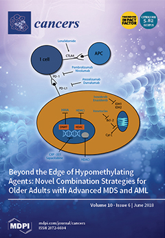
Until today, older patients with high-risk myelodysplastic syndrome (MDS) and acute myeloid leukemia (AML) have poor outcomes when treated with conventional treatment strategies. Response after a first-line treatment with hypomethylating agents (HMA) occurs in less than 50% of these patients. In our review, we focus on upcoming new treatment strategies in the field of HMA-based combination strategies. One of the current most promising approaches is preventing HMA resistance by using checkpoint inhibitors. We are sure that these new treatment options will guide the future of clinical research. Recent data also suggest a potential renaissance of intensive treatment strategies, such as therapies based on CPX-351. View this paper.
  • Issues are regarded as officially published after their release is announced to the table of contents alert mailing list.
  • You may sign up for e-mail alerts to receive table of contents of newly released issues.
  • PDF is the official format for papers published in both, html and pdf forms. To view the papers in pdf format, click on the "PDF Full-text" link, and use the free Adobe Reader to open them.
Order results
Result details
Section
Select all
Export citation of selected articles as:
14 pages, 11192 KiB  
Article
Crosstalk between ERα and Receptor Tyrosine Kinase Signalling and Implications for the Development of Anti-Endocrine Resistance
by Rugaia Z. Montaser and Helen M. Coley
Cancers 2018, 10(6), 209; https://doi.org/10.3390/cancers10060209 - 20 Jun 2018
Cited by 9 | Viewed by 3754
Abstract
Although anti-endocrine therapies have significantly advanced the treatment of breast cancer, they pose the problem of acquired drug resistance. The oestrogen receptor (ER)-expressing breast cancer cell lines MCF-7 and T47D alongside their in vitro derived resistant counterparts MCF-7-TR (tamoxifen-resistant) and T47D-FR (fulvestrant-resistant) showed [...] Read more.
Although anti-endocrine therapies have significantly advanced the treatment of breast cancer, they pose the problem of acquired drug resistance. The oestrogen receptor (ER)-expressing breast cancer cell lines MCF-7 and T47D alongside their in vitro derived resistant counterparts MCF-7-TR (tamoxifen-resistant) and T47D-FR (fulvestrant-resistant) showed dual resistance to fulvestrant and tamoxifen in the presence of upregulated HER1 and HER2 growth factor receptors. Our study demonstrated that tamoxifen resistance and fulvestrant resistance are associated with collateral sensitivity to the tyrosine kinase inhibitors (TKIs) lapatinib (p < 0.0001) and afatinib (p < 0.0001). Further, we found that over time, the TKIs reactivated ERα protein and/or mRNA in tamoxifen- and fulvestrant-resistant cells. Combinations of anti-endocrine agents with afatinib gave rise to significantly enhanced levels of apoptosis in both T47D-FR and MCF-7-TR in a synergistic manner versus additive effects of agents used singly. This was associated with p27kip1 induction for anti-endocrine-resistant cells versus parental cells. Our data supports the use of combination treatment utilising dual HER1/2 inhibitors in breast cancer patients showing resistance to multiple anti-endocrine agents. Full article
Show Figures

Graphical abstract

9 pages, 219 KiB  
Conference Report
Innovative Technologies Changing Cancer Treatment
by Sara Charmsaz, Maria Prencipe, Maeve Kiely, Graham P. Pidgeon and Denis M. Collins
Cancers 2018, 10(6), 208; https://doi.org/10.3390/cancers10060208 - 19 Jun 2018
Cited by 19 | Viewed by 4572
Abstract
Conventional therapies for cancer such as chemotherapy and radiotherapy remain a mainstay in treatment, but in many cases a targeted approach is lacking, and patients can be vulnerable to drug resistance. In recent years, novel concepts have been emerging to improve the traditional [...] Read more.
Conventional therapies for cancer such as chemotherapy and radiotherapy remain a mainstay in treatment, but in many cases a targeted approach is lacking, and patients can be vulnerable to drug resistance. In recent years, novel concepts have been emerging to improve the traditional therapeutic options in cancers with poor survival outcomes. New therapeutic strategies involving areas like energy metabolism and extracellular vesicles along with advances in immunotherapy and nanotechnology are driving the next generation of cancer treatments. The development of fields such as theranostics in nanomedicine is also opening new doors for targeted drug delivery and nano-imaging. Here we discuss the use of innovative technologies presented at the Irish Association for Cancer Research (IACR) Annual Meeting, highlighting examples of where new approaches may lead to promising new treatment options for a range of cancer types. Full article
19 pages, 1433 KiB  
Review
Hypersialylation in Cancer: Modulation of Inflammation and Therapeutic Opportunities
by Emily Rodrigues and Matthew S. Macauley
Cancers 2018, 10(6), 207; https://doi.org/10.3390/cancers10060207 - 18 Jun 2018
Cited by 134 | Viewed by 9662
Abstract
Cell surface glycosylation is dynamic and often changes in response to cellular differentiation under physiological or pathophysiological conditions. Altered glycosylation on cancers cells is gaining attention due its wide-spread occurrence across a variety of cancer types and recent studies that have documented functional [...] Read more.
Cell surface glycosylation is dynamic and often changes in response to cellular differentiation under physiological or pathophysiological conditions. Altered glycosylation on cancers cells is gaining attention due its wide-spread occurrence across a variety of cancer types and recent studies that have documented functional roles for aberrant glycosylation in driving cancer progression at various stages. One change in glycosylation that can correlate with cancer stage and disease prognosis is hypersialylation. Increased levels of sialic acid are pervasive in cancer and a growing body of evidence demonstrates how hypersialylation is advantageous to cancer cells, particularly from the perspective of modulating immune cell responses. Sialic acid-binding receptors, such as Siglecs and Selectins, are well-positioned to be exploited by cancer hypersialylation. Evidence is also mounting that Siglecs modulate key immune cell types in the tumor microenvironment, particularly those responsible for maintaining the appropriate inflammatory environment. From these studies have come new and innovative ways to block the effects of hypersialylation by directly reducing sialic acid on cancer cells or blocking interactions between sialic acid and Siglecs or Selectins. Here we review recent works examining how cancer cells become hypersialylated, how hypersialylation benefits cancer cells and tumors, and proposed therapies to abrogate hypersialylation of cancer. Full article
(This article belongs to the Special Issue Inflammation and Cancer)
Show Figures

Figure 1

18 pages, 2693 KiB  
Article
Constitutive Activation of STAT3 in Myeloma Cells Cultured in a Three-Dimensional, Reconstructed Bone Marrow Model
by Yung-Hsing Huang, Ommoleila Molavi, Abdulraheem Alshareef, Moinul Haque, Qian Wang, Michael P. Chu, Christopher P. Venner, Irwindeep Sandhu, Anthea C. Peters, Afsaneh Lavasanifar and Raymond Lai
Cancers 2018, 10(6), 206; https://doi.org/10.3390/cancers10060206 - 16 Jun 2018
Cited by 19 | Viewed by 5351
Abstract
Malignant cells cultured in three-dimensional (3D) models have been found to be phenotypically and biochemically different from their counterparts cultured conventionally. Since most of these studies employed solid tumor types, how 3D culture affects multiple myeloma (MM) cells is not well understood. Here, [...] Read more.
Malignant cells cultured in three-dimensional (3D) models have been found to be phenotypically and biochemically different from their counterparts cultured conventionally. Since most of these studies employed solid tumor types, how 3D culture affects multiple myeloma (MM) cells is not well understood. Here, we compared MM cells (U266 and RPMI8226) in a 3D culture model with those in conventional culture. While the conventionally cultured cells were present in single cells or small clusters, MM-3D cells grew in large spheroids. We discovered that STAT3 was the pathway that was more activated in 3D in both cell lines. The active form of STAT3 (phospho-STAT3 or pSTAT3), which was absent in MM cells cultured conventionally, became detectable after 1–2 days in 3D culture. This elevated pSTAT3 level was dependent on the 3D environment, since it disappeared after transferring to conventional culture. STAT3 inhibition using a pharmacological agent, Stattic, significantly decreased the cell viability of MM cells and sensitized them to bortezomib in 3D culture. Using an oligonucleotide array, we found that 3D culture significantly increased the expression of several known STAT3 downstream genes implicated in oncogenesis. Since most primary MM tumors are naturally STAT3-active, studies of MM in 3D culture can generate results that are more representative of the disease. Full article
Show Figures

Figure 1

18 pages, 2686 KiB  
Article
Oncolytic Reovirus and Immune Checkpoint Inhibition as a Novel Immunotherapeutic Strategy for Breast Cancer
by Ahmed A. Mostafa, Daniel E. Meyers, Chandini M. Thirukkumaran, Peter J. Liu, Kathy Gratton, Jason Spurrell, Qiao Shi, Satbir Thakur and Don G. Morris
Cancers 2018, 10(6), 205; https://doi.org/10.3390/cancers10060205 - 15 Jun 2018
Cited by 45 | Viewed by 8765
Abstract
As the current efficacy of oncolytic viruses (OVs) as monotherapy is limited, exploration of OVs as part of a broader immunotherapeutic treatment strategy for cancer is necessary. Here, we investigated the ability for immune checkpoint blockade to enhance the efficacy of oncolytic reovirus [...] Read more.
As the current efficacy of oncolytic viruses (OVs) as monotherapy is limited, exploration of OVs as part of a broader immunotherapeutic treatment strategy for cancer is necessary. Here, we investigated the ability for immune checkpoint blockade to enhance the efficacy of oncolytic reovirus (RV) for the treatment of breast cancer (BrCa). In vitro, oncolysis and cytokine production were assessed in human and murine BrCa cell lines following RV exposure. Furthermore, RV-induced upregulation of tumor cell PD-L1 was evaluated. In vivo, the immunocompetent, syngeneic EMT6 murine model of BrCa was employed to determine therapeutic and tumor-specific immune responses following treatment with RV, anti-PD-1 antibodies or in combination. RV-mediated oncolysis and cytokine production were observed following BrCa cell infection and RV upregulated tumor cell expression of PD-L1. In vivo, RV monotherapy significantly reduced disease burden and enhanced survival in treated mice, and was further enhanced by PD-1 blockade. RV therapy increased the number of intratumoral regulatory T cells, which was reversed by the addition of PD-1 blockade. Finally, dual treatment led to the generation of a systemic adaptive anti-tumor immune response evidenced by an increase in tumor-specific IFN-γ producing CD8+ T cells, and immunity from tumor re-challenge. The combination of PD-1 blockade and RV appears to be an efficacious immunotherapeutic strategy for the treatment of BrCa, and warrants further investigation in early-phase clinical trials. Full article
(This article belongs to the Special Issue Oncolytic Virotherapy)
Show Figures

Figure 1

15 pages, 980 KiB  
Review
The Role of Immune Checkpoint Inhibitors in Classical Hodgkin Lymphoma
by Nicholas Meti, Khashayar Esfahani and Nathalie A. Johnson
Cancers 2018, 10(6), 204; https://doi.org/10.3390/cancers10060204 - 15 Jun 2018
Cited by 27 | Viewed by 7042
Abstract
Hodgkin Lymphoma (HL) is a unique disease entity both in its pathology and the young patient population that it primarily affects. Although cure rates are high, survivorship can be linked with significant long-term morbidity associated with both chemotherapy and radiotherapy. The most significant [...] Read more.
Hodgkin Lymphoma (HL) is a unique disease entity both in its pathology and the young patient population that it primarily affects. Although cure rates are high, survivorship can be linked with significant long-term morbidity associated with both chemotherapy and radiotherapy. The most significant recent advances have been with the use of the anti-CD30-drug conjugated antibody brentuximab vedotin (BV) and inhibitors of program death 1 (PD-1). HL is genetically wired to up-regulate program death ligand 1 (PD-L1) in >95% of cases, creating a state of so-called “T cell exhaustion”, which can be reversed with immune checkpoint-inhibitor blockade. The overall and complete response rates to PD-1 inhibitors in patients with relapsed or refractory HL are 70% and 20%, respectively, with a long median duration of response of ~16 months. In fact, PD-1 inhibitors can benefit a wide spectrum of relapsed HL patients, including some who have “progressive disease” by strict response criteria. We review the biology of HL, with a focus on the immune micro-environment and mechanisms of immune evasion. We also provide the rationale supporting the use of PD-1 inhibitors in HL and highlight some of the challenges of monitoring disease response in patients treated with this immunotherapy. Full article
(This article belongs to the Special Issue Hodgkin's Lymphoma)
Show Figures

Figure 1

13 pages, 260 KiB  
Perspective
Biomarkers for Early Diagnosis and Prognosis of Malignant Pleural Mesothelioma: The Quest Goes on
by Caterina Ledda, Paola Senia and Venerando Rapisarda
Cancers 2018, 10(6), 203; https://doi.org/10.3390/cancers10060203 - 15 Jun 2018
Cited by 39 | Viewed by 10205
Abstract
Malignant pleural mesothelioma (MM) is a highly aggressive tumor characterized by a poor prognosis. Although its carcinogenesis mechanism has not been strictly understood, about 80% of MM can be attributed to occupational and/or environmental exposure to asbestos fibers. The identification of non-invasive molecular [...] Read more.
Malignant pleural mesothelioma (MM) is a highly aggressive tumor characterized by a poor prognosis. Although its carcinogenesis mechanism has not been strictly understood, about 80% of MM can be attributed to occupational and/or environmental exposure to asbestos fibers. The identification of non-invasive molecular markers for an early diagnosis of MM has been the subject of several studies aimed at diagnosing the disease at an early stage. The most studied biomarker is mesothelin, characterized by a good specificity, but it has low sensitivity, especially for non-epithelioid MM. Other protein markers are Fibulin-3 and osteopontin which have not, however, showed a superior diagnostic performance. Recently, interesting results have been reported for the HMGB1 protein in a small but limited series. An increase in channel proteins involved in water transport, aquaporins, have been identified as positive prognostic factors in MM, high levels of expression of aquaporins in tumor cells predict an increase in survival. MicroRNAs and protein panels are among the new indicators of interest. None of the markers available today are sufficiently reliable to be used in the surveillance of subjects exposed to asbestos or in the early detection of MM. Our aim is to give a detailed account of biomarkers available for MM. Full article
(This article belongs to the Special Issue Cancer Biomarkers)
11 pages, 1576 KiB  
Article
Loss of Cyclin-Dependent Kinase Inhibitor Alters Oncolytic Adenovirus Replication and Promotes More Efficient Virus Production
by Naseruddin Höti, Tamara Jane Johnson, Wasim H. Chowdhury and Ronald Rodriguez
Cancers 2018, 10(6), 202; https://doi.org/10.3390/cancers10060202 - 15 Jun 2018
Cited by 2 | Viewed by 3568
Abstract
We elucidate the role of p21/Waf-1, a cyclin-dependent kinase inhibitor, on the oncolytic infection and replication cycle of adenovirus by studying both mRNA and adenoviral proteins expression. We found that infection in the absence of p21 causes a significant increase in adenoviral genomes [...] Read more.
We elucidate the role of p21/Waf-1, a cyclin-dependent kinase inhibitor, on the oncolytic infection and replication cycle of adenovirus by studying both mRNA and adenoviral proteins expression. We found that infection in the absence of p21 causes a significant increase in adenoviral genomes and late gene expression. Similarly, the oncolytic adenoviral infected p21−/− cells have earlier formation of replication foci and robust replication kinetics that were not observed in the wild type p21/Waf-1 intact cells. These findings suggest a culmination that the presence of intact p21 in host cells causes defects in the oncolytic viral life cycle which results in the production of immature and noninfectious particles. Full article
(This article belongs to the Special Issue Oncolytic Virotherapy)
Show Figures

Figure 1

39 pages, 4806 KiB  
Review
Designer Oncolytic Adenovirus: Coming of Age
by Alexander T. Baker, Carmen Aguirre-Hernández, Gunnel Halldén and Alan L. Parker
Cancers 2018, 10(6), 201; https://doi.org/10.3390/cancers10060201 - 14 Jun 2018
Cited by 64 | Viewed by 12303
Abstract
The licensing of talimogene laherparepvec (T-Vec) represented a landmark moment for oncolytic virotherapy, since it provided unequivocal evidence for the long-touted potential of genetically modified replicating viruses as anti-cancer agents. Whilst T-Vec is promising as a locally delivered virotherapy, especially in combination with [...] Read more.
The licensing of talimogene laherparepvec (T-Vec) represented a landmark moment for oncolytic virotherapy, since it provided unequivocal evidence for the long-touted potential of genetically modified replicating viruses as anti-cancer agents. Whilst T-Vec is promising as a locally delivered virotherapy, especially in combination with immune-checkpoint inhibitors, the quest continues for a virus capable of specific tumour cell killing via systemic administration. One candidate is oncolytic adenovirus (Ad); it’s double stranded DNA genome is easily manipulated and a wide range of strategies and technologies have been employed to empower the vector with improved pharmacokinetics and tumour targeting ability. As well characterised clinical and experimental agents, we have detailed knowledge of adenoviruses’ mechanisms of pathogenicity, supported by detailed virological studies and in vivo interactions. In this review we highlight the strides made in the engineering of bespoke adenoviral vectors to specifically infect, replicate within, and destroy tumour cells. We discuss how mutations in genes regulating adenoviral replication after cell entry can be used to restrict replication to the tumour, and summarise how detailed knowledge of viral capsid interactions enable rational modification to eliminate native tropisms, and simultaneously promote active uptake by cancerous tissues. We argue that these designer-viruses, exploiting the viruses natural mechanisms and regulated at every level of replication, represent the ideal platforms for local overexpression of therapeutic transgenes such as immunomodulatory agents. Where T-Vec has paved the way, Ad-based vectors now follow. The era of designer oncolytic virotherapies looks decidedly as though it will soon become a reality. Full article
(This article belongs to the Special Issue Oncolytic Virotherapy)
Show Figures

Figure 1

15 pages, 1331 KiB  
Article
Overcoming Resistance of Human Non-Hodgkin’s Lymphoma to CD19-CAR CTL Therapy by Celecoxib and Histone Deacetylase Inhibitors
by Antoni Xavier Torres-Collado and Ali R. Jazirehi
Cancers 2018, 10(6), 200; https://doi.org/10.3390/cancers10060200 - 14 Jun 2018
Cited by 34 | Viewed by 5640
Abstract
Patients with B-cell non-Hodgkin’s lymphoma (B-NHL) who fail to respond to first-line treatment regimens or develop resistance, exhibit poor prognosis. This signifies the need to develop alternative treatment strategies. CD19-chimeric antigen receptor (CAR) T cell-redirected immunotherapy is an attractive and novel option, which [...] Read more.
Patients with B-cell non-Hodgkin’s lymphoma (B-NHL) who fail to respond to first-line treatment regimens or develop resistance, exhibit poor prognosis. This signifies the need to develop alternative treatment strategies. CD19-chimeric antigen receptor (CAR) T cell-redirected immunotherapy is an attractive and novel option, which has shown encouraging outcomes in phase I clinical trials of relapsed/refractory NHL. However, the underlying mechanisms of, and approaches to overcome, acquired anti-CD19CAR CD8+ T cells (CTL)-resistance in NHL remain elusive. CD19CAR transduced primary human CTLs kill CD19+ human NHLs in a CD19- and caspase-dependent manner, mainly via the tumor necrosis factor-related apoptosis-inducing ligand (TRAIL) apoptotic pathway. To understand the dynamics of the development of resistance, we analyzed several anti-CD19CAR CTL-resistant NHL sublines (R-NHL) derived by serial exposure of sensitive parental lines to excessive numbers of anti-CD19CAR CTLs followed by a limiting dilution analysis. The R-NHLs retained surface CD19 expression and were efficiently recognized by CD19CAR CTLs. However, R-NHLs developed cross-resistance to CD19CAR transduced human primary CTLs and the Jurkat human T cell line, activated Jurkat, and lymphokine activated killer (LAK) cells, suggesting the acquisition of resistance is independent of CD19-loss and might be due to aberrant apoptotic machinery. We hypothesize that the R-NHL refractoriness to CD19CAR CTL killing could be partially rescued by small molecule sensitizers with apoptotic-gene regulatory effects. Chromatin modifiers and Celecoxib partially reversed the resistance of R-NHL cells to the cytotoxic effects of anti-CD19CAR CTLs and rhTRAIL. These in vitro results, though they require further examination, may provide a rational biological basis for combination treatment in the management of CD19CAR CTL-based therapy of NHL. Full article
(This article belongs to the Special Issue CAR-T Cell Therapy-Novel Approaches and Challenges)
Show Figures

Figure 1

18 pages, 10279 KiB  
Review
TGF-β Sustains Tumor Progression through Biochemical and Mechanical Signal Transduction
by Robert L. Furler, Douglas F. Nixon, Christine A. Brantner, Anastas Popratiloff and Christel H. Uittenbogaart
Cancers 2018, 10(6), 199; https://doi.org/10.3390/cancers10060199 - 14 Jun 2018
Cited by 32 | Viewed by 6000
Abstract
Transforming growth factor β (TGF-β) signaling transduces immunosuppressive biochemical and mechanical signals in the tumor microenvironment. In addition to canonical SMAD transcription factor signaling, TGF-β can promote tumor growth and survival by inhibiting proinflammatory signaling and extracellular matrix (ECM) remodeling. In this article, [...] Read more.
Transforming growth factor β (TGF-β) signaling transduces immunosuppressive biochemical and mechanical signals in the tumor microenvironment. In addition to canonical SMAD transcription factor signaling, TGF-β can promote tumor growth and survival by inhibiting proinflammatory signaling and extracellular matrix (ECM) remodeling. In this article, we review how TGF-β activated kinase 1 (TAK1) activation lies at the intersection of proinflammatory signaling by immune receptors and anti-inflammatory signaling by TGF-β receptors. Additionally, we discuss the role of TGF-β in the mechanobiology of cancer. Understanding how TGF-β dampens proinflammatory responses and induces pro-survival mechanical signals throughout cancer development is critical for designing therapeutics that inhibit tumor progression while bolstering the immune response. Full article
(This article belongs to the Special Issue TGF-Beta Signaling in Cancer)
Show Figures

Figure 1

16 pages, 961 KiB  
Review
Oncolytic Viruses for Multiple Myeloma Therapy
by Christine M. Calton, Kevin R. Kelly, Faiz Anwer, Jennifer S. Carew and Steffan T. Nawrocki
Cancers 2018, 10(6), 198; https://doi.org/10.3390/cancers10060198 - 14 Jun 2018
Cited by 20 | Viewed by 6575
Abstract
Although recent treatment advances have improved outcomes for patients with multiple myeloma (MM), the disease frequently becomes refractory to current therapies. MM thus remains incurable for most patients and new therapies are urgently needed. Oncolytic viruses are a promising new class of therapeutics [...] Read more.
Although recent treatment advances have improved outcomes for patients with multiple myeloma (MM), the disease frequently becomes refractory to current therapies. MM thus remains incurable for most patients and new therapies are urgently needed. Oncolytic viruses are a promising new class of therapeutics that provide tumor-targeted therapy by specifically infecting and replicating within cancerous cells. Oncolytic therapy yields results from both direct killing of malignant cells and induction of an anti-tumor immune response. In this review, we will describe oncolytic viruses that are being tested for MM therapy with a focus on those agents that have advanced into clinical trials. Full article
(This article belongs to the Special Issue Oncolytic Virotherapy)
Show Figures

Figure 1

6 pages, 211 KiB  
Commentary
Antiviral Drugs for EBV
by Joseph S. Pagano, Christopher B. Whitehurst and Graciela Andrei
Cancers 2018, 10(6), 197; https://doi.org/10.3390/cancers10060197 - 13 Jun 2018
Cited by 50 | Viewed by 7791
Abstract
Epstein–Barr virus (EBV) infects up to 95% of the adult human population, with primary infection typically occurring during childhood and usually asymptomatic. However, EBV can cause infectious mononucleosis in approximately 35–50% cases when infection occurs during adolescence and early adulthood. Epstein–Barr virus is [...] Read more.
Epstein–Barr virus (EBV) infects up to 95% of the adult human population, with primary infection typically occurring during childhood and usually asymptomatic. However, EBV can cause infectious mononucleosis in approximately 35–50% cases when infection occurs during adolescence and early adulthood. Epstein–Barr virus is also associated with several B-cell malignancies including Burkitt lymphoma, Hodgkin lymphoma, and post-transplant lymphoproliferative disease. A number of antiviral drugs have proven to be effective inhibitors of EBV replication, yet have resulted in limited success clinically, and none of them has been approved for treatment of EBV infections. Full article
(This article belongs to the Special Issue Epstein–Barr Virus Associated Cancers)
28 pages, 2302 KiB  
Review
Endogenous Control Mechanisms of FAK and PYK2 and Their Relevance to Cancer Development
by Rayan Naser, Abdullah Aldehaiman, Escarlet Díaz-Galicia and Stefan T. Arold
Cancers 2018, 10(6), 196; https://doi.org/10.3390/cancers10060196 - 11 Jun 2018
Cited by 46 | Viewed by 7763
Abstract
Focal adhesion kinase (FAK) and its close paralogue, proline-rich tyrosine kinase 2 (PYK2), are key regulators of aggressive spreading and metastasis of cancer cells. While targeted small-molecule inhibitors of FAK and PYK2 have been found to have promising antitumor activity, their clinical long-term [...] Read more.
Focal adhesion kinase (FAK) and its close paralogue, proline-rich tyrosine kinase 2 (PYK2), are key regulators of aggressive spreading and metastasis of cancer cells. While targeted small-molecule inhibitors of FAK and PYK2 have been found to have promising antitumor activity, their clinical long-term efficacy may be undermined by the strong capacity of cancer cells to evade anti-kinase drugs. In healthy cells, the expression and/or function of FAK and PYK2 is tightly controlled via modulation of gene expression, competing alternatively spliced forms, non-coding RNAs, and proteins that directly or indirectly affect kinase activation or protein stability. The molecular factors involved in this control are frequently deregulated in cancer cells. Here, we review the endogenous mechanisms controlling FAK and PYK2, and with particular focus on how these mechanisms could inspire or improve anticancer therapies. Full article
(This article belongs to the Special Issue FAK Signaling Pathway in Cancers)
Show Figures

Figure 1

15 pages, 1147 KiB  
Article
Ensuring the Safety and Security of Frozen Lung Cancer Tissue Collections through the Encapsulation of Dried DNA
by Kevin Washetine, Mehdi Kara-Borni, Simon Heeke, Christelle Bonnetaud, Jean-Marc Félix, Lydia Ribeyre, Coraline Bence, Marius Ilié, Olivier Bordone, Marine Pedro, Priscilla Maitre, Virginie Tanga, Emmanuelle Gormally, Pascal Mossuz, Philippe Lorimier, Charles Hugo Marquette, Jérôme Mouroux, Charlotte Cohen, Sandra Lassalle, Elodie Long-Mira, Bruno Clément, Georges Dagher, Véronique Hofman and Paul Hofmanadd Show full author list remove Hide full author list
Cancers 2018, 10(6), 195; https://doi.org/10.3390/cancers10060195 - 11 Jun 2018
Cited by 5 | Viewed by 3744
Abstract
Collected specimens for research purposes may or may not be made available depending on their scarcity and/or on the project needs. Their protection against degradation or in the event of an incident is pivotal. Duplication and storage on a different site is the [...] Read more.
Collected specimens for research purposes may or may not be made available depending on their scarcity and/or on the project needs. Their protection against degradation or in the event of an incident is pivotal. Duplication and storage on a different site is the best way to assure their sustainability. The conservation of samples at room temperature (RT) by duplication can facilitate their protection. We describe a security system for the collection of non-small cell lung cancers (NSCLC) stored in the biobank of the Nice Hospital Center, France, by duplication and conservation of lyophilized (dried), encapsulated DNA kept at RT. Therefore, three frozen tissue collections from non-smoking, early stage and sarcomatoid carcinoma NSCLC patients were selected for this study. DNA was extracted, lyophilized and encapsulated at RT under anoxic conditions using the DNAshell technology. In total, 1974 samples from 987 patients were encapsulated. Six and two capsules from each sample were stored in the biobanks of the Nice and Grenoble (France) Hospitals, respectively. In conclusion, DNA maintained at RT allows for the conservation, duplication and durability of collections of interest stored in biobanks. This is a low-cost and safe technology that requires a limited amount of space and has a low environmental impact. Full article
Show Figures

Figure 1

21 pages, 950 KiB  
Review
TGF-β in T Cell Biology: Implications for Cancer Immunotherapy
by Amina Dahmani and Jean-Sébastien Delisle
Cancers 2018, 10(6), 194; https://doi.org/10.3390/cancers10060194 - 11 Jun 2018
Cited by 121 | Viewed by 12381
Abstract
Transforming Growth Factor beta (TGF-β) is a pleiotropic cytokine produced in large amounts within cancer microenvironments that will ultimately promote neoplastic progression, notably by suppressing the host’s T-cell immunosurveillance. This effect is mostly due to the well-known inhibitory effect of TGF-β on T [...] Read more.
Transforming Growth Factor beta (TGF-β) is a pleiotropic cytokine produced in large amounts within cancer microenvironments that will ultimately promote neoplastic progression, notably by suppressing the host’s T-cell immunosurveillance. This effect is mostly due to the well-known inhibitory effect of TGF-β on T cell proliferation, activation, and effector functions. Moreover, TGF-β subverts T cell immunity by favoring regulatory T-cell differentiation, further reinforcing immunosuppression within tumor microenvironments. These findings stimulated the development of many strategies to block TGF-β or its signaling pathways, either as monotherapy or in combination with other therapies, to restore anti-cancer immunity. Paradoxically, recent studies provided evidence that TGF-β can also promote differentiation of certain inflammatory populations of T cells, such as Th17, Th9, and resident-memory T cells (Trm), which have been associated with improved tumor control in several models. Here, we review current advances in our understanding of the many roles of TGF-β in T cell biology in the context of tumor immunity and discuss the possibility to manipulate TGF-β signaling to improve cancer immunotherapy. Full article
(This article belongs to the Special Issue TGF-Beta Signaling in Cancer)
Show Figures

Figure 1

14 pages, 402 KiB  
Review
Emerging Therapies and Future Directions in Targeting the Tumor Stroma and Immune System in the Treatment of Pancreatic Adenocarcinoma
by Daniel H. Ahn, Ramesh K. Ramanathan and Tanios Bekaii-Saab
Cancers 2018, 10(6), 193; https://doi.org/10.3390/cancers10060193 - 11 Jun 2018
Cited by 16 | Viewed by 4647
Abstract
Pancreatic adenocarcinoma is typically refractory to conventional treatments and associated with poor prognosis. While therapeutic advances over the past several years have improved patient outcomes, the observed benefits have been modest at best, highlighting the need for continued development of alternate treatment strategies. [...] Read more.
Pancreatic adenocarcinoma is typically refractory to conventional treatments and associated with poor prognosis. While therapeutic advances over the past several years have improved patient outcomes, the observed benefits have been modest at best, highlighting the need for continued development of alternate treatment strategies. The tumor microenvironment has been identified as being integral to oncogenesis through its direct effect on cellular pathway communication, immune inhibition, and promoting chemo-resistance. A more in depth understanding of the biology of the disease, in addition with our ability to develop more effective novel therapies have led to ongoing studies that are investigating several promising treatment options in this disease. Herein, we highlight and review the therapeutic landscape in pancreatic adenocarcinoma. Full article
(This article belongs to the Special Issue Latest Development in Pancreatic Cancer)
Show Figures

Figure 1

10 pages, 2353 KiB  
Article
Recombinant TSR1 of ADAMTS5 Suppresses Melanoma Growth in Mice via an Anti-angiogenic Mechanism
by Bhuvanasundar Renganathan, Vinoth Durairaj, Dogan Can Kirman, Paa Kow A. Esubonteng, Swee Kim Ang and Ruowen Ge
Cancers 2018, 10(6), 192; https://doi.org/10.3390/cancers10060192 - 11 Jun 2018
Cited by 9 | Viewed by 4281
Abstract
Inhibiting tumor angiogenesis is a well-established approach for anticancer therapeutic development. A Disintegrin-like and Metalloproteinase with ThromboSpondin Motifs 5 (ADAMTS5) is a secreted matrix metalloproteinase in the ADAMTS family that also functions as an anti-angiogenic/anti-tumorigenic molecule. Its anti-angiogenic/anti-tumorigenic function is independent from its [...] Read more.
Inhibiting tumor angiogenesis is a well-established approach for anticancer therapeutic development. A Disintegrin-like and Metalloproteinase with ThromboSpondin Motifs 5 (ADAMTS5) is a secreted matrix metalloproteinase in the ADAMTS family that also functions as an anti-angiogenic/anti-tumorigenic molecule. Its anti-angiogenic/anti-tumorigenic function is independent from its proteinase activity, but requires its first thrombospondin type 1 repeat (TSR1). However, it is not known if recombinant TSR1 (rTSR1) can function as an anticancer therapeutic. In this report, we expressed and purified a 75-residue recombinant TSR1 polypeptide from E. coli and investigated its ability to function as an anticancer therapeutic in mice. We demonstrate that rTSR1 is present in the blood circulation as well as in the tumor tissue at 15 min post intraperitoneal injection. Intraperitoneal delivery of rTSR1 potently suppressed subcutaneous B16F10 melanoma growth as a single agent, accompanied by diminished tumor angiogenesis, increased apoptosis, and reduced cell proliferation in the tumor tissue. Consistently, rTSR1 dose-dependently induced the apoptosis of cultured human umbilical vein endothelial cells (HUVECs) in a caspase-dependent manner. This work indicates that rTSR1 of ADAMTS5 can function as a potent anticancer therapy in mice. It thus has the potential to be further developed into an anticancer drug. Full article
(This article belongs to the Special Issue Tumor Angiogenesis: An Update)
Show Figures

Figure 1

10 pages, 664 KiB  
Commentary
WIP-YAP/TAZ as A New Pro-Oncogenic Pathway in Glioma
by Sergio Rivas, Inés M. Antón and Francisco Wandosell
Cancers 2018, 10(6), 191; https://doi.org/10.3390/cancers10060191 - 09 Jun 2018
Cited by 18 | Viewed by 5393
Abstract
Wild-type p53 (wtp53) is described as a tumour suppressor gene, and mutations in p53 occur in many human cancers. Indeed, in high-grade malignant glioma, numerous molecular genetics studies have established central roles of RTK-PI3K-PTEN and ARF-MDM2-p53 INK4a-RB pathways in promoting oncogenic capacity. Deregulation [...] Read more.
Wild-type p53 (wtp53) is described as a tumour suppressor gene, and mutations in p53 occur in many human cancers. Indeed, in high-grade malignant glioma, numerous molecular genetics studies have established central roles of RTK-PI3K-PTEN and ARF-MDM2-p53 INK4a-RB pathways in promoting oncogenic capacity. Deregulation of these signalling pathways, among others, drives changes in the glial/stem cell state and environment that permit autonomous growth. The initially transformed cell may undergo subsequent modifications, acquiring a more complete tumour-initiating phenotype responsible for disease advancement to stages that are more aggressive. We recently established that the oncogenic activity of mutant p53 (mtp53) is driven by the actin cytoskeleton-associated protein WIP (WASP-interacting protein), correlated with tumour growth, and more importantly that both proteins are responsible for the tumour-initiating cell phenotype. We reported that WIP knockdown in mtp53-expressing glioblastoma greatly reduced proliferation and growth capacity of cancer stem cell (CSC)-like cells and decreased CSC-like markers, such as hyaluronic acid receptor (CD44), prominin-1 (CD133), yes-associated protein (YAP) and transcriptional co-activator with PDZ-binding motif (TAZ). We thus propose a new CSC signalling pathway downstream of mtp53 in which Akt regulates WIP and controls YAP/TAZ stability. WIP drives a mechanism that stimulates growth signals, promoting YAP/TAZ and β-catenin stability in a Hippo-independent fashion, which allows cells to coordinate processes such as proliferation, stemness and invasiveness, which are key factors in cancer progression. Based on this multistep tumourigenic model, it is tantalizing to propose that WIP inhibitors may be applied as an effective anti-cancer therapy. Full article
Show Figures

Graphical abstract

14 pages, 39779 KiB  
Article
Dysregulated HAI-2 Plays an Important Role in Renal Cell Carcinoma Bone Metastasis through Ligand-Dependent MET Phosphorylation
by Koji Yamasaki, Shoichiro Mukai, Satoru Sugie, Takahiro Nagai, Kozue Nakahara, Toyoharu Kamibeppu, Hiromasa Sakamoto, Noboru Shibasaki, Naoki Terada, Yoshinobu Toda, Hiroaki Kataoka and Toshiyuki Kamoto
Cancers 2018, 10(6), 190; https://doi.org/10.3390/cancers10060190 - 08 Jun 2018
Cited by 5 | Viewed by 3956
Abstract
MET, a c-met proto-oncogene product and hepatocyte growth factor (HGF) receptor, is known to play an important role in cancer progression, including bone metastasis. In a previous study, we reported increased expression of MET and matriptase, a novel activator of HGF, in bone [...] Read more.
MET, a c-met proto-oncogene product and hepatocyte growth factor (HGF) receptor, is known to play an important role in cancer progression, including bone metastasis. In a previous study, we reported increased expression of MET and matriptase, a novel activator of HGF, in bone metastasis. In this study, we employed a mouse model of renal cell carcinoma (RCC) bone metastasis to clarify the significance of the HGF/MET signaling axis and the regulator of HGF activator inhibitor type-2 (HAI-2). Luciferase-transfected 786-O cells were injected into the left cardiac ventricle of mice to prepare the mouse model of bone metastasis. The formation of bone metastasis was confirmed by whole-body bioluminescent imaging, and specimens were extracted. Expression of HGF/MET-related molecules was analyzed. Based on the results, we produced HAI-2 stable knockdown 786-O cells, and analyzed invasiveness and motility. Expression of HGF and matriptase was increased in bone metastasis compared with the control, while that of HAI-2 was decreased. Furthermore, we confirmed increased phosphorylation of MET in bone metastasis. The expression of matriptase was upregulated, and both invasiveness and motility were increased significantly by knockdown of HAI-2. The significance of ligand-dependent MET activation in RCC bone metastasis is considered, and HAI-2 may be an important regulator in this system. Full article
(This article belongs to the Special Issue Targeting Bone Metastasis in Cancer)
Show Figures

Figure 1

22 pages, 1504 KiB  
Review
The Roles of p53 in Mitochondrial Dynamics and Cancer Metabolism: The Pendulum between Survival and Death in Breast Cancer?
by David E. Moulder, Diana Hatoum, Enoch Tay, Yiguang Lin and Eileen M. McGowan
Cancers 2018, 10(6), 189; https://doi.org/10.3390/cancers10060189 - 08 Jun 2018
Cited by 46 | Viewed by 14224
Abstract
Cancer research has been heavily geared towards genomic events in the development and progression of cancer. In contrast, metabolic regulation, such as aberrant metabolism in cancer, is poorly understood. Alteration in cellular metabolism was once regarded simply as a consequence of cancer rather [...] Read more.
Cancer research has been heavily geared towards genomic events in the development and progression of cancer. In contrast, metabolic regulation, such as aberrant metabolism in cancer, is poorly understood. Alteration in cellular metabolism was once regarded simply as a consequence of cancer rather than as playing a primary role in cancer promotion and maintenance. Resurgence of cancer metabolism research has identified critical metabolic reprogramming events within biosynthetic and bioenergetic pathways needed to fulfill the requirements of cancer cell growth and maintenance. The tumor suppressor protein p53 is emerging as a key regulator of metabolic processes and metabolic reprogramming in cancer cells—balancing the pendulum between cell death and survival. This review provides an overview of the classical and emerging non-classical tumor suppressor roles of p53 in regulating mitochondrial dynamics: mitochondrial engagement in cell death processes in the prevention of cancer. On the other hand, we discuss p53 as a key metabolic switch in cellular function and survival. The focus is then on the conceivable roles of p53 in breast cancer metabolism. Understanding the metabolic functions of p53 within breast cancer metabolism will, in due course, reveal critical metabolic hotspots that cancers advantageously re-engineer for sustenance. Illustration of these events will pave the way for finding novel therapeutics that target cancer metabolism and serve to overcome the breast cancer burden. Full article
(This article belongs to the Special Issue p53 Signaling in Cancers)
Show Figures

Figure 1

16 pages, 424 KiB  
Review
Gain-of-Function (GOF) Mutant p53 as Actionable Therapeutic Target
by Ramona Schulz-Heddergott and Ute M. Moll
Cancers 2018, 10(6), 188; https://doi.org/10.3390/cancers10060188 - 07 Jun 2018
Cited by 83 | Viewed by 8322
Abstract
p53 missense mutant alleles are present in nearly 40% of all human tumors. Such mutated alleles generate aberrant proteins that not only lose their tumor-suppressive functions but also frequently act as driver oncogenes, which promote malignant progression, invasion, metastasis, and chemoresistance, leading to [...] Read more.
p53 missense mutant alleles are present in nearly 40% of all human tumors. Such mutated alleles generate aberrant proteins that not only lose their tumor-suppressive functions but also frequently act as driver oncogenes, which promote malignant progression, invasion, metastasis, and chemoresistance, leading to reduced survival in patients and mice. Notably, these oncogenic gain-of-function (GOF) missense mutant p53 proteins (mutp53) are constitutively and tumor-specific stabilised. This stabilisation is one key pre-requisite for their GOF and is largely due to mutp53 protection from the E3 ubiquitin ligases Mdm2 and CHIP by the HSP90/HDAC6 chaperone machinery. Recent mouse models provide convincing evidence that tumors with highly stabilized GOF mutp53 proteins depend on them for growth, maintenance, and metastasis, thus creating exploitable tumor-specific vulnerabilities that markedly increase lifespan if intercepted. This identifies mutp53 as a promising cancer-specific drug target. This review discusses direct mutp53 protein-targeting drug strategies that are currently being developed at various preclinical levels. Full article
(This article belongs to the Special Issue p53 Signaling in Cancers)
Show Figures

Figure 1

20 pages, 640 KiB  
Review
Evolving Treatment Strategies for Elderly Leukemia Patients with IDH Mutations
by Michael J. Buege, Adam J. DiPippo and Courtney D. DiNardo
Cancers 2018, 10(6), 187; https://doi.org/10.3390/cancers10060187 - 06 Jun 2018
Cited by 27 | Viewed by 4639
Abstract
Acute myeloid leukemia (AML) is a debilitating and life-threatening condition, especially for elderly patients who account for over 50% of diagnoses. For over four decades, standard induction therapy with intensive cytotoxic chemotherapy for AML had remained unchanged. However, for most patients, standard therapy [...] Read more.
Acute myeloid leukemia (AML) is a debilitating and life-threatening condition, especially for elderly patients who account for over 50% of diagnoses. For over four decades, standard induction therapy with intensive cytotoxic chemotherapy for AML had remained unchanged. However, for most patients, standard therapy continues to have its shortcomings, especially for elderly patients who may not be able to tolerate the complications from intensive cytotoxic chemotherapy. New research into the development of targeted and alternative therapies has led to a new era in AML therapy. For the nearly 20% of diagnoses harboring a mutation in isocitrate dehydrogenase 1 or 2 (IDH1/2), potential treatment options have undergone a paradigm shift away from intensive cytotoxic chemotherapy and towards targeted therapy alone or in combination with lower intensity chemotherapy. The first FDA approved IDH2 inhibitor was enasidenib in 2017. In addition, IDH1 inhibitors are in ongoing clinical studies, and the oral BCL-2 inhibitor venetoclax shows preliminary efficacy in this subset of patients. These new tools aim to improve outcomes and change the treatment paradigm for elderly patients with IDH mutant AML. However, the challenge of how to best incorporate these agents into standard practice remains. Full article
(This article belongs to the Special Issue Treatment of Older Adults with Acute Myeloid Leukemia)
Show Figures

Figure 1

13 pages, 2494 KiB  
Article
Regulation of Constitutive Interferon-Stimulated Genes (Isgs) in Tumor Cells Contributes to Enhanced Antitumor Response of Newcastle Disease Virus-Infected Tumor Vaccines
by Mai Takamura-Ishii, Takaaki Nakaya and Katsuro Hagiwara
Cancers 2018, 10(6), 186; https://doi.org/10.3390/cancers10060186 - 06 Jun 2018
Cited by 5 | Viewed by 4373
Abstract
Newcastle disease virus (NDV) is an oncolytic virus. As immunogenicity of tumor cells is enhanced by NDV infection, recombinant NDV-infected tumor vaccines (rNDV-TV) are effective methods for inducing specific immunity. However, several tumor cells resist NDV infection, and tumor specific immunity is not [...] Read more.
Newcastle disease virus (NDV) is an oncolytic virus. As immunogenicity of tumor cells is enhanced by NDV infection, recombinant NDV-infected tumor vaccines (rNDV-TV) are effective methods for inducing specific immunity. However, several tumor cells resist NDV infection, and tumor specific immunity is not sufficiently induced by rNDV-TV. Therefore, we clarified the factor contributing to the suppression of NDV infection and attempted to improve rNDV-TV. Initially we investigated the correlation between the NDV infection rate and interferon-related gene expression in six murine tumor cell lines. A significant negative correlation was observed between the constitutive gene expression of Interferon-stimulated genes (ISGs) and NDV infectivity. The NDV infection rate was examined in each tumor cell treated with the Janus kinase (JAK) inhibitor ruxolitinib (Rux). Furthermore, we evaluated the Th1 response induction by Rux-treated rNDV-TV (rNDV-TV-Rux). In Rux-treated tumor cells, Oasl2 gene expression was significantly decreased and viral infectivity was increased. In immunized mice, the number of CD8+ cells, and those expressing the IFN-γ gene, were significantly increased as compared with Rux-untreated rNDV-TV. The infectivity of the virus was dependent on the degree of ISGs expression in tumor cells. To remedy for this problem, rNDV-TV-Rux was expected to have a Th1 immune response. Full article
(This article belongs to the Special Issue Oncolytic Virotherapy)
Show Figures

Graphical abstract

14 pages, 1308 KiB  
Review
Cutting to the Chase: How Matrix Metalloproteinase-2 Activity Controls Breast-Cancer-to-Bone Metastasis
by Marilena Tauro and Conor C. Lynch
Cancers 2018, 10(6), 185; https://doi.org/10.3390/cancers10060185 - 05 Jun 2018
Cited by 52 | Viewed by 7347
Abstract
Bone metastatic breast cancer is currently incurable and will be evident in more than 70% of patients that succumb to the disease. Understanding the factors that contribute to the progression and metastasis of breast cancer can reveal therapeutic opportunities. Matrix metalloproteinases (MMPs) are [...] Read more.
Bone metastatic breast cancer is currently incurable and will be evident in more than 70% of patients that succumb to the disease. Understanding the factors that contribute to the progression and metastasis of breast cancer can reveal therapeutic opportunities. Matrix metalloproteinases (MMPs) are a family of proteolytic enzymes whose role in cancer has been widely documented. They are capable of contributing to every step of the metastatic cascade, but enthusiasm for the use of MMP inhibition as a therapeutic approach has been dampened by the disappointing results of clinical trials conducted more than 20 years ago. Since the trials, our knowledge of MMP biology has expanded greatly. Combined with advances in the selective targeting of individual MMPs and the specific delivery of therapeutics to the tumor microenvironment, we may be on the verge of finally realizing the promise of MMP inhibition as a treatment strategy. Here, as a case in point, we focus specifically on MMP-2 as an example to show how it can contribute to each stage of breast-cancer-to-bone metastasis and also discuss novel approaches for the selective targeting of MMP-2 in the setting of the bone-cancer microenvironment. Full article
(This article belongs to the Special Issue Targeting Bone Metastasis in Cancer)
Show Figures

Figure 1

17 pages, 4041 KiB  
Article
RIPK2: New Elements in Modulating Inflammatory Breast Cancer Pathogenesis
by Alaa Zare, Alexandra Petrova, Mehdi Agoumi, Heather Armstrong, Gilbert Bigras, Katia Tonkin, Eytan Wine and Shairaz Baksh
Cancers 2018, 10(6), 184; https://doi.org/10.3390/cancers10060184 - 05 Jun 2018
Cited by 25 | Viewed by 5772
Abstract
Inflammatory breast cancer (IBC) is a rare and aggressive form of breast cancer that is associated with significantly high mortality. In spite of advances in IBC diagnoses, the prognosis is still poor compared to non-IBC. Due to the aggressive nature of the disease, [...] Read more.
Inflammatory breast cancer (IBC) is a rare and aggressive form of breast cancer that is associated with significantly high mortality. In spite of advances in IBC diagnoses, the prognosis is still poor compared to non-IBC. Due to the aggressive nature of the disease, we hypothesize that elevated levels of inflammatory mediators may drive tumorigenesis and metastasis in IBC patients. Utilizing IBC cell models and patient tumor samples, we can detect elevated NF-κB activity and hyperactivation of non-canonical drivers of NF-κB (nuclear factor kappaB)-directed inflammation such as tyrosine phosphorylated receptor-interacting protein kinase 2 (pY RIPK2), when compared to non-IBC cells or patients. Interestingly, elevated RIPK2 activity levels were present in a majority of pre-chemotherapy samples from IBC patients at the time of diagnosis to suggest that patients at diagnosis had molecular activation of NF-κB via RIPK2, a phenomenon we define as “molecular inflammation”. Surprisingly, chemotherapy did cause a significant increase in RIPK2 activity and thus molecular inflammation suggesting that chemotherapy does not resolve the molecular activation of NF-κB via RIPK2. This would impact on the metastatic potential of IBC cells. Indeed, we can demonstrate that RIPK2 activity correlated with advanced tumor, metastasis, and group stage as well as body mass index (BMI) to indicate that RIPK2 might be a useful prognostic marker for IBC and advanced stage breast cancer. Full article
(This article belongs to the Special Issue Inflammation and Cancer)
Show Figures

Figure 1

17 pages, 1227 KiB  
Review
Clinico-Pathological Importance of TGF-β/Phospho-Smad Signaling during Human Hepatic Fibrocarcinogenesis
by Katsunori Yoshida, Koichi Matsuzaki, Miki Murata, Takashi Yamaguchi, Kanehiko Suwa and Kazuichi Okazaki
Cancers 2018, 10(6), 183; https://doi.org/10.3390/cancers10060183 - 05 Jun 2018
Cited by 58 | Viewed by 7873
Abstract
Chronic viral hepatitis is a global public health problem, with approximately 570 million persons chronically infected. Hepatitis B and C viruses increase the risk of morbidity and mortality from liver cirrhosis, hepatocellular carcinoma (HCC), and extrahepatic complications that develop. Hepatitis virus infection induces [...] Read more.
Chronic viral hepatitis is a global public health problem, with approximately 570 million persons chronically infected. Hepatitis B and C viruses increase the risk of morbidity and mortality from liver cirrhosis, hepatocellular carcinoma (HCC), and extrahepatic complications that develop. Hepatitis virus infection induces transforming growth factor (TGF)-β, which influences microenvironments within the infected liver. TGF-β promotes liver fibrosis by up-regulating extracellular matrix production by hepatic stellate cells. TGF-β is also up-regulated in patients with HCC, in whom it contributes importantly to bringing about a favorable microenvironment for tumor growth. Thus, TGF-β is thought to be a major factor regulating liver fibrosis and carcinogenesis. Since TGF-β carries out regulatory signaling by influencing the phosphorylation of Smads, we have generated several kinds of phospho-specific antibodies to Smad2/3. Using these, we have identified three types of phospohorylated forms: COOH-terminally phosphorylated Smad2/3 (pSmad2C and pSmad3C), linker phosphorylated Smad2/3 (pSmad2L and pSmad3L), and dually phosphorylated Smad3 (pSmad2L/C and pSmad3L/C). TGF-β-mediated pSmad2/3C signaling terminates cell proliferation; on the other hand, cytokine-induced pSmad3L signaling accelerates cell proliferation and promotes fibrogenesis. This review addresses TGF-β/Smad signal transduction in chronic liver injuries and carcinogenic processes. We also discuss the reversibility of Smad signaling after antiviral therapy. Full article
(This article belongs to the Special Issue TGF-Beta Signaling in Cancer)
Show Figures

Figure 1

37 pages, 3829 KiB  
Review
Cancer Metastases to Bone: Concepts, Mechanisms, and Interactions with Bone Osteoblasts
by Alison B. Shupp, Alexus D. Kolb, Dimpi Mukhopadhyay and Karen M. Bussard
Cancers 2018, 10(6), 182; https://doi.org/10.3390/cancers10060182 - 04 Jun 2018
Cited by 91 | Viewed by 8962
Abstract
The skeleton is a unique structure capable of providing support for the body. Bone resorption and deposition are controlled in a tightly regulated balance between osteoblasts and osteoclasts with no net bone gain or loss. However, under conditions of disease, the balance between [...] Read more.
The skeleton is a unique structure capable of providing support for the body. Bone resorption and deposition are controlled in a tightly regulated balance between osteoblasts and osteoclasts with no net bone gain or loss. However, under conditions of disease, the balance between bone resorption and deposition is upset. Osteoblasts play an important role in bone homeostasis by depositing new bone osteoid into resorption pits. It is becoming increasingly evident that osteoblasts additionally play key roles in cancer cell dissemination to bone and subsequent metastasis. Our laboratory has evidence that when osteoblasts come into contact with disseminated breast cancer cells, the osteoblasts produce factors that initially reduce breast cancer cell proliferation, yet promote cancer cell survival in bone. Other laboratories have demonstrated that osteoblasts both directly and indirectly contribute to dormant cancer cell reactivation in bone. Moreover, we have demonstrated that osteoblasts undergo an inflammatory stress response in late stages of breast cancer, and produce inflammatory cytokines that are maintenance and survival factors for breast cancer cells and osteoclasts. Advances in understanding interactions between osteoblasts, osteoclasts, and bone metastatic cancer cells will aid in controlling and ultimately preventing cancer cell metastasis to bone. Full article
(This article belongs to the Special Issue Targeting Bone Metastasis in Cancer)
Show Figures

Figure 1

16 pages, 916 KiB  
Review
The Glucose-Regulated MiR-483-3p Influences Key Signaling Pathways in Cancer
by Felice Pepe, Rosa Visone and Angelo Veronese
Cancers 2018, 10(6), 181; https://doi.org/10.3390/cancers10060181 - 04 Jun 2018
Cited by 34 | Viewed by 5823
Abstract
The hsa-mir-483 gene, located within the IGF2 locus, transcribes for two mature microRNAs, miR-483-5p and miR-483-3p. This gene, whose regulation is mediated by the the CTNNB1/USF1 complex, shows an independent expression from its host gene IGF2. The miR-483-3p affects the Wnt/β-catenin, [...] Read more.
The hsa-mir-483 gene, located within the IGF2 locus, transcribes for two mature microRNAs, miR-483-5p and miR-483-3p. This gene, whose regulation is mediated by the the CTNNB1/USF1 complex, shows an independent expression from its host gene IGF2. The miR-483-3p affects the Wnt/β-catenin, the TGF-β, and the TP53 signaling pathways by targeting several genes as CTNNB1, SMAD4, IGF1, and BBC3. Accordingly, miR-483-3p is associated with various tissues specific physiological properties as insulin and melanin production, as well as with cellular physiological functions such as wounding, differentiation, proliferation, and survival. Deregulation of miR-483-3p is observed in different types of cancer, and its overexpression can inhibit the pro-apoptotic pathway induced by the TP53 target effectors. As a result, the oncogenic characteristics of miR-483-3p are linked to the effect of some of the most relevant cancer-related genes, TP53 and CTNNB1, as well as to one of the most important cancer hallmark: the aberrant glucose metabolism of tumor cells. In this review, we summarize the recent findings regarding the miR-483-3p, to elucidate its functional role in physiological and pathological contexts, focusing overall on its involvement in cancer and in the TP53 pathway. Full article
(This article belongs to the Special Issue p53 Signaling in Cancers)
Show Figures

Graphical abstract

28 pages, 5341 KiB  
Article
KIAA0100 Modulates Cancer Cell Aggression Behavior of MDA-MB-231 through Microtubule and Heat Shock Proteins
by Zhenyu Zhong, Vaishali Pannu, Matthew Rosenow, Adam Stark and David Spetzler
Cancers 2018, 10(6), 180; https://doi.org/10.3390/cancers10060180 - 04 Jun 2018
Cited by 9 | Viewed by 4153
Abstract
The KIAA0100 gene was identified in the human immature myeloid cell line cDNA library. Recent studies have shown that its expression is elevated in breast cancer and associated with more aggressive cancer types as well as poor outcomes. However, its cellular and molecular [...] Read more.
The KIAA0100 gene was identified in the human immature myeloid cell line cDNA library. Recent studies have shown that its expression is elevated in breast cancer and associated with more aggressive cancer types as well as poor outcomes. However, its cellular and molecular function is yet to be understood. Here we show that silencing KIAA0100 by siRNA in the breast cancer cell line MDA-MB-231 significantly reduced the cancer cells’ aggressive behavior, including cell aggregation, reattachment, cell metastasis and invasion. Most importantly, silencing the expression of KIAA0100 particularly sensitized the quiescent cancer cells in suspension culture to anoikis. Immunoprecipitation, mass spectrometry and immunofluorescence analysis revealed that KIAA0100 may play multiple roles in the cancer cells, including stabilizing microtubule structure as a microtubule binding protein, and contributing to MDA-MB-231 cells Anoikis resistance by the interaction with stress protein HSPA1A. Our study also implies that the interaction between KIAA0100 and HSPA1A may be targeted for new drug development to specifically induce anoikis cell death in the cancer cell. Full article
Show Figures

Figure 1

Previous Issue
Next Issue
Back to TopTop