Next Article in Journal
Automated Dermoscopy Image Analysis of Pigmented Skin Lesions
Next Article in Special Issue
Possible DNA Viral Factors of Human Breast Cancer
Previous Article in Journal
Thioredoxin and Cancer: A Role for Thioredoxin in all States of Tumor Oxygenation
Previous Article in Special Issue
Increased Level of Phosphorylated ShcA Measured by Chemiluminescence-Linked Immunoassay Is a Predictor of Good Prognosis in Primary Breast Cancer Expressing Low Levels of Estrogen Receptor
 
 
Correction published on 30 August 2010, see Cancers 2010, 2(3), 1642-1643.
Article

Identification of Insulin-Like Growth Factor-I Receptor (IGF-IR) Gene Promoter-Binding Proteins in Estrogen Receptor (ER)-Positive and ER-Depleted Breast Cancer Cells

1
Department of Human Molecular Genetics and Biochemistry, Sackler School of Medicine, Tel Aviv University, Tel Aviv 69978, Israel
2
Department of Clinical and Experimental Medicine, University Magna Graecia of Catanzaro, Catanzaro 88100, Italy
*
Author to whom correspondence should be addressed.
Cancers 2010, 2(2), 233-261; https://doi.org/10.3390/cancers2020233
Submission received: 23 February 2010 / Accepted: 19 March 2010 / Published: 25 March 2010
(This article belongs to the Special Issue Breast Cancer)

Abstract

:
The insulin-like growth factor I receptor (IGF-IR) has been implicated in the etiology of breast cancer. Overexpression of the IGF-IR gene is a typical feature of most primary breast cancers, whereas low IGF-IR levels are seen at advanced stages. Hence, evaluation of IGF-IR levels might be important for assessing prognosis. In the present study, we employed a proteomic approach based on DNA affinity chromatography followed either by mass spectroscopy (MS) or Western blot analysis to identify transcription factors that may associate with the IGF-IR promoter in estrogen receptor (ER)-positive and ER-depleted breast cancer cells. A biotinylated IGF-IR promoter fragment was bound to streptavidin magnetic beads and incubated with nuclear extracts of breast cancer cells. IGF-IR promoter-binding proteins were eluted with high salt and analyzed by MS and Western blots. Among the proteins that were found to bind to the IGF-IR promoter we identified zinc finger transcription factors Sp1 and KLF6, ER-α, p53, c-jun, and poly (ADP-ribosylation) polymerase. Furthermore, chromatin immune-precipitation (ChIP) analysis confirmed the direct in vivo binding of some of these transcription factors to IGF-IR promoter DNA. The functional relevance of binding data was assessed by cotransfection experiments with specific expression vectors along with an IGF-IR promoter reporter. In summary, we identified nuclear proteins that are potentially responsible for the differential expression of the IGF-IR gene in ER-positive and ER-depleted breast cancer cells.

1. Introduction

The insulin-like growth factors, IGF-I and IGF-II, are mitogenic polypeptides with important roles in cell growth and differentiation [1]. The biological activities of IGF-I and IGF-II are mediated by the IGF-I receptor (IGF-IR) and are modulated by a family of IGF-binding proteins (IGFBPs) that control ligand bioavailability [2,3,4]. The IGF-IR is essential for normal cell cycle progression and has a crucial role in tumor growth and metastasis development. Ligand-mediated activation of the IGF-IR tyrosine kinase domain leads to mitogenic and antiapoptotic effects in multiple cells and cancer-derived cell lines [5]. In the specific context of breast tumorigenesis, the IGF-IR is overexpressed in most primary tumors [4]. In addition, and consistent with its potent antiapoptotic role, IGF-IR-null cells do not undergo transformation when exposed to different oncogenes [6,7]. On the other hand, inhibition of IGF-IR signaling by IGF-IR antibodies, antisense oligonucleotides, IGF-BPs, or specific IGF-IR kinase inhibitors results in reduced cell proliferation and cancer progression in vivo and in vitro [8,9,10,11,12,13,14]. Furthermore, epidemiological studies revealed that high levels of circulating IGF-I are linked to an increased risk of developing breast cancer in premenopausal women [15,16].
Regulation of IGF-IR gene expression is mainly attained at the transcription level. The IGF-IR promoter is a TATA-less, CCAAT-less, highly GC-rich, ‘initiator’-type of promoter. IGF-IR gene transcription is dependent on a number of stimulatory zinc-finger nuclear proteins, including Sp1 [17] and KLF6 [18]. In addition, IGF-IR gene transcription is negatively regulated by several tumor suppressors, including BRCA1, p53/p63/p73, the von Hippel-Lindau protein (VHL), and the Wilms’ protein-1 (WT1) [19,20,21,22,23,24,25]. Interactions between stimulatory and inhibitory transcription factors play an important role in IGF-IR gene regulation and, therefore, were postulated to have a major impact on the proliferative status of the cell. The molecular mechanisms and specific transcription factors responsible for regulating IGF-IR gene expression in breast cancer cells, however, have not yet been identified.
The IGF-I and estrogen signaling systems were shown to act in a synergistic fashion in breast epithelial cells [25]. Estrogens regulate IGF-I signaling and the expression of several members of the IGF system [25,26,27,28,29,30,31]. Moreover, activation of estrogen receptor-α (ERα) by estrogens induces a physical interaction between ERα and IGF-IR [32] that results in activation and phosphorylation of IGF-IR and downstream signaling molecules [33,34,35,36]. The aim of this study was to identify the collections of IGF-IR promoter-binding transcription factors in ER-positive and ER-depleted breast cancer cells. Using DNA affinity chromatography, mass spectroscopy (MS), and Western blot analyses, we identified a series of known and previously unidentified transcription factors that specifically bind to the IGF-IR promoter in either cell type. The ability of selected proteins to bind and transactivate the IGF-IR promoter was confirmed by chromatin immunoprecipitation (ChIP) and transient transfection assays. In addition, we identified a number of non-DNA sequence-specific nuclear proteins that are probably involved in IGF-IR gene regulation. Identification of differentially expressed IGF-IR promoter-binding and non-binding transcription factors may help elucidate the mechanisms responsible for the differential expression of the IGF-IR gene in ER-positive and ER-depleted breast cancer cells.

2. Material and Methods

2.1. Cell Cultures

Human breast cancer-derived MCF7 cells [(ER-positive), American Type Culture Collection, Manassas, VA, USA] were grown in Dulbecco’s modified Eagle’s medium (DMEM) supplemented with 10% fetal bovine serum (FBS), 2 mM glutamine, 50 μg/mL gentamicin sulfate, and 5.6 mg/L fungizone (Sigma-Aldrich Co., St. Louis, MO, USA). The C4.12.5 cell line was derived by clonal selection of MCF7 cells that were grown in the absence of estrogen for nine months [37]. C4.12.5 cells were maintained in phenol red-free DMEM with 10% charcoal/dextran-treated FBS, 2 mM glutamine, and antibiotics. The C4.12.5 cell line was provided by Dr. Wade V. Welshons (University of Missouri, Columbia, MO, USA). Cells were incubated at 37 °C in a humidified atmosphere containing 5% CO2.

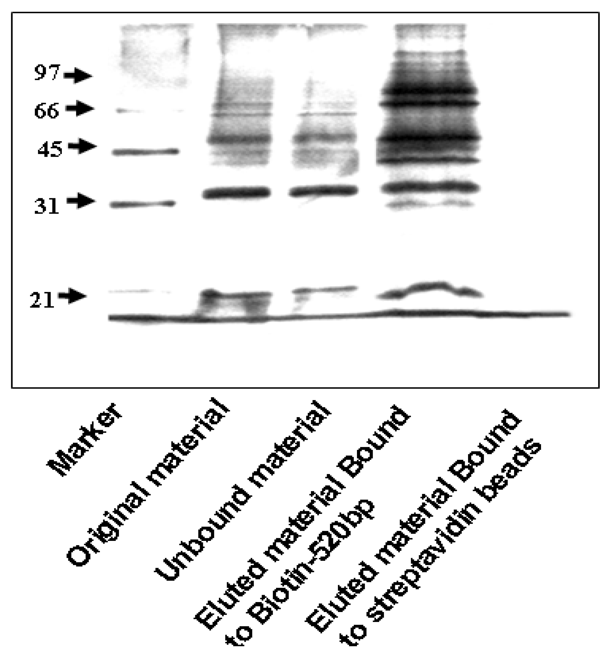
2.2. PCR and DNA Affinity Chromatography of the IGF-IR Promoter

For DNA affinity chromatography, a 511-bp human proximal IGF-IR promoter fragment extending from nucleotides −458 to +53 was labeled using a 5’-biotinylated antisense primer. This fragment includes most of the proximal 5'-flanking region and comprises the ‘initiator’ motif from which transcription starts in vivo (Nucleotide 1) (Figure 1).
Primer sequences were derived from genomic IGF-IR clone #R73P2 [38] as follows: sense, 5'-CTTTCCAGCCGCGCTGTTGTTG-3'; antisense, 5'-(Biotin)-GGTAAACAAGAGCCCCAGCCTC-3'. PCR was performed using the TermalAceTM DNA Polymerase reagent (InVitrogen, Carlsbad, CA, USA). The biotinylated PCR product was bound to streptavidin magnetic beads (Dynabeads® M-270 Streptavidin; Dynak Biotech ASA, Oslo, Norway), and incubated with nuclear extracts of MCF7 or C4.12.5 cells in the presence of double stranded poly dI-dC (Amersham Pharmacia Biotech, Piscataway, NJ, USA). Following high-salt elution, specifically-bound proteins were resolved on 10% SDS-PAGE gels and visualized by silver staining (Figure 2). Gel slices were then submitted for MS analysis at the Smoler Proteomics Center (Technion-Israel Institute of Technology, Haifa, Israel) or analyzed by Western blotting as described below.
Figure 1. IGF-IR promoter. (A) Schematic representation of the IGF-IR promoter region [5]. The initiator (INR) element is denoted by an arrow. The coding region, starting with the AUG codon, is shown in gray. The translation start site is denoted by a dashed arrow. The 5’-flanking region is denoted by a black bar, and the 5’-untranslated region (UTR) is represented by a dotted bar. The location of primers (P#1 and P#2), employed to amplify the proximal promoter is indicated. (B) PCR amplification of the human IGF-IR promoter. The PCR reaction was performed using a biotinylated antisense primer, as described under Materials and Methods section, and 4 μL of the biotinylated PCR product was loaded on a 1% agarose gel. A PCR reaction without DNA served as negative control. A 100-bp DNA ladder was used as a M.W. marker.
Cancers 02 00233 g001
Figure 2. Silver staining of IGF-IR promoter-bound proteins identified by DNA affinity chromatography. Nuclear extracts of MCF7 cells were prepared as described under Materials and Methods Section. Proteins bound to IGF-IR promoter DNA (10 μg) were electrophoresed through 10% SDS-PAGE, fixed, and stained with silver (Bio-Rad, Hercules, CA, USA). Lane 1, M.W. marker; lane 2, starting material (MCF7 nuclear extract, 4.2 μg); lane 3, unbound material (MCF7 nuclear extract that did not bind to DNA, 4.2 μg); lane 4, eluted material bound to Biotin-520 bp (MCF7 nuclear proteins that bound to IGF-IR promoter, 10 μg); and lane 5, negative control (MCF7 nuclear proteins bound to strepavidin magnetic beads, 10 μg).
Cancers 02 00233 g002

2.3. Gel Slices Proteolysis and Mass Spectroscopy Analysis

The gel slices containing the specific DNA-bound proteins were reduced with 10 mM DTT, incubated at 60 °C for 30 min, carboxyamidomethylated with 40 mM iodoacetamide at room temperature for 30 min, and trypsinized overnight at 37 °C [modified trypsin (Promega)] at a 1:100 enzyme-to-substrate ratio. The tryptic peptides were resolved by reverse-phase chromatography on 0.075 × 200-mm fused silica capillaries (J&W, Wilmington, DE, USA) packed with Reprosil reversed phase material (Dr Maisch GmbH, Germany). The peptides were eluted with a linear 90 min gradient from 5 to 45% acetonitrile containing 0.1% formic acid, followed by a wash of 95% acetonitrile for 15 min. The flow rate was 0.25 μl/min and the peptides were electrosprayed into an ion-trap mass spectrometer (Orbitrap, Thermo Fisher Scientific Inc., USA). MS was performed in a positive mode using repetitively full MS scans followed by collision induced dissociation (CID) of the 7 most dominant ions selected from the first MS scan. The MS data was clustered and analyzed using the Sequest software (Finnigan, San Jose, CA, USA) and Pep-Miner [39] searching against International Protein Index (IPI) protein IDs (part of the NR-NCBI database).

2.4. Cell Fractionation

Whole cell extracts were prepared by lysing cells in a buffer containing protease inhibitors, as described previously [20]. To prepare cytosolic and nuclear extracts, cells were washed with ice-cold phosphate-buffered saline (PBS), centrifuged, and the pellet was resuspended in buffer A [10 mM Hepes (pH 7.9), 1.5 mM MgCl2, 10 mM KCl, 0.1 mM EDTA, 0.1 mM EGTA, 1 mM dithiothreitol, 2 μg/mL aprotinin, 1 mM ortovanadate, 2 μg/mL leupeptin, 4 mM pyrophosphate, 2 μg/mL pepstatin, and 1 mM PMSF] and incubated for 30 min on ice. IGEPAL (CA-630, Sigma-Aldrich Co.) was then added to a final concentration of 3%, vortexed, and centrifuged (14,000 rpm, 30 sec), after which the supernatant containing the cytosolic fraction was saved at −70 °C. Buffer C [20 mM Hepes, 420 mM NaCl, 1 mM EDTA, 1 mM EGTA, and protease inhibitors] was added to the nuclei-containing pellet, incubated for 30 min at room temperature, and spun at 14,000 rpm for 5 min. The supernatant containing the nuclear extract was frozen at −70 °C. Protein concentrations were determined with the Bradford reagent (Bio-Rad, Hercules, CA, USA) using bovine serum albumin (BSA) as a standard.

2.5. Western Immunoblots

Samples were electrophoresed through 12%, 10%, or 7.5% SDS-PAGE gels, followed by blotting of the proteins onto nitrocellulose membranes. After blocking with either skim milk or 3% BSA, the blots were incubated overnight with the antibodies listed below, washed and incubated with the appropriate horseradish peroxidase (HRP)-conjugated secondary antibody. Antibodies against BRCA1 (C-20), TOPOII (C-15), MET (C-28), p120 (H-90), androgen receptor [(AR), N-20], IGF-IR β-subunit (C-20), Sp1 (PEP2), TCF3A (M-20), c-fos (4), ER α (MC-20), WT1 (C-19), KLF6 (R-173), lamin A/C (636), Smad2/3 (FL-425), c-Myc (9E10), p63 (4A4), E2F1 (KH95), LEF1 (C-19), TCF1A (C-21), p53 (mixture of DO-1 and Pab 1801), GSK3β (11B7), phospho c-jun (KM-1), cyclin D (H295), cyclin G (F-5), histone1 (FL-219), β-catenin (E-5), p300 (C-20), BRCA2 (H-300), TLE-1 (M-101), p21 (C-19), normal mouse IgG [NMS (SC-2025)], and normal rabbit IgG [NRS (SC-2027)] were purchased from Santa Cruz Biotechnology (Santa Cruz, CA, USA). An antibody against tubulin (Clone: B-5-1-2) was purchased from Sigma-Aldrich Co. Anti IRS-1 was kindly provided by Dr. Hagit Eldar-Finkelman (Tel Aviv University). Antibodies against poly ADP ribose polymerase [(PARP); 9542 and 9532], phospho-IGF-IR (3024), Foxo1 (9462), and phospho-Smad2 (3101) were obtained from Cell Signaling Technology (Beverly, MA, USA). In addition, the following antibodies were used: p73 (Ab-1; Oncogene Research Products, San Diego, CA, USA), c-jun (610326; BD Transduction Laboratories, Franklin Lakes, NJ, USA), APC (OP47L; Calbiochem, San Diego, CA, USA) and hemagglutinin (HA-11; Berkeley Antibody Company, Richmond, CA, USA). The secondary antibodies were HRP-conjugated bovine (1:500) or donkey (1:500) anti-goat IgG (both from Santa Cruz Biotechnology), goat anti-rabbit IgG (1:50,000) and donkey anti-mouse IgG (1:25,000; Jackson ImmunoResearch Laboratories, West Grove, PA, USA). Proteins were detected using the SuperSignal West PicoChemiluminescent Substrate (Pierce, Rockford, IL, USA).

2.6. Chromatin Immunoprecipitation Assays

The ChIP assays were performed as described previously [40]. Briefly, MCF7 cells were cross-linked with 1% formaldehyde at 37 °C for 10 min. Cells were rinsed with ice-cold PBS and centrifuged. The pellet was then resuspended in lysis buffer (1% SDS, 10 mM EDTA, 50 mM Tris-HCl, pH 8.1 and protease inhibitor mixture: 1 mM PMSF, 1 μg/mL aprotinin and 1 μg/ml pepstatin A) and sonicated for 3 min. The supernatant fraction was immunoprecipitated with anti-PARP, anti-c-jun, anti-Sp1, anti-KLF6, anti-HA, and NRS or NMS for 18 h at 4°C. The next day, Protein A/G plus agarose beads (SC-20003, Santa Cruz Biotechnology) was added for one hour at 4 °C. Immunoprecipitates were electrophoresed and immunoblotted as described under Western immunoblots. For PCR analysis of antibody-immunoprecipitated chromatin, a set of primers encompassing the IGF-IR proximal promoter region, sense, 3'-CCAGCCGCGCTGTTGTTG-5', anti-sense: 3-CCAGCCGCGCTGTTGTTG-5, and ThermolaceTM DNA polymerase kit were used.

2.7. DNA Transfections

For transient cotransfection experiments an IGF-IR promoter luciferase reporter construct including 476 bp of 5'-flanking and 640 bp of 5'-untranslated regions of the IGF-IR gene [p(−476/+640) luciferase (LUC)] was employed [41,42]. An expression vector encoding c-jun (RSV-c-jun) was provided by Dr. Lili Vardimon (Tel Aviv University). An HA-HMGA1 expression vector (in pcDNA3) was provided by Dr. G. Manfioletti (University of Trieste, Italy) [43]. A KLF6 expression vector (pCI-neo-KLF6) was provided by Dr. Scott L. Friedman (Mount Sinai School of Medicine, New York, USA). The E2F1 expression vector, pcDNA-I-E2F-1, was previously described [44]. Cells were seeded in six-well plates the day before transfection and cotransfected with 1 μg of the IGF-IR promoter reporter along with 1μg of the E2F1 expression plasmid (or empty pCDNA3), or 2 μg of the HA-HMGA1 (or empty pCDNA3), or 2 μg of the pCI-neo-KLF6 (or empty pCI-neo) using the Jet-PEI (Polyplus, Illkirch, France) transfection reagent. Cells were harvested 48 h after transfection, and luciferase activity was measured as previously described [41]. Promoter activities were expressed as luciferase values normalized to protein concentrations. Protein content was determined using the Bradford reagent (Bio-Rad, Hercules, CA, USA) using BSA as a standard.

3. Results

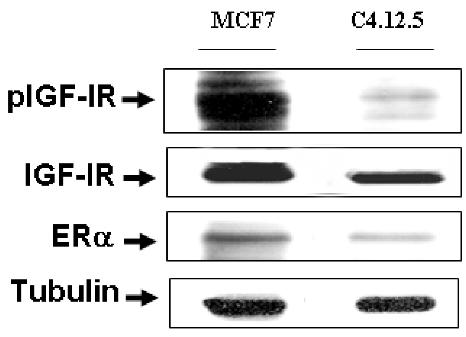
3.1. ER and IGF-IR Expression in MCF7 and C4.12.5 Cells

The involvement of the IGF-IR in breast cancer development has been well established. To address the potential regulation of IGF-IR expression during breast cancer progression, we measured in initial experiments the levels of total and phospho-IGF-IR (pIGF-IR) in syngeneic cell lines representing different stages of the disease. For this purpose, we employed the human breast cancer-derived MCF7 (ER-positive) and MCF7-derived C4.12.5 (ER-depleted) cell lines. C4.12.5 cells were generated by clonal selection of MCF7 cells that were maintained in estrogen-free conditions for nine months [37]. Although C4.12.5 cells exhibited some variability in ER expression, the passages used in the present study displayed consistently low levels of ERα (~59% of the ERα levels in MCF7 cells). Western blots using anti-IGF-IR β-subunit and anti-pIGF-IR antibodies showed that both total IGF-IR and pIGF-IR levels were significantly reduced in C4.12.5, in comparison to, MCF7 cells (76% for IGF-IR and 30% for pIGF-IR) (Figure 3). These results corroborate previous observations [40,45].
Figure 3. IGF-IR and ERα expression in MCF7 and C4.12.5 breast cancer cell lines.MCF7 and C4.12.5 cells were lysed as described under Materials and Methods. Cellular extracts (80 μg) were electrophoresed through 10% SDS-PAGE, transferred to nitrocellulose membranes, and blotted with anti-total IGF-IR-β, anti-pIGF-IR, and anti-ERα. Blots were then blotted with a tubulin antibody as a loading control.
Cancers 02 00233 g003

3.2. DNA Affinity Chromatography

The MCF7 and C4.12.5 cell lines were employed to identify the sets of IGF-IR promoter-binding proteins that may be involved in IGF-IR transcription and that are responsible for the differential expression of the gene at different stages of the disease. The rationale for these studies was the fact that regulation at the transcription level has been identified as a major control step in IGF-IR regulation [46]. To this end, we used a proteomic approach based on DNA affinity chromatography followed either by MS or Western blot analyses using antibodies against previously identified or novel IGF-IR promoter-binding proteins. The basis for selecting this specific fragment was the fact that previous studies have shown that this region is responsible for most of the promoter activity. Nuclear extracts of MCF7 and C4.12.5 cells were incubated with the PCR-amplified, biotin-labeled IGF-IR proximal promoter DNA probe, after which DNA-protein complexes were adsorbed to streptavidin beads. Bound proteins were eluted with high-salt buffer and concentrated by centrifugation at reduced pressure. The eluted material was electrophoresed through 10% SDS-PAGE and proteins were visualized by silver staining (Figure 2). A number of bands were seen in the eluted material (lane 4) whereas no proteins were detected in eluates of lysates incubated with strepavidin magnetic beads alone (lane 5). Silver stained gels were cut into slices and the specific DNA-bound proteins were analyzed by MS.

3.3. MS Analysis

MS analysis of DNA affinity chromatography-purified proteins identified a number of previously reported and several novel IGF-IR promoter binding proteins. The proteins identified were categorized using the Gi (gene index) identification tool, coverage (percentage of amino acids covered by the identified peptide), and source (Table 1). The specific proteins bound to the IGF-IR promoter were classified based on cellular localization and biological function (Table 2). Most of the proteins were localized in the nucleus, but some of them were found also in the cytoplasm as well as in the plasma membrane. Furthermore, Table 2 illustrates the variety of biological processes associated with the identified IGF-IR promoter-binding transcription factors in ER-positive and ER-depleted breast cancer cells. Out of 91 proteins identified by MS, 6 correspond to cytoskeleton-associated proteins, 6 are involved in transcription, regulation of nucleobase, nucleoside and nucleic acid metabolism, 2 in nuclear stability, chromatin structure, cycle control, and gene expression, 20 in DNA repair, breaking, replication, and cell death, 24 in RNA splicing and processing, and translation, and 33 in other functions, including proliferation, apoptosis, and proteosomal degradation.
Table 1. IGF-IR promoter-binding proteins identified by MS in MCF7 and C4.12.5 cells.
Protein NameCoverageCoverageSource
MCF7C4.12.5
4760598Adenylate kinase isozyme 2 4-Mus musculus
61743954AHNAK nucleoprotein isoform 1 1 3Homo sapiens
1061128Anpg 25 21Homo sapiens
5453541Anterior gradient 2 homolog 26 Homo sapiens
6753086Apurinic/apyrimidinic endonuclease 18Mus musculus
10947139Arginase, type I3Homo sapiens
6680748ATP synthase, H+ transporting, mitochondrial F1 complex, alpha subunit, isoform 1 3 7Mus musculus
10863945ATP-dependent DNA helicase II20 28Homo sapiens
4503841ATP-dependent DNA helicase II, 70 kDa subunit22 28Homo sapiens
1040689ATP-dependent DNA helicase Q1 (DNA-dependent ATPase Q1)_ 4Homo sapiens
42740907Clusterin isoform 2 8Homo sapiens
22208854Cytidine monophospho-N-acetylneuraminic acid synthetase3 3Mus musculus
4557515Damage-specific DNA binding protein 2 (48kD) 618Homo sapiens
45476309Damage-specific DNA binding protein 2 splicing variant D4 7 13Homo sapiens
4758138DEAD (Asp-Glu-Ala-Asp) box polypeptide 515 13Homo sapiens
506756Desmoyokin6
1706481DNA ligase III 23 37Homo sapiens
11225260DNA topoisomerase I10 11Homo sapiens
347019DnaK-type molecular chaperone hsp72-ps1 8 10Rattus norvegicus
1722884DNA-repair protein complementing XP-C cells 6Drosophila melanogaster
38201714ELAV-like 110 Homo sapiens
4503471Eukaryotic translation elongation factor 1 α 1 6 2Homo sapiens
4503477Eukaryotic translation elongation factor 1 β 2 7Homo sapiens
25453472Eukaryotic translation elongation factor 1 δ isoform 2 22 Homo sapiens
110625979Eukaryotic translation elongation factor 1 γ 6 Mus musculus
37078458Far upstream element-binding protein 1 (FUSE-binding protein 1) 11 13Mus musculus
27806887Fusion (involved in t(12;16) in malignant liposarcoma) 5 _Bos taurus
7669492Glyceraldehyde-3-phosphate dehydrogenase9Mus musculus
74736618Grainyhead-like protein 2 homolog 16Homo sapiens
51316135GTP-binding nuclear protein Ran, testis-specific isoform10 Rattus norvegicus
4504517Heat shock 27kDa protein 1 33 Homo sapiens
2495339Heat shock 70 kDa protein 1B8 10Bos taurus
8393547Heterogeneous nuclear ribonucleoprotein A1 13 11Rattus norvegicus
14043072Heterogeneous nuclear ribonucleoprotein A2/B1 isoform B119 10Homo sapiens
31559916Heterogeneous nuclear ribonucleoprotein A3 isoform a11 15Mus musculus
55956919Heterogeneous nuclear ribonucleoprotein AB isoform a9 3Homo sapiens
14249959Heterogeneous nuclear ribonucleoprotein C (C1/C2)10 2Homo sapiens
870747Heterogeneous nuclear ribonucleoprotein D (hnRNP D) 11 22Homo sapiens
10946928Heterogeneous nuclear ribonucleoprotein H1 13 8Mus musculus
14141157Heterogeneous nuclear ribonucleoprotein H3 isoform a11 Homo sapiens
13384620Heterogeneous nuclear ribonucleoprotein K 23 10Mus musculus
133274Heterogeneous nuclear ribonucleoprotein L (hnRNP L)4 4Homo sapiens
14141152Heterogeneous nuclear ribonucleoprotein M isoform a15 18Homo sapiens
5031755Heterogeneous nuclear ribonucleoprotein R isoform 2 6 6Homo sapiens
8393536High mobility group box 2 14 Rattus norvegicus
1568557Histone H2B 19 12Homo sapiens
968888HMG-113 Homo sapiens
32358HnRNP U protein 11 9Homo sapiens
37183160HRPE773 29Homo sapiens
1040689Human Diff6, H5, CDC10 homologue, KIAA00976 Homo sapiens
13385872Interleukin enhancer binding factor 2 8 13Mus musculus
4504865KH-type splicing regulatory protein (FUSE binding protein 2) 68Homo sapiens
6453818Kinesin family member 22 20Homo sapiens
2055427KSRP5 26Homo sapiens
27436946Lamin A/C isoform 1 precursor2015Homo sapiens
5031877Lamin B1821Homo sapiens
14149645Methyl CpG binding protein 2 44Mus musculus
1770458M-phase phosphoprotein 4 12 6Homo sapiens
4038587Mucin 2Homo sapiens
55956788Nucleolin 5 5Homo sapiens
58037163Nucleoporin 35 6 Mus musculus
35119NuMA protein2Homo sapiens
1483131p80 protein 3 3Homo sapiens
115497186Paraspeckle component 1 2 7Bos taurus
156523968Poly (ADP-ribose)polymerase 3236Homo sapiens
14141166Poly(rC)-binding protein 2 isoform b20 9Homo sapiens
31543419Polynucleotide kinase 3 phosphatase 13 11Homo sapiens
4506243Polypyrimidine tract-binding protein 1 isoform a8 10Homo sapiens
4502801Regulator of chromosome condensation (RCC1)1210Homo sapiens
4506583Replication protein A1, 70kDa 18 26Homo sapiens
4506585Replication protein A2, 32kDa7 Homo sapiens
4506439Retinoblastoma binding protein 7 3 Homo sapiens
15718687Ribosomal protein S3 23 Homo sapiens
7159943RNA and export factor binding protein 1-II 24 9Mus musculus
4757926RNA binding motif protein 39 isoform b 3 Homo sapiens
66346679SERPINE1 mRNA binding protein 1 isoform 1 9 9Homo sapiens
2440078SPIN protein4Homo sapiens
4826998Splicing factor proline/glutamine rich 9 11Homo sapiens
5902076Splicing factor, arginine/serine-rich 1 isoform 1 4 Homo sapiens
39930517Sterile alpha motif domain containing 1 11 17Homo sapiens
30088600Telomerase subunit EST1A 1 1Homo sapiens
4507555Thymopoietin isoform alpha 8 17Homo sapiens
4507401Transcription factor A, mitochondrial 4-Homo sapiens
14389309Tubulin alpha 6 6 12Homo sapiens
12963615Tubulin, beta 3 98Mus musculus
21361322Tubulin, beta 4 9 9Homo sapiens
1066004Typr II DNA topoisomerase beta isoform1Mus musculus
28077011Tyrosyl-DNA phodphodiesterase 14 4Mus musculus
4506387UV excision repair protein RAD23 homolog B 4 Homo sapiens
2632123Xeroderma Pigmentosum Group E Complementing protein7 17Homo sapiens
5454172X-ray repair cross complementing protein 1 9 12Homo sapiens
Gi: identification tool; Coverage: percentage of amino acids covered by the identified peptide (n = 3 independent analyses).
Table 2. Summary of functional categories of IGF-IR promoter-bound proteins identified by MS.
GeneSubcellular fractionFunction
Cytoskeleton-associated proteins
KIAA0097 ; Human Diff6Cytoplasm
Nucleus
Microtubule associated protein.
Kinesin family member 22NucleusMicrotubule-dependent molecular motor.
NuMA proteinNucleusComponent of the mitotic spindle matrix.
-
Tubulin α 6
-
Tubulin, β 3
-
Tubulin, β 4
Cytoplasm
Nucleus
Related to microtubules.
Factors involved in transcription, regulation of nucleobase, nucleoside, nucleotide and nucleic acid metabolism
-
Elongation factor-1 (EF1)
-
Eukaryotic translation elongation factor 1 β 2
-
Eukaryotic translation elongation factor 1 δ isoform 2
-
Eukaryotic translation elongation factor 1 γ
Cytoplasm
Nucleus
Involved in enzymatic delivery of aminoacyl tRNAs to the ribosome.
Poly(rC)-binding protein 2 isoform bCytoplasm
Nucleus
RNA binding protein involved in regulation of nucleobase, nucleoside, nucleotide and nucleic acid metabolism.
Telomerase subunit EST1A
Alternative name: Chromosome 17 open reading frame 31
Est1p like protein A
Ever shorter telomeres 1
Nucleus
Nucleolus
Telomere regulation.
Nuclear stability, chromatin structure, cycle control, gene expression
-
Lamin B1
-
Lamin A/C
NucleusNuclear stability, chromatin structure and gene expression.
Proteins with a known function in DNA repair, breaking, replication, transcription and cell death
Apurinic/apyrimidinic endonuclease 1NucleusDNA repair and redox regulatory activities.
ATP-dependent DNA helicase II, 70 kDa subunit
ATP-dependent (80 kda)
NucleusRepair of nonhomologous DNA ends.
ATP-dependent DNA helicase Q1 (DNA-dependent ATPaseNucleusRepair of UV-irradiated DNA damage or other mutagens.
-
Damage-specific DNA binding protein (127kDa) –DDB1
-
Damage-specific DNA binding protein 2 (48kD) – DDB2
-
Damage-specific DNA binding protein 2 splicing variant D4
Cytoplasm
Nucleus
Repair of UV-irradiated DNA damage.
-
DNA Topoisomerase I
-
DNA Topoisomerase II
-
Tyrosyl-DNA phosphodiesterase 1
Cytoplasm
Nucleus
Nucleoplasm
Type I topoisomerase breaks single DNA strands, whereas Type II topoisimerase breaks double stranded DNA.
-
High mobility group box 2
-
High Mobility Group Protein 17 (HMG17) –chromosomal protein
-
HMG1
NucleusDNA repair, recombination, replication, and transcription.
Histone 1,2 and 3NucleusPlay a central role in transcription regulation, DNA repair, DNA replication and chromosomal stability.
Poly (ADP-ribosylation) polymerase I-PARP-1NucleusDNA repair, replication, transcription and cell death.
Polynucleotide kinase 3 phosphataseNucleusDNA repair following ionizing radiation or oxidative damage.
Replication protein A1, 70kDa
Replication Protein A2, 32-KD; RPA2
NucleusReplication, recombination and DNA repair.
Ribosomal Protein S3; RPS3Cytoplasm
Nucleus
DNA repair and apoptosis.
Xeroderma Pigmentosum Group E
Complementing protein
Cytoplasm
Nucleus
Involved in DNA repair.
X-ray repair cross complementing protein 1NucleusRepair of single-strand DNA breaks.
RNA slipicing, RNA processing, Transcription, Translation and other functions
-
Dead/H-box-5
-
RNA Helicase p68
NucleusImplicated in cellular processes involving alteration of RNA secondary structure.
ELAV-like 1Cytoplasm
Nucleus
RNA-binding proteins.
Fusion (involved in t(12;16) in malignant liposarcoma)NucleusPlays a role in maintenance of genomic integrity.
hnRNP U proteinCell surface Nucleus Binds to pre-mRNA.
Interleukin enhancer binding factor 2 variantNucleus
Nucleolus
Regulates transcription of the IL2 gene during T-cell activation.
NucleolinNucleus
Nucleolus
The major nucleolar protein of growing eukaryotic cells.
RNA and export factor binding protein 1Cytoplasm
Nucleus
Nuclear mRNA export pathway.
RNA binding motif protein 39 isoform bNucleusTranscriptional coactivator for steroid nuclear receptors ESR1/ER α and ESR2/ER-β, and JUN/AP-1.
-
Splicing factor, arginine/serine-rich 1 isoform 1
-
Splicing factor proline/glutamine rich
Cytoplasm
Nucleus
Prevents exon skipping.
Others
Adenylate kinase isozyme 2Membrane
Microsomal
Mitochondria
Nucleus
Small ubiquitous enzyme.
Alkyladenine DNA glycosylase (AAG), also known as 3-methyladenine DNA glycosylase,NucleusCatalyzes the first step in base excision repair.
Arginase, type IMitochondriaPlays a role in the regulation of extra-urea cycle arginine metabolism.
ATP synthase, H+ transporting, mitochondrial F1 complex, α subunit, isoform 1Mitochondria
Cytoplasm
Nucleus
Produces ATP from ADP in the presence of a proton gradient across the membrane.
BBF2H7/FUS protein NucleusBinds both single-stranded and double-stranded DNA and promotes ATP-independent annealing of complementary single-stranded DNAs and d-loop formation in superhelical double-stranded.
ClusterinCytoplasm
Nucleus
Undefined function, being described as being both pro-apoptotic and anti- apoptotic.
Cytidine monophospho-N-acetylneuraminic acid synthetaseCytoplasm
Nucleus
Involved in the biosynthesis of sialo-glycoproteins and gangliosides.
Desmoyokin/AHNAKPlasma Membrane
Cytoplasm
Nucleus
Involved in the formation of cytoskeletal structure, calcium homeostasis, and muscle regeneration.
DNA ligase III α and βNucleusInteracts with DNA-repair protein XRCC1 and corrects defective DNA strand-break repair and sister chromatid exchange.
-
DnaK-type molecular chaperone hsp72-ps1
-
Heat shock 27 kD protein 1
-
Heat shock cognate 71 kDa protein (Heat shock 70 kDa protein 8).
Cytosol
Perinuclear
Mitochondria
Endoplasmatic
Reticulum
Nucleus
Chaperone activity, thermotolerance, inhibition of apoptosis, regulation of cell development and cell differentiation.
DNA-repair protein complementingNucleusNucleotide excision repair (NER) of DNA.
-
Far upstream element-binding protein 1 (FUSE-binding protein 1)
-
KH-type splicing regulatory protein (FUSE binding protein 2)
-
KSRP
Nucleus (Probable).Regulates MYC expression by binding to a far-upstream element (FUSE) upstream of the MYC promoter.
Grainyhead-like protein 2 homolog (Brother of mammalian grainyhead) (Transcription factor CP2-like 3)NucleusTranscription factor involved in epidermal differentiation.
GTP-binding nuclear protein Ran, testis-specific isoformCytoplasm
Nucleus
Nuclear transport pathways, required for the import of protein into the nucleus and also for RNA export.
Gyceraldehyde-3-phosphate dehydrogenaseMembrane
Cytoplasm
Nucleus
Carbohydrate metabolism and membrane trafficking in the early secretory pathway.
Methyl CpG binding protein 2NucleusNuclear proteins containing a methyl-binding domain (MBD)
M-phase phosphoprotein 4NucleolusRegulation of cell cycle, cell communication and signal transduction.
MucinCytoplasm
Mitochondria
Nucleus
Activation of Wnt target genes.
Nucleoporin 35 [Nuclear pore glycoprotein-210 (gp210)]Membrane
Cytoplasm
Nucleus
Trafficking regulator in the eukaryotic nuclear pore complex.
p80 proteinCytoplasm
Nucleus
Part of the nuclear coiled bodies (CBS) involved in assembly/disassembly of nucleoplasmic snRNPs.
Paraspeckle component 1Cytoplasm
Nucleus
Nucleolus
Regulates cooperatively with NONO and SFPQ, androgen receptor-mediated gene transcription activity.
PREDICTED: similar to RAN proteinCytoplasm
Nucleus
Inhibits GTP exchange on Ran.
Regulator of chromosome condensation (RCC1)Cytoplasm
Nucleus
Promotes the exchange of Ran-bound GDP by GTP.
Retinoblastoma binding protein 7NucleusModulates the functions ascribed to BRCA1 in transcriptional regulation, DNA repair, and/or cell cycle checkpoint control.
SERPINE1 mRNA binding protein 1 isoform 1Cytoplasm
Nucleus
Regulates mRNA stability.
SPINNucleusRegulation of cell cycle
Sterile alpha motif domain containing 1NucleusWidespread domain in signalling and nuclear proteins.
Thymopoietin γ and β ( TMPOs)NucleusDirects the assembly of the nuclear lamina.
Transcription factor A, mitochondrialMitochondriaInvolved in mitochondrial transcription regulation.
UV excision repair protein RAD23 homolog B Cytoplasm
Nucleus
Plays a role both in proteosomal degradation of misfolded proteins and DNA repair.

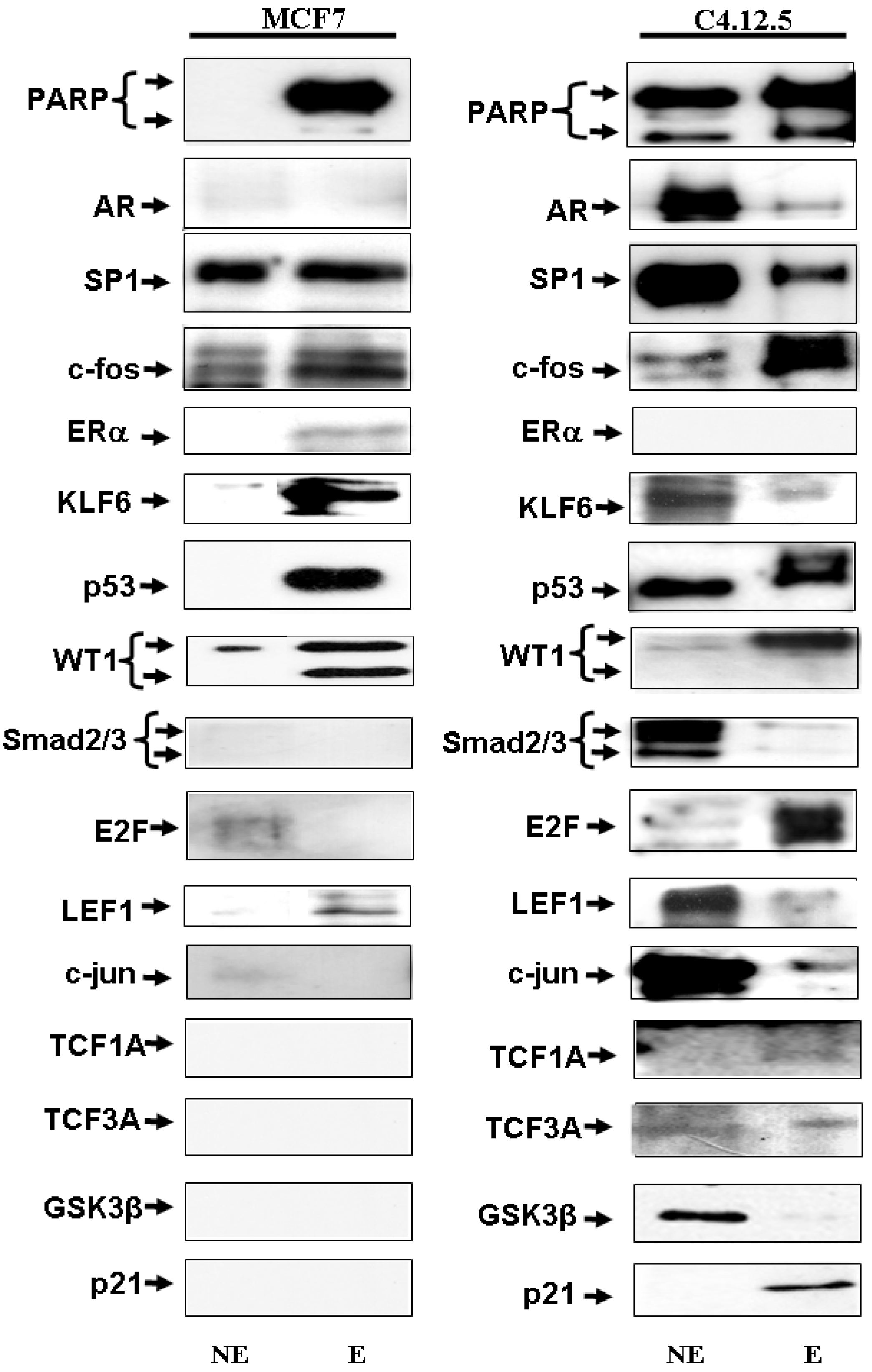
3.4. Identification of IGF-IR Promoter-Binding Transcription Factors by Western Blots

Western blot analysis of DNA affinity chromatography-purified proteins identified a number of previously reported as well as novel IGF-IR promoter binding proteins. The data are summarized in Figure 4 and Table 3 and Table 4. Consistent with their role as positive regulators of the IGF-IR gene, the binding of zinc finger proteins Sp1 and KLF6 to the IGF-IR promoter was stronger in MCF7 than in C4.12.5 cells. Similarly, ER-α binding to the IGF-IR promoter was stronger in MCF7 than in C4.12.5 cells. On the other hand, a number of transcription factors, including AR, Smad2/3, E2F1, c-jun, TCF1A, TCF3A, GSK3β, and p21 appear to bind to the promoter region only in the C4.12.5 cell line. p53, c-fos, and PARP bind to IGF-IR promoter elements in both cell lines with similar affinity. No DNA binding was detected in either cell line using antibodies against BRCA1, TOPOII, IRS-1, MET, p120, Foxo1, p73, lamin A/C, c-Myc, p63, cyclin D, cyclin G, histone1, and BRCA2.
Figure 4. IGF-IR promoter-binding transcription factors identified by Western blots in the MCF7 and C4.12.5 cell lines. DNA chromatography eluates were electrophoresed though 10% SDS-PAGE and blotted with the antibodies listed under Materials and Methods Section. The left lanes correspond to nuclear extracts (NE; 4.2 μg) and the right lanes correspond to DNA affinity chromatography eluates (E; 10 μg).
Cancers 02 00233 g004
Table 3. Differentially expressed nuclear proteins identified by Westen blots in MCF7 and C4.12.5 cells.
GeneMCF7C4.12.5Nomenclature
BRCA110.3Breast and ovarian cancer type I
TOPOII10Type II topoisomerase
IRS-I00Insulin receptor substrate-1
PARP12Poly ADP ribose polymerase
MET10Met tyrosine kinase
p12011.39Zinc finger protein 120
AR10.64Androgen receptor
Sp110.79Trans-acting transcription factor 1
Foxo110.5Forkhead Box A1
ER10.59Estrogen receptor
WT110.8Wilms' tumor suppressor gene-1
KLF611.2Kruppel-like factor 6
LaminA/C12.4Nuclear protein
Smad 211.12Smad 2/3 family
Smad 302.6
pSmad2/311.13
c-Myc11.48Myelocytomatosis oncogene
E2F110.88E2F transcription factor 1
TCF110.717T-cell factor 1
TCF3A10.002High Mobility Group box transcription factor 3
LEF111.15Lymphoid enhancer binding factor 1
p5310.96p53-related tumor suppressors
p6311.12
p7310.03
p2110.91Waf1/Cip1
GSK3α11.05Glycogen Synthase Kinase-3
c-fos10.92c-fos/c-jun family of transcription factors
c-jun110
pc-jun10.5
Cyclin G 10.13Regulatory subunits of Cdc2 p34
Cyclin D10
Histone110.74Nuclear protein
A value of 1 was assigned to the level of expression in whole MCF7 cell lysates, normalized to tubulin.
Table 4. Function of gene products identified in MCF7 and C4.12.5 cells by Western analyses.
GeneFunction
BRCA1Tumor suppressor involved in DNA damage repair, cell growth and apoptosis, and gene transcription.
TOPOIIType II topoisomerase, breaks double strands of duplex DNA.
IRIS-IMediates insulin related activities.
PARPDNA repair, replication, transcription and cell death.
METReceptor for HGF/SF.
p120Prototypical member of a subfamily of armadillo (Arm)-domain proteins involved in intercellular adhesion and in nuclei.
ARAndrogen receptor (AR), a zinc finger transcription factor of the nuclear receptor superfamily.
SP1Transcription factor of the zinc finger family.
Foxo1Transcription factor of the forkhead family.
EREstrogen receptor (ER), a DNA binding transcription factor of the nuclear receptor superfamily.
WT1Wilm’s tumor suppressor protein-1.
KLF6Kruppel-like zinc finger transcription factor-6.
LaminA/CInvolved in nuclear stability, chromatin structure and gene expression.
pSmad 2/3Smad transcription factors mediate the effect of TGF-β.
Smad 2/3
c-MycNuclear proteins involved in cell proliferation, differentiation and neoplastic disease.
E2F1Involved in cell cycle progression, apoptosis and oncogenesis.
LEF1Members of the high mobility group (HMG) DNA binding protein family of transcription factors.
TCFA1
TCF3A
P53Tumor suppressor p53, p63 and p73.
p63
p73
GSK3αInvolved in protein synthesis, cell adhesion, proliferation, and differentiation.
c-fosFos and Jun protooncogenes.
c-junp
c-jun
Cyclin G Regulatory subunits of Cdc2 p34 and related cyclin-dependent kinases (Cdks).
Cyclin D
Histone1Basic and water soluble nuclear proteins.
p21Tumor suppressor protein p21/Waf1/Cip1 acts as an inhibitor of cell cycle progression.

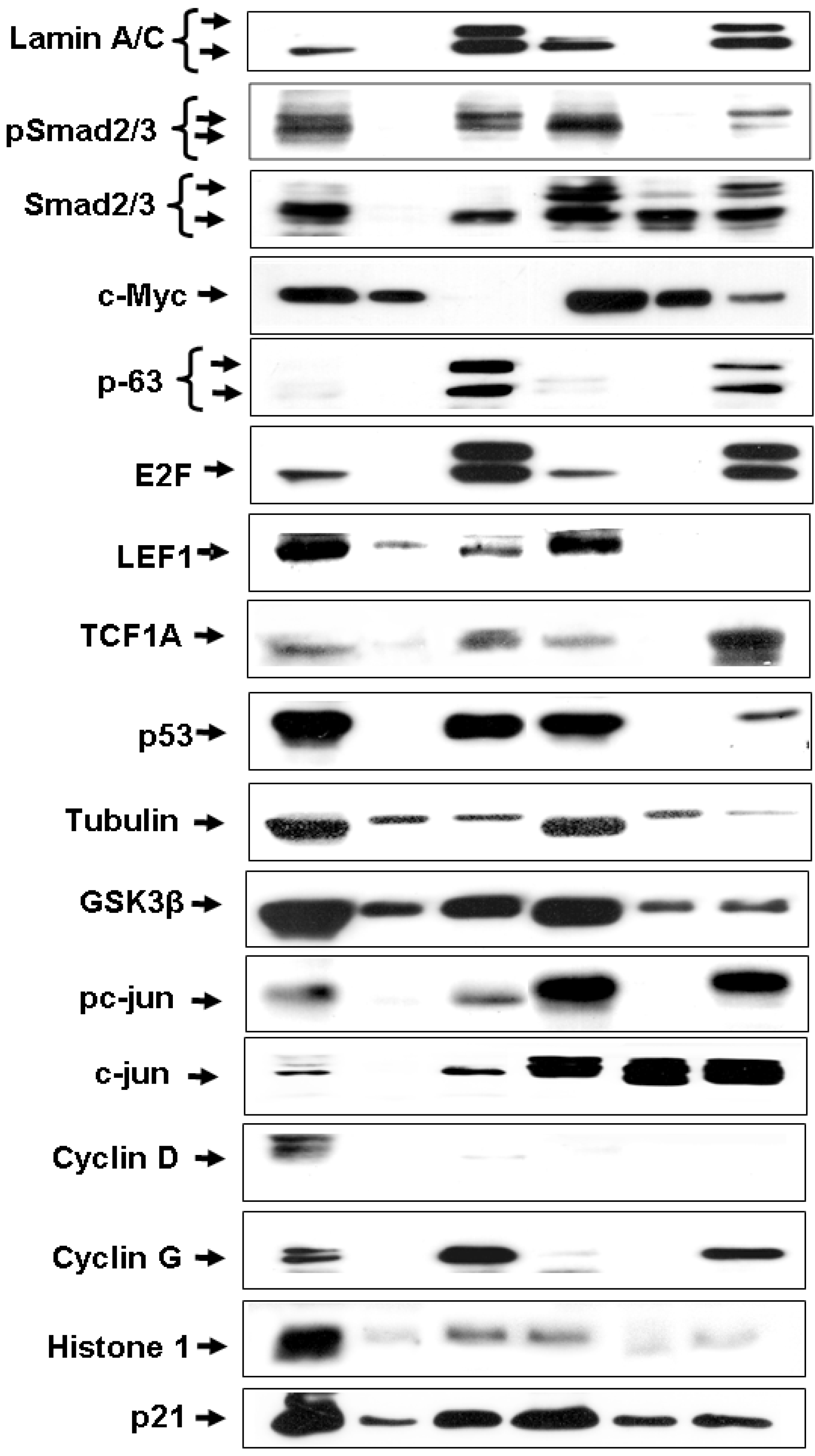
3.5. Cellular Distribution of IGF-IR Promoter-Binding Proteins
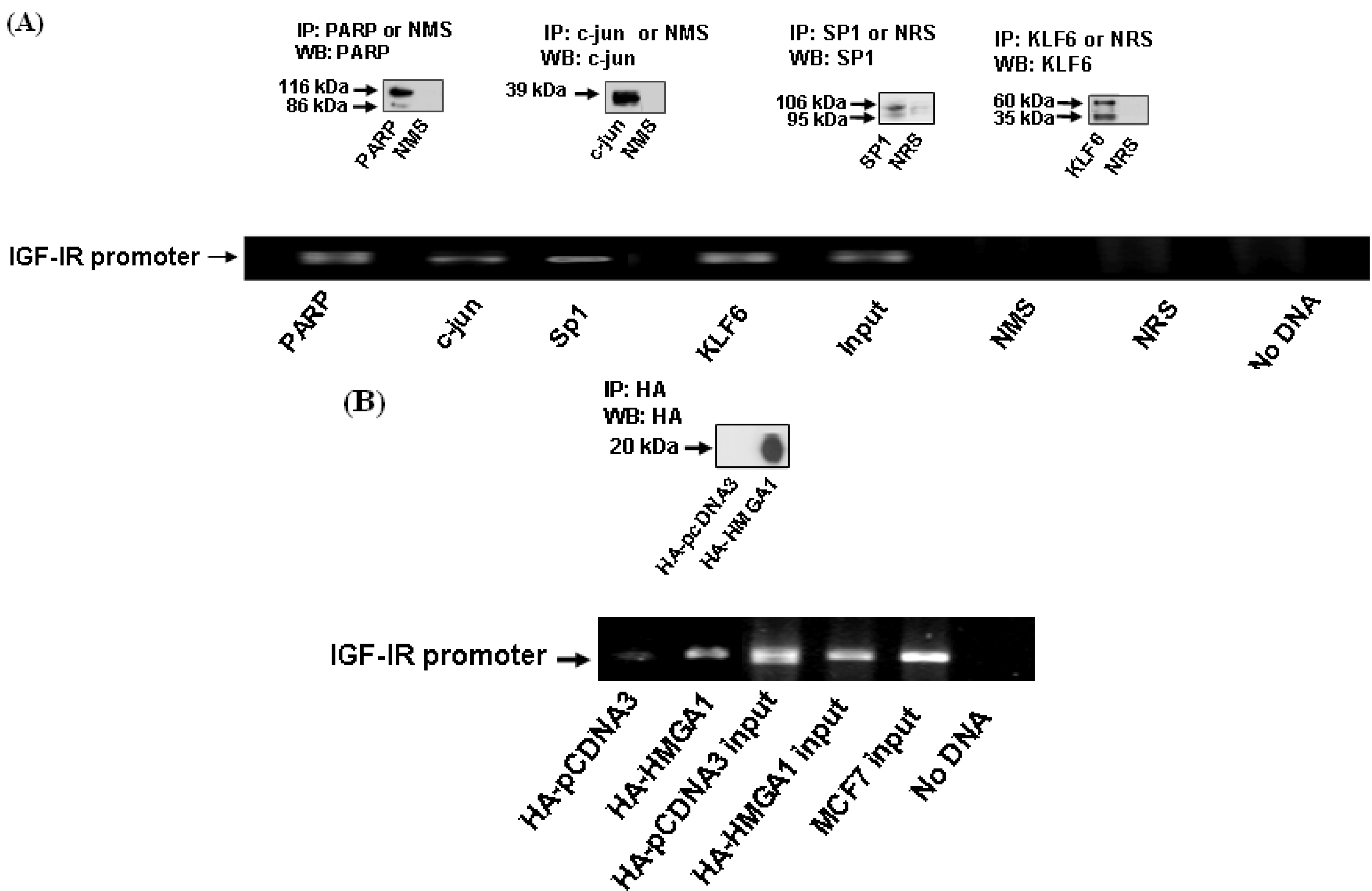
Given that differences in binding could be due to differential expression of the transcription factors in both cell lines, we next investigated the subcellular distribution and expression of IGF-IR promoter-binding transcription factors. For this purpose, MCF7 and C4.12.5 lysates were fractionated into cytoplasmic and nuclear fractions and the subcellular distribution of the different proteins was examined by Western blots (Figure 5 and Table 3, Table 4). The cytosolic marker MET [47] was used to check for cytoplasmic contamination of the nuclear fraction, and it was found to be expressed at very low levels in both cell lines. A number of proteins, including PARP (200% of levels in MCF7 cells), lamin (240%), Smad 2/3 (260%), and c-jun (1000%) had higher expression levels in C4.12.5 than in MCF7 cells. Other proteins, such as BRCA1 (30% of levels in MCF7), AR (64%), Sp1 (79%), Foxo I (50%), p73 (3%), TCF3A (0.2%), ER (59%), TCF1 (71.7%), pc-jun (50%), cyclin G (13%), and histone (74%) were expressed at lower levels in C4.12.5 compared to MCF7 cells. No differences in expression between the cell lines were detected in p120, c-fos, WT1, KLF6, pSmad2/3, c-Myc, p63, E2F, LEF1, p53, GSK3α, and p21. We were unable to detect expression of β-catenin, p300, TLE1, APC, and BRCA2 in neither cell line. Transcription factor WT1 was present in both cell lines, though its low molecular weight form appears to be specific to the MCF7 cell line.
Figure 5. Cellular distribution of transcription factors in the MCF7 and C4.12.5 cell lines. Cell lines were fractionated as described under Materials and Methods and total lysates (T; 80 μg), cytosolic fractions (C; 20 μg), and nuclear extracts (N; 20 μg) were resolved on 10% SDS-PAGE and blotted with the indicated antibodies.
Cancers 02 00233 g005

3.6. ChIP Assays

To corroborate results of DNA chromatography we measured the binding of selected proteins identified by MS or by Western blots using ChIP assays. The following proteins were chosen for ChIP assays: PARP (detected by MS and Western blots in both cell lines), Sp1 (positive control, detected in both cell lines), c-jun, (detected only in C4.12.5) and HMGA1 (detected by MS only in MCF7). Briefly, cells were lysed, sonicated for 3 min, and immunoprecipitated with the indicated antibodies (or normal mouse or rabbit sera) for 18 h (Figure 6A). In addition, MCF7 cells transfected with HA-HMGA1 (or empty pCDNA3 vector) were also immunoprecipitated with an HA monoclonal antibody (Figure 6B). For PCR analysis of immunoprecipitatd chromatin, a set of primers encompassing the human IGF-IR promoter was employed as described under Materials and Methods section. The input bands represent the amplified PCR product in the absence of antibodies (Figure 6A,B). The results of ChIP analyses confirmed that PARP, c-jun, Sp1, KLF6 and HMGA1 bound in vivo directly to the IGF-IR promoter and, thus, corroborate the results of MS and Western blot analyses.
Figure 6. ChIP assays of transcription factor binding to IGF-IR promoter DNA. (A). MCF7 cells were cross-linked with formaldehyde, lysed, sonicated, and immunoprecipitated with PARP, c-jun, Sp1, or KLF6 antibodies, or normal mouse or rabbit sera, followed by PCR amplification of precipitated chromatin using primers encompassing the IGF-IR promoter. The position of the 773 bp-amplified fragments is indicated. The input bands represent the amplified PCR product in the absence of antibodies. The insets indicate the endogenous levels of expression of the various proteins as measured by Western blots (WB) with specific antibodies or NRS or NMS as negative controls. IP, immunoprecipitation. (B). MCF7 cells were transfected with an expression vector encoding HA-HMGA1 (or empty pcDNA3 vector), after which the cells were cross linked with formaldehyde, lysed, sonicated and immunoprecipitated with an HA antibody, followed by PCR amplification of IGF-IR promoter DNA. HA-HMGA1 immunoprecipitated protein was detected by Western blots using an anti-HA antibody (inset).
Cancers 02 00233 g006

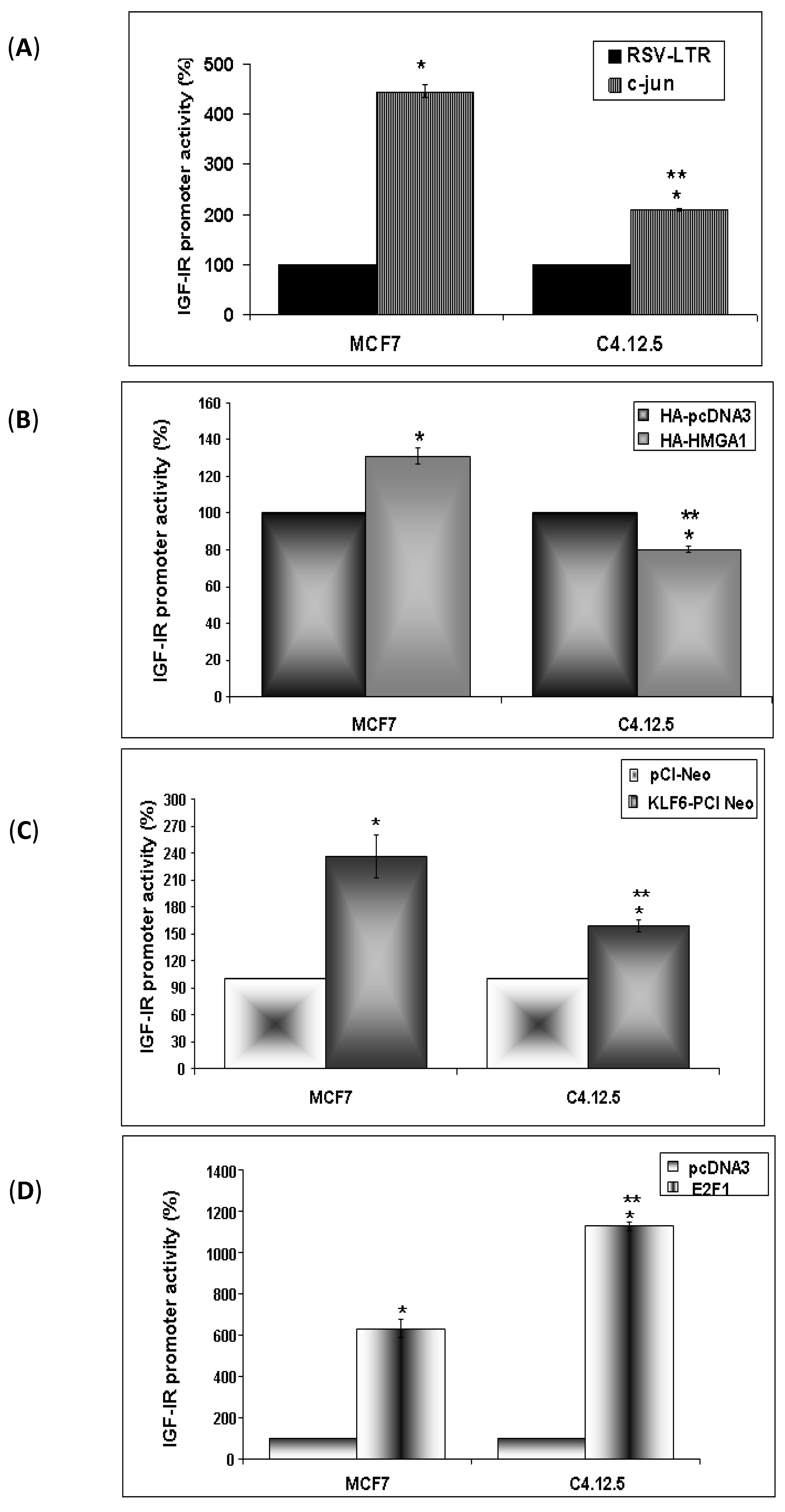
3.7. Regulation of IGF-IR Promoter Activity by c-jun, HMGA1, KLF6 and E2F1

To evaluate the potential functional implications of our findings we next assessed the ability of selected IGF-IR promoter-binding proteins to activate the promoter. For this purpose cotransfection experiments were performed in MCF7 and C4.12.5 cells using expression vectors encoding c-jun, HMGA1, KLF6 and E2F1, along with an IGF-IR promoter-luciferase reporter plasmid, p(−476/+640)LUC, which contains most of the proximal region of the IGF-IR promoter. The results of cotransfection experiments are presented in Figure 7A-D. Results obtained indicate that transcription factors c-jun, KLF6, and E2F1 induced a significant increase in IGF-IR promoter activity in both cells lines. Specifically, c-jun enhanced IGF-IR promoter activity by 445 ± 12.4% in MCF7 and by 209 ± 3.1% in C4.12.5 cells (versus empty vector), KLF6 enhanced promoter acitiviy by 237 ± 9.7% in MCF7 and by 159 ± 7.1% in C4.12.5 cells, and E2F1 stimulated promoter activity by 631.5 ± 44.5% in MCF7 and by 1127 ± 22.2% in C4.12.5 cells. On the other hand, HMGA1 had a minimal effect (131 ± 4.3% increase in MCF7 and 80.3 ± 1.7% reduction in C4.12.5 cells).
Figure 7. Regulation of IGF-IR promoter activity by c-jun, HMGA1, KLF6 or E2F1. MCF7 and C4.12.5 cells were cotransfected with the p(−476/+640)LUC reporter construct along with c-jun (A), HMGA1 (B), KLF6 (C) or E2F1(D) expression plasmids (or empty expression vectors). Forty eight hours after transfection the cells were harvested and luciferase activity was measured. Promoter activities are expressed as luciferase values normalized to protein concentration. A value of 100% was given to the promoter activity generated by the reporter plasmid in empty vector-transfected MCF-7 or C4.12.5 cells. The results represent the mean ± SEM (N = 3 independent experiments in triplicate wells); *p < 0.01 versus empty vector-tranfected cells and ∗∗ p < 0.01 versus specific vector-transfected MCF7 cells.
Cancers 02 00233 g007

4. Discussion

Breast cancer is one of the main causes of cancer-related mortality among women in economically developed regions of the world. Care of patients with breast cancer is complicated by the wide differences in the natural history of the disease, as manifested by a remarkable variability in response to treatment and survival. Over 100 potential biomarkers were reported in various studies to be differentially expressed between normal and cancerous cells and tissues. Cancer biomarkers include, among others, ER, progesterone receptor, HER2, cathepsin D, cyclin E, CA 15-3, and CA 27.29. Currently, only a few of those biomarkers are used in clinical practice. High-throughput genomic and proteomic techniques developed over the last decade offer the potential to better define the biological nature of the disease process, both for risk assessment and therapy purposes [48].
There is conflicting evidence regarding the importance of IGF-IR expression for diagnosis and prognosis of breast cancer. In different studies, IGF-IR expression was detected in 39% to 93% of the cases [49]. Whereas some studies reported that IGF-IR levels are indicative of early recurrence of the disease (i.e., poor prognosis), others considered high IGF-IR values as an indication of good prognosis [4,50,51,52,53,54]. These disparities may be due to a number of factors, including selection of patients or the diverse approaches used to detect IGF-IR, including immunoblots of membrane fractions and/or whole tissues, ligand binding assays, etc. Immunohistochemical studies revealed high IGF-IR and IRS-1 levels in control breast tissues and in well-differentiated breast carcinoma, whereas very low levels were seen in poorly differentiated cancers [3]. These results, suggesting that progression to advanced-stage disease is associated with a reduction in IGF-IR expression, indicate that IGF-IR status can be used as a marker for tumor evaluation [3]. Notably, no correlation was seen between insulin receptor levels and the differentiation stage of the tumors.
In the present study we employed a novel and reliable DNA affinity chromatography protocol in order to identify IGF-IR promoter-binding proteins in breast cancer cells. This method allowed us to detect known and new specific potential biomarkers in ER-positive and ER-depleted cells, which may reflect early and advanced stages of the disease. Our analyses identified twenty four proteins that bind to the IGF-IR promoter only in MCF7 cells whereas, on the other hand, C4.12.5 cells contain nineteen proteins that did not exhibit IGF-IR promoter binding activity in MCF7 cells. The results obtained suggest that the specific binding of different DNA-binding transcription factors and non-DNA sequence specific-binding nuclear proteins (including Smad2/3, LEF1, c-jun, TCF1A, TCF3A, GSK3β and others ) in the MCF7 and C4.12.5 cell lines could reflect differences in transcriptional regulation of the IGF-IR gene at different stages of the disease. Interestingly, sixty two proteins were shown to bind (either directly or indirectly) to IGF-IR promoter elements in both cell lines. As shown in Table 2 and Table 4, the list of proteins identified in this study includes proteins involved in apoptosis, protein synthesis, cell adhesion, proliferation, differentiation, nuclear stability, chromatin structure, DNA repair, recombination, replication, tumor suppression, and oncogenesis. In this context, it has been previously shown that the regulatory region of the IGF-IR gene constitutes a molecular target for a number of transcription factors with tumor suppressor or stimulatory activity [46]. Under normal physiological conditions, IGF-IR expression is tightly regulated by complex interactions involving positively acting (Sp1, KLF-6, ERα) and negatively acting (p53, p73, BRCA1, ATM, WT1, VHL) transcription factors [17,18,22,23,24,40,45,55,56,57,24,40,45,55]. Elevated IGF-IR levels were postulated to be the result of loss-of-function mutations of tumor suppressor genes in cancer cells [58]. The approach employed in this study allowed us to identify several previously reported IGF-IR promoter-binding proteins, including Sp1, KLF6, and WT1. Moreover, results of MS and Western blots confirmed bioinformatic predictions on transcription factor binding sites (e.g., WT1, ER, E2F, Sp1, etc.), obtained using the TRANSFAC and AliBaba2 softwares (http://www.biobase-international.com and http://www.gene-regulation.com) (data not shown). Furthermore, the fact that we were able to detect differences in promoter binding between the two cell lines, despite the lack of differences in protein expression, indicates that the DNA affinity chromatography approach used was highly specific and sensitive. Given that differences in binding could be due to differences in the availability of these proteins, we confirmed that the proteins detected in the eluates were truly present in the nuclear fractions. Several proteins, including KLF6, Smad2/3, GSK3β, c-jun, and p21, were present in both cytosolic and nuclear fractions. A number of transcription factors, including BRCA1, p120, Foxo1, p73, c-myc, p63, cyclin D and G, were undetectable in eluates of both cell lines. A potential explanation for these results is the fact that the expression of these transcription factors was very low in the nuclear fractions [59] or, alternatively, that they are engaged in multi-protein complexes and, therefore, cannot bind to IGF-IR promoter sequences.
The capacity of a number of proteins identified by MS and/or Western blots (PARP, Sp1, c-jun, HMGA1) to directly bind to IGF-IR promoter DNA in vivo was confirmed by ChIP assays. Furthermore, the functional relevance of binding data was addressed by coexpression experiments which revealed that c-jun, KLF6, and E2F1 significantly enhanced IGF-IR promoter activity. Differences in the capacity of these factors to stimulate IGF-IR promoter activity was seen between both cell lines. The luciferase assays corroborate the results obtained by Western blots and ChIP assays. We have previously shown using EMSA and ChIP that both Sp1 and ERα bind to the IGF-IR promoter and stimulate gene transcription, whereas no ERα binding was seen in C4 cells [60]. In addition, we have reported that Sp1 increased IGF-IR promoter activity with a 5.8-fold higher potency in MC7 than in C4 cells [60]. Taken together, there is a remarkable consistency between the various technological approaches used to identify IGF-IR promoter-binding transcription factors.

5. Conclusions

In summary, using DNA affinity chromatography we identified a collection of known and novel IGF-IR promoter-binding transcription factors in breast cancer cell lines. These proteins may constitute potential biomarkers characteristic of ER-positive or ER-negative tumors. The potential clinical relevance of our results need to be confirmed by future studies on biopsies from patients at early and advanced stages, and need to be correlated with conventional prognostic factors such as tumor size, lymph nodes status, histological grade, and ER status.

Abbreviations:

IGF-IR:
Insulin-like growth factor I receptor
ER:
Estrogen receptor

Acknowledgements

The authors wish to thank W.V. Welshons, S.L. Friedman, L. Vardimon, H. Eldar-Finkelman, and G. Manfioletti for providing cell lines and reagents, and the Smoler Proteomics Center at the Technion, Haifa, Israel for the MS analyses. This work was supported by the Cooperation Program in Cancer Research of the Deutsches Krebsforschungszentrum (DKFZ) and Israel’s Ministry of Science and Technology (MOST). A.B. wishes to thank the Associazione Italiana per la Ricarca sul Cancro for grant support.

References

  1. Werner, H.; LeRoith, D. The role of the insulin-like growth factor system in human cancer. Adv. Cancer Res. 1996, 68, 183–223. [Google Scholar] [CrossRef]
  2. Yee, D.; Paik, S.; Lebovic, G.S.; Marcus, R.R.; Favoni, R.E.; Cullen, K.J.; Lippman, M.E.; Rosen, N. Analysis of insulin-like growth factor I gene expression in malignancy: evidence for a paracrine role in human breast cancer. Mol. Endocrinol. 1989, 3, 509–517. [Google Scholar] [CrossRef]
  3. Schnarr, B.; Strunz, K.; Ohsam, J.; Benner, A.; Wacker, J.; Mayer, D. Down-regulation of insulin-like growth factor-I receptor and insulin receptor substrate-1 expression in advanced human breast cancer. Int. J. Cancer 2000, 89, 506–513. [Google Scholar] [CrossRef]
  4. Surmacz, E. Function of the IGF-I receptor in breast cancer. J. Mammary Gland Biol. Neoplasia 2000, 5, 95–105. [Google Scholar] [CrossRef]
  5. LeRoith, D.; Werner, H.; Beitner-Johnson, D.; Roberts, C.T., Jr. Molecular and cellular aspects of the insulin-like growth factor I receptor. Endocr. Rev. 1995, 16, 143–163. [Google Scholar]
  6. Sell, C.; Rubini, M.; Rubin, R.; Liu, J.-P.; Efstratiadis, A.; Baserga, R. Simian virus 40 large tumor antigen is unable to transform mouse embryonic fibroblasts lacking type 1 insulin-like growth factor receptor. Proc. Natl. Acad. Sci. USA 1993, 90, 11217–11221. [Google Scholar] [CrossRef]
  7. Sell, C.; Dumenil, G.; Deveaud, C.; Miura, M.; Coppola, D.; DeAngelis, T.; Rubin, R.; Efstratiadis, A.; Baserga, R. Effect of a null mutation of the insulin-like growth factor I receptor gene on growth and transformation of mouse embryo fibroblasts. Mol. Cell. Biol. 1994, 14, 3604–3612. [Google Scholar]
  8. Arteaga, C.L.; Kitten, L.J.; Coronado, E.B.; Jacobs, S.; Kull, F.C., Jr.; Allred, D.C.; Osborne, C.K. Blockade of the type I somatomedin receptor inhibits growth of human breast cancer cells in athymic mice. J. Clin. Invest. 1989, 84, 1418–1423. [Google Scholar] [CrossRef]
  9. Surmacz, E.; Guvakova, M.; Nolan, M.; Nicosia, R.; Sciacca, L. Type I insulin-like growth factor receptor function in breast cancer. Breast Cancer Res. Treat. 1998, 47, 255–267. [Google Scholar] [CrossRef]
  10. Van der Burg, B.; Isbrucker, L.; Van Selm-Miltenburg, A.J.; de Laat, S.W.; Van Zoelen, E.J. Role of estrogen-induced insulin-like growth factors in the proliferation of human breast cancer cells. Cancer Res. 1990, 50, 7770–7774. [Google Scholar]
  11. Cullen, K.I.; Yee, D.; Sly, W.S.; Perdue, J.; Hampton, B.; Lippman, M.E.; Rosen, N. Insulin-like growth factor receptor expression and function in human breast cancer. Cancer Res. 1990, 50, 48–53. [Google Scholar]
  12. Wu, J.D.; Odman, A.; Higgins, L.M.; Haugk, K.; Vessella, R.; Ludwig, D.L.; Plymate, S.R. In vivo effects of the human type I insulin-like growth factor receptor antibody A12 on androgen-dependent and androgen-independent xenograft human prostate tumors. Clin. Cancer Res. 2005, 11, 3065–3074. [Google Scholar] [CrossRef]
  13. Ji, Q.S.; Mulvihill, M.J.; Rosenfeld-Franklin, M.; Cooke, A.; Feng, L.; Mak, G.; O'Connor, M.; Yao, Y.; Pirritt, C.; Buck, E.; Eyzaguirre, A.; Arnold, L.D.; Gibson, N.W.; Pachter, J.A. A novel, potent, and selective insulin-like growth factor-I receptor kinase inhibitor blocks insulin-like growth factor-I receptor signaling in vitro and inhibits insulin-like growth factor-I receptor dependent tumor growth in vivo. Mol. Cancer Ther. 2007, 6, 2158–2167. [Google Scholar] [CrossRef]
  14. Liu, W.; Bloom, D.A.; Cance, W.G.; Kurenova, E.V.; Golubovskaya, V.M.; Hochwald, S.N. FAK and IGF-IR interact to provide survival signals in human pancreatic adenocarcinoma cells. Carcinogenesis 2008, 29, 1096–1107. [Google Scholar] [CrossRef]
  15. Hankinson, S.E.; Willett, W.C.; Colditz, G.A.; Hunter, D.J.; Michaud, D.S.; Deroo, B.; Rosner, B.; Speizer, F.E.; Pollak, M. Circulating concentrations of insulin-like growth factor-I and risk of breast cancer. Lancet 1998, 351, 1393–1396. [Google Scholar] [CrossRef]
  16. Renehan, A.G.; Zwahlen, M.; C, M.; O'Dwyer, S.T.; Shalet, S.M.; Egger, M. Insulin-like growth factor-I, IGF binding protein-3, and cancer risk: systematic review and meta-regression analysis. Lancet 2004, 363, 1346–1353. [Google Scholar] [CrossRef]
  17. Beitner-Johnson, D.; Werner, H.; Roberts, C.T.Jr.; LeRoith, D. Regulation of insulin-like growth factor I receptor gene expression by Sp1: Physical and functional interactions of Sp1 at GC boxes and at a CT element. Mol. Endocrinol. 1995, 9, 1147–1156. [Google Scholar] [CrossRef]
  18. Rubinstein, M.; Idelman, G.; Plymate, S.R.; Narla, G.; Friedman, S.L.; Werner, H. Transcriptional activation of the IGF-I receptor gene by the Kruppel-like factor-6 (KLF6) tumor suppressor protein: potential interactions between KLF6 and p53. Endocrinology 2004, 145, 3769–3777. [Google Scholar] [CrossRef]
  19. Abramovitch, S.; Glaser, T.; Ouchi, T.; Werner, H. BRCA1-Sp1 interactions in transcriptional regulation of the IGF-IR gene. FEBS Lett. 2003, 541, 149–154. [Google Scholar] [CrossRef]
  20. Idelman, G.; Glaser, T.; Roberts, C.T., Jr.; Werner, H. WT1-p53 interactions in IGF-I receptor gene regulation. J. Biol. Chem. 2003, 278, 3474–3482. [Google Scholar]
  21. Werner, H.; Karnieli, E.; Rauscher, F.J., III; LeRoith, D. Wild type and mutant p53 differentially regulate transcription of the insulin-like growth factor I receptor gene. Proc. Natl. Acad. Sci. USA 1996, 93, 8318–8323. [Google Scholar] [CrossRef]
  22. Werner, H.; Re, G.G.; Drummond, I.A.; Sukhatme, V.P.; Rauscher, F.J., III; Sens, D.A.; Garvin, A.J.; LeRoith, D.; Roberts, C.T., Jr. Increased expression of the insulin-like growth factor-I receptor gene, IGFIR, in Wilms’ tumor is correlated with modulation of IGFIR promoter activity by the WT1 Wilms’ tumor gene product. Proc. Natl. Acad. Sci. USA 1993, 90, 5828–5832. [Google Scholar] [CrossRef]
  23. Yuen, J.S.P.; Cockman, M.E.; Sullivan, M.; Protheroe, A.; Turner, G.D.H.; Roberts, I.S.; Pugh, C.W.; Werner, H.; Macaulay, V.M. The VHL tumor suppressor inhibits expression of the IGF1R and its loss induces IGF1R upregulation in human clear cell renal carcinoma. Oncogene 2007, 26, 6499–6508. [Google Scholar] [CrossRef]
  24. Nahor, I.; Abramovitch, S.; Engeland, K.; Werner, H. The p53-family members p63 and p73 inhibit insulin-like growth factor-I receptor gene expression in colon cancer cells. Growth Hormone IGF Res. 2005, 15, 388–396. [Google Scholar] [CrossRef]
  25. Lee, A.V.; Jackson, J.G.; Gooch, J.L.; Hilsenbeck, S.G.; Coronado-Heinsohn, E.; Osborne, C.K.; Yee, D. Enhancement of insulin-like growth factor signaling in human breast cancer: estrogen regulation of insulin receptor substrate-1 expression in vitro and in vivo. Mol. Endocrinol. 1999, 13, 787–796. [Google Scholar] [CrossRef]
  26. Umayahara, Y.; Kawamori, R.; Watada, H.; Imano, E.; Iwama, N.; Morishima, T.; Yamasaki, Y.; Kajimoto, Y.; Kamada, T. Estrogen regulation of the insulin-like growth factor I gene transcription involves an AP-1 enhancer. J. Biol. Chem. 1994, 269, 16433–16442. [Google Scholar]
  27. Osborne, C.K.; Coronado, E.B.; Kitten, L.J.; Arteaga, C.I.; Fuqua, S.A.W.; Ramasharma, K.; Marshall, M.; Li, C.H. Insulin-like growth factor-II (IGF-II): a potential autocrine/paracrine growth factor for human breast cancer acting via the IGF-I receptor. Mol. Endocrinol. 1989, 3, 1701–1709. [Google Scholar] [CrossRef]
  28. Stewart, A.J.; Johnson, M.D.; May, F.E.B.; Westley, B.R. Role of insulin-like growth factors and the type I insulin-like growth factor receptor in the estrogen stimulated proliferation of human breast cancer cells. J. Biol. Chem. 1990, 265, 21172–21178. [Google Scholar]
  29. Salerno, M.; Sisci, D.; Mauro, L.; Guvakova, M.A.; Ando, S.; Surmacz, E. Insulin receptor substrate 1 is a target for the pure antiestrogen ICI 182,780 in breast cancer cells. Int. J. Cancer 1999, 81, 299–304. [Google Scholar] [CrossRef]
  30. McGuire, W.; Jackson, J.G.; Figueroa, J.A.; Shimasaki, S.A.; Powell, D.R.; Yee, D. Regulation of IGFBP expression by breast cancer cells: use of IGFBP-1 as an inhibitor of insulin-like growth factor action. J. Natl. Cancer Inst. 1992, 84, 1336–1341. [Google Scholar] [CrossRef]
  31. Mathieu, M.; Vignon, F.; Capony, F.; Rochefort, H. Estradiol down-regulates the mannose-6-phosphate/insulin-like growth factor-II receptor gene and induces cathepsin-D in breast cancer cells: a receptor saturation mechanism to increase the secretion of lysosomal proenzymes. Mol. Endocrinol. 1991, 5, 815–822. [Google Scholar] [CrossRef]
  32. Kahlert, S.; Nuedling, S.; van Eickels, M.; Vetter, H.; Meyer, R.; Grohe, C. Estrogen receptor α rapidly activates the IGF-I receptor pathway. J. Biol. Chem. 2000, 275, 18447–18453. [Google Scholar]
  33. Kato, S.; Endoh, H.; Masuhiro, Y.; Kitamoto, T.; Uchiyama, S.; Sasaki, H.; Masushige, S.; Gotoh, Y.; Nishida, E.; Kawashima, H.; Metzger, D.; Chambon, P. Activation of the estrogen receptor through phosphorylation by mitogen-activated protein kinase. Science 1995, 270, 1491–1494. [Google Scholar]
  34. Martin, M.B.; Franke, T.F.; Stoica, G.E.; Chambon, P.; Katzenellenbogen, B.S.; Stoica, B.A.; McLemore, M.S.; Olivo, S.E.; Stoica, A. A role for Akt in mediating the estrogenic functions of epidermal growth factor and insulin-like growth factor I. Endocrinology 2000, 141, 4503–4511. [Google Scholar] [CrossRef]
  35. Feng, W.; Webb, P.; Nguyen, P.; Liu, X.; Li, J.; Karin, M.; Kushner, P.J. Potentiation of estrogen receptor activation function 1 (AF-1) by Src/JNK through a serine 118-independent pathway. Mol. Endocrinol. 2001, 15, 32–45. [Google Scholar] [CrossRef]
  36. Joel, P.B.; Smith, J.; Sturgill, T.W.; Fisher, T.L.; Blenis, J.; Lannigan, D.A. pp90rsk1 regulates estrogen receptor-mediated transcription through phosphorylation of Ser-167. Mol. Cell Biol. 1998, 18, 1978–1984. [Google Scholar]
  37. Oesterreich, S.; Zhang, P.; Guler, R.L.; Sun, X.; Curran, E.M.; Welshons, W.V.; Osborne, C.K.; Lee, A.V. Re-expression of estrogen receptor α in estrogen receptor α-negative MCF-7 cells restores both estrogen and insulin-like growth factor-mediated signaling and growth. Cancer Res. 2001, 61, 5771–5777. [Google Scholar]
  38. Cooke, D.W.; Bankert, L.A.; Roberts, C.T., Jr; LeRoith, D.; Casella, S.J. Analysis of the human type I insulin-like growth factor receptor promoter region. Biochem. Biophys. Res. Comm. 1991, 177, 1113–1120. [Google Scholar] [CrossRef]
  39. Beer, I.; Barnea, E.; Ziv, T.; Admon, A. Improving large-scale proteomics by clustering of mass spectrometry data. Proteomics 2004, 4, 950–960. [Google Scholar] [CrossRef]
  40. Maor, S.; Mayer, D.; Yarden, R.I.; Lee, A.V.; Sarfstein, R.; Werner, H.; Papa, M.Z. Estrogen receptor regulates insulin-like growth factor-I receptor gene expression in breast tumor cells: involvement of transcription factor Sp1. J. Endocrinol. 2006, 191, 605–612. [Google Scholar] [CrossRef]
  41. Werner, H.; Bach, M.A.; Stannard, B.; Roberts, C.T., Jr; LeRoith, D. Structural and functional analysis of the insulin-like growth factor I receptor gene promoter. Mol. Endocrinol. 1992, 6, 1545–1558. [Google Scholar] [CrossRef]
  42. Werner, H.; Rauscher, F.J., III; Sukhatme, V.P.; Drummond, I.A.; Roberts, C.T., Jr.; LeRoith, D. Transcriptional repression of the insulin-like growth factor I receptor (IGF-I-R) gene by the tumor suppressor WT1 involves binding to sequences both upstream and downstream of the IGF-I-R gene transcription start site. J. Biol. Chem. 1994, 269, 12577–12582. [Google Scholar]
  43. Sgarra, R.; Lee, J.; Tessari, M.A.; Altamura, S.; Spolaore, B.; Giancotti, V.; Bedford, M.T.; Manfioletti, G. The AT-hook of the chromatin architectural transcription factor high mobility group A1a is arginine-methylated by protein arginine methyltransferase 6. J. Biol. Chem. 2006, 281, 3764–3772. [Google Scholar] [CrossRef]
  44. Berkovich, E.; Ginsberg, D. ATM is a target for positive regulation by E2F1. Oncogene 2003, 22, 161–167. [Google Scholar] [CrossRef]
  45. Reizner, N.; Maor, S.; Sarfstein, R.; Abramovitch, S.; Welshons, W.V.; Curran, E.; Lee, A.V.; Werner, H. The WT1 Wilms' tumor suppressor gene product interacts with estrogen receptor-alpha and regulates IGF-I receptor gene transcription in breast cancer cells. J. Mol. Endocrinol. 2005, 35, 135–144. [Google Scholar] [CrossRef]
  46. Sarfstein, R.; Maor, S.; Reizner, N.; Abramovitch, S.; Werner, H. Transcriptional regulation of the insulin-like growth factor-1 receptor in breast cancer. Mol. Cell. Endocrinol. 2006, 252, 241–246. [Google Scholar] [CrossRef]
  47. Mak, H.H.; Peschard, P.; Lin, T.; Naujokas, M.A.; Zuo, D.; Park, M. Oncogenic activation of the Met receptor tyrosine kinase fusion protein, Tpr-Met, involves exclusion from the endocytic degradative pathway. Oncogene 2007, 26, 7213–7221. [Google Scholar] [CrossRef]
  48. Barginear, M.F.; Bradley, T.; Shapira, I.; Budman, D.R. Implications of applied research for prognosis and therapy of breast cancer. Crit. Rev. Oncol. Hematol. 2008, 65, 223–234. [Google Scholar] [CrossRef]
  49. Happerfield, L.C.; Miles, D.W.; Barnes, D.M.; Thomsen, L.L.; Smith, P.; Hanby, A. The localization of the insulin-like growth factor receptor 1 (IGFR-1) in benign and malignant breast tissue. J. Pathol. 1997, 183, 412–417. [Google Scholar] [CrossRef]
  50. Peyrat, J.P.; Bonneterre, J. Type 1 IGF receptor in human breast diseases. Breast Cancer Res. Treat. 1992, 22, 59–67. [Google Scholar] [CrossRef]
  51. Papa, V.; Russo, P.; Gliozzo, B.; Goldfine, I.D.; Vigneri, R.; Pezzino, V. An intact and functional soluble form of the insulin receptor is secreted by cultured cells. Endocrinology 1993, 133, 1369–1376. [Google Scholar] [CrossRef]
  52. Yee, D. The insulin-like growth factor system as a target in breast cancer. Breast Cancer Res. Treat. 1994, 32, 85–95. [Google Scholar] [CrossRef]
  53. Turner, B.C.; Haffty, B.G.; Narayanan, L.; Yuan, J.; Havre, P.A.; Gumbs, A.A.; Kaplan, L.; Burgaud, J.-L.; Carter, D.; Baserga, R.; Glazer, P.M. Insulin-like growth factor-I receptor overexpression mediates cellular radioresistance and local breast cancer recurrence after lumpectomy and radiation. Cancer Res. 1997, 57, 3079–3083. [Google Scholar]
  54. Lee, A.V.; Hilsenbeck, S.G.; Yee, D. IGF system components as prognostic markers in breast cancer. Breast Cancer Res. Treat. 1998, 47, 295–302. [Google Scholar] [CrossRef]
  55. Karnieli, E.; Werner, H.; Rauscher, F.J., III; Benjamin, L.E.; LeRoith, D. The IGF-I receptor gene promoter is a molecular target for the Ewings' sarcoma-Wilms' tumor 1 fusion protein. J. Biol. Chem. 1996, 271, 19304–19309. [Google Scholar]
  56. Bentov, I.; Narla, G.; Schayek, H.; Akita, K.; Plymate, S.R.; LeRoith, D.; Friedman, S.L.; Werner, H. Insulin-like growth factor-i regulates Kruppel-like factor-6 gene expression in a p53-dependent manner. Endocrinology 2008, 149, 1890–1897. [Google Scholar] [CrossRef]
  57. Shahrabani-Gargir, L.; Pandita, T.K.; Werner, H. Ataxia-telangiectasia mutated gene controls insulin-like growth factor I receptor gene expression in a deoxyribonucleic acid damage response pathway via mechanisms involving zinc-finger transcription factors Sp1 and WT1. Endocrinology 2004, 145, 5679–5687. [Google Scholar] [CrossRef]
  58. Werner, H.; Roberts, C.T., Jr. The IGF-I receptor gene: a molecular target for disrupted transcription factors. Genes Chromosomes Cancer 2003, 36, 113–120. [Google Scholar] [CrossRef]
  59. Kadonaga, J.T.; Tjian, R. Affinity purification of sequence-specific DNA binding proteins. Proc. Natl. Acad. Sci. USA 1986, 83, 5889–5893. [Google Scholar] [CrossRef]
  60. Maor, S.; Yosepovich, A.; Papa, M.Z.; Yarden, R.I.; Mayer, D.; Friedman, E.; Werner, H. Elevated insulin-like growth factor-I receptor (IGF-IR) levels in primary breast tumors associated with BRCA1 mutations. Cancer Lett. 2007, 257, 236–243. [Google Scholar] [CrossRef]

