
Interstitial Photodynamic Therapy—A Focused Review

Abstract
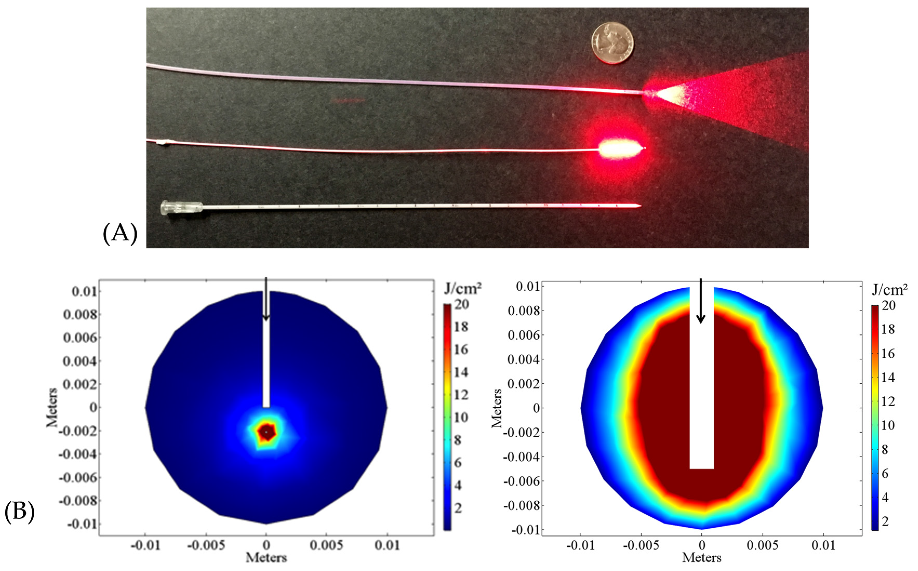
:1. Introduction
2. Interstitial PDT (I-PDT) in Prostate, Pancreatic, Head and Neck, and Brain Cancers
2.1. I-PDT in Prostate Cancer
2.1.1. Image-Based Treatment Planning in I-PDT of Prostate Cancer
2.1.2. Phase III Trial of PDT versus Active Surveillance of Prostate Cancer
2.2. I-PDT for Pancreatic Cancer
2.3. I-PDT for Locally-Advanced Head and Neck Cancer
2.4. I-PDT for Brain Cancers
3. Summary
Acknowledgments
Conflicts of Interest
Abbreviations
| PDT | photodynamic therapy |
| I-PDT | interstitial photodynamic therapy |
| RPCI | Roswell Park Cancer Institute |
| PS | photosensitizer |
| EB-PDT | external beam PDT |
| SOC | standard of care |
| MLu, Lutex | motexafin lutetium |
| TookadTM | palladium bacteriopheophorbide |
| mTHPC, FoscanTM | meso-tetrahydroxyphenylchlorin |
| 5-ALA | 5-aminolevulinic acid |
| Verteporfin | VisudyneTM |
| 3-D | three-dimensional |
| TRUS | transrectal ultrasound |
| PSA | Prostate Specific Antigen |
| BOLD | blood oxygenation level-dependent |
| MRI | magnetic resonance imaging |
| J | energy |
| J/cm2 | energy density |
| J/cm | linear energy |
| mW/cm2 | intensity |
| FEM | finite elements method |
| iDOSE | Interactive Dosimetry by Sequential Evaluation |
| MC | Monte Carlo |
| CT | computed tomography |
| LAHNC | locally advanced head and neck cancer |
| EMA | European Medicines Agency |
References
- Henderson, B.W.; Dougherty, T.J. How does photodynamic therapy work? Photochem. Photobiol. 1992, 55, 145–157. [Google Scholar] [CrossRef] [PubMed]
- Agostinis, P.; Berg, K.; Cengel, K.A.; Foster, T.H.; Girotti, A.W.; Gollnick, S.O.; Hahn, S.M.; Hamblin, M.R.; Juzeniene, A.; Kessel, D.; et al. Photodynamic therapy of cancer: An update. CA Cancer J. Clin. 2011, 61, 250–281. [Google Scholar] [CrossRef] [PubMed]
- Shafirstein, G.; Battoo, A.; Harris, K.; Baumann, H.; Gollnick, S.O.; Lindenmann, J.; Nwogu, C.E. Photodynamic Therapy of Non-Small Cell Lung Cancer. Narrative Review and Future Directions. Ann. Am. Thorac. Soc. 2016, 13, 265–275. [Google Scholar] [CrossRef] [PubMed]
- D’Cruz, A.K.; Robinson, M.H.; Biel, M.A. mTHPC-mediated photodynamic therapy in patients with advanced, incurable head and neck cancer: A multicenter study of 128 patients. Head Neck 2004, 26, 232–240. [Google Scholar] [CrossRef] [PubMed]
- Wilson, B.C.; Patterson, M.S. The physics, biophysics and technology of photodynamic therapy. Phys. Med. Biol. 2008, 53, R61–R109. [Google Scholar] [CrossRef] [PubMed]
- Bown, S.G.; Rogowska, A.Z.; Whitelaw, D.E.; Lees, W.R.; Lovat, L.B.; Ripley, P.; Jones, L.; Wyld, P.; Gillams, A.; Hatfield, A.W. Photodynamic therapy for cancer of the pancreas. Gut 2002, 50, 549–557. [Google Scholar] [CrossRef] [PubMed]
- Jerjes, W.; Upile, T.; Hamdoon, Z.; Abbas, S.; Akram, S.; Mosse, C.A.; Morley, S.; Hopper, C. Photodynamic therapy: The minimally invasive surgical intervention for advanced and/or recurrent tongue base carcinoma. Lasers Surg. Med. 2011, 43, 283–292. [Google Scholar] [CrossRef] [PubMed]
- Karakullukcu, B.; Nyst, H.J.; van Veen, R.L.; Hoebers, F.J.; Hamming-Vrieze, O.; Witjes, M.J.; de Visscher, S.A.; Burlage, F.R.; Levendag, P.C.; Sterenborg, H.J.; et al. mTHPC mediated interstitial photodynamic therapy of recurrent nonmetastatic base of tongue cancers: Development of a new method. Head Neck 2012, 34, 1597–1606. [Google Scholar] [CrossRef] [PubMed]
- Li, J.; Zhu, T.C. Determination of in vivo light fluence distribution in a heterogeneous prostate during photodynamic therapy. Phys. Med. Biol. 2008, 53, 2103–2114. [Google Scholar] [CrossRef] [PubMed]
- Mimikos, C.; Shafirstein, G.; Arshad, H. Current state and future of photodynamic therapy for the treatment of head and neck squamous cell carcinoma. World J. Otorhinolaryngol. Head Neck Surg. 2016, 2, 126–129. [Google Scholar] [CrossRef]
- Baran, T.M.; Foster, T.H. Comparison of flat cleaved and cylindrical diffusing fibers as treatment sources for interstitial photodynamic therapy. Med. Phys. 2014. [Google Scholar] [CrossRef] [PubMed]
- Robinson, D.J.; Karakullukcu, B.M.; Kruijt, B.; Kanick, C.S.; van Veen, R.P.L.; Amelink, A.; Sterenborg, H.J.C.M.; Witjes, M.J.; Tan, B.I. Optical Spectroscopy to Guide Photodynamic Therapy of Head and Neck Tumors. IEEE J. Sel. Top. Quantum Electron. 2010, 16, 854–862. [Google Scholar] [CrossRef]
- Shafirstein, G.; Baumler, W.; Lapidoth, M.; Ferguson, S.; North, P.E.; Waner, M. A new mathematical approach to the diffusion approximation theory for selective photothermolysis modeling and its implication in laser treatment of port-wine stains. Lasers Surg. Med. 2004, 34, 335–347. [Google Scholar] [CrossRef] [PubMed]
- Oakley, E.; Wrazen, B.; Bellnier, D.A.; Syed, Y.; Arshad, H.; Shafirstein, G. A new finite element approach for near real-time simulation of light propagation in locally advanced head and neck tumors. Lasers Surg. Med. 2015, 47, 60–67. [Google Scholar] [CrossRef] [PubMed]
- Davidson, S.R.; Weersink, R.A.; Haider, M.A.; Gertner, M.R.; Bogaards, A.; Giewercer, D.; Scherz, A.; Sherar, M.D.; Elhilali, M.; Chin, J.L.; et al. Treatment planning and dose analysis for interstitial photodynamic therapy of prostate cancer. Phys. Med. Biol. 2009, 54, 2293–2313. [Google Scholar] [CrossRef] [PubMed]
- Ramirez Backhaus, M.; Trassierra Villa, M.; Vera Donoso, C.D.; Jimenez Cruz, J.F. Photodynamic therapy in localised prostate cancer. Actas Urol. Esp. 2007, 31, 633–641. [Google Scholar] [PubMed]
- Verigos, K.; Stripp, D.C.; Mick, R.; Zhu, T.C.; Whittington, R.; Smith, D.; Dimofte, A.; Finlay, J.; Busch, T.M.; Tochner, Z.A.; et al. Updated results of a phase I trial of motexafin lutetium-mediated interstitial photodynamic therapy in patients with locally recurrent prostate cancer. J. Environ. Pathol. Toxicol. Oncol. 2006, 25, 373–387. [Google Scholar] [CrossRef] [PubMed]
- Trachtenberg, J.; Weersink, R.A.; Davidson, S.R.; Haider, M.A.; Bogaards, A.; Gertner, M.R.; Evans, A.; Scherz, A.; Savard, J.; Chin, J.L.; et al. Vascular-targeted photodynamic therapy (padoporfin, WST09) for recurrent prostate cancer after failure of external beam radiotherapy: A study of escalating light doses. BJU Int. 2008, 102, 556–562. [Google Scholar] [CrossRef] [PubMed]
- Betrouni, N.; Lopes, R.; Puech, P.; Colin, P.; Mordon, S. A model to estimate the outcome of prostate cancer photodynamic therapy with TOOKAD Soluble WST11. Phys. Med. Biol. 2011, 56, 4771–4783. [Google Scholar] [CrossRef] [PubMed]
- Gross, S.; Gilead, A.; Scherz, A.; Neeman, M.; Salomon, Y. Monitoring photodynamic therapy of solid tumors online by BOLD-contrast MRI. Nat. Med. 2003, 9, 1327–1331. [Google Scholar] [CrossRef] [PubMed]
- Zaak, D.; Sroka, R.; Höppner, M.; Khoder, W.; Reich, O.; Tritschler, S.; Muschter, R.; Knüchel, R.; Hofstetter, A. Photodynamic Therapy by Means of 5-ALA Induced PPIX in Human Prostate Cancer—Preliminary Results. Med. Laser Appl. 2003, 18, 91–95. [Google Scholar] [CrossRef]
- Yu, G.; Durduran, T.; Zhou, C.; Zhu, T.C.; Finlay, J.C.; Busch, T.M.; Malkowicz, S.B.; Hahn, S.M.; Yodh, A.G. Real-time in situ monitoring of human prostate photodynamic therapy with diffuse light. Photochem. Photobiol. 2006, 82, 1279–1284. [Google Scholar] [CrossRef] [PubMed]
- Wang, K.K.; Zhu, T.C. Reconstruction of in-vivo optical properties for human prostate using interstitial diffuse optical tomography. Opt. Express 2009, 17, 11665–11672. [Google Scholar] [CrossRef] [PubMed]
- Trachtenberg, J.; Bogaards, A.; Weersink, R.A.; Haider, M.A.; Evans, A.; McCluskey, S.A.; Scherz, A.; Gertner, M.R.; Yue, C.; Appu, S.; et al. Vascular targeted photodynamic therapy with palladium-bacteriopheophorbide photosensitizer for recurrent prostate cancer following definitive radiation therapy: Assessment of safety and treatment response. J. Urol. 2007, 178, 1974–1979. [Google Scholar] [CrossRef] [PubMed]
- Azzouzi, A.R.; Barret, E.; Moore, C.M.; Villers, A.; Allen, C.; Scherz, A.; Muir, G.; de Wildt, M.; Barber, N.J.; Lebdai, S.; et al. TOOKAD(®) Soluble vascular-targeted photodynamic (VTP) therapy: Determination of optimal treatment conditions and assessment of effects in patients with localised prostate cancer. BJU Int. 2013, 112, 766–774. [Google Scholar] [CrossRef] [PubMed]
- Moore, C.M.; Azzouzi, A.R.; Barret, E.; Villers, A.; Muir, G.H.; Barber, N.J.; Bott, S.; Trachtenberg, J.; Arumainayagam, N.; Gaillac, B.; et al. Determination of optimal drug dose and light dose index to achieve minimally invasive focal ablation of localised prostate cancer using WST11-vascular-targeted photodynamic (VTP) therapy. BJU Int. 2015, 116, 888–896. [Google Scholar] [CrossRef] [PubMed]
- Azzouzi, A.R.; Vincendeau, S.; Barret, E.; Cicco, A.; Kleinclauss, F.; van der Poel, H.G.; Stief, C.G.; Rassweiler, J.; Salomon, G.; Solsona, E.; et al. Padeliporfin vascular-targeted photodynamic therapy versus active surveillance in men with low-risk prostate cancer (CLIN1001 PCM301): An open-label, phase 3, randomised controlled trial. Lancet Oncol. 2016. [Google Scholar] [CrossRef]
- Nathan, T.R.; Whitelaw, D.E.; Chang, S.C.; Lees, W.R.; Ripley, P.M.; Payne, H.; Jones, L.; Parkinson, M.C.; Emberton, M.; Gillams, A.R.; et al. Photodynamic therapy for prostate cancer recurrence after radiotherapy: A phase I study. J. Urol. 2002, 168 Pt 1, 1427–1432. [Google Scholar] [CrossRef]
- Moore, C.M.; Nathan, T.R.; Lees, W.R.; Mosse, C.A.; Freeman, A.; Emberton, M.; Bown, S.G. Photodynamic therapy using meso tetra hydroxy phenyl chlorin (mTHPC) in early prostate cancer. Lasers Surg. Med. 2006, 38, 356–363. [Google Scholar] [CrossRef] [PubMed]
- Swartling, J.; Axelsson, J.; Ahlgren, G.; Kalkner, K.M.; Nilsson, S.; Svanberg, S.; Svanberg, K.; Andersson-Engels, S. System for interstitial photodynamic therapy with online dosimetry: First clinical experiences of prostate cancer. J. Biomed. Opt. 2010. [Google Scholar] [CrossRef] [PubMed]
- Swartling, J.; Hoglund, O.V.; Hansson, K.; Sodersten, F.; Axelsson, J.; Lagerstedt, A.S. Online dosimetry for temoporfin-mediated interstitial photodynamic therapy using the canine prostate as model. J. Biomed. Opt. 2016. [Google Scholar] [CrossRef] [PubMed]
- Zhu, T.C.; Finlay, J.C. Prostate PDT dosimetry. Photodiagn. Photodyn. Ther. 2006, 3, 234–246. [Google Scholar] [CrossRef] [PubMed]
- Patel, H.; Mick, R.; Finlay, J.; Zhu, T.C.; Rickter, E.; Cengel, K.A.; Malkowicz, S.B.; Hahn, S.M.; Busch, T.M. Motexafin lutetium-photodynamic therapy of prostate cancer: Short- and long-term effects on prostate-specific antigen. Clin. Cancer Res. 2008, 14, 4869–4876. [Google Scholar] [CrossRef] [PubMed]
- Samkoe, K.S.; Chen, A.; Rizvi, I.; O'Hara, J.A.; Hoopes, P.J.; Pereira, S.P.; Hasan, T.; Pogue, B.W. Imaging tumor variation in response to photodynamic therapy in pancreatic cancer xenograft models. Int. J. Radiat. Oncol. Biol. Phys. 2010, 76, 251–259. [Google Scholar] [CrossRef] [PubMed]
- Huggett, M.T.; Jermyn, M.; Gillams, A.; Illing, R.; Mosse, S.; Novelli, M.; Kent, E.; Bown, S.G.; Hasan, T.; Pogue, B.W.; et al. Phase I/II study of verteporfin photodynamic therapy in locally advanced pancreatic cancer. Br. J. Cancer 2014, 110, 1698–1704. [Google Scholar] [CrossRef] [PubMed]
- Jermyn, M.; Davis, S.C.; Dehghani, H.; Huggett, M.T.; Hasan, T.; Pereira, S.P.; Bown, S.G.; Pogue, B.W. CT contrast predicts pancreatic cancer treatment response to verteporfin-based photodynamic therapy. Phys. Med. Biol. 2014, 59, 1911–1921. [Google Scholar] [CrossRef] [PubMed]
- Lou, P.J.; Jager, H.R.; Jones, L.; Theodossy, T.; Bown, S.G.; Hopper, C. Interstitial photodynamic therapy as salvage treatment for recurrent head and neck cancer. Br. J. Cancer 2004, 91, 441–446. [Google Scholar] [CrossRef] [PubMed]
- Jager, H.R.; Taylor, M.N.; Theodossy, T.; Hopper, C. MR imaging-guided interstitial photodynamic laser therapy for advanced head and neck tumors. AJNR Am. J. Neuroradiol. 2005, 26, 1193–1200. [Google Scholar] [PubMed]
- Karakullukcu, B.; van Veen, R.L.; Aans, J.B.; Hamming-Vrieze, O.; Navran, A.; Teertstra, H.J.; van den Boom, F.; Niatsetski, Y.; Sterenborg, H.J.; Tan, I.B. MR and CT based treatment planning for mTHPC mediated interstitial photodynamic therapy of head and neck cancer: Description of the method. Lasers Surg. Med. 2013, 45, 517–523. [Google Scholar] [CrossRef] [PubMed]
- Baran, T.M.; Nazareth, D.P.; Foster, T.H. Image-guided treatment planning and dosimetry for interstitial photodynamic therapy. In Proceedings of the Biomedical Optics and 3D Imaging, Miami, FL, USA, 28 April–2 May 2012.
- Fisher, C.J.; Lilge, L. Photodynamic therapy in the treatment of intracranial gliomas: A review of current practice and considerations for future clinical directions. J. Innov. Opt. Health Sci. 2015. [Google Scholar] [CrossRef]
- Quirk, B.J.; Brandal, G.; Donlon, S.; Vera, J.C.; Mang, T.S.; Foy, A.B.; Lew, S.M.; Girotti, A.W.; Jogal, S.; LaViolette, P.S.; et al. Photodynamic therapy (PDT) for malignant brain tumors—Where do we stand? Photodiagn. Photodyn. Ther. 2015, 12, 530–544. [Google Scholar] [CrossRef] [PubMed]
- Muller, P.J.; Wilson, B.C. Photodynamic therapy for recurrent supratentorial gliomas. Semin. Surg. Oncol. 1995, 11, 346–354. [Google Scholar] [CrossRef] [PubMed]
- Beck, T.J.; Kreth, F.W.; Beyer, W.; Mehrkens, J.H.; Obermeier, A.; Stepp, H.; Stummer, W.; Baumgartner, R. Interstitial photodynamic therapy of nonresectable malignant glioma recurrences using 5-aminolevulinic acid induced protoporphyrin IX. Lasers Surg. Med. 2007, 39, 386–393. [Google Scholar] [CrossRef] [PubMed]
- Johansson, A.; Faber, F.; Kniebuhler, G.; Stepp, H.; Sroka, R.; Egensperger, R.; Beyer, W.; Kreth, F.W. Protoporphyrin IX fluorescence and photobleaching during interstitial photodynamic therapy of malignant gliomas for early treatment prognosis. Lasers Surg. Med. 2013, 45, 225–234. [Google Scholar] [CrossRef] [PubMed]
- Dupont, C.; Betrouni, N.; Mordon, S.R.; Reyns, N.; Vermandel, M. 5-ALA Photodynamic Therapy in Neurosurgery, Towards the Design of a Treatment Planning System: A Proof of Concept. IRBM 2016. [Google Scholar] [CrossRef]
- Betrouni, N.; Colin, P.; Puech, P.; Villers, A.; Mordon, S. An image guided treatment platform for prostate cancer photodynamic therapy. Conf. Proc. IEEE Eng. Med. Biol. Soc. 2013, 2013, 370–373. [Google Scholar] [PubMed]
- Cassidy, J.; Betz, V.; Lilge, L. Treatment plan evaluation for interstitial photodynamic therapy in a mouse model by Monte Carlo simulation with FullMonte. Front. Phys. 2015. [Google Scholar] [CrossRef]
- Weersink, R.A.; Bogaards, A.; Gertner, M.; Davidson, S.R.; Zhang, K.; Netchev, G.; Trachtenberg, J.; Wilson, B.C. Techniques for delivery and monitoring of TOOKAD (WST09)-mediated photodynamic therapy of the prostate: Clinical experience and practicalities. J. Photochem. Photobiol. B 2005, 79, 211–222. [Google Scholar] [CrossRef] [PubMed]
- Qiu, H.; Kim, M.M.; Penjweini, R.; Zhu, T.C. Macroscopic singlet oxygen modeling for dosimetry of Photofrin-mediated photodynamic therapy: An in-vivo study. J. Biomed. Opt. 2016. [Google Scholar] [CrossRef] [PubMed]
- Hasan, T. Using cellular mechanisms to develop effective combinations of photodynamic therapy and targeted therapies. J. Natl. Compr. Cancer Netw. 2012, 10 (Suppl. 2), S23–S26. [Google Scholar]
- Kimura, M.; Miyajima, K.; Kojika, M.; Kono, T.; Kato, H. Photodynamic Therapy (PDT) with Chemotherapy for Advanced Lung Cancer with Airway Stenosis. Int. J. Mol. Sci. 2015, 16, 25466–25475. [Google Scholar] [CrossRef] [PubMed]
- Postiglione, I.; Chiaviello, A.; Palumbo, G. Enhancing photodynamyc therapy efficacy by combination therapy: Dated, current and oncoming strategies. Cancers 2011, 3, 2597–2629. [Google Scholar] [CrossRef] [PubMed]
- Fan, H.T.; Wang, L.; Zhang, P.; Liu, S.B. Photodynamic therapy in spinal metastases: A qualitative analysis of published results. Int. Surg. 2015, 100, 712–719. [Google Scholar] [CrossRef] [PubMed]

| Drug | Drug Dose (mg/kg) | λ (nm) | Laser Settings | # of Patients | Results/Findings | Reference |
|---|---|---|---|---|---|---|
| ALA (*) | 20 | 633 | 250 J/cm | 14 | Significant reduction in prostate-specific antigen (PSA) values was found. | Zack et al., 2003 [21] |
| MLu | 2 | 732 | 150 J/cm | 18 | The 2 mg/kg MLu dose was found too low for effective treatment. | Verigos et al., 2006 [17] |
| MLu | 2 | 732 | 150 mW/cm with 100 J/cm2 measured with isotropic detectors | 3 | Pilot study of diffuse reflectance spectroscopy (DRS) for tumor blood oxygenation and diffuse correlation spectroscopy (DCS) for tumor blood flow. Hemoglobin concentration decreased by 50% following I-PDT. | Yu et al., 2006 [22] |
| MLu | 2 | 732 | 150 mW/cm | 4 | Simulations showed wide variation in light intensity in I-PDT treatment of prostate cancer. | Li and Zhu 2008 [9] |
| MLu | 2 | 732 | 150 mW/cm | 1 | Numerical simulations demonstrate significant variation in optical properties in the target tumor. | Wang and Zhu 2009 [23] |
| TookadTM (**) | 2 | 763 | 230–360 J/cm | 6 | Phase I study. Treatment was found to be safe and well tolerated. | Trachtenberg et al., 2007 [24] |
| TookadTM | 4,6 | 763 | 200–300 J/cm | 4 | Retrospective analysis of clinical trials to examine drug dose, energy fluence and time on I-PDT; Best result with 4 mg/kg and 200 J/cm. | Gross et al., 2003 [20]; Davidson et al., 2009 [15]; Betrouni et al., 2011 [19] |
| TookadTM | 4, 6 | 753 | 200J/cm | 83 | Negative biopsy after 6 months for 61/83 (74%); 4mg/kg and 200 J/cm were optimal for 38/46 (82.6%). | Azzouzi et.al., 2013 [25] |
| TookadTM | 2, 4, 6 | 753 | 200 J/cm | 40 | Phase II trial using 4 mg/kg activated with 753-nm light at a dose of 200 J/cm resulted in a treatment effect of 95% of the planned treatment volume in 12 men and negative biopsy after 6 months for 10/12 or 83.3%. | Moore et al., 2015 [26] |
| TookadTM | 4 | 753 | 150 mW/cm 200 J/cm | 206/PDT, 207/active surveillance | Phase III trial; negative biopsy after 24 months in 49% (101) of patients who received PDT versus 14% (28) in the active surveillance group. | Azzouzi et. al., 2016 [27] |
| mTHPC (***) | 0.15 | 652 | 100–150 mW | 14 | Phase I study, following radiotherapy treatment; partial gland was treated. Up to 91% necrosis or 49% necrosis if one lobe only; cited need for improved dosimetry. | Nathan et. al., 2002 [28] |
| mTHPC | 0.15 | 652 | 100 J/cm | 6 | Early study, after 8–10 I-PDT treatments PSA level fell by 67%. | Moore et al., 2006 [29] |
| mTHPC | 0.15 | 652 | 5 J/cm2 Calculated from lesion size measured with MRI | 4 | Online dosimetry, dose plans were provided with fiber positions and light dose was based on model; Results were that 5 J/cm2 was too low a light dose. | Swartling et al., 2010 [30] |
| Drug | Drug Dose (mg/kg) | λ (nm) | Laser Settings | # of Patients | Results/Findings | Reference |
|---|---|---|---|---|---|---|
| mTHPC | 0.15 | 652 | 100 mW per fiber; 20–40 J/cm | 16 | Tumors regrew at edges of necrotic regions. Median survival: 9.5 months. | Bown et al., 2002 [6] |
| Verteporfin | 0.4 | 690 | 150 mW/cm; 5–40 J/cm per fiber | 15 | No necrosis at 5 J/cm; at 40 J/cm, necrosis was >12 mm in diameter; considerable variation depending on dose; median survival: 8.8 months. | Huggett et al., 2014 [35] |
| Drug | Drug Dose (mg/kg) | λ (nm) | Laser Settings | # of Patients | Results/Findings | Reference |
|---|---|---|---|---|---|---|
| mTHPC | 0.15 | 652 | 100 mW/cm, 20 J/cm, flat cut | 45 | Median overall survival 14 months for responders (73%), versus 2 months for non-responders. | Lou et al., 2004 [37] |
| mTHPC | 0.15 | 652 | 100 mW/cm, 20 J/cm, flat cut | 14 | Median overall survival 14 months. | Jager et al., 2005 [38] |
| mTHPC | 0.15 | 652 | 200 J per site (10 mm) at 100 mW. Flat-cut fiber | 21 | Improvement in palliation (9/11), 60% overall survival after 45 months. | Jerjes et al., 2011 [7] |
| mTHPC | 0.15 | 652 | 100 mW/cm, 30 J/cm, Cylindrical diffuser fiber | 20 | Median overall survival 15 months. | Karakullukcu et al., 2012 [8] |
| Drug | Drug Dose (mg/kg) | λ (nm) | Laser Settings | # of Patients | Results/Findings | Reference |
|---|---|---|---|---|---|---|
| ALA | 20 | 633 | Up to six cylindrical diffusers; total 4320–11,520 J (at 200 mW/cm) | 10 | Adult patients with recurrent malignant glioma; median survival 15 months. | Beck et al., 2007 [44] |
| ALA | 20 or 30 | 635 | 4–6 cylindrical diffusers; total 5700–12,960 J; 720 J/cm (at 150-200 mW/cm) | 5 | Survival >29 months in three responders, <9 months in two non-responders. | Johansson et al., 2013 [45] |
© 2017 by the authors. Licensee MDPI, Basel, Switzerland. This article is an open access article distributed under the terms and conditions of the Creative Commons Attribution (CC BY) license ( http://creativecommons.org/licenses/by/4.0/).
Share and Cite
Shafirstein, G.; Bellnier, D.; Oakley, E.; Hamilton, S.; Potasek, M.; Beeson, K.; Parilov, E. Interstitial Photodynamic Therapy—A Focused Review. Cancers 2017, 9, 12. https://doi.org/10.3390/cancers9020012
Shafirstein G, Bellnier D, Oakley E, Hamilton S, Potasek M, Beeson K, Parilov E. Interstitial Photodynamic Therapy—A Focused Review. Cancers. 2017; 9(2):12. https://doi.org/10.3390/cancers9020012
Chicago/Turabian StyleShafirstein, Gal, David Bellnier, Emily Oakley, Sasheen Hamilton, Mary Potasek, Karl Beeson, and Evgueni Parilov. 2017. "Interstitial Photodynamic Therapy—A Focused Review" Cancers 9, no. 2: 12. https://doi.org/10.3390/cancers9020012