Prickly Pear-Like Three-Dimensional Porous MoS2: Synthesis, Characterization and Advanced Hydrogen Evolution Reaction


1
Department of Chemistry, School of Chemistry and Bioengineering, University of Science & Technology Beijing, Beijing 100083, China
2
Research Center for Bioengineering and Sensing Technology, Beijing Key Laboratory for Bioengineering and Sensing Technology, School of Chemistry and Bioengineering, University of Science & Technology Beijing, Beijing 100083, China
3
Hubei Key Laboratory of Pollutant Analysis & Reuse Technology, Hubei Normal University, Huangshi 435002, China
*
Authors to whom correspondence should be addressed.
Catalysts 2018, 8(6), 235; https://doi.org/10.3390/catal8060235
Submission received: 8 April 2018
/
Revised: 28 May 2018
/
Accepted: 29 May 2018
/
Published: 4 June 2018
(This article belongs to the Special Issue Active Sites in Catalytic Reaction)
Abstract
:Herein, we hydrothermally synthesize a type of prickly pear-like three-dimensional (3D) porous MoS2 (ZT-MoS2), using a zinc oxide (ZnO) rod deposited on quartz glass substrates, as a template for an advanced hydrogen evolution reaction (HER) catalyst. Microscopic and spectroscopic tools comprehensively characterize the morphology of the ZT-MoS2 nanostructure, which exhibits adequate edge active sites and defects, as well as a high component of active octahedral MoS2 (1T-MoS2). Electrochemical characterizations reveal the good HER performance of the ZT-MoS2 that presents a good overpotential of 110 mV, and a Tafel slope of 63 mV·dec−1, superior to most of the previously reported MoS2-based HER catalysts. This work contributes to the design and fabrication of 3D MoS2 with enhanced HER performance, which holds great promise for fuel cells and energy conversion.
1. Introduction
Nowadays, as an environmentally-friendly and abundant energy carrier, hydrogen is attracting intense interest for sustainable energy resources due to the energy crisis [1]. Hydrogen evolution from water splitting is the main strategy to generate hydrogen, and platinum (Pt)-based catalysts are generally employed to enhance hydrogen evolution reaction (HER) [2,3,4,5]. However, their high-cost, insufficient reservoir, and instability limit the widespread application of Pt-based catalysts. Thus, it is urgent to exploit efficient alternative catalysts, such as various transition metals and their derivatives, as well as metal-free catalysts [6,7,8]. Two-dimensional (2D) materials with a large specific surface area, good charge migration rate, controllable electronic properties, good stackability, and mechanical flexibility provide a unique advantage to HER catalytic activity [9]. Molybdenum sulfide (MoS2), a type of two-dimensional layered material, has attracted much attention for its unique physical and chemical properties [10]. Increasing theoretical and experimental evidence reveals that the hydrogen adsorption energy on the edge of naturally van der Waals layered MoS2 is calculated to be close to that of Pt, so that MoS2 has been considered as a promising substitute for Pt-based catalysts [11,12,13]. The edge sites of MoS2 were responsible for hydrogen evolution catalytic activity, and it possessed a lateral dimension size-dependent pattern. The deficiencies of aggregation and low conductivity of bulk MoS2 limit its HER catalytic activity and widespread application [14,15].
Various efficient MoS2 nanostructures with numerous active sites and good conductivity are continuously being explored. Firstly, this is an efficient strategy that introduces active edge sites into MoS2; for example, MoS2 nanoplates [16], MoS2 nanoparticles [17], MoS2 quantum dots [18], and three dimensional (3D) MoS2 [19] with numerous edge sites were developed. Secondly, MoS2 nanostructures can be doped with conductive elements or deposited on highly conductive matrices to improve their conductivity. For example, Se-doped MoS2 and Au-doped MoS2 were designed [20,21]; MoS2 nanoparticles were conjugated on carbon nanotubes [22] and graphene [23], to form MoS2-based heterogeneous composites. Moreover, this method offers a new avenue to improving the catalytic ability that fabricates MoS2 with an adequate octahedral MoS2 (1T-MoS2) component [24].
Among these, a 3D MoS2 nanostructure with both considerable active sites and good substrate transfer ability has received intense attention. To synthesize 3D MoS2, efficient templates are a prerequisite to control the growth of MoS2. Recently, zinc oxide (ZnO) is emerging as an attractive template for constructing 3D materials, since it can be easily and controllably synthesized to produce the desired morphological structures. Various complicated nanostructures, such as silica, gold, and carbon nanotubes, using a ZnO nanostructure as a template have been reported [25,26,27,28].
Herein, using ZnO as a template, a prickly pear-like 3D porous MoS2 (ZT-MoS2) was synthesized by a facile hydrothermal synthesis route (Scheme 1). The MoS2 nanostructures were hydrothermally synthesized in the presence of the ZnO template deposited on the quartz glass substrate. Numerous small MoS2 nanoparticles and ZnO NRs modified on the suface of big MoS2 microbeads. The removal of the ZnO from the nanocomposites generated the prickly pear-like 3D ZT-MoS2 nanostructure. The synthesized ZT-MoS2 exhibited an outstanding HER performance with a good overpotential of 110 mV, and a small Tafel slope of 63 mV·dec−1, as well as extraordinary stability. It was superior to pure MoS2 without using ZnO as template (P-MoS2), and to most previously reported MoS2-based HER catalysts.
2. Results and Discussion
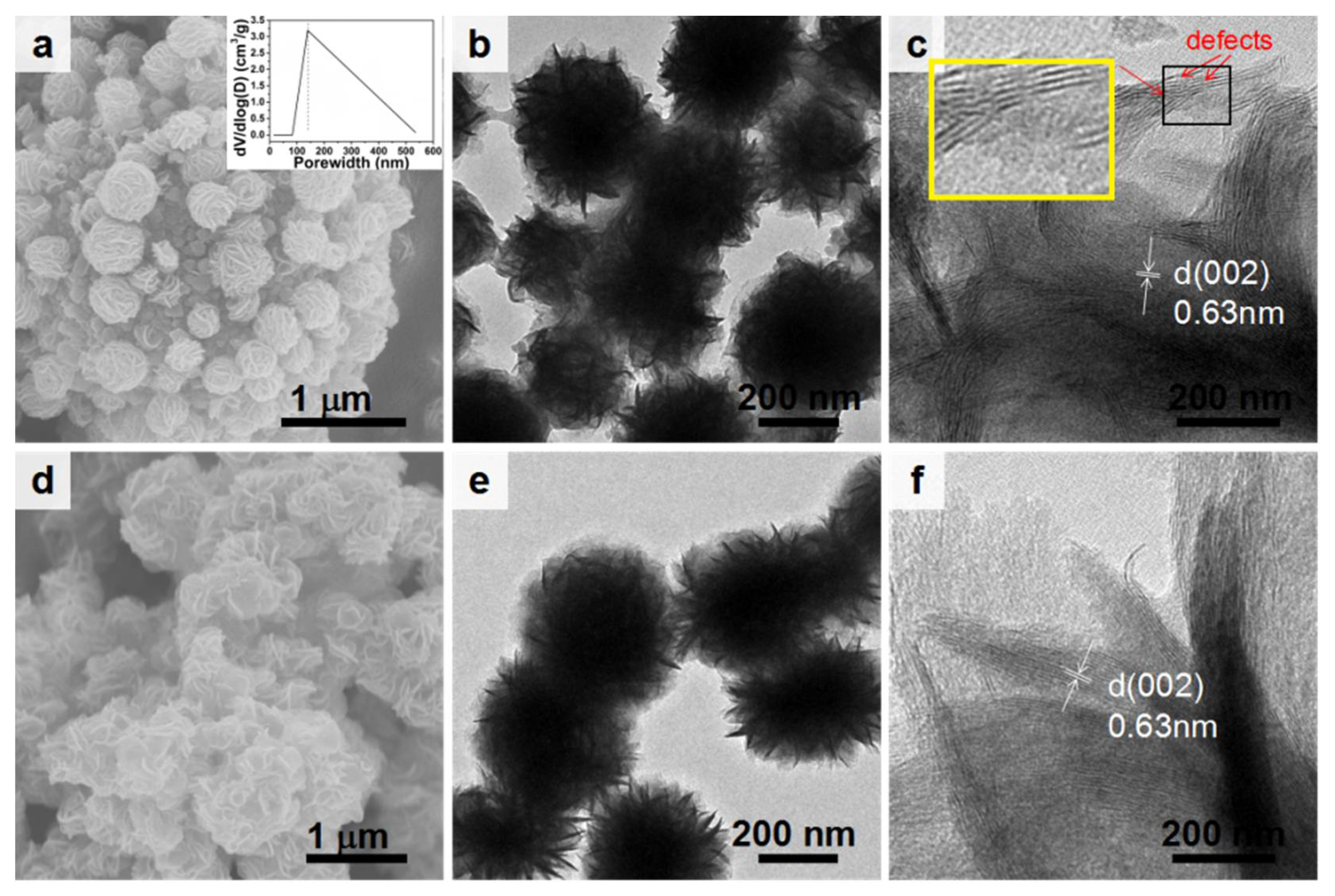
SEM, TEM, and high-resolution TEM (HRTEM) measurements (Figure 1) characterized the morphologies of the P-MoS2 and ZT-MoS2. The ZT-MoS2 presented a prickly pear-like structure, with numerous small MoS2 nanoparticles assembled on the surface of a 2.5 μm porous MoS2 microbead (Figure 1a). The pore size of the porous microbeads in the ZT-MoS2 was about 135 nm (inset in Figure 1a), which was similar to the diameter of the ZnO template (Figure S1). The S-to-Mo ratio was about 2:1 (Figure S2), indicating the successful synthesis of MoS2. Zhang et al. reported that possible intergrowth would be generated between ZnO and MoS2 [29]. However, the Energy Dispersive X-ray Spectrometer (EDS) analysis (Figure S2) showed there was no Zn element, which might be caused by the different synthesis processes. It was found that the ZnO template significantly affected the morphology of ZT-MoS2 (Figure S3), and the HCl-mediated etching process could effectively remove the ZnO from the ZnO-MoS2 nanostructure (Figure 1a, Figures S2 and S4). The P-MoS2 was a nanoflower structure with rippled and corrugated leaf structures (Figure 1d). The size of surface MoS2 nanoparticles decorated on the porous MoS2 microbeads in ZT-MoS2 was about 350 nm (Figure 1b) (with an average surface area of 462.1 ± 5.2 m2 g−1, Table S1), which was smaller than that of P-MoS2 (450 nm) (Figure 1e). It indicated that the ZnO controllably confines the growth of the surface MoS2 nanoparticles. Both the ZT-MoS2 (Figure 1c) and P-MoS2 (Figure 1f) exhibited an interplanar spacing of 6.3 Å assigned to the d spacing of (002) planes of MoS2, suggesting that the ZnO did not influence the component of MoS2 [30]. Compared to the smooth and regular lattice of the P-MoS2, ZT-MoS2 had more delaminated MoS2 (inset in Figure 1c). The resulting defects were beneficial to HER performance.
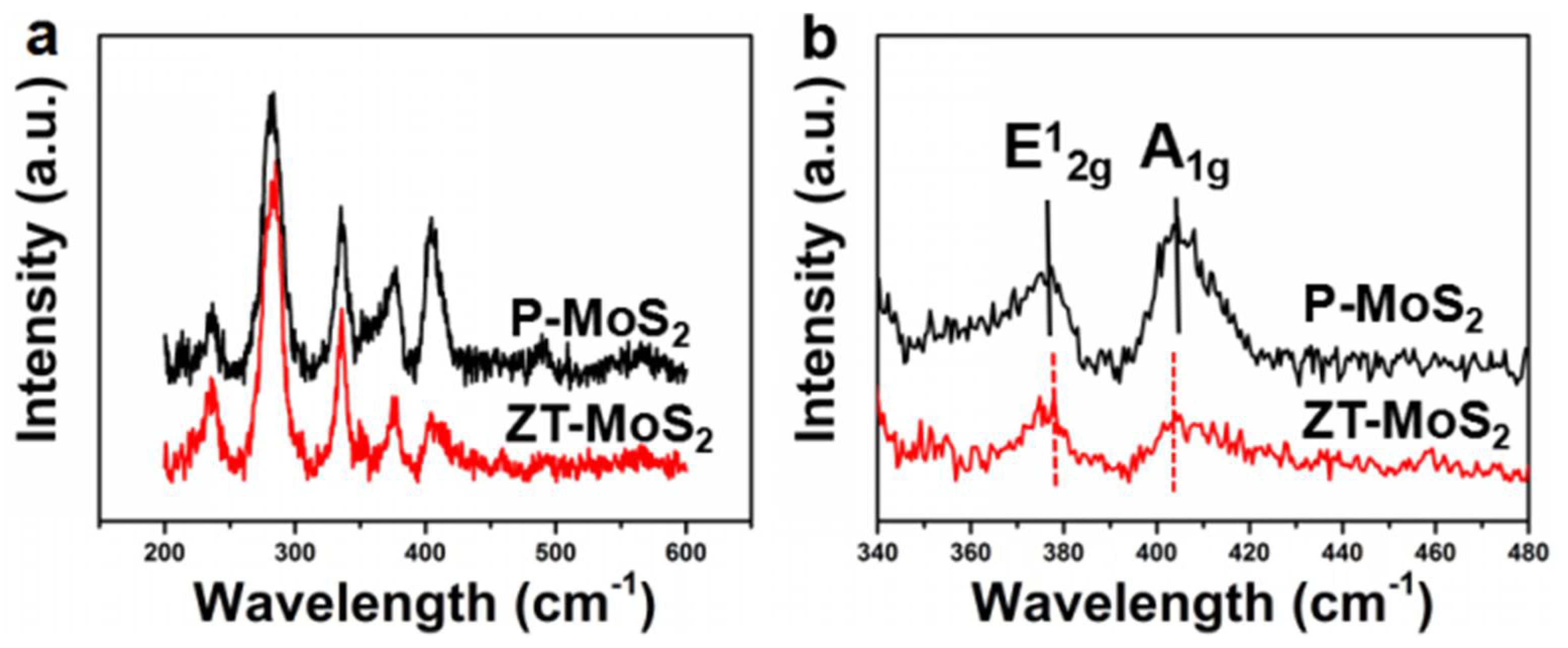
The composition and phase of the ZT-MoS2 and P-MoS2 were investigated by Raman spectroscopy analysis. ZT-MoS2 presented two obvious characteristic peaks at 378 and 404 cm−1, attributed to in-plane vibration E12g modes and out-of-plane vibration A1g modes, respectively (red curve in Figure 2a) [31,32]. The characteristic peaks at 284, 490, and 234 cm−1 assigned to the E1g mode forbidden in a back-scattering geometry on the surface perpendicular to the c-axis, two longitudinal acoustic (2LAM) modes, and structural-defect-induced scattering (red curve in Figure 2a) were also observed [33]. The P-MoS2 exhibited similar characteristic peaks (black curve in Figure 2a) compared to the ZT-MoS2. However, the ZT-MoS2 revealed a larger E12g/A1g ratio and a wider half-wavelength of E12g and A1g peaks than that of the P-MoS2 (Figure 2b), which indicated that more edge structures existed in ZT-MoS2 nanostructures [29]. Additionally, ZT-MoS2 showed a red shift of E12g and a blue shift of A1g compared to that of P-MoS2, illustrating the thinner layered structure of ZT-MoS2 when compared with P-MoS2. It could be anticipated that both the larger edge structure and thinner layered structure would lead to more active sites and endow ZT-MoS2 with better HER performance than P-MoS2 [34,35].
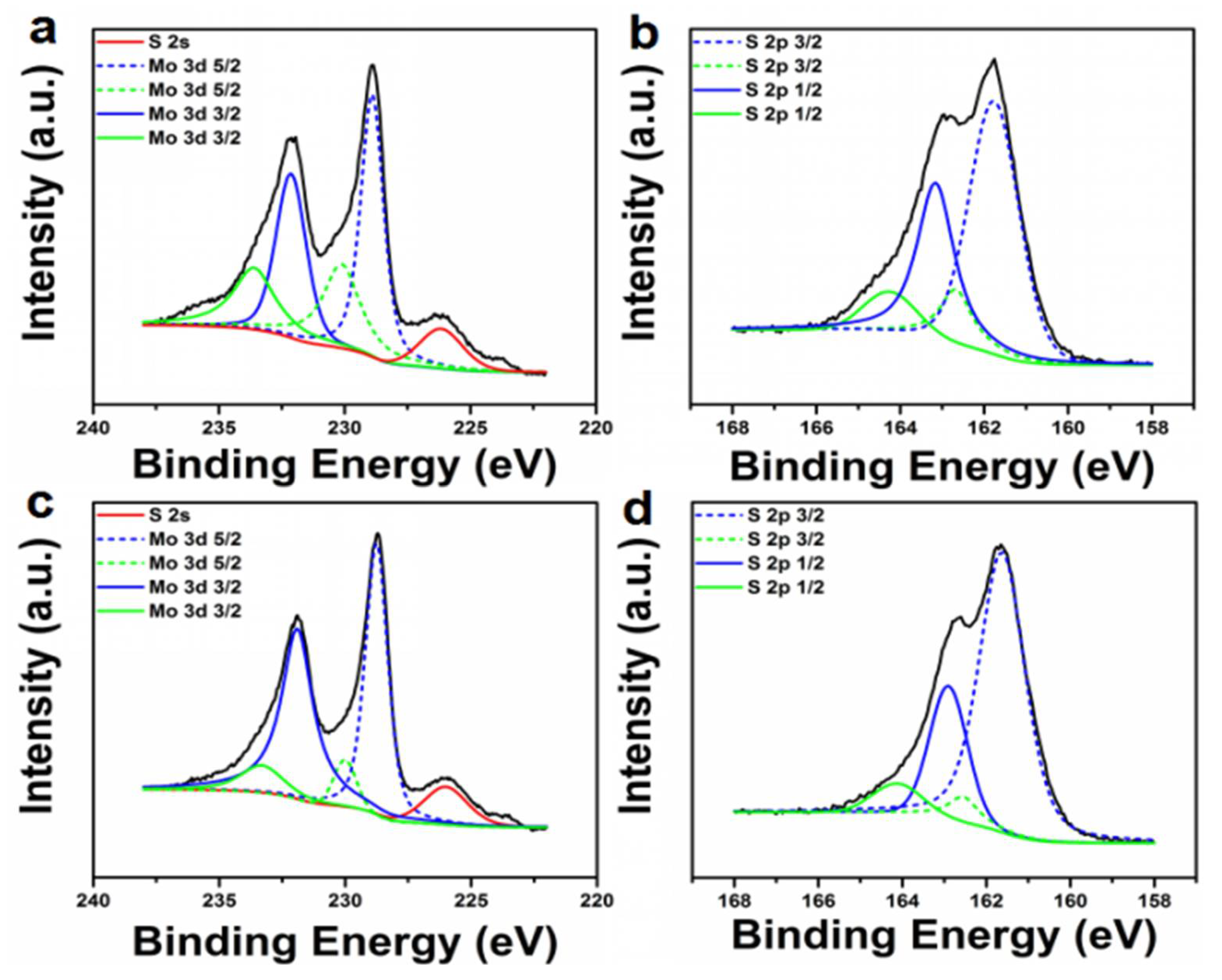
The high-resolution XPS of the Mo 3d peak and the corresponding S 2p peak of the ZT-MoS2 and P-MoS2 were depicted in Figure 3. The Mo 3d of ZT-MoS2 shown in Figure 3a could be deconvoluted into four peaks of 228.9, 230.1, 232.1, and 233.6 eV assigned to Mo4+ 3d5/2, Mo5+ 3d5/2Mo4+ 3d3/2, and Mo5+ 3d3/2, respectively. The peak of 226.2 eV ascribed to S2− 2s was also observed. Mo4+ 3d5/2 and Mo4+ 3d3/2 are related to trigonal prismatic MoS2 (2H-MoS2), while Mo5+ 3d5/2 and Mo5+ 3d3/2 are associated with the1T-MoS2 [36,37]. These results illustrated the co-existence of 2H-MoS2 and 1T-MoS2 in the synthesized ZT-MoS2. The high-resolution XPS of S 2p presented characteristic peaks at S2− 2p3/2 and S2− 2p1/2 associated with 2H-MoS2 at 161.6 and 162.9 eV, while S2− 2p3/2 and S2− 2p1/2 related to 1T-MoS2 at 162.5 and 164.1 eV (Figure 3b), which further confirmed the co-existence of 1T-MoS2 and 2H-MoS2 in ZT-MoS2 [38]. The P-MoS2 exhibited higher peaks of Mo4+ 3d5/2 and Mo4+ 3d3/2 related to 2H-MoS2 than the ZT-MoS2(Figure 3c), and the characteristic peaks of S2− 2p3/2 and S2− 2p1/2 assigned to 2H-MoS2 at 161.6 and 162.9 eV were also larger than that of ZT-MoS2 (Figure 3d). It was calculated that ZT-MoS2 consisted of 60.7 ± 5.2% 2H-MoS2 and 39.3 ± 4.3% 1T-MoS2, while P-MoS2 was composed of 70.5 ± 3.8% 2H-MoS2 and 29.5 ± 4.7% 1T-MoS2. The high 1T-MoS2 in ZT-MoS2 can also contribute to the advanced HER catalytic activity [39].
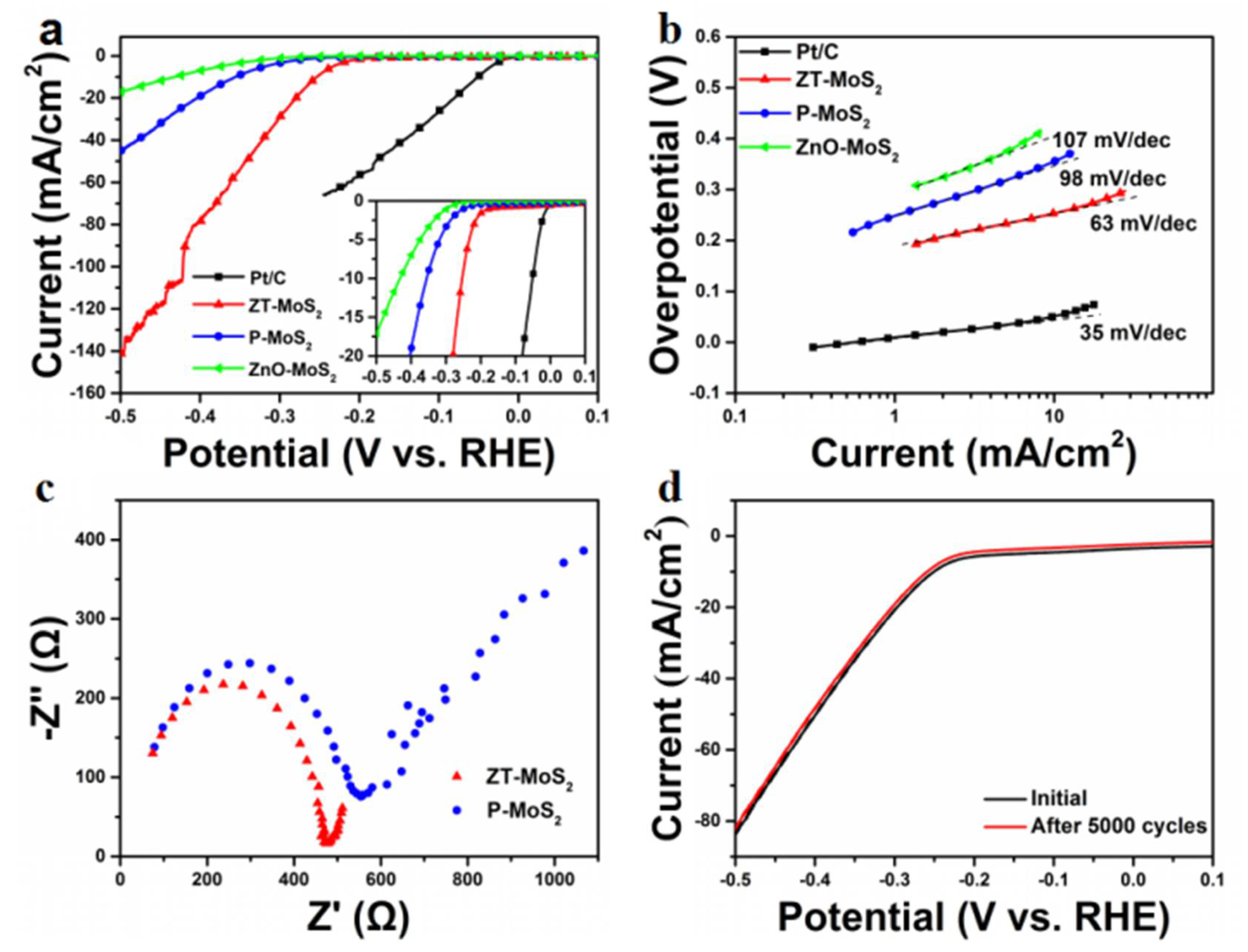
As shown in Figure 4a, the onset overpotential of ZT-MoS2 was 110 ± 5 mV, which was smaller than that of P-MoS2 (200 ± 4 mV) and ZnO-MoS2 (240 ± 4 mV). The onset overpotential of ZT-MoS2 also competed with previously reported MoS2-based HER catalysts [29,40]. The potential of the current density reached to 10 mA·cm−2 was further analyzed. The potential of ZT-MoS2 was 250 mV when the current density reached 10 mA·cm−2, which was smaller than the 370 and 435 mV of P-MoS2 and ZnO-MoS2. The Tafel slope of the Pt/C shown in Figure 4b was 35 mV·dec−1, consistent with previous reports [20,23]. ZT-MoS2 exhibited a Tafel slope of 63 mV·dec−1, smaller than the 98 mV·dec−1 of P-MoS2 and 107 mV·dec−1 of ZnO-MoS2, suggesting that the HER reaction rate of ZnO-MoS2 would increase faster along with the increase of the overpotential when compared with P-MoS2 and ZnO-MoS2 [41]. The electrochemical impedance spectroscopy (EIS) measurements (Figure 4c) revealed a smaller impedance of the ZT-MoS2 than the P-MoS2. It demonstrated that ZT-MoS2 dramatically enhanced the electron transfer and reaction mass accessibility to the active sites. Furthermore, we carried out a long-term cycling test for more than 5000 cycles to investigate the stability of ZT-MoS2. The negligible difference before and after 5000 cycles indicated the remarkable stability of ZT-MoS2 during the long-term electrochemical catalytic process (Figure 4d). The turnover frequency (TOF) for the active sites of the ZT-MoS2 catalyst was calculated using the roughness factor method according to the following equation [42]. The ZT-MoS2 presented a TOF of 1.25 s−1. It was 1.81-fold higher than the TOF of P-MoS2 (0.69 s−1), further indicating the advanced HER catalytic activity of ZT-MoS2 (Figure S5).
These results revealed the superb HER catalytic ability of ZT-MoS2, which can be explained as follows: firstly, the unique prickly pear-like 3D porous MoS2 structures possess many edge active sites confirmed by HRTEM (Figure 1e) and Raman Spectrum analysis (Figure 2b), which contribute to the advanced HER performance (Figure 4a,b). Moreover, adequate defects (Figure 1e) and abundant 1T-MoS2 (Figure 3a,b) were included in ZT-MoS2, enabling the advanced HER catalytic activity. Lastly, the porous 3D structure facilitated the reaction mass transfer during the reaction, which creates the ready accessibility of active sites to the substrate, while the surface of the nanostructure also contributed to its good performance (Table S1). In comparison, both the MoS2 microbeads without decorated MoS2 nanoparticles and the MoS2 microbeads decorated with crowded MoS2 nanoparticles (Figure S3) exhibited inferior HER to ZT-MoS2 (Figure S6). The negligible difference before and after a 5000 cycles long-term test (Figure 4d) indicated the extraordinary stability of the synthesized ZT-MoS2. It suggested that the numerous edge active sites and the good accessibility of active sites to the reaction substrate were significant factors in the efficient HER performance.
3. Materials and Methods
3.1. Reagent
Zinc nitrate hexahydrate (Zn(NO3)2·6H2O), hexamethylenetetramine (C6H12N4), zinc acetate dihydrate (C4H10O6Zn), thiourea (H2NCSN2), and hexaammonium heptamolybdate tetrahydrate ((NH4)6Mo7O24·4H2O) were purchased from Sigma (St. Louis, MO, USA). Potassium hexacyanoferrate (III) (K3Fe(CN)6), potassium hexacyanoferrate (II) trihydrate (K4Fe(CN)6·3H2O), potassium chloride (KCl), hydrochloric acid (HCl), sulfuric acid (H2SO4), and ethanol absolute were purchased from Sinopharm Chemical Reagent Co., Ltd. (Shanghai, China). All reagents used in this study were of analytical grade.
3.2. Preparation of ZnO Nanorods (NRs)
The ZnO NRs were synthesized in accordance with a method set out in a previous report, with some modification [43]. Briefly, ZnO NRs were synthesized on quartz glass substrates with dimensions of 5.5 × 2.5 cm. Firstly, a droplet of 2 mL zinc acetate (5 mM) in ethanol was spin-coated on the quartz glass substrates three times, and then the quartz glass was annealed at 300 °C for 30 min in a tube furnace. The resulting quartz glass was kept in the aqueous solution containing zinc nitrate hexahydrate (50 mM) and hexamethylenetetramine (50 mM) at 100 °C for 10 h in a sealed Schott-Duran bottle. Following this, the substrates were then rinsed thoroughly with ultrapure water and dried overnight at 60 °C in an oven.
3.3. Fabrication of ZT-MoS2 and P-MoS2
(NH4)6Mo7O24·4H2O (1 mmol) and thiourea (30 mmol) were dissolved in ultrapure water (40 mL) to form a homogeneous solution, and the solution was transferred to a 50 mL Teflon-lined stainless-steel autoclave. Different amounts of prepared quartz glass (0.5, 1, and 2) were immersed into the Teflon-lined stainless-steel autoclave and kept at 180 °C for 24 h to obtain different ZT-MoS2 (termed as ZT-MoS2-H, ZT-MoS2 and ZT-MoS2-T, respectively). The solution was naturally cooled down to room temperature, and 3 mL 10% HCl solution was added to etch the ZnO NRs. The resulting ZT-MoS2 dispersed in the solution was generated by centrifuging at 8000 rpm, which was washed thoroughly in the ultrapure water, and then dried at 60 °C by a vacuum freeze dryer to obtain about 4 mg ZT-MoS2 powder each time.
(NH4)6Mo7O24·4H2O (1 mmol) and thiourea (30 mmol) were dissolved in ultrapure water (40 mL) to form a homogeneous solution and the solution was transferred to a 50 mL Teflon-lined stainless-steel autoclave. The solution was naturally cooled down to room temperature and 3 mL HCl solution was added. The resulting P-MoS2 dispersed in the solution was generated by centrifuging at 8000 rpm, which was washed thoroughly in the ultrapure water, and then dried at 60 °C by a vacuum freeze dryer to obtain P-MoS2 powder.
3.4. Characterization
The morphologies of these products were observed under scanning electron microscopy (SEM) (HITACHI S-4800, Tokyo, Japan) and transmission electron microscopy (TEM) (JEM-2010, JEOL Ltd., Tokyo, Japan, 200 kV). High-resolution TEM images were taken using a JEOL 2100F microscope (JEOL Ltd., Tokyo, Japan, 200 kV) with an accelerating voltage of 200 kV. X-ray photoelectron spectroscopy (XPS) measurements were performed using an AXIS ULTRADLD instrument (Kratos, Manchester, UK) equipped with an Al Kα X-ray source. Raman spectra were recorded on an InVia-Reflex Raman microscope (Renishaw, London, UK) with a laser excitation wavelength of 532 nm. The porosity was performed with a nitrogen adsorption–desorption isotherm using a surface area analyzer (QuadraSorb SI 2000-08, Quantachrome Instruments, Boynton Beach, FL, USA).
3.5. Electrochemical Measurements
Electrochemical measurements were performed using an electrochemical station (CHI 852C, Shanghai Chenhua Instrument Co., Shanghai, China) in a three-electrode system. A three-electrode system was employed, consisting of a saturated calomel electrode (SCE) as the reference electrode, a graphite rod as the counter electrode, and a glass carbon rotating disk electrode (RDE) or glassy carbon electrode (GCE) loaded on the catalyst as the working electrode. Linear sweep voltammetry (LSV) measurements were run in 0.5 M H2SO4 (purged with pure N2) at a scan rate of 5 mV·s−1 and at 1400 rpm [17]. For durability measurement, the LSV was performed at a scan rate of 50 mV·s−1 from 0.2 to −0.6 V for 5000 cycles.
Typically, 2 mg of the catalyst was dispersed in 2 mL of ultrapure water to form homogeneous ink under sonication. Then, 20 μL of the catalyst ink was loaded onto the RDE (3 mm in diameter, loading ~0.283 mg·cm−2). 5μL of 1 wt % Nafion solution was dropped onto the electrode after the ink was dried. Cyclic Voltammetry (CV) measurements using GCE as the working electrode were run in 0.5 M H2SO4 (purged with pure N2) at scan rates of 150, 120, 90, 60, 30, and 10 mV·s−1, respectively. The EIS measurements were recorded in the same configuration using the 1 mmol·L−1 K3Fe(CN)6, 1 mmol·L−1 K4Fe(CN)6·3H2O, 0.1 mol·L−1 KCl as the electrolyte at η = 0.2 V from 10−2 to 5 × 10⁵ Hz with a voltage amplitude of 5 mV.
4. Conclusions
In conclusion, a prickly pear-like 3D porous MoS2 nanostructure with advanced HER performance was developed by using a simple and ZnO-mediated hydrothermal synthesis route. Comprehensive microscopic and spectroscopic measurements including SEM, TEM, HRTEM, Raman spectra, and XPS were employed to characterize the morphology and components of the ZT-MoS2. The prickly pear-like 3D porous ZT-MoS2 consisted of numerous small MoS2 nanoparticles decorated on large porous MoS2 microbeads. It displayed many edge active sites and defects as well as abundant 1T-MoS2. The ZT-MoS2 exhibited a superior HER catalytic activity compared with structures using P-MoS2 as a template, and most of the previously reported MoS2-based HER catalysts, such as MoS2/MoSe2 films [31] and Core-shell MoO3-MoS2 nanowires [41]. Additionally, ZT-MoS2 presented extraordinary stability during the long-term cycling test. This work paves a new avenue for the controllable design of efficient 3D MoS2 with advanced HER performance, which can be easily extended to other analogous materials.
Supplementary Materials
The following are available online at https://www.mdpi.com/2073-4344/8/6/235/s1, Table S1: HER activities of synthesized MoS2 catalysts, Figure S1: (a) SEM image and (b) corresponding magnified SEM image of ZnO NRs, Figure S2: Energy Dispersive X-ray Spectrometer (EDS) spectrum of ZT-MoS2, Figure S3: SEM image of different ZT-MoS2: (a) ZT-MoS2-H, (b) ZT-MoS2 and (c) ZT-MoS2-T, Figure S4: SEM image of MoS2 before HCl etching, Figure S5: Electrochemical measurement for determining TOF: (a) a cyclic voltammetry (CV) curve of ZT-MoS2 at different scan rates. (b) Current density of CV experiment at overpotential 500 mV vs. RHE as a function of scan rates, Figure S6: Polarization curves of different ZT-MoS2: (a) ZT-MoS2-H, (b) ZT-MoS2 and (c) ZT-MoS2-T.
Author Contributions
H.D. and X.C. conceived the project. H.L. designed the experiment and synthesized the material; W.D. and K.Z. carried out the characterization of materials; X.C. and C.L. performed the electrochemical Measurements; H.L. and X.C. analyzed the data and wrote the main manuscript text; H.D. modified the manuscript. All authors have given approval to the final version of the manuscript.
Acknowledgments
The work was supported by National Key R&D Program of China (Grant Nos. 2016YFC0106602 and 2016YFC0106601); National Natural Science Foundation of China (Grant Nos. 21645005, 21475008); the Open Research Fund Program of Beijing Key Lab of Plant Resource Research and Development, Beijing Technology and Business University (PRRD-2016-YB2); the Open Research Fund Program of Hubei Key Laboratory of Pollutant Analysis & Reuse Technology, Hubei Normal University (PA160105).
Conflicts of Interest
The authors declare no conflicts of interest.
References
- Dresselhaus, M.S.; Thomas, I.L. Alternative energy technologies. Nature 2001, 414, 332–337. [Google Scholar] [CrossRef] [PubMed]
- Bard, A.J.; Fox, M.A. Artificial photosynthesis: Solar splitting of water to hydrogen and oxygen. Acc. Chem. Res. 1995, 28, 141–145. [Google Scholar] [CrossRef]
- Walter, M.G.; Warren, E.L.; McKone, J.R.; Boettcher, S.W.; Mi, Q.X.; Santori, E.A.; Lewis, N.S. Solar water splitting cells. Chem. Rev. 2010, 110, 6446–6473. [Google Scholar] [CrossRef] [PubMed]
- Norskov, J.K.; Christensen, C.H. Chemistry—Toward efficient hydrogen production at surfaces. Science 2006, 312, 1322–1323. [Google Scholar] [CrossRef] [PubMed]
- Greeley, J.; Jaramillo, T.F.; Bonde, J.; Chorkendorff, I.B.; Norskov, J.K. Computational high-throughput screening of electrocatalytic materials for hydrogen evolution. Nat. Mater. 2006, 5, 909–913. [Google Scholar] [CrossRef] [PubMed]
- Hou, Y.D.; Laursen, A.B.; Zhang, J.S.; Zhang, G.G.; Zhu, Y.S.; Wang, X.C.; Dahl, S.; Chorkendorff, I. Layered nanojunctions for hydrogen-evolution catalysis. Angew. Chem. Int. Ed. 2013, 52, 3621–3625. [Google Scholar] [CrossRef] [PubMed]
- Li, Y.G.; Hasin, P.; Wu, Y.Y. NixCO3-XO4 nanowire arrays for electrocatalytic oxygen evolution. Adv. Mater. 2010, 22. [Google Scholar] [CrossRef] [PubMed]
- Casado-Rivera, E.; Volpe, D.J.; Alden, L.; Lind, C.; Downie, C.; Vazquez-Alvarez, T.; Angelo, A.C.D.; DiSalvo, F.J.; Abruna, H.D. Electrocatalytic activity of ordered intermetallic phases for fuel cell applications. J. Am. Chem. Soc. 2004, 126, 4043–4049. [Google Scholar] [CrossRef] [PubMed]
- Di, J.; Yan, C.; Handoko, A.D.; Seh, Z.W.; Li, H.; Liu, Z. Ultrathin two-dimensional materials for photo- and electrocatalytic hydrogen evolution. Mater. Today 2018. [Google Scholar] [CrossRef]
- Benck, J.D.; Hellstern, T.R.; Kibsgaard, J.; Chakthranont, P.; Jaramillo, T.F. Catalyzing the hydrogen evolution reaction (HER) with molybdenum sulfide nanomaterials. ACS Catal. 2014, 4, 3957–3971. [Google Scholar] [CrossRef]
- Karunadasa, H.I.; Montalvo, E.; Sun, Y.J.; Majda, M.; Long, J.R.; Chang, C.J. A molecular MoS2 edge site mimic for catalytic hydrogen generation. Science 2012, 335, 698–702. [Google Scholar] [CrossRef] [PubMed]
- Laursen, A.B.; Kegnaes, S.; Dahl, S.; Chorkendorff, I. Molybdenum sulfides-efficient and viable materials for electro- and photoelectrocatalytic hydrogen evolution. Energy Environ. Sci. 2012, 5, 5577–5591. [Google Scholar] [CrossRef]
- Wang, T.Y.; Liu, L.; Zhu, Z.W.; Papakonstantinou, P.; Hu, J.B.; Liu, H.Y.; Li, M.X. Enhanced electrocatalytic activity for hydrogen evolution reaction from self-assembled monodispersed molybdenum sulfide nanoparticles on an Au electrode. Energy Environ. Sci. 2013, 6, 625–633. [Google Scholar] [CrossRef]
- Kibsgaard, J.; Chen, Z.B.; Reinecke, B.N.; Jaramillo, T.F. Engineering the surface structure of MoS2 to preferentially expose active edge sites for electrocatalysis. Nat. Mater. 2012, 11, 963–969. [Google Scholar] [CrossRef] [PubMed]
- Jaramillo, T.F.; Jorgensen, K.P.; Bonde, J.; Nielsen, J.H.; Horch, S.; Chorkendorff, I. Identification of active edge sites for electrochemical H2 evolution from MoS2 nanocatalysts. Science 2007, 317, 100–102. [Google Scholar] [CrossRef] [PubMed]
- Yan, Y.; Xia, B.Y.; Ge, X.M.; Liu, Z.L.; Wang, J.Y.; Wang, X. Ultrathin MoS2 nanoplates with rich active sites as highly efficient catalyst for hydrogen evolution. ACS Appl. Mater. Interfaces 2013, 5, 12794–12798. [Google Scholar] [CrossRef] [PubMed]
- Dong, H.F.; Liu, C.H.; Ye, H.T.; Hu, L.P.; Fugetsu, B.S.; Dai, W.H.; Cao, Y.; Qi, X.Q.; Lu, H.T.; Zhang, X.J. Three-dimensional nitrogen-doped graphene supported molybdenum disulfide nanoparticles as an advanced catalyst for hydrogen evolution reaction. Sci. Rep. 2015, 5, 17542. [Google Scholar] [CrossRef] [PubMed]
- Xu, S.J.; Li, D.; Wu, P.Y. One-pot, facile, and versatile synthesis of monolayer MoS2/WS2 quantum dots as bioimaging probes and efficient electrocatalysts for hydrogen evolution reaction. Adv. Funct. Mater. 2015, 25, 1127–1136. [Google Scholar] [CrossRef]
- Zhou, W.J.; Zhou, K.; Hou, D.M.; Liu, X.J.; Li, G.Q.; Sang, Y.H.; Liu, H.; Li, L.G.; Chen, S.W. Three-dimensional hierarchical frameworks based on MoS2 nanosheets self-assembled on graphene oxide for efficient electrocatalytic hydrogen evolution. ACS Appl. Mater. Interfaces 2014, 6, 21534–21540. [Google Scholar] [CrossRef] [PubMed]
- Ren, X.P.; Ma, Q.; Fan, H.B.; Pang, L.Q.; Zhang, Y.X.; Yao, Y.; Ren, X.D.; Liu, S.Z. A Se-doped MoS2 nanosheet for improved hydrogen evolution reaction. Chem. Commun. 2015, 51, 15997–16000. [Google Scholar] [CrossRef] [PubMed]
- Shi, Y.; Wang, J.; Wang, C.; Zhai, T.T.; Bao, W.J.; Xu, J.J.; Xia, X.H.; Chen, H.Y. Hot electron of Au nanorods activates the electrocatalysis of hydrogen evolution on MoS2 nanosheets. J. Am. Chem. Soc. 2015, 137, 7365–7370. [Google Scholar] [CrossRef] [PubMed]
- Li, P.; Yang, Z.; Shen, J.X.; Nie, H.G.; Cai, Q.R.; Li, L.H.; Ge, M.Z.; Gu, C.C.; Chen, X.; Yang, K.Q.; et al. Subnanometer molybdenum sulfide on carbon nanotubes as a highly active and stable electrocatalyst for hydrogen evolution reaction. ACS Appl. Mater. Interfaces 2016, 8, 3543–3550. [Google Scholar] [CrossRef] [PubMed]
- Li, Y.G.; Wang, H.L.; Xie, L.M.; Liang, Y.Y.; Hong, G.S.; Dai, H.J. MoS2 nanoparticles grown on graphene: An advanced catalyst for the hydrogen evolution reaction. J. Am. Chem. Soc. 2011, 133, 7296–7299. [Google Scholar] [CrossRef] [PubMed]
- Voiry, D.; Salehi, M.; Silva, R.; Fujita, T.; Chen, M.W.; Asefa, T.; Shenoy, V.B.; Eda, G.; Chhowalla, M. Conducting MoS2 nanosheets as catalysts for hydrogen evolution reaction. Nano Lett. 2013, 13, 6222–6227. [Google Scholar] [CrossRef] [PubMed]
- Krishna, K.S.; Vivekanandan, G.; Ravinder, D.; Eswaramoorthy, M. ZnO: A versatile template to obtain unusual morphologies of silica, gold and carbon nanostructures. Chem. Commun. 2010, 46, 2989–2991. [Google Scholar] [CrossRef] [PubMed]
- Zeng, H.B.; Cai, W.P.; Liu, P.S.; Xu, X.X.; Zhou, H.J.; Klingshirn, C.; Kalt, H. ZnO-based hollow nanoparticles by selective etching: Elimination and reconstruction of metal-semiconductor interface, improvement of blue emission and photocatalysis. ACS Nano 2008, 2, 1661–1670. [Google Scholar] [CrossRef] [PubMed]
- Gong, W.; Chen, W.S.; He, J.P.; Tong, Y.; Liu, C.; Su, L.; Gao, B.W.; Yang, H.K.; Zhang, Y.; Zhang, X.J. Substrate-independent and large-area synthesis of carbon nanotube thin films using ZnO nanorods as template and dopamine as carbon precursor. Carbon 2015, 83, 275–281. [Google Scholar] [CrossRef]
- Liu, J.P.; Jiang, J.; Bosman, M.; Fan, H.J. Three-dimensional tubular arrays of MnO2-NiO nanoflakes with high areal pseudocapacitance. J. Mater. Chem. 2012, 22, 2419–2426. [Google Scholar] [CrossRef]
- Zhang, K.N.; Zhang, Y.; Zhang, T.N.; Dong, W.J.; Wei, T.X.; Sun, Y.; Chen, X.; Shen, G.Z.; Dai, N. Vertically coupled ZnO nanorods on MoS2 monolayers with enhanced Raman and photoluminescence emission. Nano Res. 2015, 8, 743–750. [Google Scholar] [CrossRef]
- Kong, D.S.; Wang, H.T.; Cha, J.J.; Pasta, M.; Koski, K.J.; Yao, J.; Cui, Y. Synthesis of MoS2 and MoSe2 films with vertically aligned layers. Nano Lett. 2013, 13, 1341–1347. [Google Scholar] [CrossRef] [PubMed]
- Rao, C.N.R.; Matte, H.; Maitra, U. Graphene analogues of inorganic layered materials. Angew. Chem. Int. Ed. 2013, 52, 13162–13185. [Google Scholar] [CrossRef] [PubMed]
- Wang, H.T.; Lu, Z.Y.; Kong, D.S.; Sun, J.; Hymel, T.M.; Cui, Y. Electrochemical tuning of MoS2 nanoparticles on three-dimensional substrate for efficient hydrogen evolution. ACS Nano 2014, 8, 4940–4947. [Google Scholar] [CrossRef] [PubMed]
- Lee, C.; Yan, H.; Brus, L.E.; Heinz, T.F.; Hone, J.; Ryu, S. Anomalous lattice vibrations of single- and few-layer MoS2. ACS Nano 2010, 4, 2695–2700. [Google Scholar] [CrossRef] [PubMed]
- Hinnemann, B.; Moses, P.G.; Bonde, J.; Jorgensen, K.P.; Nielsen, J.H.; Horch, S.; Chorkendorff, I.; Norskov, J.K. Biornimetic hydrogen evolution: MoS2 nanoparticles as catalyst for hydrogen evolution. J. Am. Chem. Soc. 2005, 127, 5308–5309. [Google Scholar] [CrossRef] [PubMed]
- Xie, J.F.; Zhang, H.; Li, S.; Wang, R.X.; Sun, X.; Zhou, M.; Zhou, J.F.; Lou, X.W.; Xie, Y. Defect-rich MoS2 ultrathin nanosheets with additional active edge sites for enhanced electrocatalytic hydrogen evolution. Adv. Mater. 2013, 25. [Google Scholar] [CrossRef] [PubMed]
- Shi, Y.M.; Wang, Y.; Wong, J.I.; Tan, A.Y.S.; Hsu, C.L.; Li, L.J.; Lu, Y.C.; Yang, H.Y. Self-assembly of hierarchical MoSx/CNT nanocomposites (2 < x < 3): Towards high performance anode materials for lithium ion batteries. Sci. Rep. 2013, 3, 2169. [Google Scholar] [CrossRef] [PubMed]
- Pingli, Q.; Guojia, F.; Weijun, K.; Fei, C.; Qiao, Z.; Jiawei, W.; Hongwei, L.; Xingzhong, Z. In situ growth of double-layer MoO3/MoS2 film from MoS2 for hole-transport layers in organic solar cell. J. Mater. Chem. A 2014, 2, 2742–2756. [Google Scholar] [CrossRef]
- Dai, W.H.; Dong, H.F.; Fugetsu, B.; Cao, Y.; Lu, H.T.; Ma, X.L.; Zhang, X.J. Tunable fabrication of molybdenum disulfide quantum dots for intracellular microRNA detection and multiphoton bioimaging. Small 2015, 11, 4158–4164. [Google Scholar] [CrossRef] [PubMed]
- Lukowski, M.A.; Daniel, A.S.; Meng, F.; Forticaux, A.; Li, L.S.; Jin, S. Enhanced hydrogen evolution catalysis from chemically exfoliated metallic MoS2 nanosheets. J. Am. Chem. Soc. 2013, 135, 10274–10277. [Google Scholar] [CrossRef] [PubMed]
- Chen, Z.B.; Cummins, D.; Reinecke, B.N.; Clark, E.; Sunkara, M.K.; Jaramillo, T.F. Core-shell MoO3-MoS2 nanowires for hydrogen evolution: A functional design for electrocatalytic materials. Nano Lett. 2011, 11, 4168–4175. [Google Scholar] [CrossRef] [PubMed]
- Merki, D.; Hu, X.L. Recent developments of molybdenum and tungsten sulfides as hydrogen evolution catalysts. Energy Environ. Sci. 2011, 4, 3878–3888. [Google Scholar] [CrossRef]
- Behranginia, A.; Asadi, M.; Liu, C.; Yasaei, P.; Kumar, B.; Phillips, P.; Foroozan, T.; Waranius, J.C.; Kim, K.; Abiade, J.; et al. Highly efficient hydrogen evolution reaction using crystalline layered three-dimensional molybdenum disulfides grown on graphene film. Chem. Mater. 2016, 28, 549–555. [Google Scholar] [CrossRef]
- Handoko, A.D.; Liew, L.-L.; Lin, M.; Sankar, G.; Du, Y.; Su, H.; Dong, Z.; Goh, G.K.L. Elucidation of thermally induced internal porosity in zinc oxide nanorods. Nano Res. 2018, 11, 2412–2423. [Google Scholar] [CrossRef]
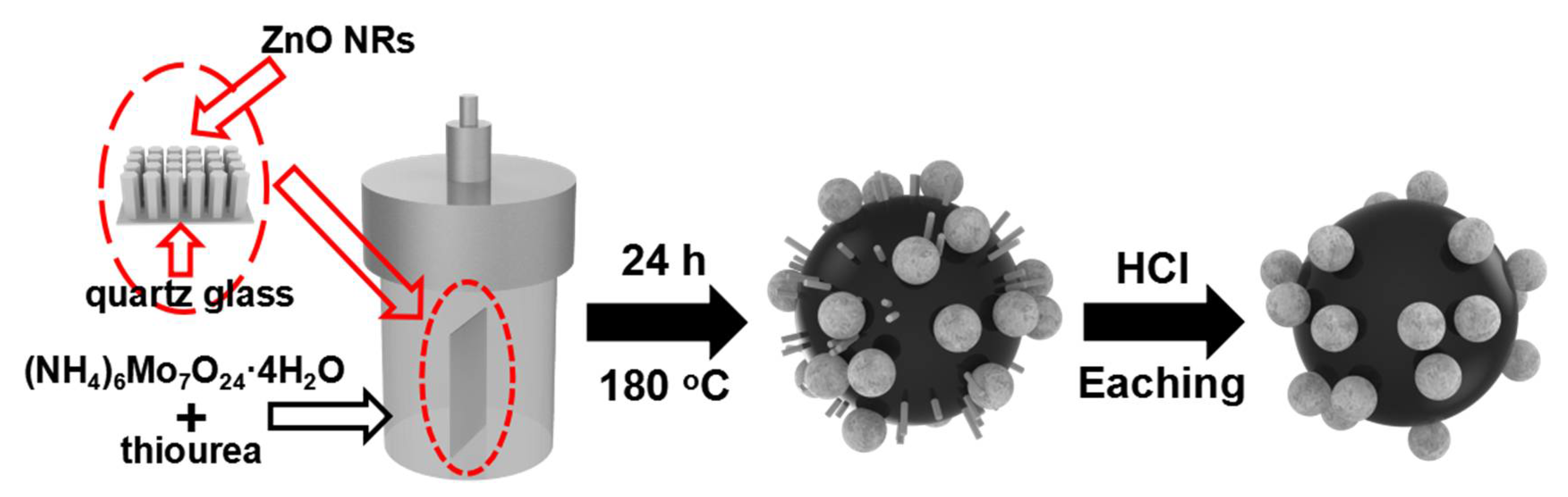
Scheme 1.
Schematic illustration of the synthesis procedure for the ZT-MoS2.

Figure 1.
Scanning electron microscopy (SEM) images of (a) ZT-MoS2 and (d) P-MoS2; transmission electron microscopy (TEM) images of (b) ZT-MoS2 and (e) P-MoS2; high-resolution TEM (HRTEM) images of (c) ZT-MoS2 and (f) P-MoS2. Inset a: porous size analysis of ZT-MoS2.
Figure 1.
Scanning electron microscopy (SEM) images of (a) ZT-MoS2 and (d) P-MoS2; transmission electron microscopy (TEM) images of (b) ZT-MoS2 and (e) P-MoS2; high-resolution TEM (HRTEM) images of (c) ZT-MoS2 and (f) P-MoS2. Inset a: porous size analysis of ZT-MoS2.

Figure 2.
(a) Raman spectra of the P-MoS2 and ZT-MoS2 after HCl etching, (b) the corresponding high resolution of E12g and A1g in (a).
Figure 2.
(a) Raman spectra of the P-MoS2 and ZT-MoS2 after HCl etching, (b) the corresponding high resolution of E12g and A1g in (a).

Figure 3.
High resolution X-ray photoelectron spectroscopy (XPS) spectrum recorded for the Mo 3D of (a) ZT-MoS2 and (c) P-MoS2, and S 2p of (b) ZT-MoS2 (d) P-MoS2. The data were the average of three measurements.
Figure 3.
High resolution X-ray photoelectron spectroscopy (XPS) spectrum recorded for the Mo 3D of (a) ZT-MoS2 and (c) P-MoS2, and S 2p of (b) ZT-MoS2 (d) P-MoS2. The data were the average of three measurements.

Figure 4.
Electrochemical characterizations of as-product catalysts. (a) Polarization curves of Pt/C, ZT-MoS2, P-MoS2, ZnO-MoS2. (b) Tafel plots of Pt/C, ZT-MoS2, P-MoS2, ZnO-MoS2. (c) Electrochemical impedance spectroscopy (EIS) Nyquist plots of ZT-MoS2 and P-MoS2. (d) Durability test of ZT-MoS2 through 5000 cycles linear sweep voltammetry (LSV).

© 2018 by the authors. Licensee MDPI, Basel, Switzerland. This article is an open access article distributed under the terms and conditions of the Creative Commons Attribution (CC BY) license (http://creativecommons.org/licenses/by/4.0/).
Share and Cite
MDPI and ACS Style
Lu, H.; Chen, X.; Dai, W.; Zhang, K.; Liu, C.; Dong, H. Prickly Pear-Like Three-Dimensional Porous MoS2: Synthesis, Characterization and Advanced Hydrogen Evolution Reaction. Catalysts 2018, 8, 235. https://doi.org/10.3390/catal8060235
AMA Style
Lu H, Chen X, Dai W, Zhang K, Liu C, Dong H. Prickly Pear-Like Three-Dimensional Porous MoS2: Synthesis, Characterization and Advanced Hydrogen Evolution Reaction. Catalysts. 2018; 8(6):235. https://doi.org/10.3390/catal8060235
Chicago/Turabian StyleLu, Huiting, Xin Chen, Wenhao Dai, Kai Zhang, Conghui Liu, and Haifeng Dong. 2018. "Prickly Pear-Like Three-Dimensional Porous MoS2: Synthesis, Characterization and Advanced Hydrogen Evolution Reaction" Catalysts 8, no. 6: 235. https://doi.org/10.3390/catal8060235
Note that from the first issue of 2016, this journal uses article numbers instead of page numbers. See further details here.