Synthesis and Crystal Structure of 1-Chloro-2-methyl-4-nitrobenzene

1
Department of Chemistry, Quaid-I-Azam University, Islamabad 45320, Pakistan
2
Department of Chemistry, University of Otago, P.O. Box 56, Dunedin 9054, New Zealand
*
Author to whom correspondence should be addressed.
Crystals 2012, 2(1), 137-143; https://doi.org/10.3390/cryst2010137
Submission received: 29 December 2011
/
Revised: 12 March 2012
/
Accepted: 13 March 2012
/
Published: 19 March 2012
Abstract
:The title compound (3) was prepared from 4-chloroaniline in good yield on successive oxidation and methylation and its crystal and molecular structure is reported. The compound crystallizes in the monoclinic space group P 21/n with unit cell dimensions a = 13.5698(8), b = 3.7195 (3), c = 13.5967 (8) Å, ß = 91.703(3) °, V = 685.96 (10) Å3. The molecule is essentially planar with a dihedral angle of 6.2(3) ° between the nitro group and the phenyl ring. The crystal structure is stabilised by π...π contacts between adjacent benzene rings together with C–H...O hydrogen bonds and close Cl...O contacts.
1. Introduction
1-Chloro-2-methyl-4-nitrobenzene, also called 2-chloro-5-nitrotoluene, belongs to the family of chlorinated nitroaromatic compounds. These are important building blocks for synthesis of diverse heterocycles and a number of industrial chemicals. Thus, the isomeric 2-chloro-6-nitrotoluene is a common intermediate in the synthesis of industrial chloronitrotoluenes, as well as of pharmaceuticals such as the bronchodilatory compound vasicine [1]. It is toxic to the fresh water flea D. magna and freshwater protozoa T. pyriformis, suggesting it as a harmful pollutant [2,3]. Similarly, 1-chloro-4-nitrobenzene is used in the industrial production of azo and sulfur dyes, drugs and pesticides [4]. It is found in industrial wastes [5] and is a serious environmental pollutant [6], causes methemoglobinemia in humans and animals [7] and is weakly mutagenic and carcinogenic [8]. Several Pseudomonas species have been reported to be able to reduce mono-nitro compounds to the corresponding anilines under aerobic conditions [9]. The bacterial strain LW1 (family Comamonadaceae), utilizes 1-chloro-4-nitrobenzene as a sole source of carbon, nitrogen, and energy and transforms it into 2-amino-5-chlorophenol [10]. The title compound was prepared as a key starting material towards some heterocyclic compounds and to study its biodegradation pathway.
2. Results and Discussion
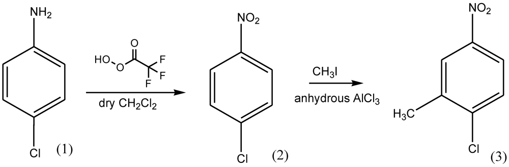
An efficient synthesis of the target compound was carried out according to the route depicted in Figure 1. It started from 4-chloroaniline (1) which, on oxidation using peroxy trifluoroacetic acid in dry dichloromethane, was converted to 4-nitrochlorobenzene (2) in nearly quantitative yield. Friedel-Crafts alkylation of (2) using methyl iodide in the presence of anhydrous aluminum chloride afforded the title compound (3) Figure (1).
Figure 1.
Synthesis of 1-chloro-2-methyl-4-nitrobenzene (3).

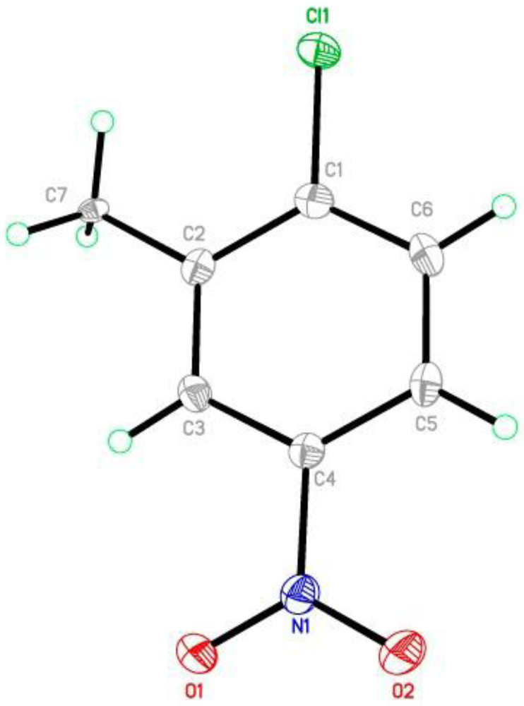
The molecular structure of 1-chloro-2-methyl-4-nitrobenzene shown in Figure 2 is close to planar with the chloro- and methyl- substituents lying 0.022(4) and 0.067(4) Å from the meanplane of the benzene ring (rms deviation = 0.0022 Å). The nitro group is inclined at 6.2(3) ° to the ring plane. The hydrogen atoms of the methyl group are disordered over two positions of equal occupancy. Bond distances in the molecule are normal [11], and bond lengths and angles are similar to those reported for the structure of 2-chloro-3,5-dinitro-p-xylene [12,13] when the difference in data collection temperature and the precision of the measurements are taken into account.
Figure 2.
The structure of 3 showing the atom numbering, with displacement ellipsoids drawn at the 50% probability level [16]. For clarity in all of the figures, the H atoms of only one component of the disordered methyl group are shown.
Figure 2.
The structure of 3 showing the atom numbering, with displacement ellipsoids drawn at the 50% probability level [16]. For clarity in all of the figures, the H atoms of only one component of the disordered methyl group are shown.

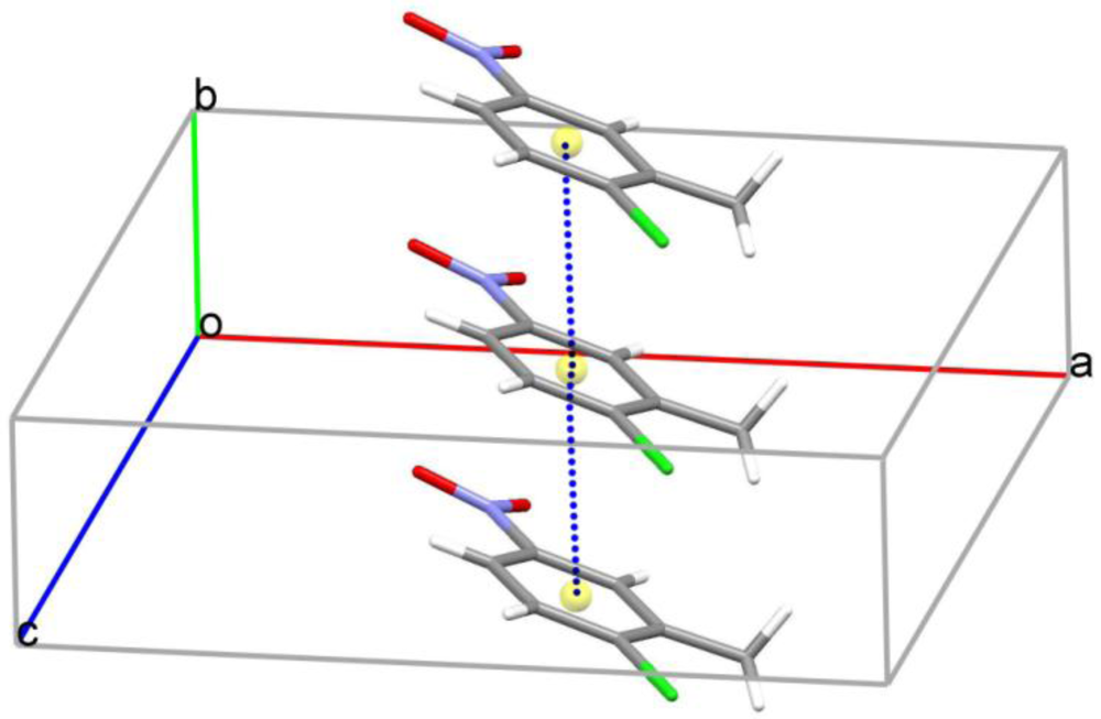
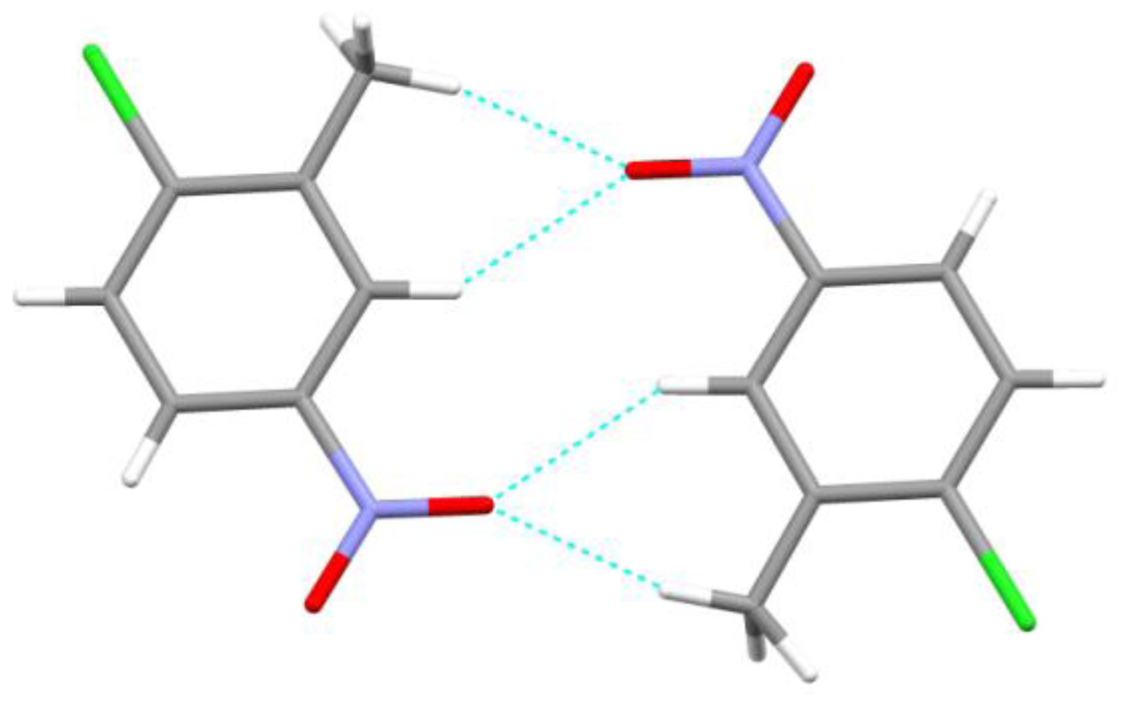
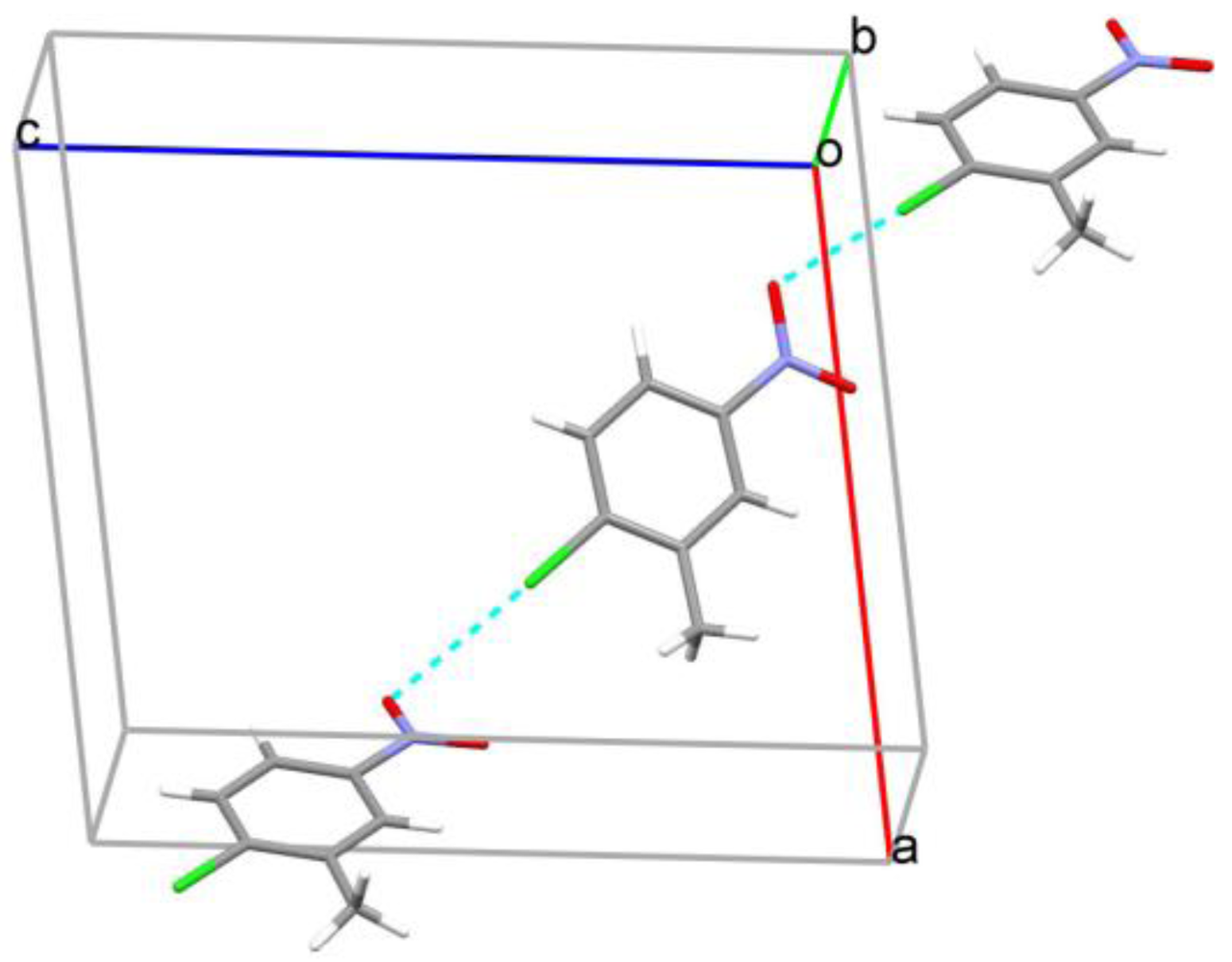
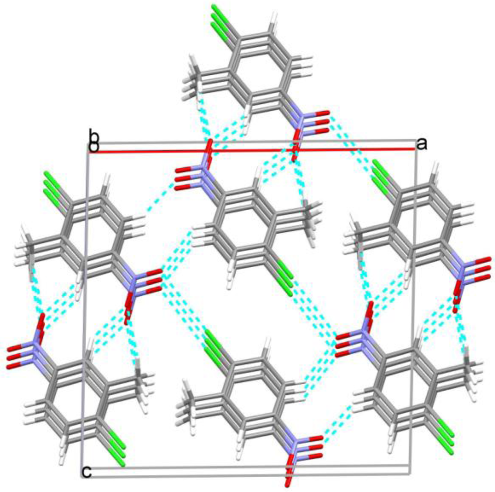
In the crystal structure, π…π contacts stack the molecules along the a axis with centroid to centroid distances 3.719(4) Å, Figure 3. In addition, bifurcated C–H...O hydrogen bonds, Table 1, link adjacent molecules into centrosymmetric dimers generating R21(6), R22(10) and R22(14) ring motifs [14], Figure 4. Cl...O contacts (3.215(3) Å) [15] link the molecules into chains running approximately along the [010] diagonal, Figure 5. These and additional C–H...O contacts generate an extensive three dimensional network with layers of molecules stacked along the b axis, Figure 6.
Figure 3.
π…π stacking interactions [17] between adjacent aromatic rings in 3.
Figure 3.
π…π stacking interactions [17] between adjacent aromatic rings in 3.

Figure 4.
Inversion related dimers formed from C–H…O hydrogen bonds [17].
Figure 4.
Inversion related dimers formed from C–H…O hydrogen bonds [17].

Figure 5.
Short Cl…O contacts forming chains along the [010] diagonal [17].
Figure 5.
Short Cl…O contacts forming chains along the [010] diagonal [17].

Figure 6.
Overall crystal packing for 3 showing layers of molecules stacked along the b axis [17].
Figure 6.
Overall crystal packing for 3 showing layers of molecules stacked along the b axis [17].


| Bond | D–H | H···A | D···A | D–H···A |
|---|---|---|---|---|
| C5—H5···O2 i | 0.95 | 2.69 | 3.129(4) | 109 |
| C7—H7D···O1 ii | 0.98 | 2.20 | 3.137(4) | 161 |
| C3—H3···O1 ii | 0.95 | 2.57 | 3.320(4) | 136 |
Symmetry codes: (i) −x + 1/2, y − 1/2, −z + 1/2; (ii) −x + 1, −y, −z
3. Experimental Section
Synthesis of 1-Chloro-2-methyl-4-nitrobenzene (3). A solution of peroxytrifluoroacetic acid was prepared by addition of 17.0 mL. (0.12 mole) of trifluoroacetic anhydride to a suspension of 2.7 mL. (0.1 mole) of 90% hydrogen peroxide in 50 ml of dichloromethane cooled in an ice-bath. The resulting solution was stirred for five minutes and the cooling bath was removed. To this solution was added dropwise over a 30 minute period 2.35 g. (0.025 mole) of 4-chloroaniline in 10 mL of dichloromethane. After the addition was complete, the reaction mixture was refluxed for one hour. It was successively washed with water and 10% sodium carbonate solution. The dichloromethane extract was dried and concentrated to afford 4-nitrochlorobenzene (2) which was converted to 1-chloro-2-methyl-4-nitrobenzene (3) using methyl iodide and anhydrous aluminum chloride according to standard procedure m.p. 46–47 °C (lit. 40–44 °C). A single crystal of the title compound was grown by slow evaporation of a chloroform solution of the compound over 5 days.
X-ray Data Collection and Structure Solution. Data for 3 were collected on a Bruker APEX II CCD diffractometer using graphite monochromated Mo-Kα radiation, λ = 0.71073 Å at 92 K. The crystal was mounted on a mylar loop in a thin film of Paratone N oil. Intensity data were collected using 60 second ω scans with the APEX2 program and processed with SAINT with absorption corrections applied using SADABS [18]. The structure was solved by direct methods using SHELXS and refined against ׀F׀2 using SHELXL [16] and TITAN [19]. Crystals were very weakly diffracting which results in the measured fraction of data being somewhat low. However solution and refinement proceeded smoothly and residuals and standard uncertainties reported for the structure are acceptable.
Crystal Data for 3. C7H6NO2Cl, Mr = 171.58, yellow, rectangular plate, 0.34 × 0.13 × 0.08 mm. Monoclinic, P21/n, a = 13.5698(8), b = 3.7195 (3), c = 13.5967 (8) Å, β = 91.703(3) °, V = 685.96 (10) Å3, Z = 4, F(000) = 352, T = 91(2) K, ρcalc = 1.661,μ = 0.494 mm-1, 6202 reflections measured, Rint = 0.0536, 1229 merged reflections, 987 with I > 2σ(I), 101 parameters, R indices [I > 2σ(I)]: R1 = 0.0498, wR2 = 0.1482, [all data] R1 = 0.0607, wR2 = 0.1546, w = 1/[σ2(Fo2) + (0.0846P)2] where P = (Fo2 + 2Fc2)/3, largest diff. peak and hole 0.497 and −0.376 e Å−3. CSD deposition number: CCDC 859847
4. Conclusions
A nitroaromatic compound was synthesized as an intermediate towards some heterocycles and for biodegradation studies. The crystal structure of 3 is stabilized by C–H…O hydrogen bonds together with short Cl…O and π…π contacts
Acknowledgments
We thank the University of Otago for purchase of the diffractometer.
References and Notes
- Katritzky, A.R.; Oliferenko, P.; Oliferenko, A.; Lomaka, A.; Karelson, M. Nitrobenzene toxicity; QSAR correlations and mechanistic interpretations. J. Phys. Org. Chem. 2003, 16, 811–817. [Google Scholar]
- Ramos, E.U.; Vaal, M.A.; Hermens, J.L.M. Structure-toxicity relationships for benzenes evaluated with Tetrahymena pyriformis. Environ. Toxicol. Pharmacol. 2001, 11, 149–158. [Google Scholar]
- Harttner, D.R. Toxicity of Nitroaromatic Compounds; Hemisphere Publishing Corp.: Washington, D.C., USA, 1985. [Google Scholar]
- Schackmann, A.; Müller, R. Reduction of nitroaromatic compounds by different Pseudomonas species under aerobic conditions. Appl. Microbiol. Biotechnol. 1991, 34, 809–813. [Google Scholar]
- Shimizu, M.; Yasui, T.; Matsumoto, N. Structural specificity of aromatic compounds with special reference to mutagenic activity in Salmonellatyphimurium-a series of chloro- or fluoro-nitrobenzene derivatives. Mutat. Res. 1983, 116, 217–238. [Google Scholar] [CrossRef]
- Spiess, T.F.; Desiere, P.; Fischer, J.C.; Spain, H.-J.; Lenke, H. A new 4-nitrotoluene degradation pathway in a Mycobacterium strain. Appl. Environ. Microbiol. 1998, 64, 446–452. [Google Scholar]
- Susarla, S.; Masunaga, S.; Yonezawa, Y. Transformations of chloronitrobenzenes in anaerobic sediment. Chemosphere 1996, 32, 967–977. [Google Scholar] [CrossRef]
- Takenaka, S.; Murakami, S.; Shinke, R. Metabolism of 2-aminophenol by Pseudomonas sp. AP-3: modified meta-cleavage pathway. Arch. Microbiol. 1998, 170, 132–137. [Google Scholar] [CrossRef]
- Weisburger, E.K.; Russfield, F.; Homburger, J.; Weisburger, J.H.; Boger, E.; Van Dongen, C.G.; Chu, K.C. Testing of twenty-one environmental aromatic amines or derivatives for long-term toxicity or carcinogenicity. Environ. Pathol. Toxicol. 1978, 2, 325–356. [Google Scholar]
- Katsivela, E.; Wray, V.; Pieper, D.H.; Wittich, R. Initial reactions in the biodegradation of 1-chloro-4-nitrobenzene by a newly isolated bacterium, Strain LW1. Appl. Environ. Microbiol. 1999, 65, 1405–1412. [Google Scholar]
- Allen, F.H.; Kennard, O.; Watson, D.G.; Brammer, L.; Orpen, A.G.; Taylor, R. Tables of Bond Lengths determined by X-Ray and Neutron Diffraction. Part I. Bond Lengths in Organic Compounds. J. Chem. Soc. Perkin Trans. 1987, 2, S1–S19. [Google Scholar]
- Henichart, J.-P.; Bernier, J.-L.; Vaccher, C.; Houssin, R.; Warin, V.; Baert, F. Cyclisation de dinitrodiarylamines en nitro-3 carbazoles inhibition sterique de la substitution nucleophile SNAr par effet. Tetrahedron Lett. 1979, 20, 945–948. [Google Scholar]
- Henichart, J.-P.; Bernier, J.-L.; Vaccher, C.; Houssin, R.; Warin, V.; Baert, F. Acces aux dinitro-2,4 diarylamines par substitution nucleophile SNAr-limites de la reaction et application a la synthese de cyanocarbazoles. Tetrahedron 1980, 36, 3535–3541. [Google Scholar] [CrossRef]
- Bernstein, J.; Davis, R.E.; Shimoni, L.; Chang, N.-L. H bond motifs. Patterns in Hydrogen Bonding: Functionality and Graph Set Analysis in Crystals. Angew. Chem. Int. Ed. Engl. 1995, 34, 1555–1573. [Google Scholar]
- Politzer, P.; Lane, P.; Concha, M.C.; Ma, Y.; Murray, J.S. An overview of halogen bonding. J. Mol. Model. 2007, 13, 305–311. [Google Scholar] [CrossRef]
- Sheldrick, G.M. A short history of SHELX. Acta Cryst. 2008, A64, 112–122. [Google Scholar]
- Macrae, C.F.; Bruno, I.J.; Chisholm, J.A.; Edgington, P.R.; McCabe, P.; Pidcock, E.; Rodriguez-Monge, L.; Taylor, R.; van de Streek, J.; Wood, P.A. Mercury CSD 2.0—New features for the visualization and investigation of crystal structures. J. Appl. Cryst. 2008, 41, 466–470. [Google Scholar] [CrossRef]
- APEX2, SAINT and SADABS. Bruker AXS Inc.: Madison, Wisconsin, USA, 2009.
- Hunter, K.A.; Simpson, J. TITAN2000; University of Otago: Dunedin, New Zealand, 1999. [Google Scholar]
© 2012 by the authors; licensee MDPI, Basel, Switzerland. This article is an open-access article distributed under the terms and conditions of the Creative Commons Attribution license (http://creativecommons.org/licenses/by/3.0/).
Share and Cite
MDPI and ACS Style
Saeed, A.; Simpson, J. Synthesis and Crystal Structure of 1-Chloro-2-methyl-4-nitrobenzene. Crystals 2012, 2, 137-143. https://doi.org/10.3390/cryst2010137
AMA Style
Saeed A, Simpson J. Synthesis and Crystal Structure of 1-Chloro-2-methyl-4-nitrobenzene. Crystals. 2012; 2(1):137-143. https://doi.org/10.3390/cryst2010137
Chicago/Turabian StyleSaeed, Aamer, and Jim Simpson. 2012. "Synthesis and Crystal Structure of 1-Chloro-2-methyl-4-nitrobenzene" Crystals 2, no. 1: 137-143. https://doi.org/10.3390/cryst2010137