
A New Hemihydrate of Valacyclovir Hydrochloride
by
Shuai Zhang
1, 
Meiqi Zheng
1,
Mengqing Zhou
1,
Tian Chen
2,
Naixing Wang
2,
Zhaoxia Zhang
1 and
Guoqing Zhang
1,* 1
Engineering Research Center for Eco-Dyeing and Finishing of Textiles, Ministry of Education, Zhejiang Sci-Tech University, Hangzhou 310018, China
2
Zhejiang Charioteer Pharmaceutical Co., Ltd., Taizhou 317321, China
*
Author to whom correspondence should be addressed.
Crystals 2017, 7(5), 140; https://doi.org/10.3390/cryst7050140
Submission received: 28 March 2017
/
Revised: 9 May 2017
/
Accepted: 11 May 2017
/
Published: 16 May 2017
(This article belongs to the Special Issue Selected Papers from "The Sixth Annual Conference of Chinese Crystallographic Society")
Abstract
:Several crystal forms of valacyclovir hydrochloride, including two anhydrous and three hydrates, were investigated in this study. At the same time, a new hemihydrate of valacyclovir hydrochloride was first discovered and its properties were characterized by PXRD, TGA, DSC, and Raman in this study. The hemihydrate shows a distinctive PXRD pattern and a melting point of 209 °C with a water weight loss of 2.42% from the thermal analysis. The Raman spectra show a few distinctive peaks in the region of 1250–1400 cm−1 due to different crystal forms. The thermostability testing suggests it is a stable crystal form and remain the same for several months under high temperature and humidity. All these crystal forms show good dissolubility in the water at room temperature with excess 100 mg/mL.
1. Introduction
Polymorphism of a drug compound usually results from the possibility of at least two different spatial arrangements of the molecules in the crystal lattice, or in some cases, from variations in molecular conformation, including crystalline and amorphous forms as well as solvate and hydrate forms [1,2,3,4].
Due to difference in the solid–state structure, polymorphs can be imparted with different chemical and physical properties, including melting point, chemical reactivity, apparent solubility, dissolution rate, and optical and mechanical properties of solid dosage forms [5,6,7], which may affect subsequent process and/or manufacture of drug products.
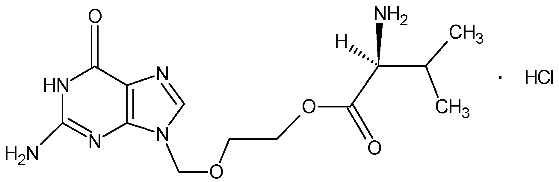
Valacyclovir hydrochloride (VCV), [L-Valine, 2-[(2-amino-1, 6-dihydro-6-oxo-9H-purin-9-yl) methoxy] ethyl ester, monohydrochloride], as shown in Figure 1, is the L-valyl ester prodrug of acyclovir and has demonstrated antiviral activity against herpes simplex virus types, 1 (HSV-1) and 2 (HSV-2) and varicella-zoster virus (VZV) both in vitro and in vivo [8,9,10,11,12,13]. Although VCV has been used as one of the most important antiviral drugs, study on its polymorphism and pseudopolymorphism is still in negligible quantity. So far, two anhydrous and several hydrates crystalline forms of valacyclovir hydrochloride were reported in the US patent 6107302, US 6849736 B2 and US 0021444 as well as their preparation processes [14,15,16]. Based on the complexity and influence on the subsequent drug dosage, it is necessary to further investigate the polymorphism of VCV.
In this work, a new crystal form—hemihydrate—was obtained by recrystallization technology, together with the other four crystal forms of VCV were investigated using powder X-ray diffraction (PXRD), differential scanning calorimetry (DSC), and thermogravimetric analysis (TGA). In addition, the testing for apparent solubility and thermostability was also carried out for deep understanding their pharmaceutical potential.
2. Results and Discussion
According to above experiments, the five solid forms of VCV were successfully prepared and their physicochemical properties were also characterized by PXRD, DSC, and TGA.
2.1. X-ray Diffraction
Although crystal structure determination from an analysis of single-crystal diffraction can provide the fundamental structural understanding of polymorphic solids, the difficulty of preparing a suitable single-crystal precludes this technique from being used on a routine basis for characterization of most drug crystal forms. Since most drug substances were obtained as micro-crystalline powders, the PXRD technology becomes the predominant tool for the routine characterization of polymorphs [4]. In this study, no any suitable single-crystal of the five VCV crystal forms was successfully prepared under current experiment conditions. The X-ray diffraction data relevant to the five powder crystalline forms are summarized in Table 1.
From the diffraction patterns, one can see that Form I, II, IV and V are consistent as previously reported crystal forms. However, the PXRD pattern of the Form III—with characteristic diffraction peaks at 2θ 3.5°, 6.9°, 7.9°, 8.5°, 9.3°, 13°, 14.4°, 16.3°, 20.8°, 24.5°, and 27.2°—is completely distinctive from those of the other four forms. In particular, Form III exhibited several very characteristic peaks at 6.9°, 7.9°, 13°, 20.8° and 23.5°, where no any peak can be detected in the patterns of other forms. It indicates that Form III should be a new polymorphic form of VCV. In order to confirm this point, thermal analysis including DSC and TGA technology were used to further analyze the new polymorphic form.
2.2. Differential Scanning Calorimetry
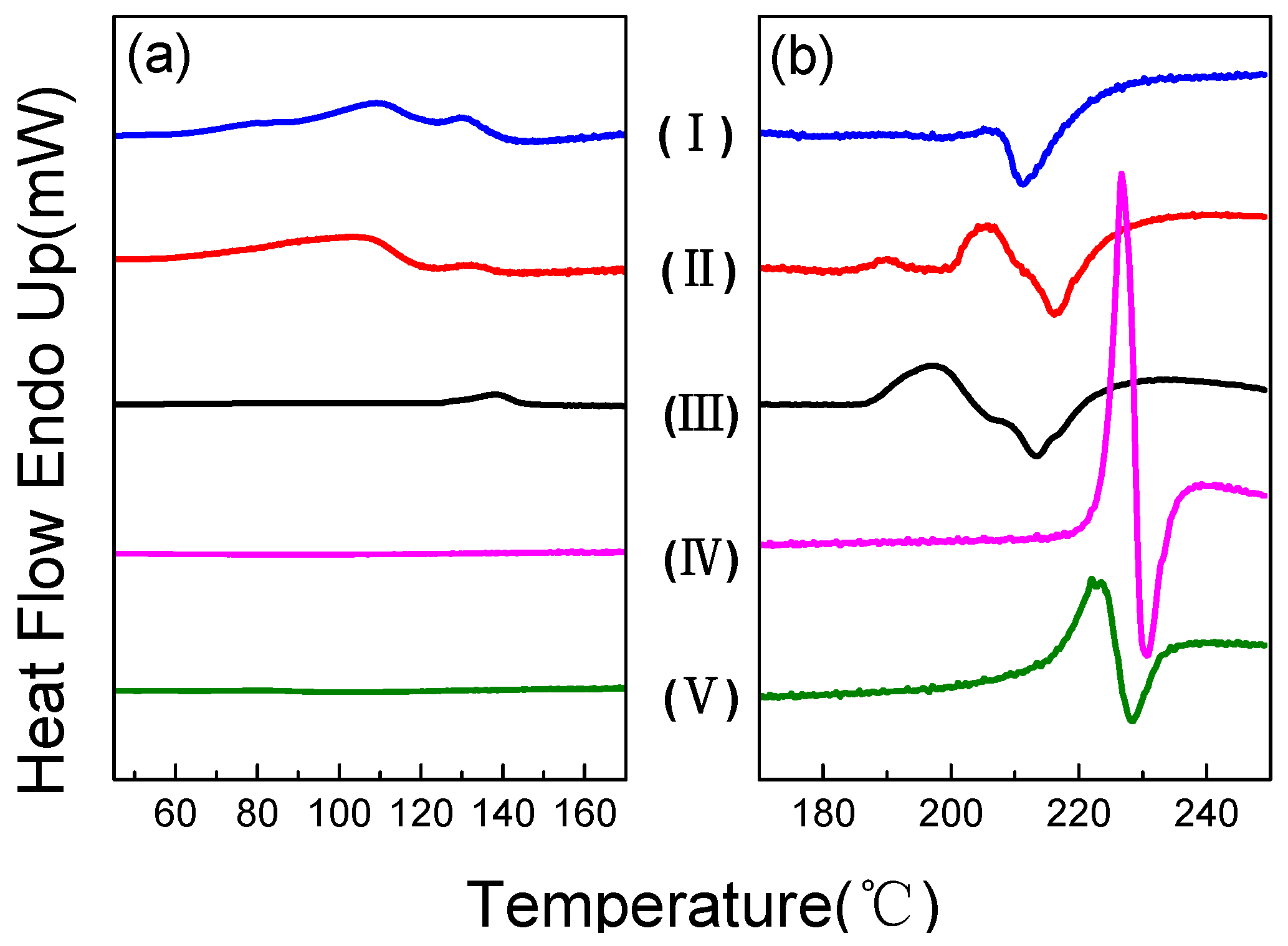
The DSC thermograms of the five crystal forms of VCV are presented in Figure 2. For Form I, II, and III—different crystal forms—as hydrates containing a certain of crystal water, there are significant endothermic peaks on their DSC scans because of water loss in the temperature range of 60–140 °C (Figure 2a). DSC curves of Form I and II showed two partially overlapping endothermic peaks at 70 and 130 °C respectively, which means there are two different types of crystal water in the crystal structure, whereas only one endothermic event at about 141 °C is seen for Form III. In addition, there are not any endothermic peaks in 60–140 °C on the runs of Form IV and Form V, which indicates that both of them are anhydrous compounds.
The melt/decomposition events of the five forms were shown in Figure 2b. For Form I, the melting and decomposition simultaneously occurs at about 205 °C because there is only a single exothermic peak on the DSC curve, while Forms II, IV, and V have two significant melting and decomposition stages, respectively. Melting point is an important parameter for a polymorphic drug, which to some extent can be used to identify different crystal form. The melting temperatures of the four forms were estimated and listed in Table 2. Form III has a solid-solid transformation at about 197 °C, which means it is probably already turned into another crystalline form after the dehydration process.
2.3. Thermogravimetric Analysis
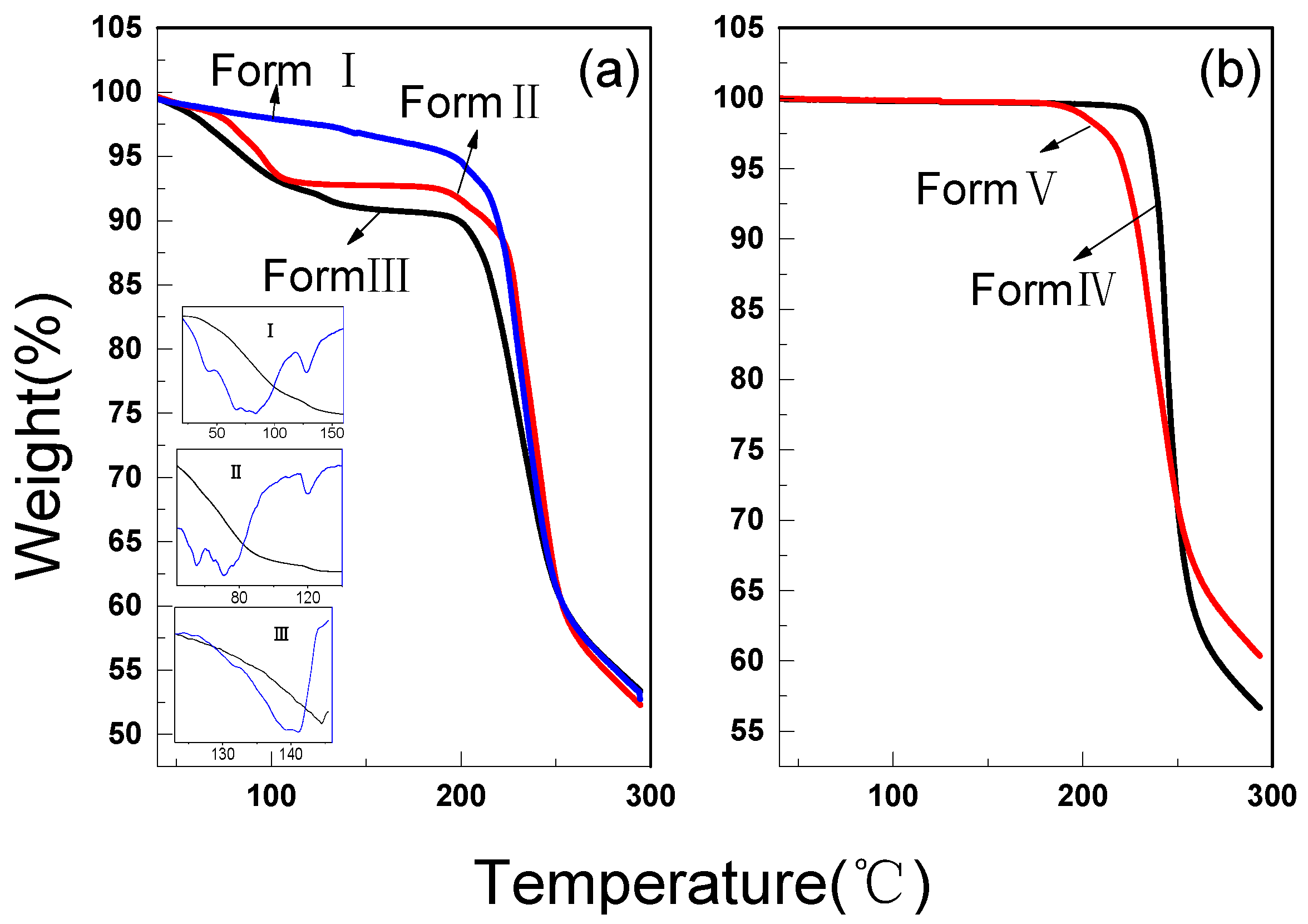
The water contents of all the forms were determined by TGA and the curves are illustrated in Figure 3. The three hydrates showed significant water release steps from Figure 3a, and Form I and II were respectively calculated to be 6.93 wt% and 4.78 wt% in the temperature range of 50–140 °C, but it is 2.42 wt% for Form III in a higher temperature range of 123–145 °C. The above results are respectively consistent with water content theoretically calculated from sesquihydrate, monohydrate, and hemihydrate. Whereas it is clear that Form IV and V had no any weight loss in the same temperature zone from Figure 3b, this suggest they are anhydrous crystal forms.
In addition, the profiles of the derivative curves insert in Figure 3a show there are two weight loss stages for Forma I and II while Form III has only one. These results are totally corresponding with the DSC profiles shown in Figure 2. In fact, each water lease stage usually stands for a specific combination mode between water and VCV molecules. This suggests that there are two bonding modes among water with VCV molecular in VCV hydrates for Form I and II but only one for Form III. Form DSC and TGA results of Form III, the single water loss process actually confirms that it is a pure hydrate crystalline form with half crystal water.
2.4. Raman Spectroscopy
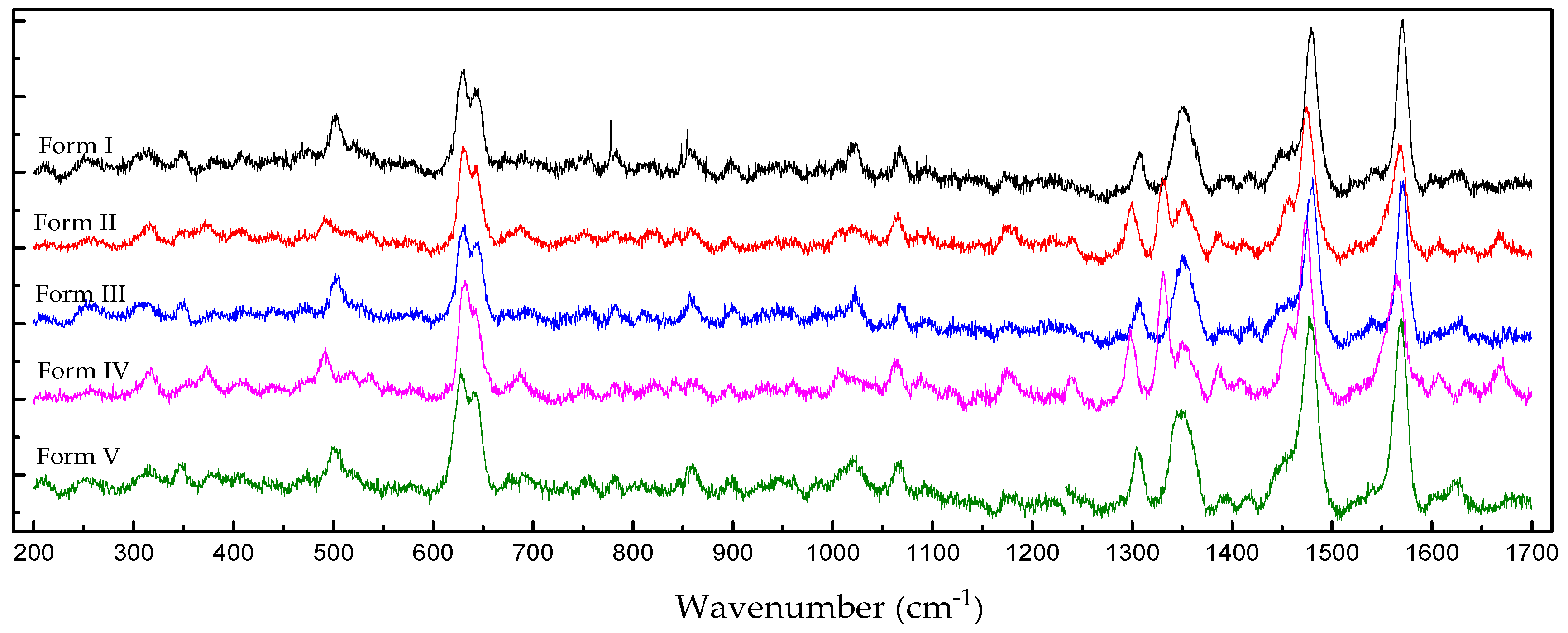
As well known, Raman spectroscopy is a reliable technology to differentiate crystal forms. From the Raman spectra of the five VCV crystal forms in Figure 4, the most distinctive region can be seen between 1250 and 1400 cm−1. In this region, Form I and V show three peaks, Form II and IV have four peaks, but Form III has only two peaks.
From Table 3, Form I has two peaks at 1308.5 and 1351.3 cm−1 with a small peak at 1392.3 cm−1, whereas Form II has three peaks in the region at 1299.4, 1331.7, and 1352.3 cm−1 with a small peak at 1387.3 cm−1. The new crystal Form III shows two peaks at 1307.1 and 1353.3 cm−1. The anhydrate Form IV has four peaks at 1298.4, 1331.7, 1352.8, and 1386.8 cm−1, whereas another anhydrate form V shows three peaks at 1306.1, 1348.8, and 1393.36 cm−1. It is clear that different crystal forms exhibit distinctive Raman spectra.
2.5. Apparent Solubility
As well known, the solid-state structure has a significant influence on the apparent solubility of the drug substance. In this study, the apparent solubility of polymorphic solids of VCV were measured and listed in Table 4. It is obvious that all the crystal forms have excellent dissolubility with excess 100 mg/mL in the ultrapure water. However, the two anhydrous Forms—IV and V—show best apparent solubility with about 100 mg/mL compared with the other hydrates. The results of the present study are consistent with the commonly observed trend that the aqueous solubility of hydrate compounds can be significantly less than their anhydrous forms [17].
Once the solubility measurements had been completed, the remaining crystals were collected and subjected to PXRD testing. The results suggest that Forms I, II, III, and V did not transform into other crystals forms, but Form IV spontaneously converted to Form I during the time of solubility equilibrium. It is easily to understand why solubility of Form IV is close to those hydrates.
2.6. Stability Studies
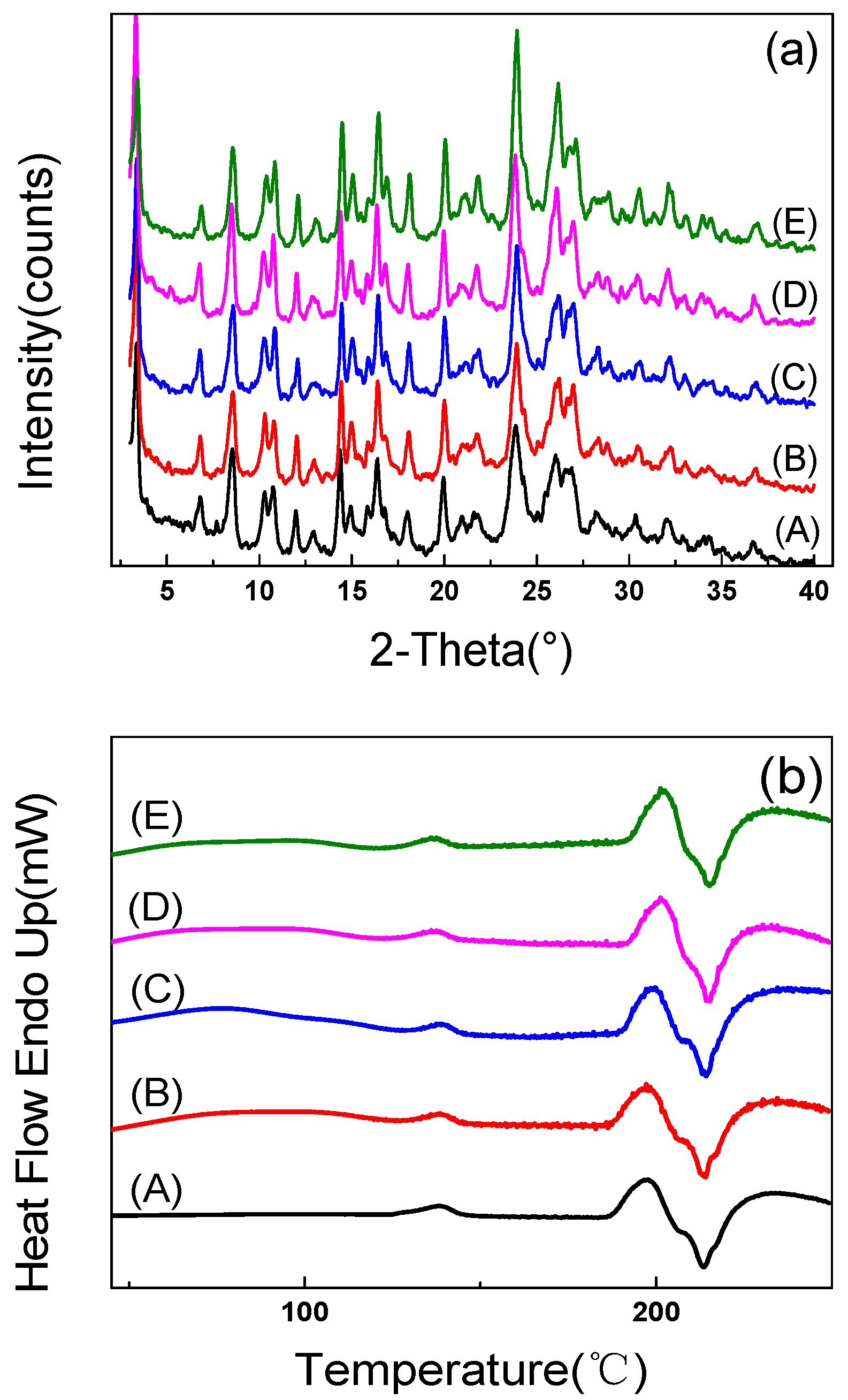
Stability of a drug compound is also an important property during pharmaceutical production. A stability study for Form III was performed by storing some sample under a condition of 40 °C, 75% RH for 16 weeks. During the testing, several batches of specimens were in turn taken out at intervals for PXRD and DSC measurements to monitor their structure changes. As shown in Figure 5a, no impurity diffraction peaks were detected in the PXRD patterns of all the specimens, which means the crystal structure still retained its original form.
From Figure 5b, broad endothermic peaks can be seen in the temperature range of 50–110 °C on the DSC curves of specimens after stored for four weeks, which means a water lease process. However, considering relevant unchanged PXRD patterns, the presence of the water does not change the crystal structure of the hemihydrate, so it should be not crystal water but absorption water. In addition, it is noticed that no recognizable distinction was viewed from the melting points of all the specimens, which further suggests that their crystal form is not changed after a long storage time. Above results indicate that hemihydrate of VCV has good stability and suitable for use in solid drug dosage.
3. Materials and Methods
3.1. Materials
VCV raw material was obtained from Zhejiang Charioteer Medical Technology Co., Ltd. (Zhejiang, China). Solvents—such as ethanediol, trichloromethane, acetone, ethyl alcohol, and isopropanol—were purchased from Tianjing YongDa Chemical Reagent Co., Ltd. (Tianjing, China) with a purity exceeding 99.5%. All aqueous solutions were prepared using Milli-Q deionized (DI) Water with a resistivity of 10.2 MΩ·cm and total organic content <5 ppb.
3.2. Preparation of Polymorphs and Hydrates
The five crystal forms—including two anhydrous and three hydrates—were prepared and named as Forms I, II, III, IV, and V respectively (as shown in Table 5).
Form I, II, IV, and V of VCV could be obtained by recrystallizing from the mixed solvent of water and isopropanol. The VCV solution with 20 wt% raw VCV was first heated up to 80 °C and refluxed for one hour then cooled down to room temperature until crystals precipitation in full. During this process, Forms I, II, IV, and V can be prepared respectively by adjusting the percentage of water in mixed solvent. In fact, Form I can be obtained from mixed solvent with 10 wt% water, Form II in a water content range of 6–8 wt%, and Form IV when the water content is lower than 5%. In particular, Form V was obtained in the mixture with 8% water content by quenching to zero.
For the preparation of Form III, 1 g VCV raw material was dissolved in 10 mL ethanediol at room temperature, the solution was then heated up to 40 °C under stirring. A mixture of 20 mL trichloromethane and 20 mL isopropanol was prepared and cooled to zero. The VCV solution was then dropped into the mixed solvent with a rate of 1 mL/min, the crystals would settle out soon during this process. At last, the precipitation was filtered and dried in an oven at 50 °C.
3.3. Powder X-ray Diffraction Measurements
Powder X-ray diffraction (PXRD) patterns of all the forms of VCV were collected on a powder X-ray diffractometer (Thermal ARL X’TRA, Thermal Technology Co., Santa Rosa, CA, USA) equipped with Cu Kα radiation (λ = 1.54178 Å), operating with a 40 mA current and 40 kV voltage. The data was recorded at a continuous scanning rate of 1.2°/min over a 2θ angular range of 3–40° with a 0.02° step size.
3.4. Thermal Analysis
Thermal analysis was carried out using a differential scanning calorimetry (PYRIS Diamond DSC, PerkinElmer Co., Waltham, MA, USA) calibrated using the indium and zinc standards. These VCV samples were respectively weighted and encapsulated in aluminum pans and then heated from 40–250 °C at a heating rate of 5°/min under nitrogen gas flow of 20 mL/min.
Thermogravimetric analysis (TGA) was performed on a thermogravimetric analyzer (PYRIS 1 TGA, PerkinElmer Co., Waltham, MA, USA) which is calibrated using standard weight and the Curie temperature of magnetic metal. About 4 mg of samples were placed in a platinum crucible and heated from ambient temperature to 300 °C at a rate of 10 °C/min with a 20 mL/min dry nitrogen purge.
3.5. Raman Measurement
Raman spectra were recorded on a */PI Action Tri Vista Raman microscope (Princeton Instruments, Trenton, NJ, USA) equipped with a 20× objective and utilizing a 633 nm laser. The scan range was 100 to 3600 cm−1, using three scans of 30 s length each per spectrum.
3.6. Apparent Solubility Measurement
The chromatography analysis was performed on an Agilent 1100 (Agilent Co., Santa Clara, CA, USA) HPLC equipped with an Agilent G1316B UV Detector and an octadecylsilyl silica gel C18 column with a pore size of 10 nm. The mobile phase consisted of 0.02 mol/L monopotassium phosphate and methyl alcohol (4:1, v/v) at a flow rate of 1.0 mL/min. The column eluent was monitored at 280 nm for 10 min and the injection volume was 20 μL at a column temperature of 25 (±2) °C.
In a typical testing, a sample was respectively weighted 40, 80, 120, 160, 200, 240, and 280 mg and added carefully into glass vials with 1.0 mL ultrapure water, then oscillated for 15 hours at 25 °C until the solution reached apparent equilibrium. Afterwards, 20 μL solution was collected through 0.22 μm syringe filters and diluted into 10.0 mL mobile phase for HPLC testing.
3.7. Stability Testing
In order to evaluate the stability of the new crystal form product, a long-term stability testing was carried out under certain temperature and humility. During this process, Form III sample was stored under an accelerated condition of 40 °C and 75% RH for 16 weeks. The crystal transformation of the sample, including crystal structure and thermal behavior, was monitored periodically by PXRD and DSC.
4. Conclusions
In this study, a few of VCV crystal forms were obtained by recrystallization in different mixed solvents. At the same time, a new polymorphic form—hemihydrate of VCV—was first discovered and its physicochemical properties were investigated by PXRD, DSC, TGA, and Raman.
The PXRD pattern for the new crystal form is completely different from the other polymorphs reported previously. It has melting point measured to be 209 °C from the DSC curve, and a weight loss of 2.42 wt% calculated from TGA profile in the temperature range of 123–145 °C, which confirms that it is a hemihydrate of VCV.
The stability testing suggested the new polymorphic form is a stable crystal form and will remain the same for several months under high temperature and humidity. The results suggest that it has potential to be an active pharmaceutical ingredient (API) using in the drug dosage.
Supplementary Materials
Supplementary File 1Acknowledgments
The authors would like to express the gratitude to the financial support from the Young Researchers Foundation of Zhejiang Provincial Top Key Academic Discipline of Chemical Engineering and Technology, Zhejiang Sci-Tech University (ZYG2015007), Zhejiang Provincial Natural Science Foundation under Grant No. LY16B010002.
Author Contributions
Guoqing Zhang, Shuai Zhang, and Mengqing Zhou conceived and designed the experiments; Shuai Zhang, Mengqing Zhou, Meiqi Zheng, and Zhaoxia Zhang performed the experiments; Shuai Zhang and Mengqing Zhou analyzed the data; Tian Chen and Naixing Wang contributed reagents/materials/analysis tools; Shuai Zhang, Mengqing Zhou, and Guoqing Zhang wrote the paper.
Conflicts of Interest
The authors report no conflicts of interest and are responsible for the content and writing of the paper.
References
- Vega, D.; Petragalli, A.; Fernández, D.; Ellena, J.A. Polymorphism on Leflunomide: Stability and Crystal Structures. J. Pharm. Sci. 2005, 95, 1075–1083. [Google Scholar] [CrossRef] [PubMed]
- Nagai, K.; Ushio, T.; Miura, H.; Nakamura, T.; Moribe, K.; Yamamoto, K. Four new polymorphic forms of suplatast tosilate. Int. J. Pharm. 2014, 460, 83–91. [Google Scholar] [CrossRef] [PubMed]
- Sohn, Y.T.; Kim, S.H. Polymorphism and pseudopolymorphism of acyclovir. Arch. Pharm. Res. 2008, 31, 231–234. [Google Scholar] [CrossRef] [PubMed]
- Brittain, H.G. Polymorphism in Pharmaceutical Solids; Drugs and the Pharmaceutical Science: New York, NY, USA, 1999; pp. 76–81. ISBN 9781420073218. [Google Scholar]
- Haleblian, J.K. Characterization of habits and crystalline modification of solids and their pharmaceutical applications. J. Pharm. Sci. 1975, 64, 1269–1288. [Google Scholar] [CrossRef] [PubMed]
- Lutker, K.M.; Quinones, R.; Xu, J.; Ramamoorthy, A.; Matzger, A.J. Polymorphs and hydrates of acyclovir. J. Pharm. Sci. 2010, 100, 949–963. [Google Scholar] [CrossRef] [PubMed]
- Haleblian, J.; McCrone, W. Pharmaceutical Applications of Polymorphism. J. Pharm. Sci. 1969, 58, 911–929. [Google Scholar] [CrossRef] [PubMed]
- Granero, G.E.; Amidon, G.L. Stability of valacyclovir: Implications for its oral bioavailability. Int. J. Pharm. 2006, 317, 4–18. [Google Scholar] [CrossRef] [PubMed]
- Shinkai, I.; Ohta, Y. Valacyclovir. Bioorgan. Med. Chem. 1996, 4, 1–2. [Google Scholar] [CrossRef]
- Beutner, K.R. Valacyclovir: A review of its antiviral activity, pharmacokinetic properties, and clinical efficacy. Antivir. Res. 1995, 28, 281–290. [Google Scholar] [CrossRef]
- Yadav, M.; Upadhyay, V.; Singhal, P.; Goswami, S.; Shrivastav, P.S. Stability evaluation and sensitive determination of antiviral drug, valacyclovir and its metabolite acyclovir in human plasma by a rapid liquid chromatography–tandem mass spectrometry method. J. Chromatogr. B 2009, 877, 680–688. [Google Scholar] [CrossRef] [PubMed]
- De Clercq, E. Milestones in the discovery of antiviral agents: Nucleosides and nucleotides. Acta Pharm. Sin. B 2012, 2, 535–548. [Google Scholar] [CrossRef]
- Jadhav, A.S.; Pathare, D.B.; Shingare, M.S. Development and validation of enantioselective high performance liquid chromatographic method for Valacyclovir, an antiviral drug in drug substance. J. Pharm. Biomed. Anal. 2007, 43, 1568–1572. [Google Scholar] [CrossRef] [PubMed]
- Carter, B.H.; Partin, J.M.; Varlashkin, P.G. Guanine Derivative. U.S. Patent 6,107,302, 22 August 2000. [Google Scholar]
- Wizel, S.; Aronhime, J.; Niddam-Hildesheim, V.; Dolitzky, B.Z. Crystalline Forms of Valacyclovir Hydrochloride. U.S. Patent 6,849,736 B2, 1 February 2005. [Google Scholar]
- Shepherd, M.; Mckay, B. Valacyclovir Polymorphs and a Process for the Preparation Thereof. U.S. Patent 0,021,444 A1, 25 January 2007. [Google Scholar]
- Shefter, E.; Higuchi, T. Dissolution behavior of crystalline solvated and nonsolvated forms of some pharmaceuticals. J. Pharm. Sci. 1963, 52, 781–791. [Google Scholar] [CrossRef] [PubMed]
Figure 1.
Molecular structure scheme of VCV.

Figure 2.
DSC thermograms of all the polymorphic forms of VCV, (a) water lease stage and (b) melting/decomposition stage.
Figure 2.
DSC thermograms of all the polymorphic forms of VCV, (a) water lease stage and (b) melting/decomposition stage.

Figure 3.
TGA curves of VCV polymorphs: (a) Form I, Form II and Form III; (b) Form IV and Form V, the insert is the profiles of the derivative curves.
Figure 3.
TGA curves of VCV polymorphs: (a) Form I, Form II and Form III; (b) Form IV and Form V, the insert is the profiles of the derivative curves.

Figure 4.
The Raman spectra of the five forms of valacyclovir (all spectra were collected at room temperature).
Figure 4.
The Raman spectra of the five forms of valacyclovir (all spectra were collected at room temperature).

Figure 5.
The PXRD patterns (a) and DSC curves (b) for Form III stored for 16 weeks, several specimens were taken out and tested at (A) 0 week, (B) 4 weeks, (C) 8 weeks, (D) 12 weeks, (E) 16 weeks.
Figure 5.
The PXRD patterns (a) and DSC curves (b) for Form III stored for 16 weeks, several specimens were taken out and tested at (A) 0 week, (B) 4 weeks, (C) 8 weeks, (D) 12 weeks, (E) 16 weeks.

Table 1.
Reflections of the five crystal forms of VCV.
| Form I | Form II | Form III | Form IV | Form V |
|---|---|---|---|---|
| 2θ(°) Intensity (%) | 2θ(°) Intensity (%) | 2θ(°) Intensity (%) | 2θ(°) Intensity (%) | 2θ(°) Intensity (%) |
| 3.66 100 | 3.56 100 | 3.46 100 | 3.66 63.1 | 3.50 33.6 |
| 8.58 2.6 | 8.58 5.2 | 6.94 93.1 | 8.66 41.1 | 6.58 58.7 |
| 9.48 6.0 | 9.38 5.2 | 7.88 13.2 | 10.62 13.8 | 9.18 100 |
| 10.60 9.8 | 10.52 8.6 | 8.46 10.9 | 10.92 48.5 | 11.32 18.8 |
| 10.86 24.4 | 10.78 15.8 | 9.30 13.3 | 12.18 18.3 | 15.34 13.3 |
| 13.36 7.8 | 12.08 0.9 | 10.46 9.5 | 13.32 12.1 | 15.62 16.9 |
| 16.44 3.9 | 13.26 6.1 | 11.64 8.4 | 14.56 51.1 | 16.16 10.4 |
| 20.20 2.0 | 14.42 2.5 | 13.02 11.5 | 15.20 14.3 | 17.02 16.1 |
| 21.42 2.9 | 16.36 5.3 | 14.44 12.9 | 16.48 52.2 | 19.14 18.8 |
| 21.80 7.2 | 20.04 2.5 | 15.34 9.2 | 16.96 22.5 | 19.66 9.0 |
| 24.00 3.2 | 21.34 2.2 | 16.25 11 | 18.16 33.8 | 21.26 19.6 |
| 26.76 2.1 | 23.92 9.3 | 20.78 11.4 | 20.10 34.1 | 22.86 15.0 |
| 27.28 5.2 | 25.94 1.8 | 23.47 6.6 | 20.62 7.1 | 24.16 10.8 |
| 26.82 2.4 | 23.90 9.9 | 21.28 15.3 | 26.30 8.4 | |
| 27.22 4.1 | 24.54 19.5 | 21.88 25.7 | 27.34 17.8 | |
| 26.18 11.0 | 22.72 8.8 | 27.86 15.3 | ||
| 27.16 19.6 | 24.02 100 | 28.76 3.4 | ||
| 27.80 11.6 | 24.46 15.9 | |||
| 25.13 6.8 | ||||
| 25.94 21.1 | ||||
| 26.26 60.8 | ||||
| 26.92 10.6 | ||||
| 27.26 8.4 | ||||
| 28.10 7.9 | ||||
| 28.38 6.5 | ||||
| 29.00 16.2 | ||||
| 29.70 8.8 | ||||
| 30.64 18.1 |
Table 2.
Differential scanning calorimetry data for all the forms of VCV.
| Items | Form I | Form II | Form III | Form IV | Form V |
|---|---|---|---|---|---|
| Melting point (°C) | 205 | 206 | 209 | 227 | 223 |
| Transition 1 (°C) | 53–139 | 60–140 | 124–141 | / | / |
| Transition 2 (°C) | / | 183–196 | 186–206 | / | / |
Table 3.
Summary of Raman Peak Positions for the Five Forms of Valacyclovir.
| Raman Shift (cm−1) | Form I | Form II | Form III | Form IV | Form V |
|---|---|---|---|---|---|
| 400–600 | Three peaks at 409.4, 472.6 and 501.8 | Four peaks at 409.9, 470.4, 493.1 and 536.4 | Three peaks at 471.5, 504.0 and 527.7 | Four peaks at 409.4, 439.9, 490.3 and 538.1 | Five peaks at 409.4, 438.1, 470.9, 503.5 and 578.6 |
| 600–800 | Four peaks at 630.7, 644.8, 754.3 and 777.3 | Five peaks at 630.2, 642.1, 687.9, 751.2 and 785.2 | Five peaks at 629.6, 642.6, 691.1, 752.7 and 782.5 | Five peaks at 634.5, 643.2, 685.7, 754.8 and 783.1 | Five peaks at 626.9, 940.9, 692.2, 751.7 and 780.4 |
| 1000–1200 | Five peaks at 1023.0, 1067.8, 1097.1 and 1171.3 | Three peaks at 1019.4, 1065.3 and 1177.8 | Four peaks at 1021.9, 1067.8, 1096.6 and 1176.3 | Four peaks at 1010.1, 1064.2, 1089.4 and 1176.3 | Four peaks at 1019.9, 1067.3, 1091.5 and 1172.3 |
| 1200–1400 | Three peaks at 1308.5, 1351.3 and 1392.3 | Four peaks at 1299.4, 1331.7, 1352.3 and 1387.3 | Two peaks at 1307.1 and 1353.3 | Four peaks at 1298.4, 1331.7, 1352.8 and 1386.8 | Three peaks at 1306.1, 1348.8 and 1393.3 |
| 1400–1700 | Five peaks at 1416.7, 1450.0, 1479.7, 1571.0 and 1629.3 | Six peaks at 1411.3, 1454.5, 1473.8, 1569.5, 1606.6 and 1666.5 | Five peaks at 1420.7, 1452.5, 1480.7, 1570.6 and 1629.3 | Six peaks at 1457.4, 1473.8, 1565.1, 1607.0, 1633.6 and 1667.0 | Five peaks at 1416.2, 1451.0, 1477.7, 1568.5 and 1625.9 |
Table 4.
Apparent Solubility of all the Forms of VCV in Water (25 °C).
| Crystals | Apparent Solubility (mg/mL) | Crystal form after Solubility Determination |
|---|---|---|
| Form I | 145 | Form I |
| Form II | 143 | Form II |
| Form III | 118 | Form III |
| Form IV | 147 | Form I |
| Form V | 161 | Form V |
Table 5.
The Five Crystal Forms of VCV, Including Two Anhydrous and Three Hydrates.
| Number | Form I | Form II | Form III | Form IV | Form V |
|---|---|---|---|---|---|
| Crystal form | Sesquihydrate | Monohydrate | Hemihydrate | Anhydrous one | Anhydrous two |
© 2017 by the authors. Licensee MDPI, Basel, Switzerland. This article is an open access article distributed under the terms and conditions of the Creative Commons Attribution (CC BY) license (http://creativecommons.org/licenses/by/4.0/).
Share and Cite
MDPI and ACS Style
Zhang, S.; Zheng, M.; Zhou, M.; Chen, T.; Wang, N.; Zhang, Z.; Zhang, G. A New Hemihydrate of Valacyclovir Hydrochloride. Crystals 2017, 7, 140. https://doi.org/10.3390/cryst7050140
AMA Style
Zhang S, Zheng M, Zhou M, Chen T, Wang N, Zhang Z, Zhang G. A New Hemihydrate of Valacyclovir Hydrochloride. Crystals. 2017; 7(5):140. https://doi.org/10.3390/cryst7050140
Chicago/Turabian StyleZhang, Shuai, Meiqi Zheng, Mengqing Zhou, Tian Chen, Naixing Wang, Zhaoxia Zhang, and Guoqing Zhang. 2017. "A New Hemihydrate of Valacyclovir Hydrochloride" Crystals 7, no. 5: 140. https://doi.org/10.3390/cryst7050140
Note that from the first issue of 2016, this journal uses article numbers instead of page numbers. See further details here.