Article

The Discovery of an Iridium(III) Dimer Complex as a Potent Antibacterial Agent against Non-Replicating Mycobacterium smegmatis

1
Institute of Modern Biopharmaceuticals, College of Pharmaceutical Sciences, Southwest University, Chongqing 400715, China
2
College of Chemistry and Pharmaceutical Engineering, Nanyang Normal University, Nanyang 473061, Henan, China
3
School of Pharmacy, Chongqing Medical and Pharmaceutical College, Chongqing 401331, China
*
Authors to whom correspondence should be addressed.
Polymers 2018, 10(3), 297; https://doi.org/10.3390/polym10030297
Submission received: 12 February 2018 / Revised: 4 March 2018 / Accepted: 6 March 2018 / Published: 9 March 2018
(This article belongs to the Special Issue Antimicrobial Polymers)

Abstract

:
Novel agents are urgently needed to rapidly kill drug-resistant Mycobacterium tuberculosis. Noble metal complexes, particularly polypyridyl iridium complexes serving as therapeutic agents, have attracted considerable interest recently, due to their significant cytotoxic or antimicrobial activities. Here, we reported an polypyridyl iridium dimer complex [Ir(ppy)2Cl]2 (3), with ppy = phenylpyridine, which was found to be active against both exponential growing and non-replicating M. smegmatis, with minimum inhibitory concentration values of 2 μg/mL, and exhibited rapid bactericidal kinetics, killing pathogens within 30–60 min. Moreover, 3 was demonstrated to generate a large amount of reactive oxygen species and to be effective in drug-resistant strains. Taken together, the selectively active iridium(III) dimer complex showed promise for use as a novel drug candidate for the treatment of M. tuberculosis infection.

Graphical Abstract

1. Introduction

Mycobacterium tuberculosis is an important human pathogen that causes life-threatening infections, claiming around 1.5 million lives each year [1]. With the emergence of multidrug resistance, M. tuberculosis poses a serious public health threat [2]. It is estimated that about 450,000 individuals developed the multidrug-resistant tuberculosis (MDR-TB) in 2012, and only fewer than 20% of MDR-TB patients accessed treatment [3]. Therefore, there is a growing unmet medical need to discover novel agents to kill M. tuberculosis rapidly, resulting in the fast reduction of the bacterial burden and restriction of the development of drug resistance [4,5,6].
Polypyridyl late transition metal complexes haven shown remarkable applications in chemical biology and medicinal chemistry over the last decade [7,8,9]. However, only very recently has there been comprehensive interest in their antimicrobial properties. Noble metal complexes, particularly iridium complexes have been extensively explored as anticancer agents due to their unique modular system, the recognition and binding properties of which can be easily varied by ligand-exchange reactions [10,11,12,13,14]. To the best of our knowledge, only limited iridium complexes have been reported as antibacterial agents to date [15]. Recently, Karpin and co-workers reported that iridium complexes with hydrophobic l-amino acids have antibiotic activity against Mycobacterium spp. [16]. However, these reported iridium complexes serving as antimicrobial agents suffer from limitations with respect to their high MIC values, and their possessing bacteriostatic, rather than bactericidal, activity, regardless of the bacterial growth state. We were therefore seeking iridium complexes with new scaffolds that could be employed as potent antibacterial agents against Mycobacterium tuberculosis.
A careful examination of the literature directed our focus to the classic polypyridyl iridium(III) dimer complexes [17,18,19]; we here investigated antimicrobial activities of the phenylpyridyl iridium dimer complexes, [Ir(pq)2Cl]2 (1) and[Ir(ppy)2Cl]2 (3), with pq = quinoline and ppy = phenylpyridine, respectively. Both of these complexes have been frequently used as starting materials for the syntheses of heteroleptic iridium(III) polypyridyl complexes. Moreover, the bridging chlorine ligands are labile, similarly to the chlorine in cisplatin, which is a common chemotherapy medication used to treat a number of cancers. In addition, we also studied the antimicrobial activity of [Ir(ppy)2(dppz)]2 (2), with dppz = dipyrido[3,2-a:2′,3′-c]phenazine, which possesses an analogous structure to the classical metallo-intercalator [Ru(phen)2(dppz)]2+ [20], phen = 1,10-phenanthroline, as shown in Figure 1B.
Mycobacterium smegmatis is a fast-growing and non-pathogenic organism closely related to M. tuberculosis. They share vital physiological features, such as dormancy and regulation mechanisms [20]. In this study, we investigated the antibacterial activities of the complexes against drug-susceptible and drug-resistant M. smegmatis. The results showed that 3 could be used as a rapidly killing agent, and the bactericidal effect of 3 on M. smegmatis was owing to ROS production. The dramatic killing of drug-susceptible and -resistant M. smegmatis suggests that cyclometalated iridium dimer complexes may provide a leading structure for the further development of highly potent bacterial agents against M. tuberculosis.

2. Materials and Methods

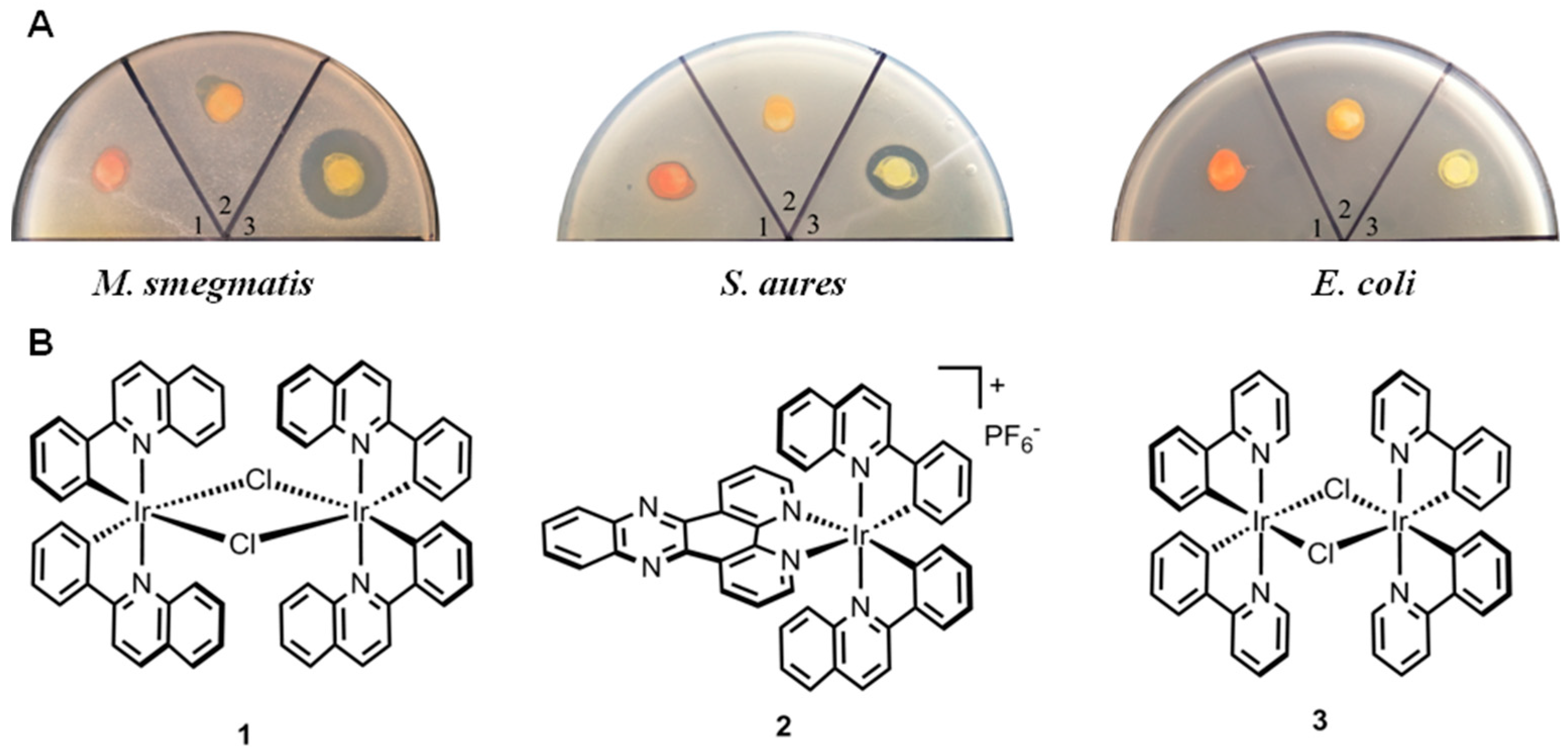
2.1. Tested Compounds

The iridium complexes 1 [21], 2 [22] and 3 [23] were synthesized according to the published procedures. 1H-NMR spectra were recorded on a Bruker Advance (400 MHz) (Bruker, Karlsruhe, Germany) at ambient temperature, and were consistent with the respective reported literature. Complexs 1, 2 and 3 were solubilized in dimethyl sulfoxide (DMSO). 2,2′-bipyridyl and thiourea were purchased from Sangon Biotech Co. (Shanghai, China), and norfloxacin was purchased from Sigma-Aldrich (St. Louis, MO, USA).

2.2. Strains and Media

M. smegmatis mc2 155 was cultured in Middlebrook 7H9 broth (Becton Dickinson, Shanghai, China) supplemented with Tween 80 (0.05% w/v), glycerol (0.5%) and glucose (0.5%) or was grown on 7H10 agar supplemented with glycerol (1%) and glucose (0.5%). Staphylococcus aureus ATCC 33591(MRSA), S. aureus ATCC 25923(MSSA), Escherichia coli ATCC25922 and Pseudomonas aeruginosa PAO1 were grown in Tryptic soy broth (TSB) medium. Cryptococcus neoformans H99 and Candida albicans ATCC90028 were grown in Yeast Extract Peptone Dextrose (YPD) medium.

2.3. Compound Susceptibility Testing

Sensitivity of bacteria to iridium complexes was measured using the disk diffusion method as described previously [24]. The minimum inhibitory concentration (MIC) of complexes 13 was determined by broth microdilution methodology as recommended by the clinical and laboratory standards Institute (CLSI) guidelines. Briefly, cultures were incubated in 96-well microtiter plates in the presence of eight two-fold serial dilution of complexes 13. Freshly prepared 105 bacterial cells or 102 fungal cells were added and incubated for 24 or 48 h at 37 °C, respectively. The MIC was defined as the lowest concentration of compounds with no visible growth.

2.4. Starvation Conditions

The nutrient starvation culture was prepared as described previously [25]. Briefly, exponential phase cultures were pelleted and washed twice with PBS before being resuspended in PBS at 107 Colony-Forming Units (CFU)/mL. Cells were then transferred to standing flasks and incubated at 37 °C with constant rolling at 110 rpm for 10 days. The cultures were then diluted to 106 CFU/mL and treated with 20 μg/mL of 3 or 10 μg/mL (10× MIC) of norfloxacin. In parallel, exponential growth cultures in 7H9 medium was exposed to the same concentration of compounds and the same treatment time to determine the bactericidal effect on replicating cells. Bactericidal activity was determined by CFU enumeration on 7H10 agar.

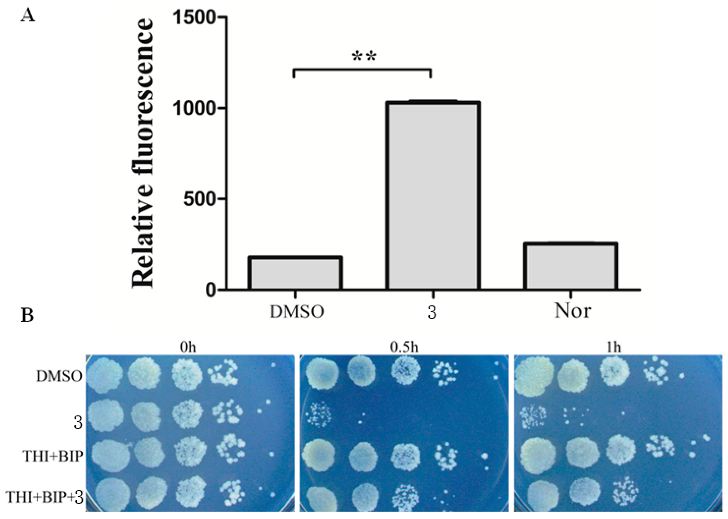
2.5. Measurement of Intracellular Reactive Oxygen Species (ROS)

ROS were measured using the Reactive Oxygen Species Assay Kit (Beyotime Institute of Biotechnology, Shanghai, China) as previously described [26,27]. Briefly, M. smegmatis was grown to exponential-phase and cultivated in the presence of indicated concentrations of compounds. Following 2 h incubation, a final concentration of 10 μM DCFH-DA was added to cultures for 20 min at 37 °C. They were then washed twice with 1× PBS and resuspended in PBS. DMSO and norfloxacin were served as controls. Fluorescence was analyzed using a Tecan Infinite 200 PRO microplate reader (Tecan, Shanghai, China).

2.6. Protection by Iron Chelator and Hydroxyl Radical Scavenger

To examine the protection provided by iron chelator and hydroxyl radical scavenger, M. smegmatis was prepared as above and treated with 2,2′-bipyridyl (250 μM; 50% MIC) and thiourea (100 mM; 50% MIC) 10 min prior to initiation of antimicrobial treatment. Growth inhibition was determined by measuring the viable cells at the indicated periods of time.

2.7. Generation of Norfloxacin-Resistant Mutants

Norfloxacin-resistant strains were obtained by the multistep selection method as described previously [28]. M. smegmatis was cultured in the presence of subinhibitory concentration of norfloxacin (1 μg/mL). After 48 h incubation at 37 °C, cells at 109 CFUs were plated on 7H10 medium containing 4 μg/mL of norfloxacin. Colonies of first-step mutant strains were cultured in 7H9 medium without drug and then by plating 109 CFUs containing 8 μg/mL of norfloxacin to generate second-step mutants. This process was repeated, and three mutants were isolated, named N4, N8, N16.

3. Results and Discussion

3.1. Complex 3 Displays Selective Activity against M. smegmatis

The antibacterial activities of 13 were investigated by using the Kirby-Bauer disk diffusion assays. Accordingly, gram-positive strain Staphylococcus aures, gram-negative strain E. coli and M. smegmatis were plated on an agar dish, and disks soaked with solution of the complexes 13 (50 μg). As shown in Figure 1A, 2 did not exhibit activity to any bacteria, which could be attributed to the absence of labile chlorine ligand. Compared to 1, 3 displayed much more antibacterial activity against M. smegmatis and S. aures, which suggests that less bulky ligands may be more beneficial for antimicrobial activity. While 3 displayed potent activity against M. smegmatis, the minimum inhibitory concentration (MIC) was determined. As shown in Table 1, treatment with 3 inhibited the growth of M. smegmatis with a MIC of 2 μg/mL.
Considering that M. smegmatis is an established surrogate for screening compounds with inhibitory activity against M. tuberculosis, and the obvious antibacterial activity of 3 towards M. smegmatis, we only focused on the work of 3 in the following study. Accordingly, to further evaluate the antibacterial activity of 3, it was used to screen activity against a wide range of pathogenic bacteria, such as E. coli, P. aeruginosa, and S. aureus (MRSA or MSSA), and pathogenic fungi such as C. albicans and C. neoformans. MIC against microbial pathogens was beyond 16 μg/mL (Table 1), demonstrating that 3 exhibited selective activity against M. smegmatis.

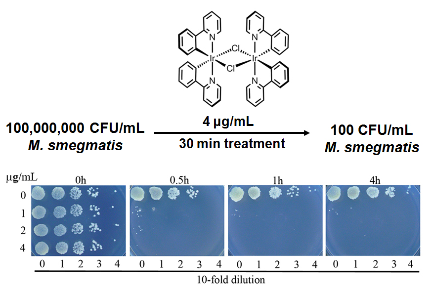
3.2. Complex 3 Displays Potent Bactericidal Activity against M. smegmatis

To further explore the activities of complex 3 against M. smegmatis, we performed time killing experiments. Complex 3 displayed potent bactericidal activity against M. smegmatis, resulting in a 3-log reduction in viable cells 0.5 h of treatment with compound concentrations of 2 μg/mL (Figure 2). No viable cells were observed at 1h at 2 μg/mL. On the basis of these results, it can be concluded that 3 is a fast killing agent with great potential.

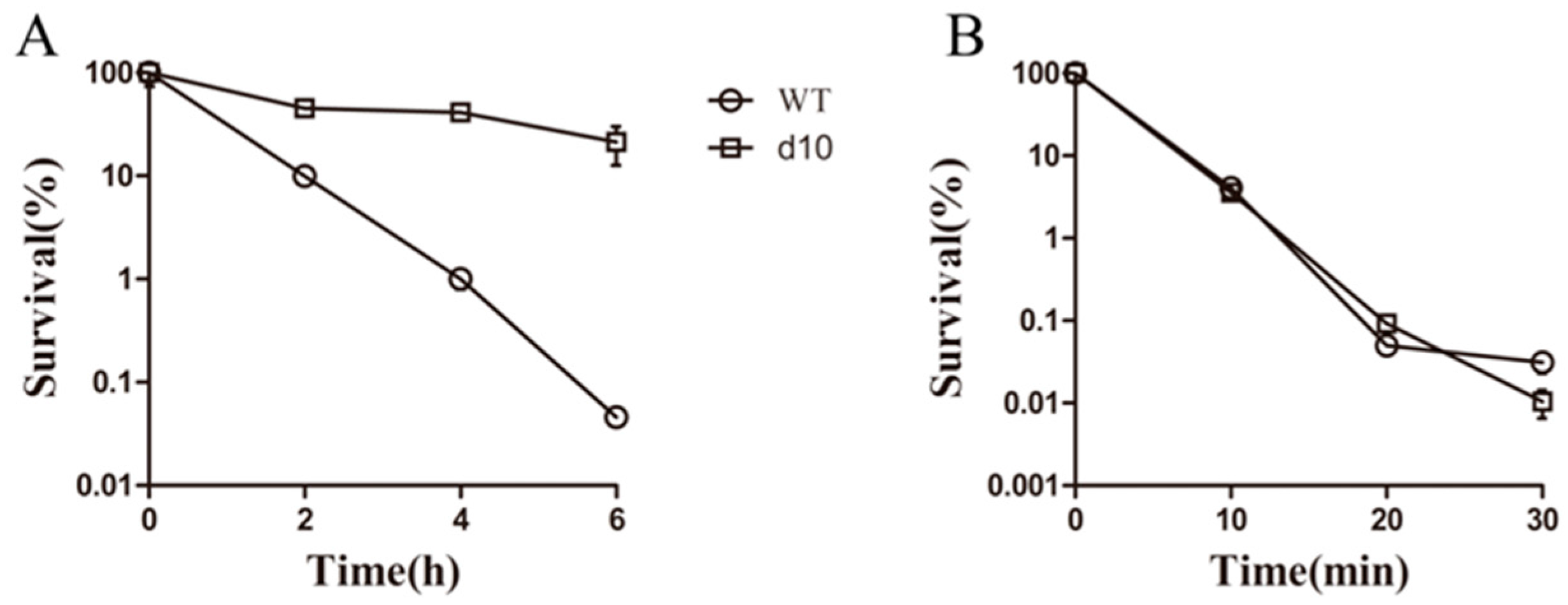
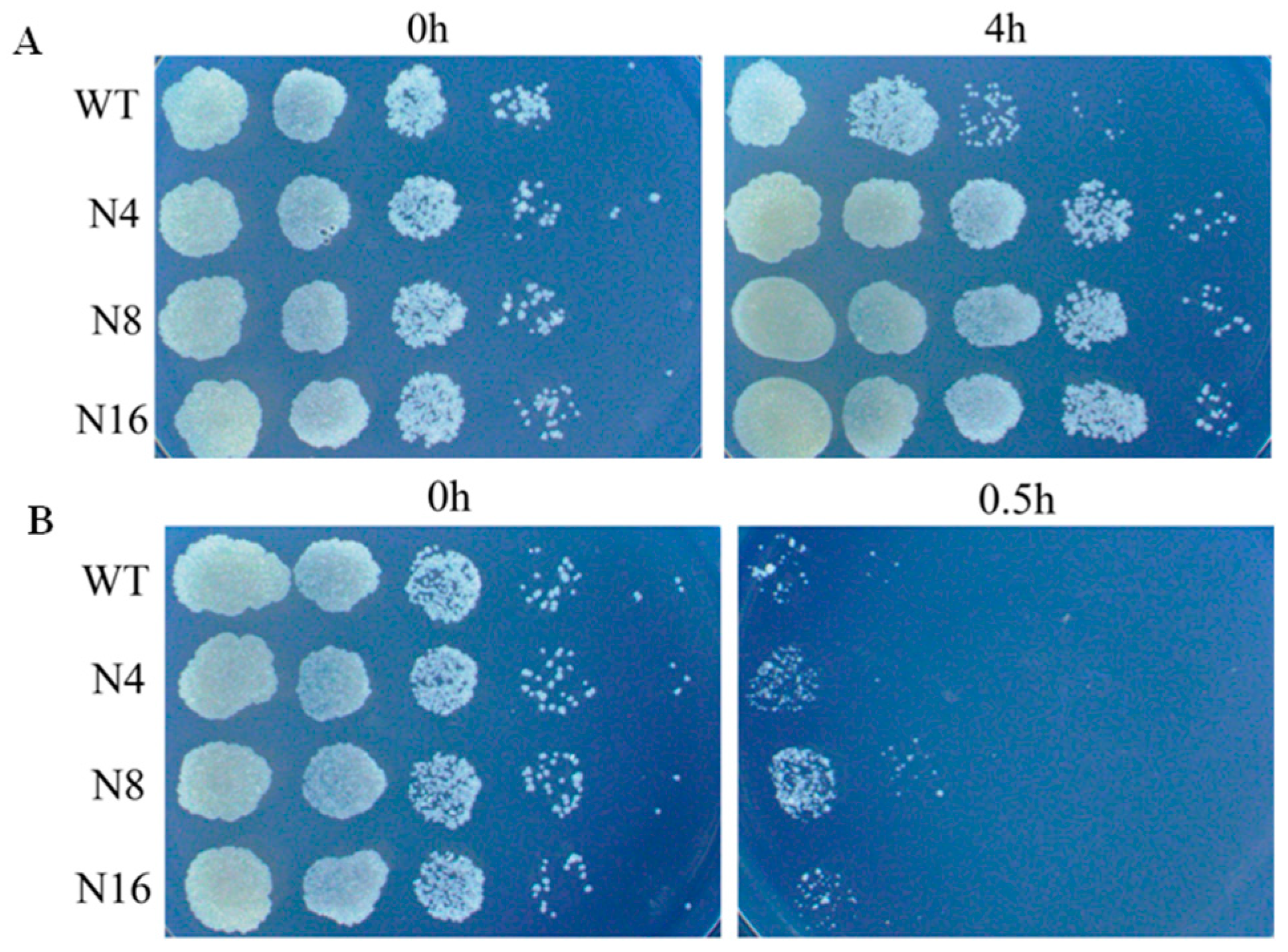
3.3. Complex 3 Displays Activity against Non-Replicating M. smegmatis

It has been reported that most antitubercular drugs exhibit reduced bactericidal activities against non-replicating starved bacilli, contributing to latent infection. Nutrient-deprived M. smegmatis is one of the established models for studying non-replicating starved states. Exposure to 20 μg/mL (10× MIC) 3 led to a 3-log CFU decrease under both culture conditions (Figure 3B). 3 killed non-replicating bacteria and showed equal efficacy against rapidly growing cells. The killing efficacy of 3 was greater that of norfloxacin (Figure 3A), which is known to retain bactericidal activity under non-replicating states, although it is significantly less active against non-replicating than against exponential-phase cells. These results demonstrated that 3 exhibited potent activity against non-replicating M. smegmatis and may target processes that are essential for survival even under non-replicating conditions.

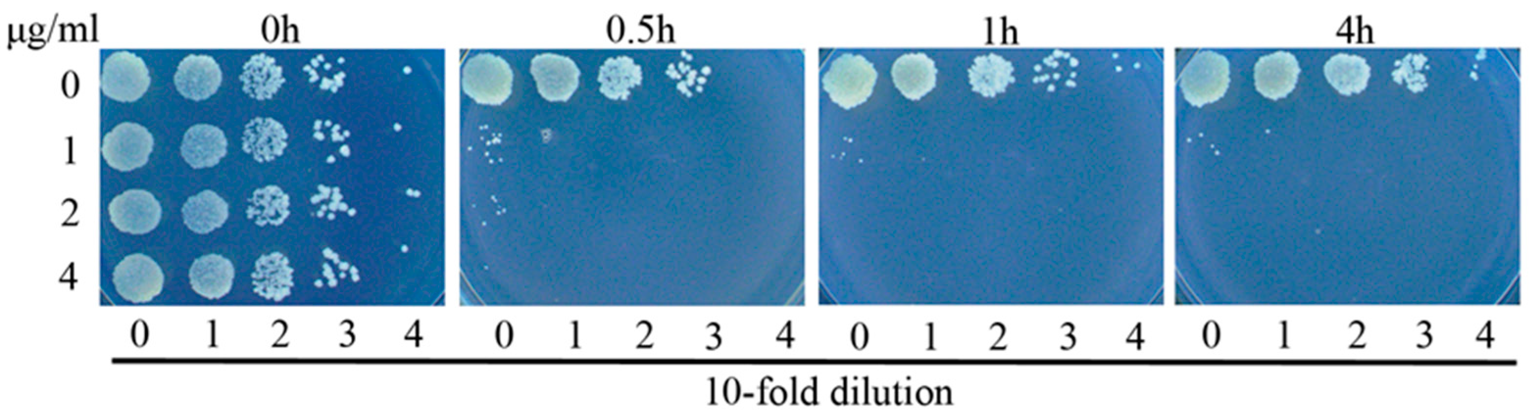
3.4. Complex 3 Was Active against Norfloxacin-Resistant Strains

One of the main obstacles to TB eradication is the high prevalence of drug-resistant strains. To assess of the effect of 3 on drug-resistant strains, norfloxacin-resistant strains were treated with 3. Laboratory-generated resistant strains were obtained by spontaneous mutation under different concentrations of norfloxacin. Three resistant strains with MICs 8 to 16 folds greater than that of WT strains were isolated (N4, N8 and N16) (Figure 4A). 3 displayed similar efficacy against norfloxacin-resistant strains, resulting in a 100-fold to 1000-fold decrease in CFU in 0.5 h treatment (Figure 4B). The lack of cross-resistance with currently used drug class suggested that 3 may retain activity against drug-resistant strains and may have novel modes of action.

3.5. Antibacterial Mechanism of Complex 3

The mechanism of Iridium(III) complexes killing bacterial pathogens remains largely unknown. These complexes can bind DNA and RNA, interact with cell wall, as well as generate ROS. Given that 3 is a rapid killing agent and is redox-active, it is likely to generate ROS via electron transfer to oxygen, as previously reported. We examined the induction of cellular oxidative stress by 3 (1 μg/mL) on M. smegmatis compared with a negative control (DMSO). It could be observed that 3 did indeed induce strong oxidative stress (p < 0.01), and the oxidative stress induced by 3 was stronger than that of norfloxacin, which is known to exert its antimicrobial activity by inducing cellular oxidative stress on bacteria (Figure 5A).
To further examine the role of ROS generation in complex 3-mediated killing, iron chelator Biphyridyl and radical scavenger thiourea, which are able to alleviate the effect of ROS on cell viability, were added to the culture in the presence of compound concentration of 1 μg/mL. Cotreatment with sub-inhibitory concentrations of biphyridyl and thiourea did not affect the growth of S. smegmatis. However, the same cotreatments reduced 3-mediated killing, resulting in 10 and 100 fold reduction of efficacy after 0.5 and 1 h treatment, respectively (Figure 5B). Taken together, these results suggested the involvement of ROS in the 3-mediated lethality.

4. Conclusions

In summary, we here reported that a polypyridyl iridium dimer complex 3 displayed potent and selective activity against M. smegmatis. Furthermore, 3 retained activity against laboratory-generated norfloxacin-resistant strain of M. smegmatis. Mode of action studies indicated that the antimicrobial activity of 3 was potentially due to the production of ROS. On the basis of our findings, it can be concluded that cyclometalated iridium dimer complexes may provide a leading structure for the further development of highly potent bacterial agents against M. tuberculosis infection.

Acknowledgments

Research reported in this study was financially supported by the following: the National Natural Science Foundation of China (Grant 21601146); the Fundamental Research Funds for the Central Universities (Grant XDJK2017B011).

Author Contributions

Guojian Liao conceived and conducted the experiments and wrote the manuscript; Xixi Peng and Zhengyuan Ye also conducted partial experiments; Ting Li and Xiaohong Xiang analyzed the data; Chen Fu conceived the experiment and wrote the manuscript. All authors have given approval to the final version of the manuscript.

Conflicts of Interest

The authors declare no conflict of interest.

References

  1. Cano-Muniz, S.; Anthony, R.; Niemann, S.; Alffenaar, J.C. New approaches and therapeutic options for Mycobacterium tuberculosis in a dormant state. Clin. Microbiol. Rev. 2018, 31, e00060-17. [Google Scholar] [PubMed]
  2. Bloemberg, G.V.; Keller, P.M.; Stucki, D.; Trauner, A.; Borrell, S.; Latshang, T.; Coscolla, M.; Rothe, T.; Homke, R.; Ritter, C.; et al. Acquired resistance to bedaquiline and delamanid in therapy for tuberculosis. N. Engl. J. Med. 2015, 373, 1986–1988. [Google Scholar] [CrossRef] [PubMed]
  3. Manson, A.L.; Cohen, K.A.; Abeel, T.; Desjardins, C.A.; Armstrong, D.T.; Barry, C.E., 3rd; Brand, J.; TBResist Global Genome Consortium; Chapman, S.B.; Cho, S.N.; et al. Genomic analysis of globally diverse Mycobacterium tuberculosis strains provides insights into the emergence and spread of multidrug resistance. Nat. Genet. 2017, 49, 395–402. [Google Scholar] [CrossRef] [PubMed]
  4. Hoagland, D.T.; Liu, J.; Lee, R.B.; Lee, R.E. New agents for the treatment of drug-resistant Mycobacterium tuberculosis. Adv. Drug Deliv. Rev. 2016, 102, 55–72. [Google Scholar] [CrossRef] [PubMed]
  5. Zumla, A.; Maeurer, M.; Host-Directed Therapies, N.; Chakaya, J.; Hoelscher, M.; Ntoumi, F.; Rustomjee, R.; Vilaplana, C.; Yeboah-Manu, D.; Rasolof, V.; et al. Towards host-directed therapies for tuberculosis. Nat. Rev. Drug Discov. 2015, 14, 511–512. [Google Scholar] [CrossRef] [PubMed]
  6. Zhang, Y.; Algburi, A.; Wang, N.; Kholodovych, V.; Oh, D.O.; Chikindas, M.; Uhrich, K.E. Self-assembled cationic amphiphiles as antimicrobial peptides mimics: Role of hydrophobicity, linkage type, and assembly state. Nanomedicine 2017, 13, 343–352. [Google Scholar] [CrossRef] [PubMed]
  7. Liao, G.; Ye, Z.; Liu, Y.; Fu, B.; Fu, C. Octahedral ruthenium(II) polypyridyl complexes as antimicrobial agents against mycobacterium. PeerJ 2017, 5, e3252. [Google Scholar] [CrossRef] [PubMed]
  8. Lu, L.; Liu, L.J.; Chao, W.C.; Zhong, H.J.; Wang, M.; Chen, X.P.; Lu, J.J.; Li, R.N.; Ma, D.L.; Leung, C.H. Identification of an iridium(III) complex with anti-bacterial and anti-cancer activity. Sci. Rep. 2015, 5, 14544. [Google Scholar] [CrossRef] [PubMed]
  9. Fu, C.; Wenzel, M.; Treutlein, E.; Harms, K.; Meggers, E. Proline as chiral auxiliary for the economical asymmetric synthesis of ruthenium(II) polypyridyl complexes. Inorg. Chem. 2012, 51, 10004–10011. [Google Scholar] [CrossRef] [PubMed]
  10. Frezza, M.; Hindo, S.; Chen, D.; Davenport, A.; Schmitt, S.; Tomco, D.; Dou, Q.P. Novel metals and metal complexes as platforms for cancer therapy. Curr. Pharm. Des. 2010, 16, 1813–1825. [Google Scholar] [CrossRef] [PubMed]
  11. Li, F.F.; Collins, J.G.; Keene, F.R. Ruthenium complexes as antimicrobial agents. Chem. Soc. Rev. 2015, 44, 2529–2542. [Google Scholar] [CrossRef] [PubMed]
  12. Medici, S.; Peana, M.; Nurchi, V.M.; Lachowicz, J.I.; Crisponi, G.; Zoroddu, M.A. Noble metals in medicine: Latest advances. Coord. Chem. Rev. 2015, 284, 329–350. [Google Scholar] [CrossRef]
  13. Adam, A.M.A.; Refat, M.S.; Mohamed, M.A. Synthesis and spectroscopic characterizations of noble metal complexes (gold, silver, platinum) in the presence of selenium, and their biological applications as antibacterial, antifungal, and anticancer. Res. Chem. Intermed. 2015, 41, 965–1000. [Google Scholar] [CrossRef]
  14. Ma, D.L.; He, H.Z.; Leung, K.H.; Chan, D.S.H.; Leung, C.H. Bioactive luminescent transition-metal complexes for biomedical applications. Angew. Chem. Int. Ed. 2013, 52, 7666–7682. [Google Scholar] [CrossRef] [PubMed]
  15. Leung, C.H.; Zhong, H.J.; Chan, D.S.H.; Ma, D.L. Bioactive iridium and rhodium complexes as therapeutic agents. Coord. Chem. Rev. 2013, 257, 1764–1776. [Google Scholar] [CrossRef]
  16. Karpin, G.W.; Merola, J.S.; Falkinham, J.O. Transition metal-alpha-amino acid complexes with antibiotic activity against Mycobacterium spp. Antimicrob. Agents Chemother. 2013, 57, 3434–3436. [Google Scholar] [CrossRef] [PubMed]
  17. King, K.A.; Spellane, P.J.; Watts, R.J. Excited-state properties of a triply ortho-metalated iridium(III) complex. J. Am. Chem. Soc. 1985, 107, 1431–1432. [Google Scholar] [CrossRef]
  18. Lowry, M.S.; Hudson, W.R.; Pascal, R.A.; Bernhard, S. Accelerated luminophore discovery through combinatorial synthesis. J. Am. Chem. Soc. 2004, 126, 14129–14135. [Google Scholar] [CrossRef] [PubMed]
  19. Zeglis, B.M.; Pierre, V.C.; Barton, J.K. Metallo-intercalators and metallo-insertors. Chem. Commun. 2007, 4565–4579. [Google Scholar] [CrossRef] [PubMed]
  20. Chaturvedi, V.; Dwivedi, N.; Tripathi, R.P.; Sinha, S. Evaluation of Mycobacterium smegmatis as a possible surrogate screen for selecting molecules active against multi-drug resistant Mycobacterium tuberculosis. J. Gen. Appl. Microbiol. 2007, 53, 333–337. [Google Scholar] [CrossRef] [PubMed]
  21. Kang, T.S.; Mao, Z.F.; Ng, C.T.; Wang, M.D.; Wang, W.H.; Wang, C.M.; Lee, S.M.Y.; Wang, Y.T.; Leung, C.H.; Ma, D.L. Identification of an iridium(III)-based inhibitor of tumor necrosis factor-alpha. J. Med. Chem. 2016, 59, 4026–4031. [Google Scholar] [CrossRef] [PubMed]
  22. Shao, F.W.; Elias, B.; Lu, W.; Barton, J.K. Synthesis and characterization of iridium(III) cyclometalated complexes with oligonucleotides: Insights into redox reactions with DNA. Inorg. Chem. 2007, 46, 10187–10199. [Google Scholar] [CrossRef] [PubMed]
  23. Ru, J.X.; Guan, L.P.; Tang, X.L.; Dou, W.; Yao, X.; Chen, W.M.; Liu, Y.M.; Zhang, G.L.; Liu, W.S.; Meng, Y.; et al. Turn-on phosphorescent chemodosimeter for Hg2+ based on a cyclometalated ir(III) complex and its application in time-resolved luminescence assays and live cell imaging. Inorg. Chem. 2014, 53, 11498–11506. [Google Scholar] [PubMed]
  24. Li, Y.H.; Su, T.T.; Zhang, Y.; Huang, X.L.; Li, J.; Li, C. Liposomal co-delivery of daptomycin and clarithromycin at an optimized ratio for treatment of methicillin-resistant staphylococcus aureus infection. Drug Deliv. 2015, 22, 627–637. [Google Scholar] [PubMed]
  25. Nyka, W. Studies on the effect of starvation on mycobacteria. Infect. Immun. 1974, 9, 843–850. [Google Scholar] [PubMed]
  26. Li, Q.M.; Xie, L.X.; Long, Q.X.; Mao, J.X.; Li, H.; Zhou, M.L.; Xie, J.P. Proteasome accessory factor c (pafC) is a novel gene involved in Mycobacterium intrinsic resistance to broad-spectrum antibiotics—Fluoroquinolones. Sci. Rep. 2015, 5. [Google Scholar] [CrossRef] [PubMed]
  27. Liu, X.M.; Lian, J.M.; Hu, C.H.; Deng, C. Betahistine co-treatment ameliorates dyslipidemia induced by chronic olanzapine treatment in rats through modulation of hepatic ampkα -srebp-1 and pparα-dependent pathways. Pharmacol. Res. 2015, 100, 36–46. [Google Scholar] [CrossRef] [PubMed]
  28. Safi, H.; Lingaraju, S.; Amin, A.; Kim, S.; Jones, M.; Holmes, M.; McNeil, M.; Peterson, S.N.; Chatterjee, D.; Fleischmann, R.; et al. Evolution of high-level ethambutol-resistant tuberculosis through interacting mutations in decaprenylphosphoryl-β-D-arabinose biosynthetic and utilization pathway genes. Nat. Genet. 2013, 45, 1190–1197. [Google Scholar] [CrossRef] [PubMed]
Figure 1. Anti-bacterial activity of complexes 13 as determined by the disk diffusion assay. (A) Strains including S. aureus ATCC 33591(MRSA), E. coli ATCC25922 and M. smegmatis mc2 155; (B) Chemical structures of iridium complex 13.
Polymers 10 00297 g001
Figure 2. Complex 3 is bactericidal against M. smegmatis. Bacterial cells were inoculated in 7H9 medium, and cultured either without drug or in the presence of 3 at various concentrations. At the indicated time points, aliquots of cell suspension were transferred and plated on drug-free 7H9 medium CFU after 24 more hours of incubation.
Polymers 10 00297 g002
Figure 3. Complex 3 displays potent activity against non-replicating mycobacteria. Ten-days-starved and exponential-phase M. smegmatis cultures were treated in triplicate with norfloxacin (A) or 3 (B) at several time points. Cultures were washed twice with PBS and their viability assessed by plating followed by CFU counting. Values represent the means ± standard errors of triplicate determinations.
Polymers 10 00297 g003
Figure 4. Complex 3 shows activity against norfloxacin-resistant strains. (A) Laboratory-generated mutants (N4, N8 and N16) are resistant to norfloxacicn; (B) WT and norfloxacin-resistant strains are susceptible to 3. The MIC of WT against norfloxacin is 2 μg/mL, the MIC of resistant strains is 16–32 μg/mL.
Polymers 10 00297 g004
Figure 5. Complex 3 triggers endogenous ROS production in M. smegmatis. (A) Percentages of intracellular increase in ROS generation in the presence of 10 times of MIC of 3. Norfloxacin represents the positive control for ROS production. Data are shown as mean ± SD of triplicate wells. ** p < 0.01; (B) Effects of a ferrous chelator and a hydroxyl radical scavenger on 3 lethality. Exponentially growing M. smegmatis cells were preincubated with 250 μM biphyridyl and 100 mM thioura for 10 min before they were treated with 10 times of MIC of 3 for 0.5 or 1 h. At least three replicate experiments were performed, and each had similar results.
Polymers 10 00297 g005
Table 1. Activity of 3 against pathogenic microorganisms.
Organism and GenotypeMIC (μg/mL)
M. smegmatis2
S. aureus (MSSA)16
S. aureus (MRSA)32
P. aeruginosa>64
E. coli>64
C. albicans>64
C. neoformans>64

